Hypothalamic Inflammation in Obesity, Insulin Resistance and Ageing
Inaugural-Dissertation
zur Erlangung des Doktorgrades
der Mathematisch-Naturwissenschaftlichen Fakultät der Universität zu Köln
vorgelegt von Eva Tsaousidou aus Thessaloniki
Köln 2015
2 Berichterstatter: Prof. Dr. Jens C. Brüning
Prof. Dr. Sigrun I. Korsching
Tag der mündlichen Prüfung: 20. April 2015
Table of Contents
Abbreviations ... 5
Zusammenfassung... 8
1 Introduction ... 9
1.1 Obesity ... 9
1.2 Energy homeostasis... 9
1.2.1 Insulin signalling pathway ... 10
1.2.2 Leptin signalling pathway ... 11
1.3 CNS in energy homeostasis regulation... 12
1.3.1 Arcuate nucleus in energy homeostasis regulation ... 12
1.3.2 Insulin and leptin signalling in the CNS... 14
1.4 Insulin and leptin resistance ... 16
1.5 Metabolic Inflammation, or Metaflammation ... 16
1.5.1 Metabolic inflammation: Where and when? ... 16
1.5.2 Hypothalamic inflammation ... 18
1.6 Ageing ... 27
1.6.1 Caloric restriction ... 27
1.6.2 Insulin/IGF signalling in ageing ... 28
1.7 Scientific objectives ... 29
2 Materials & Methods... 30
2.1 Animal care and generation of mice ... 30
2.2 Genotyping ... 30
2.2.1 Isolation of genomic DNA ... 30
2.2.2 Polymerase chain reaction ... 30
2.3 Phenotyping ... 31
2.3.1 Insulin tolerance test ... 31
2.3.2 Glucose tolerance test ... 31
2.3.4 Behavioural analysis for learning and memory ... 32
2.3.5 Indirect calorimetry, physical activity and food intake ... 32
2.3.6 Body composition and Bone mineral Density ... 32
2.4 Electrophysiology ... 33
2.4.1 Animals and brain slice preparation for electrophysiological experiments ... 33
2.4.2 Perforated patch recordings ... 33
2.5 Immunohistochemistry ... 35
2.6 Data analysis and statistical methods... 37
2.7 Chemicals and Enzymes ... 38
3 Results ... 40
3.1 Hypothalamic inflammation in obesity and insulin resistance ... 40
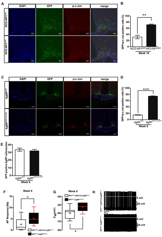
3.1.1 Activation of JNK signalling in AgRP neurons occurs in obesity and increases firing of these cells ... 40
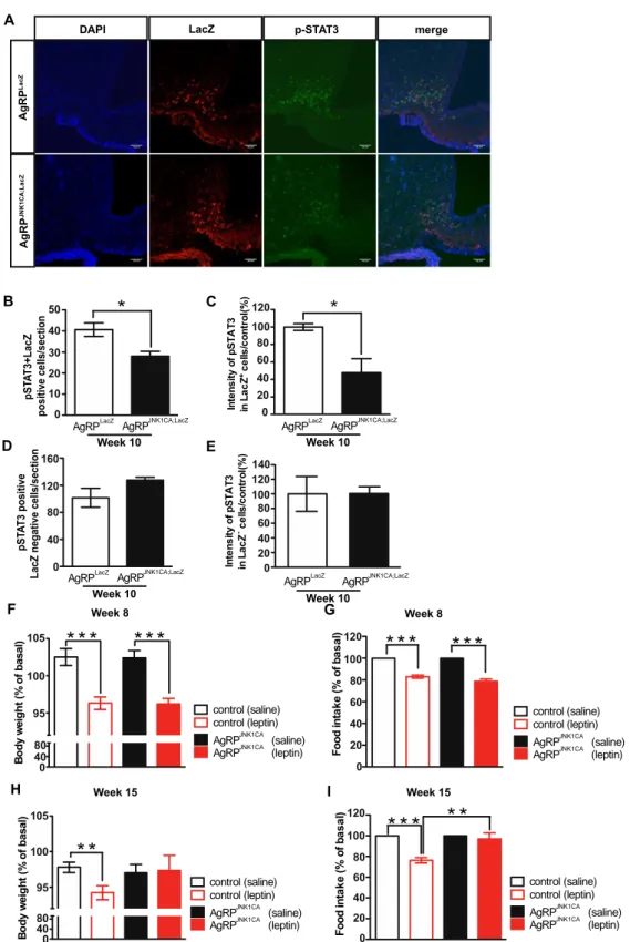
3.1.2 JNK activation in AgRP neurons causes cellular and systemic leptin resistance .. 44
3.1.3 Activation of JNK signalling in AgRP neurons causes obesity ... 46
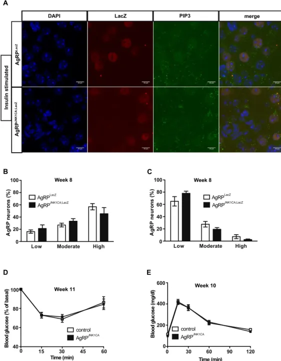
3.1.4 Unaltered glucose homeostasis in AgRPJNK1CA mice ... 48
3.1.5 Activation of IKK2 signalling increases the firing rate of AgRP neurons ... 50
3.1.6 Activation of IKK2 signalling in AgRP neurons does not affect leptin sensitivity or body weight ... 52
3.1.7 Impaired glucose homeostasis in AgRPIKK2CA/CA mice ... 54
3.2 Hypothalamic inflammation in ageing ... 56
3.2.1 Extended median and maximum lifespan in JNK1ΔNes mice ... 56
3.2.2 Decreased body weight in JNK1ΔNes mice up to 1 year of age ... 57
3.2.3 Enhanced insulin sensitivity in JNK1ΔNes mice ... 58
4
3.2.4 Increased adiposity in JNK1ΔNes mice ... 59
3.2.5 Increased energy expenditure in JNK1ΔNes mice ... 60
3.2.6 Declined Bone Mineral Density in JNK1ΔNes mice ... 62
3.2.7 Improved learning plasticity in JNK1ΔNes mice ... 62
4 Discussion ... 65
4.1 Hypothalamic inflammation in obesity and insulin resistance ... 65
4.1.1 JNK1 activation in AgRP neurons results in leptin resistance and obesity ... 66
4.1.2 IKK2 activation in AgRP neurons triggers insulin resistance ... 67
4.1.3 JNK1 or IKK2 activation result in increased firing of AgRP neurons ... 68
4.2 Hypothalamic inflammation in ageing ... 69
4.3 Targeting inflammation, is it the solution? ... 72
4.3.1 Strategies and positive effects of hypothalamic inflammation amelioration ... 72
4.3.2 Negative effects of inflammation amelioration ... 74
4.4 Conclusions and Future perspectives ... 76
Bibliography ... 78
Abbreviations
aCSF artificial cerebrospinal fluid ACTH adrenocorticotropic hormone AgRP agouti-related peptide AMP adenosine monophosphate AMPK AMP-activated protein kinase AP action potential
ApoE apolipoprotein E ARC arcuate nucleus
ATM adipose tissue macrophage ATP adenosine triphosphate BAT brown adipose tissue BBB blood-brain-barrier BMI body mass index BSA bovine serum albumin CA constitutively active CNS central nervous system
Cre site-specific recombinase from phage 1 (causes recombination) CRH corticotropin-releasing hormone
DAPI 4’, 6-diamidino-2-phenylindole DIO diet-induced obesity
DMH dorsomedial nucleus of the hypothalamus DNA deoxyribonucleic acid
DREADD designer receptors exclusively activated by designer drugs GFP green fluorescent protein
ER endoplasmic reticulum FFA free fatty acid
fl floxed
FoxO forkhead box
g gram
Gab Grb2 protein-associated binder
GABA γ-aminobutyric acid
GLP1R glucagon-like peptide 1 receptor GLUT glucose transporter
Grb growth factor receptor binding GTP guanosine triphosphate
GTT glucose tolerance test
GWAS genome-wide association studies
h hour
HFD high-fat diet
HPA hypothalamic-pituitary-adrenal axis HRP horse-radish peroxidase
ICV intracerebroventricular IGF1 insulin-like growth factor 1 IκB inhibitor of NFκB
IKK IκB kinase
IKK2CA IKK2 constitutively active
IL interleukin
6 IR insulin receptor
IRS insulin receptor substrate ITT insulin tolerance test JAK2 janus kinase 2
JNK c-Jun N-terminal kinase JNK1CA JNK1 consitutively active
KATP ATP-dependent potassium channel
L liter
LepR leptin receptor
LH lateral hypothalamic area LPS lipopolysaccharide
m milli
MAPK mitogen-activated protein kinase MC(1-5)R melanocortin 1-5 receptor
min minute
mTOR mammalian target of rapamycin
n nano
NCD normal chow diet
NFκB nuclear factor kappa-light-chain-enhancer of activated B cells NPY neuropeptide Y
NTS nucleus of the solitary tract PBN parabrachial nucleus PBS phosphate buffered saline PCR polymerase chain reaction
PDK1 phosphoinositide-dependent kinase 1 PFA paraformaldehyde
PI3K phoshoinositol-3 kinase
PIP2 phosphatidyl-inositol-(4,5)-bisphosphate PIP3 phosphatidyl-inositol-(3,4,5)-trisphosphate PCK protein kinase C
POMC proopiomelanocortin
PVH paraventricular nucleus of the hypothalamus PTP1B protein-tyrosince phosphatase 1B
RNA ribonucleic acid
ROS reactive oxygen species RT room temperature
SEM standard error of the mean SFA spike frequency adaptation SFO subfornical organ
SOCS3 suppressor of cytokine signalling 3 STAT signal transducer of transcription TLR toll-like receptor
TNF tumor necrosis factor
TRH thyrotropin-releasing hormone UPR unfolded protein response
VMH ventromedial nucleus of the hypothalamus WHO World Health Organization
α-MSH α-melanocyte-stimulating hormone
μ micro
Abstract
In this study, the role of hypothalamic inflammation in obesity, insulin resistance and the regulation of the ageing process is investigated. Activation of c-Jun N-terminal kinase (JNK)1- and inhibitor of nuclear factor kappa-B kinase (IKK)2-dependent signalling plays a crucial role in the development of obesity-associated insulin and leptin resistance not only in peripheral tissues but also in the CNS. This study demonstrates that constitutive JNK1 activation in agouti-related peptide (AgRP)- expressing neurons of the hypothalamus is sufficient to induce weight gain and adiposity in mice as a consequence of hyperphagia. JNK1 activation increases spontaneous action potential firing of AgRP cells and causes both neuronal leptin resistance in a molecular level and resistance in the anorexigenic and body weight regulating effects of leptin. Similarly, activation of IKK2 signalling in AgRP neurons also increases firing of these cells but fails to cause obesity and leptin resistance. In contrast to JNK1 activation, IKK2 activation blunts insulin signalling in AgRP neurons and impairs systemic glucose homeostasis. Collectively, these experiments reveal both overlapping and non-redundant effects of JNK- and IKK-dependent signalling in AgRP neurons, which cooperate in the manifestation of the metabolic syndrome.
JNK1 ablation in the CNS has been demonstrated to resemble the effects of caloric restriction, a dietary intervention that delays ageing. In this study, JNK1 ablation in the CNS results in extended median and maximum lifespan in mice, protection from high-fat diet induced insulin resistance and increased energy expenditure but also increased adiposity and decreased bone mineral density.
Hypothalamic inflammation amelioration via JNK1 and/or IKK2 inhibition are potential future therapeutic targets to counteract obesity- and ageing-associated diseases.
8
Zusammenfassung
In dieser Studie wird die Rolle von hypothalamischer Entzündung bei Adipositas, Insulinresistenz und der Regulation von Alterungsprozessen untersucht. Die Aktivierung der c-Jun N-terminal Kinase (JNK)1 und die der Inhibitor der Nuklear- Faktor-Kappa-B Kinase (IKK)2-abhängigen Signalübertragung spielt bei der Entwicklung von adipositasassoziierter Insulin- und Leptinresistenz in peripheren Geweben aber auch im ZNS eine entscheidende Rolle. Diese Arbeit zeigt, dass konstitutive JNK1-Aktivierung in Agouti-assoziierten Peptid (AgRP)-exprimierenden Neuronen des Hypothalamus ausreichend ist um Gewichtszunahme und Adipositas in Mäusen aufgrund von Hyperphagie zu induzieren. Die Aktivierung von JNK1 erhöht spontane Aktionspotentialimpulse der AgRP-Zellen und verursacht sowohl neuronale Leptinresistenz auf molekularer Ebene als auch Resistenzen des appetitzügelnden sowie körpergewichtsregulierenen Effektes von Leptin. Ebenso erhöht die Aktivierung der IKK2-Signalübertragung das Feuern in AgRP-Neuronen, verursacht jedoch weder Adipositas noch Leptinresistenz. Im Gegensatz zur JNK1-Aktivierung dämpft die Aktivierung von IKK2 Insulinsignalübertragung in AgRP-Neuronen und beeinträchtigt die systemische Glucosehomöostase. Zusammengefasst zeigen die Experimente sowohl den überlappenden als auch nichtredundanten Effekt der JNK- und IKK abhängigen Signalübertragung in AgRP-Neuronen, welche kooperativ zur der Erscheinungsform des metabolischen Syndroms beitragen.
Es wurde gezeigt, dass JNK1-Ablation im ZNS den Effekten von Kalorienrestriktion, einer den Alterungsprozess verlangsamenden Ernährungsintervention gleicht. In dieser Studie führt JNK1-Ablation im ZNS zu erhöhter mittlerer und maximaler Lebensdauer, Schutz vor von fettreicher Ernährung induzierter Insulinresistenz sowie Energieverbrauch, jedoch auch zu erhöhter Adipositas sowie geringerer Knochendichte. Eine Verbesserung der hypothalamischen Entzündung durch Inhibierung von JNK1 und IKK2 stellen potentielle therapeutische Ziele dar um adipositas- und altersassoziierter Krankheiten entgegenzuwirken.
1 Introduction
1.1 Obesity
Overweight, defined as body mass index (BMI) greater or equal to 25 kg/m2, and obesity, BMI greater or equal to 30 kg/m2, are conditions of excess fat accumulation that affect more than one third of the global adult population [source: World Health Organization (WHO), 2015].
Obesity and its associated pathologies, such as type 2 diabetes and cardiovascular diseases (Katzmarzyk et al., 2003), have reached epidemic proportions enhancing the necessity to develop effective therapeutic strategies against them.
Various environmental factors, including consumption of energy-dense food and sedentary lifestyle, have been held responsible for the obesity epidemic, however the genome-wide association studies (GWAS) and studies in families and twins (Farooqi and O’Rahilly, 2006) revealed that the truth is also lying in our genes (Locke et al., 2015) and the epigenetic modifications we carry (Slomko et al., 2012). In relatively similar environmental backgrounds some individuals are more prone to develop obesity and obesity-associated diseases and the effect of dietary interventions has been proven to be ineffective in the long-run for obese patients as 90% of them return to their initial body weight in a 5-year period (Safer, 1991).
Obesity is not only a major health concern but also an immense economic burden with the health care costs for obesity and its associated diseases expanding every year, reaching an annual impact of 2 trillion dollars globally, paralleling expenses associated with smoking and war according to the WHO. Collectively, obesity is a global threat and its initiation and manifestation arise from interactions between multiple genes and the environment that in turn affect behaviour and energy homeostasis.
1.2 Energy homeostasis
Life is a process that relies on energy. Energy homeostasis is the balance between energy intake and expenditure to ensure steady body weight and its dysregulation can lead to excessive fat accumulation, and eventually obesity.
Introduction
10
In order to ensure steady body weight orexigenic, promoting appetite, and anorexigenic, suppressing appetite, hormones have been identified to communicate signals of energy status in mammals. Two of the most important hormones for energy homeostasis regulation are insulin and leptin.
Insulin is secreted by pancreatic β-cells in response to increased blood glucose levels to promote glucose uptake and suppress hepatic glucose production and also to regulate glycogenesis, lipogenesis and protein synthesis (Roth et al., 2012). Upon increased blood glucose concentrations, glucose enters the β-cells, it is metabolized into adenosine triphosphate (ATP) resulting in the closure of ATP-dependent potassium (KATP) channels, cell depolarization and subsequent influx of Ca2+ ions which in turn leads to the exocytosis of insulin-containing vesicles and the release of insulin into the circulation (Roth et al., 2012; Taniguchi et al., 2006). In the prediabetic state, the β-cells produce more insulin, resulting in hyperinsulinemia, to compensate for the decreased efficacy of insulin in exerting its effects, a condition termed insulin resistance. The β-cells cannot sustain the excessive insulin secretion for long, as it leads to their exhaustion, impaired glucose homeostasis and the development of type 2 diabetes (Muoio and Newgard, 2008).
Leptin is anorexigenic, secreted from adipocytes in proportion to the adiposity of an organism communicating the current energy status to the central nervous system (CNS) (Friedman and Mantzoros, 2015). Importantly, leptin deficiency (Halaas et al., 1995; Ozata et al., 1999) as well as lack of a functional leptin receptor (Clément et al., 1998; Montague et al., 1997; Zhang et al., 1994) in mice and humans results in morbid obesity. Although initially leptin was considered a potential treatment against obesity, it has been demonstrated that obese individuals have increased plasma leptin levels and develop resistance to its anorexigenic action (Friedman, 2011).
Insulin and leptin communicate their signals to the central nervous system to regulate energy and glucose homeostasis (Cummings and Overduin, 2007; Belgardt and Brüning, 2010). Leptin and insulin signalling in the regulation of energy and glucose homeostasis will be introduced further in detail.
1.2.1 Insulin signalling pathway
Upon binding of insulin to the insulin receptor (IR) the insulin signalling cascade is activated [reviewed in (Guo, 2014)]. The insulin receptor consists of two
Introduction
heterodimers of the α- and β- subunits, which are products of a single gene and are derived from the same polypeptide that is subjected to proteolytic processing. The insulin-binding site is on the α-subunits and the insulin-regulated tyrosine kinase activity domain is on the β-subunits. Conformational changes upon binding of insulin to the α-subunits of the IR activate the tyrosine kinase activity of the β-subunits resulting in autophosphorylation of tyrosine residues and in turn recruitment of the insulin receptor substrates (IRS1-4) and the growth factor receptor binding (Grb)2 protein-associated binder (Gab) proteins. IRS proteins, when phosphorylated, are docking platforms for the phosphatidylinositol 3 kinase (PI3K), Grb2 and the SH2 domain containing phosphatase (Shp)-2. PI3K phosphorylates phosphatidylinositol- (4,5)-bisphophate (PIP2) to phosphatidylinositol-(3,4,5)-trisphosphate (PIP3) and PIP3 in turn activates downstream targets including the Akt, which co-localizes with phosphoinositide-dependent protein kinase 1 (PDK1) leading to phosphorylation and activation of Akt. Akt, among other proteins, can phosphorylate FoxO1 thereby triggering its translocation out of the nucleus, thus regulating gene transcription (Figure 1). Activation of the IRS-PI3K cascade is crucial for most of insulin’s well- known effects, including glucose transporter translocation, glycogen synthesis, protein synthesis and the regulation of gene transcription [reviewed in (Boucher et al., 2014)].
1.2.2 Leptin signalling pathway
Leptin signalling is initiated upon binding of leptin to its receptor (LepR) to initiate the JAK/STAT pathway, among others. Activation of LepRb, the long form of leptin receptor mediating its anorexigenic effects (de Luca et al., 2005), leads to recruitment of JAK2, which phosphorylates the receptor and itself. Phosphorylated LepRb recruits and phosphorylates STAT3, triggering STAT3 dimerization and nuclear localization, where it regulates gene expression (Figure 1). Leptin has been also demonstrated to trigger the mitogen-activated protein kinase (MAPK), adenosine monophosphate- activated protein kinase (AMPK) and PI3K pathways (Figure 1) [reviewed in (Bjørbaek and Kahn, 2004)].
Introduction
12
1.3 CNS in energy homeostasis regulation
The major orchestrator of energy homeostasis is the hypothalamus. The importance of the hypothalamus in energy homeostasis regulation has been demonstrated since the 1940s with the first lesion experiments in rats from Hetherington and Ranson (Hetherington and Ranson, 1940). The hypothalamus is connected to brain areas that affect behaviours such as reward and food foraging either directly or through inter- neurons (Heisler et al., 2003; Leinninger et al., 2009).
The major hypothalamic areas responsible for energy homeostasis are situated around the third ventricle and above the median eminence and are the ventromedial hypothalamus (VMH), dorsomedial hypothalamic nucleus (DMH), paraventricular nucleus (PVN), lateral hypothalamic nucleus (LH) and the arcuate nucleus (ARC).
The hypothalamus is a region sensitive to blood-born signals due to a permeable blood-brain-barrier (BBB), and to its position adjacent to the third ventricle, thus allowing hypothalamic communication with the periphery. The VMH is very important in the feeding response to insulin-induced hypoglycemia and is home to many glucose sensing neurons that can be glucose-excited or glucose-inhibited (Verberne et al., 2014). The DMH is a target site of projections from other hypothalamic regions and is pivotal to appetite regulation, circadian rhythm and thermogenesis (Chao et al., 2011; Chou et al., 2003). The PVN regulates the hypothalamic-pituitary-adrenal axis (HPA) by the release of corticotropin-releasing hormone (CRH) in response to stress and thyrotropin-releasing hormone (TRH) and also modulates the autonomic nervous system (Biag et al., 2012). Furthermore, the PVN harbours second-order neurons with melanocortin 4 receptors (MC4R) regulated by neurons of the arcuate nucleus. The LH is responsible for circadian rhythm (Goodless-Sanchez et al., 1991) and additionally for hedonic and reward-related behaviours (Sternson, 2013). The ARC is the best characterized hypothalamic region concerning energy homeostasis and will be discussed further in detail.
1.3.1 Arcuate nucleus in energy homeostasis regulation
Multiple neuronal populations regulate food intake and energy expenditure via tight- coordination in intra- and extra-hypothalamic brain areas and the most thoroughly studied hypothalamic region for energy homeostasis is the ARC adjacent to the third ventricle and over the median eminence. The ARC is considered a circumventricular
Introduction
organ due to its position, a permeable blood-brain barrier (BBB) and extensive vasculature.
In the ARC two neuronal populations with opposing roles reside, the orexigenic neuropeptide Y (NPY) and Agouti-related peptide (AgRP)-co-expressing neurons and the anorexigenic pro-opiomelanocortin (POMC) neurons. The AgRP and POMC neurons are first-order neurons that receive diverse circulating signals relevant to energy homeostasis, such as hormones and nutrients, from the periphery. AgRP and POMC neurons act on second-order neurons with MC4R residing predominantly in the PVN forming the melanocortin system (Cowley et al., 1999; Elmquist et al., 1999). The anorexigenic POMC neurons activate MC4R-expressing neurons to decrease food intake and increase energy expenditure whereas the AgRP neurons increase food intake and decrease energy expenditure by antagonizing POMC action on MC4R (Kim et al., 2014b). Importantly, Mc4r mutations are one of the most common monogenic mutations resulting in obesity in humans (Yeo et al., 1998).
A variety of proteins, including α-, β- and γ-melanocyte stimulating hormone (MSH) and adrenocorticotrophin (ACTH), are generated by the Pomc gene encoding the protein precursor Pro-opiomelanocortin which undergoes post-translational modification processes and mutations of the gene result in obesity (Krude et al., 1998). The best-studied anorexigenic peptide mediating its effects on energy expenditure and food intake is α-MSH by binding and activating MC4Rs (Ollmann et al., 1997). POMC-deficient mice are obese (Yaswen et al., 1999) and acute ablation of POMC neurons in adult mice results in hyperphagia and obesity (Gropp et al., 2005). Interestingly, early postnatal ablation of POMC neurons results in decreased food intake but also decreased energy expenditure resulting in an obese phenotype (Greenman et al., 2013).
The orexigenic AgRP neurons integrate peripheral signals and release NPY, AgRP and the inhibitory neurotransmitter γ-aminobutyric acid (GABA) to mediate their effects on other neuronal populations and in turn exert behavioural responses.
AgRP and NPY bind to MC4Rs and NPYRs, respectively, to inhibit the activity of MC4R-expressing neurons (Gerald et al., 1996; Cowley et al., 1999) through G- protein dependent and independent pathways (Ghamari-Langroudi et al., 2015).
Ablation of AgRP neurons in adult mice induces hypophagia and starvation (Gropp et al., 2005; Luquet et al., 2005; Luquet et al., 2007) but surprisingly, early postnatal ablation or AgRP mutations result in unchanged food intake and body weight
Introduction
14
(Erickson et al., 1996; Gropp et al., 2005; Luquet et al., 2005; Qian et al., 2002). In order to succumb the absence of functional AgRP neurons compensatory mechanisms are proposed to be taking place during development (Wu and Palmiter, 2011; Wu et al., 2012) and the importance of GABA as a pivotal inhibitory neurotransmitter is pointed out. GABAergic signalling by AgRP neurons through direct synaptic innervations in the ARC inhibits POMC neurons (Cowley et al., 2001; Horvath et al., 1997) and the parabranchial nucleus (PBN) in the hindbrain (Wu et al., 2009).
Additionally, the oxytocin (OXT) and Sim1 neurons in the PVN receive GABAergic input from the AgRP neurons (Atasoy et al., 2012; Krashes et al., 2014). Furthermore, AgRP neuron-specific deletion of vesicular GABA transporter genes results in a lean phenotype and protection from HFD-induced obesity demonstrating the importance of GABAergic signalling (Tong et al., 2008). Although the AgRP neuropeptide is able to antagonize the melanocortin pathway, the orexigenic effects of AgRP neurons are not affected by MC4R deletion (Krashes et al., 2011) again proposing the main role of GABAergic signalling to exert the orexigenic effects of AgRP neurons.
Acute neuronal activation of AgRP neurons, by approaches such as the designer receptors exclusively activated by designer drugs (DREADD) and optogenetic stimulation, resulted in robust hyperphagia and provided strong evidence for the orexigenic role of AgRP neurons (Aponte et al., 2011; Atasoy et al., 2012;
Krashes et al., 2011). Of note, the AgRP neurons are not only tightly-wired to the PVN and intra-hypothalamic areas as the DMH and LH but also to extra- hypothalamic areas, as demonstrated by anatomical and functional analyses of AgRP neuronal projections (Betley et al., 2013; Broberger et al., 1998) adding up to the complexity of neuronal circuits regulating feeding behaviour. Collectively, the AgRP neurons are potent regulators of energy homeostasis, participating in the melanocortin pathway, with GABAergic signalling playing a crucial role in exerting their orexigenic effects.
1.3.2 Insulin and leptin signalling in the CNS
Insulin’s functions are not only profound in the periphery, regulating glycogenesis, lipogenesis and protein synthesis (Roth et al., 2012), but also in the CNS, where insulin receptors (IR) are widely expressed (Havrankova et al., 1978). Brain-specific IR-deficient mice have increased food intake and fat mass, developing mild obesity
Introduction
(Brüning et al., 2000). Furthermore, CNS insulin signalling is important for the control of peripheral fat metabolism (Koch et al., 2008; Scherer et al., 2011).
Although IR deletion in AgRP and POMC neurons does not affect energy homeostasis in mice, IR deletion in AgRP neurons results in defective suppression of hepatic glucose production (Könner et al., 2007). Additionally, in a study using mice with hypothalamic deficiency of IRs, re-expression of IR in AgRP and POMC neurons had distinct effects, with insulin signalling in AgRP neurons decreasing hepatic glucose production whereas in POMC neurons increasing hepatic glucose production, energy expenditure and locomotor activity (Lin et al., 2010).
Insulin signalling not only modulates neuronal gene expression by activation of the PI3K pathway (Niswender et al., 2003) but also PIP3 has been shown to bind to KATP channels antagonizing the action of ATP, hyperpolarizing and thus silencing the neurons (MacGregor et al., 2002; Plum et al., 2006). Insulin further exerts its anorexigenic role in AgRP and POMC neurons by triggering Pomc expression and suppressing Agrp by phosphorylating and excluding FoxO1 from the nucleus (Kim et al., 2006). FoxO1 was also demonstrated to activate Gpr17 to activate AgRP neurons and regulate food intake (Ren et al., 2012) but recently Gpr17 deficient mice were reported to have unaltered food intake, body weight and glucose homeostasis compared to their control littermates (Mastaitis et al., 2015).
Leptin signalling in the CNS has been extensively studied, especially its role in AgRP and POMC neurons. Neuron-specific leptin deficient mice are obese (de Luca et al., 2005) together with POMC- and AgRP-restricted leptin deficient mice (Balthasar et al., 2004; van de Wall et al., 2008). Leptin is able to depolarize POMC neurons and simultaneously hyperpolarize AgRP neurons (Cowley et al., 2001) being also able to modulate synaptic plasticity in the hypothalamus (Pinto et al., 2004).
Furthermore, leptin has been demonstrated to increase POMC and decrease AgRP mRNA levels (Mizuno and Mobbs, 1999; Mizuno et al., 1998). Collectively, leptin’s regulatory functions are diverse and broad affecting not only gene expression but also synaptic plasticity and neuronal activity.
Importantly, there are distinct leptin and insulin responsive POMC neuronal subpopulations (Williams et al., 2010) adding to the complexity of the hypothalamic regulation of energy homeostasis.
Introduction
16 1.4 Insulin and leptin resistance
Obesity and type 2 diabetes are underlined by a deteriorated efficacy of leptin and insulin to exert their anorexigenic and glucoregulatory functions, conditions termed leptin and insulin resistance (Könner and Brüning, 2012). Leptin and insulin resistance develop despite the high plasma concentrations of both hormones in obese individuals through different mechanisms, such as metabolic inflammation, which will be introduced later.
Chronic leptin signalling induces high levels of suppressor of cytokine signalling 3 (SOCS3), which has been shown to blunt leptin signalling by inhibiting the leptin-induced tyrosine phosphorylation of JAK2 (Bjørbaek et al., 1999). High-fat feeding can also induce the protein-tyrosine phosphatase 1B (PTP1B) (Zabolotny et al., 2008) and protein kinase C (PKC) contributing to insulin resistance (Figure 1) (Benoit et al., 2009). Furthermore, activation of inflammatory signalling which in turn induces, among others, c-Jun N-terminal kinase 1 (JNK1) and nuclear factor kappa- light-chain-enhancer of activated B cells (NFκB) has been implicated in leptin and insulin resistance and will be introduced further in detail.
1.5 Metabolic Inflammation, or Metaflammation
The last two decades metabolic inflammation, or metaflammation, has been in focus as a mechanism to explain the pathophysiology of obesity and its hallmarks, insulin and leptin resistance (Gregor and Hotamisligil, 2011). Differing from classical inflammation, which is activated by pathogens and trauma, metaflammation is chronic and low-grade. Metaflammation is observed after high-fat diet (HFD) consumption and during obesity in multiple organs, as the adipose tissue, liver, pancreas, muscle and brain and results in insulin resistance and impaired energy homeostasis (Hotamisligil et al., 1993; Cai et al., 2005; Ehses et al., 2007;Saghizadeh et al., 1996; De Souza et al., 2005). The mechanisms and time-course of metaflammation initiation are of great importance and will be introduced further.
1.5.1 Metabolic inflammation: Where and when?
Metaflammation was first described in the adipose tissue (Hotamisligil et al., 1993) and gained a lot of attention with multiple studies demonstrating its apparent role in
Introduction
obesity-associated pathologies (Sam and Mazzone, 2014), as the manifestation of insulin resistance after HFD consumption (Xu et al., 2003). The role of immune cells (Mraz and Haluzik, 2014) and especially adipose tissue macrophages (ATMs) in metaflammation is essential (McNelis and Olefsky, 2014). ATMs are either resident and get activated or infiltrate the adipose tissue attracted by signals from dying adipocytes, which reached their oxygen diffusion limit. ATMs are shown to foster the remodelling of the adipose tissue when it reaches its capacity limits during obesity (McNelis and Olefsky, 2014). Interestingly, obese people can remain healthy and insulin sensitive when they can safely store their excess energy, keeping the levels of
‘toxic’ circulating FFA low and avoiding ectopic lipid accumulation in liver and muscle (Blüher, 2010). Furthermore, the polarization of ATMs, into M1 or M2, is important in the regulation of insulin sensitivity, affected by the primary inflammatory response, but is hard to define because ATMs can simultaneously express M1 and M2 markers (Bourlier et al., 2008). Collectively, the impact of metaflammation in obesity and insulin resistance is being thoroughly studied (Gregor and Hotamisligil, 2011), making it an important target for therapeutics.
Metaflammation is detected after some hours of HFD consumption or lipid infusion with changes in the circulating mononuclear cells, liver, muscle and brain (Ghanim et al., 2009; Aljada et al., 2004; Watt et al., 2006; Thaler et al., 2012).
Inflammatory pathways are activated involving, most commonly NFκB and JNK pointing to the direction that inflammation is triggered by nutrients long before obesity arises. Indeed, fatty acids (Shi et al., 2006; Schaeffler et al., 2009) can directly activate TLR4 signalling to induce insulin resistance and mice lacking TLR4 are protected from HFD-induced insulin resistance. ΗFD can induce gut microbiota to exacerbate inflammation in mice via the TLR4 signalling pathway changing also the permeability of the gut (Kim et al., 2012; Schaeffler et al., 2009). Changes in gut permeability enhance endotoxemia, seen as increases in circulating lipopolysaccharide (LPS) and tumour necrosis factor α (TNFα) concentrations to initiate insulin resistance and obesity (Cani et al., 2007; Cani et al., 2008). It is evident that HFD-induced metaflammation is initiated before obesity arises and it is a rapid and multifaceted process with multiple organs participating in this nutrient response.
To understand the complex interplay between metabolic organs during metaflammation, their endocrine role has to be considered. Leptin and insulin
Introduction
18
communicate the energy status to the CNS comprising the two master regulators of energy and glucose homeostasis (Myers and Simerly, 2010). Changes in circulating leptin and insulin levels but also nutrients and cytokines are sensed by defined hypothalamic areas and importantly the arcuate hypothalamic nucleus (Vogt and Brüning, 2013;Milanski et al., 2009; Thaler et al., 2012), where the orexigenic AgRP and the anorexigenic POMC neurons reside. The AgRP neurons are the first to sense small fluctuations in plasma metabolic signals to regulate food intake and short term high-fat feeding increases SOCS3 to cause leptin resistance, already after 48h of HFD consumption (Olofsson et al., 2013).
Collectively, the importance of metaflammation in obesity-associated complications is highly appreciated. Importantly, as metaflammation is initiated so fast, it can be defined as a nutrient response. The role of CNS, particularly the hypothalamus, is vital in orchestrating energy homeostasis since it can sense slight changes in the circulation and is rapidly affected by metaflammation. CNS metaflammation is an important target for the development of therapies against obesity and obesity–associated pathologies.
1.5.2 Hypothalamic inflammation
It has been a decade since the first observations of hypothalamic metabolic inflammation after HFD consumption (De Souza et al., 2005). There are still a lot of open questions regarding the fine mechanisms that regulate energy homeostasis which are affected by the activated inflammatory pathways leading to vast amounts of research conducted in order to define them. Hypothalamic metaflammation is associated with multiple stimuli/signalling pathways like ER stress and activated inflammatory receptors resulting in central insulin and leptin resistance. The focus here will be on the hypothalamus, although metaflammation is also induced in other regions as the amygdala of DIO rats, related to insulin resistance and ER stress (Castro et al., 2013) and the subfornical organ (SFO), a brain region regulating blood pressure (de Kloet et al., 2014).
De Souza et al. were the first to demonstrate that hyperlipidic diet leads to increased pro-inflammatory cytokines and inflammatory responsive proteins in the hypothalamus, such as JNK and NFκB (De Souza et al., 2005). In the same year Zhang et al. reported increased NFκB activity and neural oxidative stress in the brain
Introduction
after HFD consumption linking it to increased risk for dementia (Zhang et al., 2005).
Here, the role of ER stress, different inflammatory mediators and cell types that mediate hypothalamic metaflammation will be introduced.
1.5.2.1 ER stress in hypothalamic inflammation
The importance of ER stress and the unfolded protein response (UPR) in metabolic regulation in the periphery is well appreciated (Hotamisligil, 2008) and it was first demonstrated in the hypothalamus by Ozcan et al. 2009 (Ozcan et al., 2009). Reduced ER capacity was shown to lead to a significant augmentation of obesity on a HFD (Ozcan et al., 2009). ER stress and UPR can be activated to a different degree by a variety of stimuli. For example, LPS can stimulate ER stress and UPR whereas TNFα activates ER stress to a certain degree but fails to induce a complete UPR in the hypothalamus (Denis et al., 2010) and palmitate is able to induce ER stress in the neuronal cell model mHypoE44 (Mayer and Belsham, 2010). Induction of ER stress in the hypothalamus by thapsigargin inhibits the anorexigenic effects of leptin and insulin, whereas treatment with the chemical chaperone 4-phenyl butylic acid significantly improves leptin and insulin sensitivity in diet-induced obese mice (Won et al., 2009). Furthermore, even short-term brain ER stress is sufficient to induce glucose intolerance, systemic insulin resistance, increased blood pressure and elevated sympathetic tone (Purkayastha et al., 2011).
Lipids are important regulators of hypothalamic inflammation and a recent study revealed that hypothalamic ER stress can be activated by ceramides, leading to sympathetic inhibition, reduced brown adipose tissue (BAT) thermogenesis and weight gain (Contreras et al., 2014). The ceramide action is abolished by genetic overexpression of GRP78/BiP in the VMH. GRP78 overexpression reduces hypothalamic ER stress and increases BAT thermogenesis, leading to weight loss and enhanced leptin and insulin sensitivity (Contreras et al., 2014). Inactivation of fatty acid synthase in the hypothalamus prevents diet-induced obesity (DIO) and systemic inflammation and neuron-specific deletion of PPARδ -a lipid sensor that regulates genes involved in lipid metabolism- leads to increased susceptibility to DIO, leptin resistance and hypothalamic inflammation in low-fat diet (Kocalis et al., 2012).
Stearic acid, a saturated fatty acid, is able to cause hypothalamic inflammation and results in reduced oxygen consumption and blunted peripheral insulin signal
Introduction
20
transduction (Arruda et al., 2011) and impaired insulin secretion from the pancreas (Calegari et al., 2011).
To understand the mechanistic basis of ER stress-induction, the significance of ER and mitochondria interactions has been investigated. Depletion of Mfn2, a protein vital for ER-mitochondria interactions, in POMC neurons results in ER stress-induced leptin resistance, hyperphagia, reduced energy expenditure and defective POMC processing, effects that are reversed by inhibition of ER stress by chemical chaperone treatment (Schneeberger et al., 2013).
Collectively, ER stress in CNS metaflammation is activated to a different degree by a variety of stimuli resulting in insulin and leptin resistance and diminishing BAT thermogenesis. ER-mitochondria interactions, pivotal for regulating energy balance and chaperone treatment, can reverse the detrimental effects of hypothalamic ER stress.
1.5.2.2 Pathways and mediators of hypothalamic inflammation
Obesity is accompanied by increased concentrations of circulating cytokines, pro- inflammatory interleukins and lipids, which can reach the brain and initiate inflammatory pathways, modulating hypothalamic function and regulation of energy homeostasis, a condition termed hypothalamic inflammation (Thaler et al., 2013).
Hypothalamic inflammation involves multiple pathways converging in the activation of inflammatory mediator proteins, as JNK and NFκB. Here, important players of inflammatory pathways in the hypothalamus will be introduced.
TNFα
TNFα is a pleiotropic cytokine showing increased levels in obesity that is implicated in metabolic inflammation in many tissues (Gregor and Hotamisligil, 2011). TNFα in the hypothalamus can induce insulin and leptin resistance activating NFκB, JNK and SOCS3 (Romanatto et al., 2007). TNFα acts on the hypothalamus to increase PTP1B expression and activity via the NFκB pathway and cause insulin and leptin resistance (Ito et al., 2012) having also a dose dependent effect in regulating energy balance.
Furthermore, although low doses of ICV administration of TNF induce leptin resistance (Arruda et al., 2011) and a dysfunctional increase in insulin secretion and markers of apoptosis in pancreatic islets (Calegari et al., 2011), higher doses
Introduction
reproduce features of cancer-induced cachexia including reduced food intake (Arruda et al., 2011). Of note, hypothalamic administration of TNFα has been also demonstrated to reduce the expression of thermogenic genes in BAT (Arruda et al., 2011). Taken together, TNFα has dose-dependent orexigenic and anorexigenic effects in the hypothalamus and mediates insulin and leptin resistance.
Interleukins
Pro-inflammatory interleukins (ILs), as IL6, are found in obese patients with increased plasma concentrations. The role of diet-induced pro-inflammatory IL signalling in the brain is highly complex. Central administration of IL4 during HFD increases pro-inflammatory cytokine gene expression and causes excess weight gain (Oh-I et al., 2010). Interestingly, transgenic mice secreting human IL6 predominantly from the brain and lung are more insulin sensitive, with human IL6 enhancing central leptin action (Sadagurski et al., 2010). In human patients massive reduction of body mass after bariatric surgery promotes a partial reversal of hypothalamic dysfunction accompanied by increased IL6 concentration in the CSF (van de Sande-Lee et al., 2011). Furthermore, IL6 and IL1 have been demonstrated to mediate the anorexigenic effects of glucagon-like peptide 1 receptor (GLP1R) (Shirazi et al., 2013), with IL1 signalling being important in mediating glucose-induced anorexia (Mizuno et al., 2013). Pharmacological inhibition of IL1 and IL6 attenuates central exendin 4 effects on reducing food intake and body weight (Shirazi et al., 2013). Collectively, IL signalling in the hypothalamus exerts pro- and anti-inflammatory effects, which can suppress or enhance insulin sensitivity, respectively.
Toll-like Receptors
Inflammatory responses associated with high-fat feeding are also mediated by Toll- like Receptors (TLRs), key players in innate immunity. Saturated fatty acids can signal through TLR4 resulting in ER stress in the hypothalamus and obesity (Milanski et al., 2009). TLR4 pharmacological inhibition or loss of function mutation, protect mice from DIO (Milanski et al., 2009). Furthermore, genetic ablation of the downstream mediator of TLR4 signalling, MyD88, in the brain also protects mice from DIO and leptin resistance, caused either by HFD or central palmitate administration (Kleinridders et al., 2009). In contrast, intact TLR4 signalling protects cells from diet-induced apoptotic signals (Moraes et al., 2009) and TLR2-deficient
Introduction
22
mice exhibit mature onset obesity and susceptibility to HFD-induced weight gain, with TLR2 being increased with age or HFD in POMC neurons (Shechter et al., 2013). Collectively, hypothalamic TLR signalling is pleiotropic and is not always connected to positive energy balance.
NFκB
NFκB is a transcription factor family acting as an integral immune response regulator.
NFκB consists of five members, which form homo- and heterodimers (RelA or p65, RelB, c-Rel, p50 and p52). NFκB dimers are associating with proteins of the inhibitor of NFκB family (IκB) and kept inactive. Activation of NFκB is mediated by the IκB kinase (IKK) complex, which induces polyubiquytilation and degradation of the IκB proteins, allowing NFκB activation and translocation to the nucleus for the regulation of gene expression [reviewed in (Pasparakis, 2009)]. IKK signalling has been demonstrated to induce insulin resistance by serine phosphorylation of IRS proteins and increased NFκB activation in the liver results in insulin resistance and glucose intolerance (Cai et al., 2005). Haploinsufficient IKK2 knockout mice as well as hepatocyte- and myeloid-cell specific IKK2 deficient mice, are protected against systemic insulin resistance and glucose intolerance (Yuan et al., 2001; Arkan et al., 2005; Cai et al., 2005).
Activation of NFκB/IKK2 in the brain results in central insulin and leptin resistance (Zhang et al., 2008) and conversely, inactivation of NFκB in the mediobasal hypothalamus and AgRP neurons protects against obesity and glucose intolerance (Zhang et al., 2008). IKK2 is activated in the brain by HFD (Posey et al., 2009) being downstream of IL4 action and central administration of IL4 increases cytokine gene expression and causes excess weight gain, effects that are blocked by PS1145, an IKK2 inhibitor (Oh-I et al., 2010). NFκB is also linked to defective hypothalamic autophagy caused by high-fat feeding, resulting in hyperphagia and reduced energy expenditure (Meng and Cai, 2011). Defective autophagy in AgRP and POMC neurons has been linked to increased food intake and impaired lipolysis, respectively (Kaushik et al., 2011;Kaushik et al., 2012).
Maternal perinatal high-fat feeding and intake of trans fatty acids during lactation also leads to NFκB activation and impaired glucose homeostasis (Pimentel et al., 2012; Rother et al., 2012; Melo et al., 2014).
Introduction
Neuronal apoptosis and neurogenesis are two mechanisms that are also implicated in the CNS metaflammation and diet-induced changes in the hypothalamus. It has been demonstrated that HFD induces apoptosis in neurons and a reduction of synaptic inputs in the ARC and the LH (Moraes et al., 2009). High-fat feeding has been shown to suppress neurogenesis by increasing apoptosis in new neurons (McNay et al., 2012). NFκB was identified as a critical mediator of stress with antineurogenic actions (Koo et al., 2010). Similarly, high-fat feeding has been shown to impair differentiation and result in depletion of hypothalamic stem cells upon NFκB activation (Li et al., 2012). Of note, leptin deficiency also results in partial loss of hypothalamic stem cells (McNay et al., 2012). Pierce et al. contributed to the deeper understanding of neurogenesis in metaflammation by demonstrating that the AgRP neurons are capable of de novo neurogenesis under neurodegenerative conditions suggesting this as a potential compensatory mechanism contributing to a more plastic control of energy balance (Pierce and Xu, 2010). Furthermore, it was demonstrated that NFκB-dependent gene expression establishes a growth inhibition in the post-lesioned brain that limits structural regeneration of neuronal circuits, pointing to a similar role in diet-induced hypothalamic changes (Engelmann et al., 2014).
Of note, neuronal androgen receptors regulate hypothalamic insulin signalling by repressing NFκB-mediated induction of PTP1B (Yu et al., 2013) and ERα protects premenopausal females from metabolic complications of inflammation and obesity- related diseases (Morselli et al., 2014).
NFκB also demonstrates anorexigenic effects in the brain, making its action more complicated and difficult to define in the context of metabolic diseases. NFκB attenuates the glucocorticoid effect to stimulate the expression of AgRP and NPY under ER stress in mouse hypothalamic cultures (Hagimoto et al., 2013).
Furthermore, RelA is able to bind to the POMC promoter region and activate transcription (Shi et al., 2013). Under HFD-induced chronic inflammation, the POMC promoter gets methylated and RelA cannot bind to it in order to activate the anorexigenic peptide transcription (Shi et al., 2013). Lastly, NFκB is tightly connected to illness- and leptin-induced anorexia and weight loss (Jang et al., 2010).
Collectively, NFκB activation is implicated in insulin and leptin resistance in the hypothalamus linked also to defective autophagy and neuronal apoptosis but has been also shown to mediate anorexigenic effects.
Introduction
24 JNK
The c-Jun N-terminal kinase (JNK) family consists of three proteins, JNK1-3, with several isoforms due to alternative splicing (Weston and Davis, 2007). The JNK proteins are serine/threonine kinases that are activated by a variety of stimuli, including cytokines, resulting in the phosphorylation of c-Jun and the induction of the transcription factor activator protein 1 (AP1) (Weston and Davis, 2007). JNK is activated in obesity and it has been shown to interfere with insulin signalling by phosphorylating and inhibiting IRS1 (Aguirre et al., 2002). JNK1 null mice are protected against diet-induced obesity and mice with ablation of JNK1 in myeloid cells are protected from diet-induced insulin resistance (Bogoyevitch, 2006; Vallerie et al., 2008).
Hypothalamic JNK, similar to NFκB, is activated by maternal high-fat feeding during pregnancy and trans fatty acid intake during lactation resulting in impaired glucose metabolism in adult mice (Pimentel et al., 2012; Rother et al., 2012; Melo et al., 2014). Genetic JNK inactivation in the brain results in improved insulin sensitivity, protection from hepatic steatosis after high-fat feeding (Belgardt et al., 2010) and suppression of DIO by increasing energy expenditure connected with the HPA axis (Sabio et al., 2010). Acute inhibition of central JNK1 improves impaired glucose homeostasis and is associated with sensitization to hypothalamic insulin signalling independent of leptin levels (Benzler et al., 2013). JNK is also tightly connected to leptin resistance as it was demonstrated that JNK inhibition in the ARC reinstates the anorexigenic effects of leptin, in DIO leptin resistant mice (Koch et al., 2014). Interestingly, in this study it was also shown that even leptin deficient mice acquire leptin resistance upon HFD consumption pointing to the role of activated inflammatory pathways, independent of hyperleptinemia to be able to cause leptin resistance (Koch et al., 2014). Palmitate is an upstream mediator of JNK activation and can cause ER stress through a JNK-dependent pathway that activates eIF2 and XBP1 (Mayer and Belsham, 2010).
JNK has been also shown to have a pleiotropic role in the central regulation of energy homeostasis, demonstrating specific anorexigenic effects. JNK inhibition in hypothalamic explants stimulates AgRP and NPY expression (Unger et al., 2010).
JNK inhibits AgRP and NPY antagonizing the orexigenic effects of glucocorticoids (Unger et al., 2010). Similar to NFκB, JNK has orexigenic and anorexigenic effects depending on the level of activation and upstream stimuli.
Introduction
1.5.2.3 Complex environment: Microglia, astrocytes and endothelial cells The role of different brain cell types that participate in metabolic inflammation has been extensively studied the last years. Microglia, astrocytes, endothelial cells interact with each other and the neurons to regulate energy homeostasis and are all distinctly affected by diet-induced obesity.
Microglia, the macrophages of the brain, have been demonstrated for the first time to participate in CNS metaflammation by Tapia-Gonzalez et al. who showed that neonatal overnutrition results in microglial activation not only in hypothalamic areas but also the cerebellum of rats (Tapia-González et al., 2011). Thaler et al. determined that already 1 to 3 days after HFD consumption reactive gliosis and markers of neuronal injury become evident in the arcuate nucleus of rodents and that gliosis is also present in obese humans, as assessed by MRI studies (Thaler et al., 2012). HFD- associated microglia activation is reversible with exercise as demonstrated by Yi et al.
assessed in HFD-fed LDL1R-/- mice after treadmill running (Yi et al., 2012a).
Reversal of microglial activation is also possible after a switch to normal chow diet (NCD) for 4 weeks, following a 16-week HFD-feeding (Berkseth et al., 2014).
Recently the origin of microglia was determined, by Buckman et al. who demonstrated the recruitment of peripheral immune cells in the brain, most of them being CD45+ and CD11b+, characteristics of macrophages/microglia (Buckman et al., 2014).
Astrogliosis, as a marker of neuronal injury in the metabolic syndrome, has been observed after HFD consumption in brain areas including the medial preoptic, PVN and DMH and less in VMH, LH and AHA (Buckman et al., 2013). Furthermore, astrogliosis was detected in obese Zucker rats (Tomassoni et al., 2013) and as a result of neonatal overnutrition (Fuente-Martín et al., 2013) but already in 1999 Plagemann et al. observed astrogliosis due to hyperinsulinemia in the rat brain, pointing out the relationship between insulin signalling and astrocyte activation (Plagemann et al., 1999). Astrocytes can also be activated by saturated long chain fatty acids, such as palmitic acid, independent of the presence of microglia (Gupta et al., 2012).
Furthermore, astrocyte activation is regulated by glucolipids, such as lactosylceramide (Mayo et al., 2014) and TLR4 signalling in astrocytes can induce pro-inflammatory signalling by NFκB, MAPK and JAK/STAT1 pathways with the crosstalk signal capable of modulating the response of surrounding cells (Gorina et al., 2011).
Introduction
26
The role of endothelial cells and angiogenesis in diet-induced obesity was demonstrated by Yi et al., who observed that high-fat high-sucrose diet results in increased length and density of the blood vessels in the ARC and increased formation of new arterial vessels (Yi et al., 2012b). Similarly, type 2 diabetes patients have more arterioles, suggesting that the same mechanisms are responsible for these changes in both rodents and humans (Yi et al., 2012b).
Collectively, the role of microglia, astrocytes and endothelial cells is pivotal during the initiation and manifestation of diet-induced obesity and has to be considered in order to understand the interplay with the neuronal populations that reside in regions responsible for energy homeostasis regulation.
Figure 1. Activation of inflammatory pathways results in insulin and leptin resistance. Binding of insulin leads to a conformational change of the IR, resulting in activation of the endogenous kinase activity, in turn IRS proteins bind to the phosphorylated residues, and are phosphorylated by the IR.
Shp2 and Grb2 are activated and Grb2 triggers the MAPK signalling pathway. Phosphorylation of IRS proteins allows for activation of PI3K, which subsequently phosphorylates the membrane lipid PIP2 to generate PIP3. PIP3 binds to KATP channels, leaving them open and resulting in hyperpolarization of the neurons. PIP3 accumulation recruits and allows binding of both PDK1 and AKT. PDK1 phosphorylates and thereby activates AKT, which mediates most of insulin´s effect on glucose and glycogen metabolism, as well as activating protein translation and gene transcription. Binding of leptin leads to recruitment of JAK2, autophosphorylation and phosphorylation of LepR. After Jak2-mediated phosphorylation of STAT3, pSTAT3 dimers activate transcription of target genes. One of these genes is SOCS3, and the SOCS3 protein in a feedback loop binds to JAK2 and thereby inhibits STAT3 phosphorylation. Additionally, JAK2 is able to directly activate IRS/PI3K signalling, leading to AKT activation. Increased SFAs, FFAs, glucose, cytokines in the circulation during obesity can signal through TLR, cytokine and nutrient receptors and result in the activation of inflammatory pathways, such as IKK2 and JNK1. Furthermore, ER stress and autophagy defect can also result in IKK2 and JNK1 activation. Both JNK1 and IKK2 have been shown to inducing insulin resistance and activation leptin resistance. It still remains elusive whether JNK1 and/or IKK2 activation per se, without any environmental trigger such as high-fat feeding, could still have the same effect in insulin and leptin signalling.
Introduction
1.6 Ageing
Ageing is a process during which deteriorative changes decrease an organism’s ability to survive. These deteriorative changes might occur due to molecular damage, for example caused by oxidative stress, but can also be influenced by genetic variation (Kenyon, 2005). Ageing is associated with pathologies as neoplastic, neurodegenerative and immune diseases (Niccoli and Partridge, 2012) and very importantly the metabolic syndrome (Ford et al., 2010; Hildrum et al., 2007), termed as the metabolic dysfunctions including obesity, insulin resistance and type 2 diabetes.
Improvements in sanitation and medicine keep increasing the average human lifespan since the 1800’s (Oeppen and Vaupel, 2002) but there is still a lot to answer concerning the biology of ageing and the determinants of maximum lifespan. On one hand, scientific proof exists that decreasing the caloric intake accompanied by increased insulin sensitivity, termed as caloric restriction, can extend the maximum lifespan (Masoro, 2005). Amelioration of hypothalamic inflammation has also been connected to lifespan extension (Zhang et al., 2013) and resembles some effects of caloric restriction, such as increased insulin sensitivity and reduced growth hormone (Belgardt et al., 2010). On the other hand the disruption of insulin/insulin-like growth factor 1 (IGF1) signalling also extends lifespan, creating a controversy with caloric restriction (Katic and Kahn, 2005). The concept of caloric restriction and the importance of insulin/IGF1 signalling in ageing will be introduced further in detail.
1.6.1 Caloric restriction
The only dietary intervention able to increase maximum lifespan is caloric restriction and was described for the first time almost a century ago (Osborne et al., 1917).
Caloric restriction (CR) delays the onset of ageing-associated pathologies (Bronson and Lipman, 1991; Maeda et al., 1985; Roe et al., 1995) and is accompanied by increased insulin sensitivity (Gresl et al., 2003). Other common features of CR include decreased levels of growth hormone (GH), thus retardation of growth, and suppressed IGF1 signalling (Anderson et al., 2009).
CR, the reduction of caloric intake without malnutrition (Masoro, 2005), has been applied successfully in many model organisms including yeast, C. elegans, fruit fly, rat, mouse and non-human primates (Taormina and Mirisola, 2014; Colman et al.,
Introduction
28
2009; Weindruch and Walford, 1982), although the primate studies still present controversial results (Mattison et al., 2012).
It has been proposed that not only the reduction in caloric intake is essential, varying from 20-40% in the studies using mammals, but also the diet composition is of great significance for the lifespan extension (Taormina and Mirisola, 2014).
Indeed, protein, methionine and tryptophan restriction were also used as diets that mimic the effects of caloric restriction and were demonstrated to also result in extended lifespan (Malloy et al., 2006; De Marte and Enesco, 1986; Parrella et al., 2013).
The free radical theory provides an attractive explanation for CR’s longevity promoting effects, suggesting that free radical reactivity results in cumulative damage to lipids, proteins and DNA and eventually, senescence. Reduced energy intake, decreases mitochondrial respiration and eventually reactive oxygen species (ROS) production leading to less chronic inflammation resulting in lifespan extension (López-Torres et al., 2002; Sohal et al., 1994).
1.6.2 Insulin/IGF signalling in ageing
Ageing appears to be stochastic and the mechanisms underlying it remain poorly understood but growing evidence supports the important role of insulin/IGF signalling in the ageing process [reviewed in (Katic and Kahn, 2005)].
Although severe reduction in insulin/IGF1 signalling can result in perinatal lethality, insulin resistance and type 2 diabetes (Accili et al., 1996; Brüning et al., 2000; Joshi et al., 1996; Liu et al., 1993), moderate alterations in insulin/IGF1 signalling can extend lifespan in C. elegans (Kenyon et al., 1993; Wolkow et al., 2000) and Drosophila melanogaster (Clancy et al., 2001; Tatar et al., 2001) and in mice (Blüher et al., 2003; Selman et al., 2008). Specifically, deletion of the insulin receptor in the adipose tissue (Blüher et al., 2003) results in lifespan extension although the mechanism behind this remains elusive and disruption of IRS1 in the brain only results in lifespan extension in female mice (Selman et al., 2008).
Furthermore, low plasma levels of IGF1 and growth hormone, as found in the Dwarf mice, are related to life extension (Bartke et al., 2001; Brown-Borg et al., 1996;
Flurkey et al., 2001).
Introduction
1.7 Scientific objectives
In this study we investigate the role of hypothalamic inflammation in the fine balances that regulate energy and glucose homeostasis, as a first aim, and ageing, as a second aim. Specifically, while both inhibition of JNK1 and IKK2, two major inflammatory mediators, in the hypothalamus protects from high-fat diet-associated pathologies, it has not yet been demonstrated in which hypothalamic neuronal population JNK1 and/or IKK2 action deregulates energy and/or glucose homeostasis, and whether neuron-restricted JNK1 and/or IKK2 activation are sufficient to alter energy and glucose homeostasis. Here, we use two models of targeted mouse mutagenesis to constitutively activate JNK1 (AgRPJNK1CA) or IKK2 (AgRPIKK2CA/CA) in the AgRP orexigenic neurons of the arcuate hypothalamic nucleus in order to examine their role in the regulation of energy and glucose homeostasis, examining insulin and leptin signalling in a molecular and systemic level.
The second aim of this study is to reveal whether conditional ablation of JNK1 in the CNS and pituitary by Nestin Cre (JNK1ΔNes) affects the ageing process in mice.
The phenotype of JNK1ΔNes mice resembles the effects of caloric restriction, an intervention that delays ageing. Specifically, JNK1ΔNes mice show reduced body weight under normal chow and high-fat diet conditions and improved insulin sensitivity, glucose tolerance and impaired somatic growth with decreased circulating levels of IGF1 and decreased expression of growth hormone in the pituitary (Belgardt et al., 2010). Furthermore, JNK1ΔNes mice are protected from hepatic steatosis and present with an anti-inflammatory gene expression pattern in the adipose tissue (Belgardt et al., 2010). The fact that JNK1ΔNes phenotype characteristics resemble the features of caloric restriction prompted us to further investigate the impact of JNK1 ablation in the central nervous system in a longitudinal study.
Collectively, the following mutant mouse models are used in this study:
-Mice with AgRP neuron-specific JNK1 constitutive activation (AgRPJNK1CA) -and mice with AgRP neuron-specific IKK2 constitutive activation (AgRPIKK2CA/CA) to investigate the effects of inflammation in energy and glucose homeostasis.
-Mice with Nestin-specific JNK1 ablation (JNK1ΔNes)
to investigate the effects of hypothalamic inflammation in ageing.
2 Materials & Methods
2.1 Animal care and generation of mice
All animal procedures were conducted in compliance with protocols approved by local government authorities (Bezirksregierung Köln, Cologne, Germany) and were in accordance with NIH guidelines. Mice were housed in groups of 3–5 at 22–24°C using a 12h light/12h dark cycle. Animals were fed NCD (Teklad Global Rodent 2018; Harlan) containing 53.5 % carbohydrates, 18.5 % protein, and 5.5 % fat (12 % of calories from fat) or HFD (HFD; C1057; Altromin) containing 32.7 % carbohydrates, 20 % protein, and 35.5 % fat (55.2 % of calories from fat). Animals had ad libitum access to water at all times, and food was only withdrawn if required for an experiment.
Only male mice were used in these studies to avoid the effect of different stages of estrous cycle on glucose homeostasis. NPYGFP, R26StopFLJNK1CA, R26StopFLIKK2CA, Z/EG, LacZ, AgRPCre, NestinCre and JNK1fl/fl mice have been described previously (Könner et al., 2007; Sasaki et al., 2006; Novak et al., 2000;
Tong et al., 2008; Belgardt et al., 2010).
2.2 Genotyping
2.2.1 Isolation of genomic DNA
Tail biopsies were obtained at postnatal day 19-21. The samples were incubated for 5h in 500μl Tail Lysis Buffer (100mM Tris pH 8.5, 5mM EDTA, 0.2 % (w/v) SDS, 0.2M NaCl) containing 1 % ProteinaseK (Roche,Germany) at 56°C. DNA was precipitated by adding an equal volume of isopropanol, mixed and pelleted by centrifugation, was washed with 70 % (v/v) Ethanol, dried at RT and redissolved in 50μl ddH2O.
2.2.2 Polymerase chain reaction
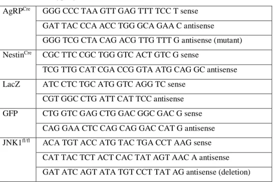
For genotypic analysis, polymerase chain reaction (PCR) was performed on tail DNA using the primers given in Table 1. For PCR DreamTaq PCR MasterMix and DNA polymerase (Thermo Scientific, Walldorf, Germany) was used. Standard PCR