Mechanisms of Cell-autonomous Resistance to Toxoplasma gondii in Mouse and Man
Inaugural-Dissertation
zur
Erlangung des Doktorgrades
der Mathematisch-Naturwissenschaftlichen Fakultät der Universität zu Köln
vorgelegt von Aliaksandr Khaminets aus Slonim, Weißrussland
Köln 2010
Berichterstatter: Prof. Dr. Jonathan C. Howard
Prof. Dr. Thomas Langer
Tag der mündlichen Prüfung: 31. 05. 2010
1.1 Infection and immunity ... 1
1.2 Interferons and their role in cell-autonomous immunity ... 2
1.3 Cellular responses to interferons ... 4
1.4 Immunity-related GTPases ... 7
1.4.1 Nomenclature and expression ... 7
1.4.2 Biochemical properties of IRG proteins ... 8
1.4.3 Cellular localisation of IRG proteins ... 9
1.4.4 Roles of IRG proteins in immunity ... 10
1.5 Toxoplasma gondii as a model pathogen to study human and mouse cell- autonomous response ... 12
1.6 Aims of the study ... 16
2 Material and Methods ... 18
2.1 Reagents and cells. ... 18
2.1.1 Chemicals, reagents and accessories ... 18
2.1.2 Equipment ... 18
2.1.3 Materials ... 18
2.1.4 Enzymes and proteins ... 19
2.1.5 Kits ... 19
2.1.6 Vectors and constructs used in the present study ... 19
2.1.7 Cell lines, bacterial and protozoan strains ... 19
2.1.8 Serological reagents ... 21
2.2 Molecular Biology ... 23
2.2.1 Agarose gel electrophoresis ... 23
2.2.2 Generation of the expression constructs ... 23
2.2.3 Cloning of PCR amplification products ... 25
2.2.4 Purification of DNA fragments from agarose gels ... 25
2.2.5 Ligation ... 25
2.2.6 Preparation of competent cells ... 26
2.2.7 Transformation of competent bacteria ... 26
2.2.10 Site-directed mutagenesis ... 27
2.2.11 DNA sequencing ... 27
2.3 Cell biology ... 28
2.3.1 Transfection ... 28
2.3.2 Immunocytochemistry ... 28
2.3.3 In vitro passage of T. gondii ... 28
2.3.4 Infection of cells with T. gondii for immunocytochemistry ... 29
2.3.5 T. gondii lysis ... 29
2.3.6 Live cell imaging ... 29
2.3.7 Inhibition of signalling pathways and microtubule polymerisation ... 30
2.3.8 Synchronisation of T. gondii infection ... 30
2.3.9 T. gondii proliferation assay ... 31
2.3.10 Quantification of IRG protein signal intensity at T. gondii PV ... 31
2.3.11 Western blotting ... 32
2.3.12 Colorimetric cell viability assay ... 32
2.3.13 Propidium iodide staining ... 33
2.3.14 Pulse-chase analysis ... 33
2.3.15 Immunoprecipitation ... 34
3 Results I ... 35
3.1 Time-course of endogenous IRG protein loading onto avirulent ME49 T. gondii vacuoles in mouse fibroblasts ... 35
3.2 Time-course of ectopically expressed IRG protein loading onto avirulent ME49 T. gondii vacuoles in mouse fibroblasts ... 37
3.3 Sequential loading of multiple IRG proteins on to the PV of avirulent ME49 T. gondii ... 38
3.4 Vacuolar loading with IRG proteins is independent of major signalling
systems and microtubules ... 41
3.6 Reduced loading of IRG proteins onto the PVM of virulent T. gondii strains ... 46 3.7 Coinfection of mouse fibroblasts with T. gondii of different virulence and virulence-associated T. gondii proteins ROP18, ROP16 and ROP5 do not affect loading of PVM with IRG proteins ... 48 3.8 IRG complex formation is required for efficient loading onto T. gondii PV ... 51 3.9 Vacuolar loading with IRG proteins is required for T. gondii elimination
... 53
4 Results II ... 59 4.1 IDO-dependent and IDO-independent mechanisms of resistance against T. gondii in human cells ... 59 4.2 Disruption of T. gondii vacuoles is not apparent in IFNγ-induced primary human fibroblasts ... 61 4.3 IFNγ stimulates death of distinct types of T. gondii-infected human cells
... 63 4.4 Human cells, stimulated with IFNγ and infected with T. gondii, undergo necrosis but not apoptosis ... 66 4.5 Addition of tryptophan inhibits necrosis of human fibroblasts during IDO-dependent but not during IDO-independent course of IFNγ-mediated control of T. gondii ... 70 4.6 Pharmacological inhibition of IDO rescues T. gondii growth in IFNγ- stimulated HeLa cells but not in primary human fibroblasts ... 72
5 Discussion ... 75 5.1 Heterogeneity of IRG protein loading onto vacuoles with avirulent T.
gondii ... 75
5.2 Regulation of IRG proteins by Atg5 in IFNγ-stimulated mouse
fibroblasts ... 77
5.4 Passive vs. active model of IRG protein association with avirulent T.
gondii PV ... 80
5.5 Reduced accumulation of IRG proteins on virulent T. gondii PVs is independent of ROP5, ROP16 and ROP18 virulence determinants of the parasite ... 82
5.6 Loading of IRG proteins onto T. gondii vacuoles is crucial for parasite elimination in IFNγ-induced mouse fibroblasts ... 85
5.7 Amount of cultured cells determines the mechanism of T. gondii growth inhibition in IFNγ-stimulated primary human fibroblasts ... 86
5.8 T. gondii infection stimulates necrosis of IFNγ-induced primary human cells ... 87
5.9 IDO-dependent and IDO-independent mechanisms of IFNγ-induced T. gondii growth restriction in human cells ... 89
6 Appendix ... 92
6.1 Figure 32. Association of wt, catalytic interface and secondary patch mutants of Irga6-cTag1 with ME49 T. gondii PV ... 92
6.2 Permeabilisation of avirulent T. gondii to cytoplasmic Cherry in IFNγ- stimulated infected MEFs ... 94
6.3 IFNγ induces death of T. gondii-infected human cells ... 95
6.4 Analysis of Hs27 cell viability by PI staining ... 96
7 References ... 97
8 Summary ... 124
9 Zusammenfassung ... 125
10 Acknowledgements ... 127
11 Erklärung ... 128
12 Teilpublikationen ... 129
1 Introduction
1.1 Infection and immunity
Organisms evolve by interacting with each other and their environment. Numerous intricate interplays between organisms proved to be the source and the result of adaptations shaping life on the planet. Forms of relations between different species, also called symbiosis (Greek syn stands for “with” and biosis stands for “living”), were classified into mutualism (both sides benefit from relationship), commensalism (one side benefits while the other is not positively or negatively affected) and parasitism (one side exploits and harms the other). Various pathogens use their hosts in order to survive and reproduce whereas hosts attempt to eliminate the unwelcome guests. This underlies the everlasting battle between the host and the parasite leading to the co-evolution of species (Roy and Mocarski, 2007).
Hosts possess an arsenal of mechanisms, called the immunity system, to defend themselves from pathogens. Host barriers, such as plasma membranes or outer cell layers, represent the most ancient and primitive means of protection against parasites.
Additionally, various molecules (e.g. complement, lysozyme, lactoferrin etc.) and processes (e.g. phagocytosis), capable of exerting antiparasitic action, account for a large portion of innate immune system. These immune mechanisms constitute a first line of defense against invasion which is deployed in a fast but unspecific manner (Janeway et al., 2008). In contrast to innate immunity, adaptive immunity (or acquired), found only in vertebrates (Medzhitov and Janeway, 1997), has a lag phase after initiation till the system is fully functional. Adaptive immunity is characterized by generation of high-specificity receptors to the antigens via somatic mechanisms of amplification and diversification, and by the phenomenon of immunological memory.
The latter allows recognizing and mounting the immune response more rapidly and efficiently after repeated exposure to the same infectious agent (Janeway et al., 2008).
Central to triggering of the immune programs is recognition of the parasite. This step is achieved by the pattern recognition receptors (PRRs), present on cellular membranes (e.g. Toll-like receptors, scavenger receptors) or in the cytoplasmic space (e.g. CARD helicases, NOD-like proteins) which recognise the pathogen-associated molecular patterns (PAMPs) (e.g. LPS, DNA, double-stranded RNA) (Medzhitov, 2001; Akira, Uematsu, and Takeuchi, 2006; Fritz et al., 2006; Lee and Kim, 2007).
Recognition of the parasite signals to the immune system to escalate the inflammatory
response, resulting in the recruitment of immune cells at the locus of invasion, secretion of cell-derived immune mediators and increased permeability of blood vessels. All these processes ensure containment and subsequent clearance of infection (Roy and Mocarski, 2007; Janeway et al., 2008).
Regulation of cell-to-cell communication, connection between innate and adaptive systems and the magnitude of the immune response are tightly regulated by cytokines.
1.2 Interferons and their role in cell-autonomous immunity
Cytokines (Greek cyto means “cell”, kinos means “movement”) are a group of peptides and proteins, secreted by both hematopoietic and none-hematopoietic cells, implicated in modulation of all steps of the immune response. Cytokines could be subdivided into chemokines, hematopoietins, tumor necrosis factor (TNF) family and interferons (Janeway et al., 2008). A prominent role in regulation of the immune system is orchestrated by interferons. Since the discovery of interferon as an antiviral drug (Isaacs and Lindenmann, 1957; Isaacs, Lindenmann, and Valentine, 1957), the cytokine was assigned a plethora of functions discussed hereafter. The family of the interferons include type I: IFNα (14-20, depending on species) (van Pesch et al., 2004), IFNβ (Mogensen et al., 1999), IFNω (Hauptmann and Swetly, 1985), IFNτ (Bazer, Spencer, and Ott, 1997), IFNε (Pestka, Krause, and Walter, 2004), IFNδ (Lefevre et al., 1998); type II: IFNγ (Bancroft, 1993) and type III: IFNλ (Kotenko et al., 2003). Type I interferons are secreted by virtually all types of cells, however the major producers of IFNα and IFNω are hematopoietic cells whereas fibroblasts are the main source of IFNβ (Bach, Aguet, and Schreiber, 1997). In addition to T-cells and NK cells, being the major contributors of IFNγ, NKT, B- and professional antigen presenting cells (APCs) have also been reported to secrete the cytokine (Schroder et al., 2004).
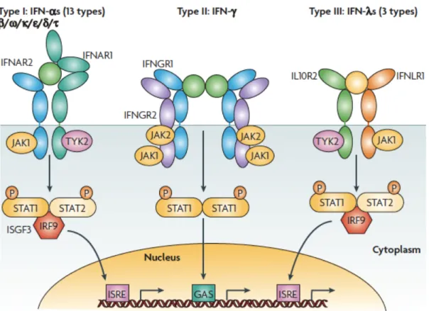
Interferons engage with specific receptors present on the cell surfaces (Figure 1)
leading to activation of the associated kinases JAK1, JAK2 and TYK2 which in turn
phosphorylate and thereby activate transcription factors STAT1 and STAT2. STAT1
and STAT2 translocate into the nucleus and bind to GAS elements in the promoter
regions driving expression of IFN-regulated genes. Some of those genes encode
transcription factors (IRF) mediating the next waves of the transcription events from
the promoter ISRE elements (Schroder et al., 2004; Borden et al., 2007).
It is now appreciated that interferons, acting in autocrine and paracrine fashion, induce multiple processes such as stimulation of intracellular and extracellular networks, regulation of resistance to infections, amplification of innate and acquired immune response, modulation of survival and death of normal and tumor cells (Schroder et al., 2004; Borden et al., 2007).
Figure 1 Receptor activation or ligand–receptor complex assembled by type I, type II or type III interferons.
Type I interferons (α, β ω, κ, ε, δ, τ) interact with IFN (α, β and ω) receptor 1 (IFNAR1) and IFNAR2; type II IFNγ with IFNγ receptor 1 (IFNGR1) and IFNGR2; and type III IFNλs with IFNλ receptor 1 (IFNLR1; also known as IL28RA) and interleukin 10 receptor 2 (IL10R2; also known as IL10RB). Type II IFNγ is an antiparallel homodimer exhibiting a two-fold axis of symmetry. It binds two IFNGR1 receptor chains, assembling a complex that is stabilized by two IFNGR2 chains. These receptors are associated with two kinases from the JAK family: JAK1 and TYK2 for type I and III IFNs; JAK1 and JAK2 for type II IFN. IFNAR2, IFNLR1, IL10R2, IFNGR1 and IFNGR2 are classical representatives of this family, while IFNAR1 is atypical as its extracellular domain is duplicated. GAS, IFNγ-activated site; IRF9, IFN regulatory factor 9; ISGF3, IFN-stimulated gene factor 3, refers to the STAT1–STAT2–IRF9 complex; ISRE, IFN-stimulated response element; P, phosphate; STAT1/2, signal transducers and activators of transcription ½ (Borden et al., 2007).
Interferons regulate cell-autonomous response to pathogens. Cell-autonomous
immunity designates the ability of a cell to cope with infection within the boundaries
of the cell and without involvement of the ambient cellular environment. Because
interferons are derived from outside of the cells to engage with the specific plasma
membrane receptors in order to arm the cells with mechanisms of defense against
invasion, this type of immunity could not be referred to as entirely autonomous.
1.3 Cellular responses to interferons
Interferons increase cellular concentration of proteins, many of which have proved to be resistance factors and have been implicated in innate and adaptive components of the immune system. The examples of immunity-related proteins regulated by interferons are numerous and their list is constantly growing (Boehm et al., 1997).
Expression of MHC class I and class II molecules and other components of the antigen presentation system of the antigen-presenting cells (APCs) is increased upon induction with interferons. Furthermore, these cytokines are involved in generation of reactive oxygen and nitrogen species by enhancing intracellular activities of the corresponding enzymes (e.g. gp91-phox and nitric oxide synthase (iNOS)). In macrophages, natural resistance-associated macrophage protein 1 (NRAMP1), a mediator of the natural resistance against intracellular pathogens, is constitutively expressed and, additionally, could be strongly upregulated by IFNγ and LPS. Double- stranded RNA activated protein kinase (PKR), oligoadenylate synthetase (OAS) and specific adenosine deaminase (dsRAD), enzymes upregulated by interferons, exert a characteristic direct antiviral effects at the cell-autonomous level (Boehm et al., 1997;
Schroder et al., 2004; Borden et al., 2007). Depletion of the cellular sources of tryptophan, mediated by the enzymes indoleamin 2,3-dioxygenase (IDO) and tryptophan 2,3-dioxygenase (TDO), has been reported to execute the antimicrobial and immunomodulatory function of the interferons (Pfefferkorn, 1984; Boehm et al., 1997; Munn et al., 1999; Schmidt et al., 2009).
Mx proteins, induced strictly by type I interferons, are potent antiviral factors, which are believed to directly bind to the virion components (e.g. influenza, Thogoto virus) and interfere with the viral trafficking, assembly or replication (Lindenmann, Lane, and Hobson, 1963; Lindenmann et al., 1978; Kochs et al., 1998; Kochs and Haller, 1999a; Kochs and Haller, 1999b; Haller and Kochs, 2002). Mx proteins are 70-80 kDa homologous to dynamin GTPases (present in all vertebrates, Mx1 and Mx2 in mice, MxA and MxB in humans), displaying some dynamin-like properties, such as GTP-dependent oligomerisation and micromolar affinities for GTP and GDP (Richter et al., 1995; Hinshaw, 2000; Kochs et al., 2002; Song and Schmid, 2003).
The family of guanylate-binding proteins (GBPs) is represented by 65-67 kDa
interferon inducible GTPases (present in all vertebrates, 10 members in mice, 7 in humans) (Degrandi et al., 2007; Olszewski, Gray, and Vestal, 2006; Robertsen et al., 2006). Resembling dynamins, GBP proteins bind nucleotides with micromolar affinity and demonstrate di- or tetramerisation and cooperative hydrolysis of GTP (Praefcke and McMahon, 2004). In contrast to Mx proteins, GBPs are strongly induced by IFNγ whereas type I interferons, TNFα and IL-1β have only a slight effect on the protein expression (Nantais et al., 1996; Boehm et al., 1998; Guenzi et al., 2001; Guenzi et al., 2003). GBPs have been implicated in resistance against vesicular stomatitis (VSV), Encephalomyocarditis (ECMV) and Hepatitis C viruses (Anderson et al., 1999; Carter, Gorbacheva, and Vestal, 2005; Itsui et al., 2009) as well as in regulation of vasculorgenesis and cell proliferation (Guenzi et al., 2001; Gorbacheva et al., 2002; Guenzi et al., 2003). Interestingly, recent localisation and overexpression studies of GBP proteins suggested their role in resistance against T. gondii (Degrandi et al., 2007) and Chlamydia trachomatis (Tietzel, El-Haibi, and Carabeo, 2009).
Very large inducible GTPases (VLIGs) are 280 kDa proteins induced in mouse cells by IFNγ and to some extent by IFNβ. The G domain of VLIGs displays some homology to Mx proteins and GBPs, but the functions of the VLIGs in resistance are still to be clarified (Klamp et al., 2003).
Autophagy (macroautophagy) is a conserved cellular degradation system, specialized in removal of damaged organelles, clearance of aggregated proteins or turnover of any part of the cell cytoplasm (Mizushima, 2002; Xie and Klionsky, 2007;
Mizushima et al., 2008). In addition to starvation, microorganisms, protein aggregates and numerous drugs, IFNγ has been reported to induce autophagy (Gutierrez et al., 2004; Deretic, 2005; Deretic, 2006). Atg1-27 proteins (defined in yeasts, but most of the proteins have homologs in mammals) orchestrate the process of the phagophore formation around the target, its conversion into auphagosome and fusion with the lysosomal compartment to give rise to autolysosomes (Figure 2) (Deretic, 2005).
Interestingly, an Atg5/Atg7-independent pathway of autophagy has been recently reported (Nishida et al., 2009). Autophagy has been implicated in direct elimination of intracellular pathogens such as Mycobacterium tuberculosis, Listeria monocytogenes, Shigella flexneri etc. (Deretic, 2005), however certain pathogens (e.g.
Francisella tularensis, Staphylococcus aureus) exploit the process of autophagy in
order to survive and replicate (Checroun et al., 2006; Schnaith et al., 2007).
Autophagy may lead to death of the cell, designated as programmed cell death type II (Golstein and Kroemer, 2007), characterized by massive accumulations of autophagic vesicles in the cytosol (Levine, 2005; Pyo et al., 2005; Feng et al., 2008b; Kroemer and Levine, 2008). Apoptosis (programmed cell death type I) carries features of chromatic condensation and fragmentation, shrinkage and blebbing of the plasma membrane and formation of apoptotic bodies (Golstein and Kroemer, 2007).
Interferons have been shown to induce expression of death-associated proteins (e.g.
TNFα-related apoptosis inducing ligand (TRAIL/Apo2L), caspase-4 and caspase-8) and in certain models stimulate apoptotic cell death (Boehm et al., 1997; Chin et al., 1997; Bernabei et al., 2001; Inagaki et al., 2002; Chawla-Sarkar et al., 2003).
Necrosis (or programmed cell death type III) is defined by rupture and dilatation of cytoplasmic organelles and the plasma membrane (Golstein and Kroemer, 2007).
Necrotic cell death has been recently reported to occur upon infection of IFNγ- stimulated mouse cells with avirulent T. gondii (Zhao et al., 2009b).
Figure 2 Schematic depiction of autophagy.
(A and B) Cytosolic material is sequestered by an expanding membrane sac, the phagophore (C)
resulting in the formation of a double-membrane vesicle, an autophagosome; (D) the outer membrane of the autophagosome subsequently fuses with a lysosome, exposing the inner single membrane of the autophagosome to lysosomal hydrolases; (E) the cargo-containing membrane compartment is then lysed, and the contents are degraded (Xie and Klionsky, 2007).
1.4 Immunity-related GTPases 1.4.1 Nomenclature and expression
The families of GBP and IRG proteins (immunity-related or p47 GTPases) have been found to dominate during cellular response to IFNγ (Boehm et al., 1998). Within the last decade IRG genes and their products have been extensively studied leading to the accumulation of a great amount of information (Taylor, 2004; MacMicking, 2005;
Martens and Howard, 2006; Taylor, 2007). The homologs of the IRG genes have been identified in various species including amphioxus, fish, dog, mouse, rat and man (Bekpen et al., 2005; Li et al., 2009). In C57BL/6 mice the IRG family is represented by 21 genes located on chromosomes 7, 11, 18 and is divided on phylogenetic principles into Irga, Irgb, Irgc, Irgd and Irgm subgroups (Bekpen et al., 2005). The best-characterized members of the family are Irgb6 (TGTP/Mg21), Irga6 (IIGP1), Irgd (IRG-47), Irgm1 (LRG-47), Irgm2 (GTPI), Irgm3 (IGTP) and Irgb10. Irgm1, Irgm2 and Irgm3 have a GX
4GMS sequence within the canonical G1 motif of the GTP-binding domain and are therefore designated as GMS proteins whereas the rest of the family harbors GX
4GKS and is called GKS proteins (Bekpen et al., 2005;
Boehm et al., 1998). The GMS subgroup of IRG proteins stands out from the rest of the family not only structurally but also functionally, since they have been implicated in regulation of GKS proteins in IFNγ-induced mouse cells (Hunn et al., 2008; Papic et al., 2008).
Due to the presence of GAS and ISRE sites in promoters of the IRG genes (with exception of Irgc), their expression is strongly upregulated by IFNγ and to some extent by IFNα/β and LPS in a STAT1-dependent manner (Taylor et al., 1996;
Boehm et al., 1998; Collazo et al., 2002; Zerrahn et al., 2002; MacMicking, Taylor, and McKinney, 2003; Bekpen et al., 2005; Lapaque et al., 2006). Other cytokines elevate the protein levels relatively inefficiently (Lafuse et al., 1995; Boehm et al., 1998). Additionally, Irga6 is constitutively expressed in mouse liver, driven by a tissue-specific promoter (Zeng, Parvanova, and Howard, 2009).
IRGC and IRGM, found on chromosomes 5 and 19, respectively, are the only
transcribed IRG genes present in humans (Bekpen et al., 2005). Neither of the human
IRG sequences is regulated by inferferons. Irgc is highly conserved in all mammals and expressed specifically in haploid spermatids during spermatogenesis in testis (Bekpen et al., 2005; Rohde, 2007). The G domain of human IRGM is truncated, and expression of IRGM is driven by an ERV9 repetitive element. Thus in contrast to mice, humans possess no functional IFNγ-inducible IRG system indicating the marked disparities in immune mechanisms between these species (Bekpen et al., 2005; Coers, Starnbach, and Howard, 2009).
1.4.2 Biochemical properties of IRG proteins
Cells are equipped with GTPases involved in various physiological processes like membrane trafficking (e.g. dynamin), cell signaling (e.g. Ras, G-proteins), and translation during protein synthesis (e.g. EF-Tu) (Leipe et al., 2002). These proteins have a common GTP-binding fold. Conformational changes in two flexible switch regions signal the rest of the protein to render the GTPases active or inactive when bound to GTP or GDP, respectively (Vetter and Wittinghofer, 2001). Guanine- nucleotide exchange factors (GEF) activate the GTPases by catalyzing the release of GDP from the G domain while GTPase activating proteins (GAPs) accelerate GTP hydrolysis and thereby inactivate the proteins. Guanine-nucleotide dissociation inhibitors (GDI) bind the GDP-form of the enzymes and keep them in an inactive state (Vetter and Wittinghofer, 2001; Martens and Howard, 2006).
Irga6 is the only member of the IRG GTPase family, that has been crystallized so far, and its structure currently serves as a model for the other IRG proteins (Figure 3) (Ghosh et al., 2004). The structure shows a classical globular Ras-like G domain, inserted between N- and C-terminal helical regions. The GTP-binding domain includes universally conserved G1 (GX
4GKS), G3 (DXXG) and G4 (N(T/Q)KXD) motifs (Ghosh et al., 2004; Martens and Howard, 2006). Bacterially expressed and purified Irga6 showed micromolar affinities for the nucleotides with affinity for GDP about 10 times higher than for GTP. Additionally, recombinant Irga6 shows GTP- dependent oligomerisation and cooperativity during GTP hydrolysis (Uthaiah et al., 2003).
Mutational analysis of the Irga6 molecule revealed the pattern of surface residues
forming the putative interface (catalytic interface) essential for oligomerisation and
GTP hydrolysis in vitro (Pawlowski et al., in preparation). The ability to hydrolyse
GTP has also been shown for Irgb6, Irgd, Irgm1 and Irgm3 proteins (Taylor et al., 1996; Carlow, Teh, and Teh, 1998; Pawlowski, 2009; Tiwari et al., 2009).
Oligomerisation of Irga6 in vitro accelerates GTP hydrolysis, and a catalytic mechanism based on activation in trans between two G domains has been proposed.
GKS proteins are subjected to the regulation by GMS proteins in vivo preventing the former from spontaneous activation and aggregation on intracellular membranes (Hunn et al., 2008; Papic et al., 2008). Thus, the GMS subgroup of the IRG proteins serves as GDIs, keeping the GKS proteins in inactive GDP-bound state.
Figure 3 3D structure of Irga6.
Ribbon presentation of one Irga6 molecule of the Irga6-GDP dimer is shown with the G domain (S1- H5) colored in light-blue and the N- and C-terminal helical regions colored in cyan (aA-aC) and dark- blue (aF-aL). The linker helix aE connecting the G domain and C-terminal helical region is shown in gray. GDP and Mg2+ are shown as atomic stick figure and yellow sphere. The topology is shown schematically using the same color code (Ghosh et al., 2004).
1.4.3 Cellular localisation of IRG proteins
In IFNγ-induced mouse cells IRG proteins distribute among distinct cellular
compartments, Irgm1 to the Golgi (Martens et al., 2004) and the endolysosomal
compartments (Zhao et al., 2010), Irgm2 to the Golgi (Hunn et al., 2008; Martens and
Howard, 2006), Irgm3 (Taylor et al., 1997) and Irga6 (Martens et al., 2004; Zerrahn et
al., 2002) to the endoplasmic reticulum. Irga6 and Irgb6 (Boehm et al., 1998; Martens
et al., 2004; Martens and Howard, 2006) as well as Irgb10 have large cytosolic pools
of proteins whereas Irgm proteins are predominantly membrane bound ((Martens and Howard, 2006) and unpublished data).
After infection of IFNγ-induced cells with avirulent T. gondii 6 IRG proteins (Irgb6, Irga6, Irgd, Irgm2, Irgm3 and Irgb10) have been found in high density on the parasitophorous vacuole membrane (PVM) (Martens et al., 2005; Khaminets et al., 2010). Irgm1 has never been reported on the PVM, but accumulates on phagocytic cups and phagosomes in cells phagocytosing M. tuberculosis or latex beads (Butcher et al., 2005a; Martens et al., 2005; Shenoy et al., 2007). Additionally, Irgm1 has been reported to colocalise with autophagosome markers monodasylcadaverine (MDC) and microtubule-associated protein 1 light chain 3 (LC3) suggesting a role in autophagy (Gutierrez et al., 2004; Singh et al., 2006). Furthermore Irgb10 is targeted to the inclusions of Chlamydia trachomatis but not of Chlamydia muridarum in infected mouse fibroblasts (Coers et al., 2008).
To date it is not explicitly clear what mediates targeting of most of IRG proteins to the subcellular compartments. It has been reported that Irgm1 localises to Golgi via amphipathic helix αK (Martens et al., 2004; Tiwari et al., 2009) and myristoylation of Irga6 is partially dispensable for targeting to the ER but is required for efficient loading onto the T. gondii PV ((Martens et al., 2004; Papic et al., 2008) and unpublished data).
1.4.4 Roles of IRG proteins in immunity
IRG proteins have been implicated in resistance to various pathogens (Table 1)
(Martens and Howard, 2006; Taylor, 2007). Mice, deficient in the following members
of the family, were susceptible to distinct bacterial and protozoan pathogens: Irgm1 to
T. gondii (Collazo et al., 2001), Leishmania major (Taylor, 2004), Trypanosoma cruzi
(Santiago et al., 2005), Listeria monocytogenes (Collazo et al., 2001), Mycobacterium
tuberculosis (MacMicking, Taylor, and McKinney, 2003), Mycobacterium avium
(Feng et al., 2004), Salmonella typhimurium (Henry et al., 2007), Chlamydia
trachomatis (Coers et al., 2008); Irgm3 to T. gondii (Taylor et al., 2000), Leishmania
major (Taylor, 2004), Chlamydia trachomatis (Coers et al., 2008); Irgd to T. gondii
(Collazo et al., 2001), Irgb10 to Chlamydia trachomatis (Bernstein-Hanley et al.,
2006). In many cases loss of resistance to the parasites in vivo correlated with
inability to control infection in cell culture. The role in cell-autonomous immunity
against the pathogens was demonstrated for Irgm1 (MacMicking, Taylor, and McKinney, 2003; Butcher et al., 2005a; Santiago et al., 2005; Coers et al., 2008), Irgm2 (Miyairi et al., 2007), Irgm3 (Butcher et al., 2005a; Ling et al., 2006; Coers et al., 2008), Irga6 (Martens et al., 2005; Nelson et al., 2005), Irgb6 (Zhao et al., 2009b) and Irgd (Koga et al., 2006).
Table 1 Summary of the evidence supporting the role of IRG proteins in host resistance (Taylor, 2007).
Several models of the mechanism of IRG protein function in elimination of intracellular parasites have been proposed (Martens and Howard, 2006; Taylor, 2007): (1) disruption of the pathogen containing vacuoles, in a manner similar to the vesiculating action of dynamins, which leads to death of the enclosed parasite (Martens et al., 2005; Ling et al., 2006; Melzer et al., 2008; Zhao et al., 2009b); (2) initiation of autophagy, which either directly kills or disposes of the already dead parasite following disruption of the parasite-containing vacuole (Ling et al., 2006); (3) maturation of the pathogen-containing vacuoles via promotion of acidification (MacMicking, Taylor, and McKinney, 2003; Shenoy et al., 2007); (4) direct stimulation of autophagy of the pathogenic vacuoles followed by fusion with the lysosomal compartment for degradation (Gutierrez et al., 2004; Deretic, 2005).
Additionally, IRG proteins were proposed to regulate heamatopoiesis (Feng et al.,
2004; Feng et al., 2008a), macrophage motility (Henry et al., 2007; Henry et al.,
2010) and antigen presentation (Bougneres et al., 2009), however, it is currently
unclear how these processes are affected by the absence of single IRG GTPases and
therefore these hypotheses require deeper analysis (Hunn and Howard, 2010). Recent
genome-wide association studies in humans have linked sequence polymorphism of
the human IRGM locus with increased risk for Crohn’s disease (Parkes et al., 2007;
Fisher et al., 2008). Overexpression of Irgb6 and Irgm2 in various cellular systems has suggested anti-viral function of the proteins in combating VSV virus (Carlow, Teh, and Teh, 1998) and coxsackie virus (Zhang et al., 2003), respectively.
1.5 Toxoplasma gondii as a model pathogen to study human and mouse cell- autonomous response
T. gondii is an obligate protozoan parasite assigned to the phylum Apicomplexa along with the causative agents of malaria (Dubey, 1977). Infections in humans are typically asymptomatic but may be lethal for immunocompromised individuals (Frenkel, 1988; Luft and Remington, 1988) and can lead to severe congenital defects during pregnancy (Wilson et al., 1980). In organisms with a healthy immune system T. gondii avoids complete elimination by entering the dormant state (cysts) in tissues like brain and muscles where it waits to be reactivated. The parasite has a complex life cycle (Figure 4).
Figure 4 Life cycle of Toxoplasma gondii, depicting stages and modes of transmission of the parasite.
Members of the cat family serve as definitive hosts, and sexual development only occurs within the small intestine, where micro- and macrogametes form within enterocytes following a round of mitotic replication (A-E stages). Fusion of the gametes yields a diploid zygote that is shed as a resistant spore (oocyst) in the feces. Meiosis occurs within the environment, yielding eight haploid progeny.
Transmission (image a shows an unsporulated oocyst) occurs when oocysts contaminate food or water.
Ingestion by a variety of warm-blooded hosts leads to acute infection, typified by fast-growing tachyzoites (image b shows a single vacuole with eight haploid parasites). Development of tissue cysts results in long-lasting chronic infection (image c shows a tissue cyst from mouse brain), which can also be transmitted by carnivorous feeding or scavenging (Sibley and Ajioka, 2008).
Acute toxoplasmosis is marked by rapid replication of tachyzoites in any infected non-feline vertebrate host. As mentioned above, the immune system attempts to eliminate infection, and this causes the pathogen to convert into the persistent state, called bradyzoite. Sexual stage of the cycle occurs in the members of Felidae (definitive host). Ingested by cats, bradyzoites infect intestinal epithelial cells and proceed through the stages of gametogenesis, gamete fusion and miotic division to give rise to oocysts, each containing 8 sporozoites. These are distributed in the environment in the faeces. When eaten by any vertebrate, oocysts or bradyzoites turn into tachyzoites to multiply and disseminate throughout the host (Pfefferkorn, 1990).
T. gondii is able to infect virtually all nucleated cells. The process of infection involves attachment of the parasite onto the cell surface followed by active penetration driven by a motility system present in the pathogen (Figure 5) (Joiner et al., 1990; Joiner and Dubremetz, 1993; Dobrowolski and Sibley, 1997).
Invading parasites become covered and eventually enclosed by the host plasma membrane, which forms the parasitophorous vacuole membrane (PVM) (Suss-Toby, Zimmerberg, and Ward, 1996). Secretory organelles, micronemes, rhopries and dense granules are sequentially secreted at consecutive steps during invasion to ensure successful establishment of the intracellular niche (Dubremetz et al., 1993;
Dubremetz and Schwartzman, 1993; Carruthers and Sibley, 1997). The PVM is largely devoid of host cell proteins which are actively excluded by the parasite to render the organelle nonfusogenic with the lysosomal compartment of the cell (Sibley, Weidner, and Krahenbuhl, 1985; Joiner et al., 1990; Dubremetz and Schwartzman, 1993; Mordue et al., 1999). Furthermore, the PVM mediates interaction with endoplasmic reticulum and mitochondria (Sinai, Webster, and Joiner, 1997). Thus, survival and multiplication of T. gondii strictly depends on the PV, isolating the parasite from the cytosolic environment and mediating nutrient acquisition (Moulder, 1985; Pfefferkorn, 1990; Schwab, Beckers, and Joiner, 1994).
The outcome of infection in mice, the major intermediate host, is dependent on
virulence of the infecting T. gondii. Phylogenetic analysis of the North American and
European T. gondii isolates initially grouped the parasites into 3 clonal lineages: type
I, type II and type III (Howe and Sibley, 1995; Su et al., 2003; Sibley and Ajioka, 2008). However, the genetic diversity of the parasite populations proved to be much greater, in particular after isolation of the South American T. gondii strains (Lehmann et al., 2006; Khan et al., 2007; Sibley and Ajioka, 2008). Genetically different strains displayed distinct virulence phenotypes, primarily measured by the amount of tachyzoites required to kill laboratory mice. Type I T. gondii strains are acutely virulent with 100% lethality following infection with a single parasite (LD
1001) whereas type II and type III strains have been designated as avirulent (LD
5010
4-10
5) (Sibley and Boothroyd, 1992; Saeij, Boyle, and Boothroyd, 2005). Several rhoptry proteins, ROP5, ROP16 and ROP18, secreted upon invasion into the host cell, have been found to be major virulence determinants of T. gondii. The precise functions and mechanisms of action of these virulence factors are still under debate (El Hajj et al., 2006; Taylor et al., 2006; Saeij et al., 2006; El Hajj et al., 2007; Saeij et al., 2007).
Figure 5 Schematic representation of host cell infection with T. gondii.
Attachment of the parasite to the cell surface and secretion of the microneme content (proteins essential for invasion, e.g. TgAMA1 (green)) form the moving junction (MJ) that begins at the anterior end of the parasite and then migrates posteriorly. Rhoptries are then discharged to deliver other essential components of the invasion system into the host cell (RON and ROP proteins). The resulting invagination of host plasma membrane creates a parasitophorous vacuole membrane (PVM) that completely envelops intracellular parasite (Alexander et al., 2005).
IFNγ is indispensable for survival of mice during infection with T. gondii
(Remington and Merigan, 1968; Suzuki et al., 1988; Scharton-Kersten et al., 1996).
Recognition of T. gondii-associated molecular patterns triggers IL-12 production by dendritic cells, neutrophils and macrophages (Johnson and Sayles, 1997; Scanga et al., 2002; Aliberti et al., 2003; Sher et al., 2003; Yarovinsky and Sher, 2006). IL-12, in turn, stimulates IFNγ secretion by T- and NK cells to activate an immune response to the parasite (Gazzinelli et al., 1994a; Gazzinelli et al., 1994b; Hunter et al., 1995;
Denkers, 2003). In a STAT1-dependent manner IFNγ regulates cell-autonomous immunity to T. gondii by upregulating cellular concentrations of resistance proteins (Boehm et al., 1998; Collazo et al., 2002). To date, in mouse system, several IRG proteins (Irgm1, Irgm3, Irga6, Irgb6 and Irgd) have been implicated in resistance to T.
gondii, analysed in vivo and in cell culture (Martens and Howard, 2006; Taylor, 2007). In IFNγ-stimulated mouse cells, IRG proteins bind to the PV and actively participate in its disruption, presumably by vesiculation of the vacuolar membrane.
This leads to demise of the parasite and necrotic death of the infected cell (Martens et al., 2005; Melzer et al., 2008; Zhao et al., 2009b). It was also proposed that, in activated macrophages, disruption of the T. gondii PVM mediates recognition of the parasite by the autophagic machinery resulting in killing or degrading of already dead pathogens in infected cells (Ling et al., 2006). In another report a toxoplasmacidal effect was achieved independently of IFNγ by autophagy-mediated fusion of PVs with lysosomes in CD40-induced macrophages (Andrade et al., 2006; Subauste and Wessendarp, 2006). However, this paradigme has later been confronted by the group of Yap in the report that emphasized the primary role of IFNγ in resistance to T.
gondii (Zhao et al., 2007).
Association of IRG proteins with T. gondii PVs appears to be crucial in IFNγ- mediated parasite elimination but till now the process of protein loading onto the parasite-containing vacuoles has not been subjected to comprehensive analysis.
In contrast to mouse, in human cells resistance to the parasite has been considered essentially dependent on tryptophan depletion by IDO since growth of T. gondii could be completely restored by addition of excess tryptophan to the cells or by pharmacological IDO inhibition prior to infection (Pfefferkorn, 1984; Habara-Ohkubo et al., 1993; Daubener et al., 2001; Bekpen et al., 2005; Könen-Waisman and Howard, 2007; Heseler et al., 2008). However, in human endothelial cells and macrophages T.
gondii growth restriction has been shown to be mediated independently of IDO via
unclarified mechanisms (Woodman, Dimier, and Bout, 1991; MacKenzie et al., 1999). It was also reported that human leukocytes could spontaneously eliminate T.
gondii by phagocytosis of the viable parasites (Wilson and Remington, 1979).
Oxygen and nitrogen species (Murray and Cohn, 1979; Adams et al., 1990; Aline, Bout, and Dimier-Poisson, 2002) and iron depletion (Dimier and Bout, 1998) are the other mechanisms that could contribute to resistance to T. gondii.
T. gondii interferes with the immune response at nearly every step of development of resistance to infection (Lang et al., 2006; Plattner and Soldati-Favre, 2008).
Infection by the parasite is associated with dysregulation and decreased production of the major proinflammatory cytokines IL-12 and IFNγ (Butcher et al., 2001; Sher et al., 2003; Butcher et al., 2005b; Lee et al., 2008); T. gondii also triggers generation of anti-inflammatory cytokines, such as IL-10, TGF-β to antagonize the IL- 12/IFNγ−mediated Th1 response (Bermudez, Covaro, and Remington, 1993; Khan et al., 1997; Langermans et al., 2001; Yamamoto et al., 2009). It has been reported that infection with the parasite blocks translocation of STAT1 into the nucleus (Luder et al., 2001), modifies NFκB (Gazzinelli et al., 1996; Butcher and Denkers, 2002;
Denkers, 2003; Molestina and Sinai, 2005a; Molestina and Sinai, 2005b; Shapira et al., 2005) and PI3 kinase signaling (Kim and Denkers, 2006), protects the cells from apoptosis (Goebel, Gross, and Luder, 2001; Keller et al., 2006; Carmen and Sinai, 2007), inhibits production of reactive nitrogen and oxygen species (Luder et al., 2003;
Ding et al., 2004; Denkers and Butcher, 2005) and impede antigen presentation (Luder et al., 1998; McKee et al., 2004). Finally, it was suggested that virulent T.
gondii strains were able to attenuate the immune response by interfering with the process of IRG protein loading onto the PV (Hunn, 2007; Zhao et al., 2008; Zhao et al., 2009a; Zhao et al., 2009c; Khaminets et al., 2010).
1.6 Aims of the study
Experiments in mice and cell culture demonstrated that IRG proteins are the key factors mediating IFNγ-regulated resistance to T. gondii (Martens and Howard, 2006;
Taylor, 2007). At the single-cell level IRG proteins accumulate on the parasite- containing vacuoles and participate in disruption of the PVM leading to elimination of the pathogen and necrotic death of the cell (Martens et al., 2005; Ling et al., 2006;
Melzer et al., 2008; Zhao et al., 2008; Zhao et al., 2009b). Therefore, association of
IRG GTPases with T. gondii PVs appears to be central in resistance to the parasite in cells and determines the outcome of the infection in vivo. Despite the fundamental importance of the process to date there has been a gap of knowledge in understanding various aspects of the mechanism of vacuolar loading.
In the first part of this work I attempt to document the dynamics of the process of multiple IRG protein loading onto T. gondii PVs; to understand if the process of GTPase association with the vacuoles is carried out in passive or active fashion; to analyse loading of IRG proteins onto the vacuoles of T. gondii strains of different virulence and to study the involvement of host cell autophagic regulator Atg5 and major T. gondii virulence determinants ROP5, ROP16 and ROP18 in the process of loading. In addition, using various cellular models displaying deficiency in the process of loading I set out to provide the evidence linking IRG protein association with T. gondii PVs to elimination of the parasite in infected IFNγ-stimulated cells.
Tryptophan depletion by IDO has been reported to play a pivotal role in restricting T. gondii growth in IFNγ-stimulated human cells (Pfefferkorn, 1984; Daubener et al., 2001). However, based on published data (Woodman, Dimier, and Bout, 1991;
MacKenzie et al., 1999) and knowledge of the colossal variability of IFNγ-mediated programs (Boehm et al., 1997; Schroder et al., 2004; Borden et al., 2007), alternative mechanisms of resistance to T. gondii are conceivable, providing the impetus for further research.
In the second part of this study the possible existence of an IDO-independent immunity mechanism to T. gondii functioning in human cells is tested. Furthermore, to get an insight into the IDO-independent mechanisms in human cells I envisage and test a two-step scenario documented for the mouse system (Zhao et al., 2009b):
disruption of the parasite-containing vacuole followed by the death of infected cell.
2 Material and Methods 2.1 Reagents and cells
2.1.1 Chemicals, reagents and accessories
All chemicals were purchased from Aldrich (Steinheim), Amersham-Pharmacia (Freiburg), Applichem (Darmstadt), Baker (Deventer, Netherlands), Boehringer Mannheim (Mannheim), Fluka (Neu-Ulm), GERBU (Gaiberg), Merck (Darmstadt), Pharma-Waldhof (Düsseldorf), Qiagen (Hilden), Riedel de Haen (Seelze), Roth (Karlsruhe), Serva (Heidelberg), Sigma-Aldrich (Deisenhofen) or ICN biochemicals, Oxoid, (Hampshire UK). Developing and fixing solutions for Western Blot detection were from Amersham Pharmacia (Freiburg), Luminol from Sigma Aldrich (Deisenhofen), Coumaric acid from Fluka (Neu-Ulm). Deionised and sterile water (Seral TM) was used for all the buffers and solutions, ultra pure water derived from Beta 75/delta UV/UF from USF Seral Reinstwassersysteme GmbH, (Baumbach) equipped with UV (185/254 nm) and ultrafiltration (5x10
3kDa cut off), or from Milli- QSynthesis (Millipore).
2.1.2 Equipment
Centrifuges used were: Biofuge 13, Heraeus; Sigma 204; Sigma 3K10; Labofuge 400R, Heraeus; Sorvall RC-5B, Du Pont instruments; Optima TLX Ultracentrifuge, Beckmann and Avanti J-20 XP, Beckman. BioRAD Gel dryer, Model-583; BioRad Power pack 300 or 3000; electrophoresis chambers from FMC Bioproducts (Rockland Maine US); Gel Electrophoresis Chamber, Cambridge electrophoresis; Biorad Mini Protean II; PTC-100, MJ Research Inc.; ÄKTA P-920, OPC-900, Frac-950, Amersham; Centrifuge tubes 15ml, TPP Switzerland; 50ml Falcon, BectonDickenson;
Zeiss Axioplan II microscope equipped with AxioCam MRm camera (Zeiss); Zeiss Axiovert 200M motorized microscope equipped with AxioCam MRm camera (Zeiss).
ELISA reader (Vmax, Molecular Devices), Harvester Filtermate 196 (Packard), Direct Beta Counter Matrix 9600 (Packard), Typhoon TRIO Variable Mode Imager (GE Healthcare).
2.1.3 Materials
Sterile filters FP 030/3 0.2 μm and ME 24 0.2 μm (Schleicher und Schüll, Dassel);
Nitrocellulose transfer membrane PROTRAN (Schleicher und Schu ü ll, Dassel); 3MM
Whatmann Paper (purchased via LaboMedic); 100 Sterican 0.50 x 16 mm hypodermic needles (Braun AG, Melsungen); 0.2 μm and 0.45 μm sterile filters (Schleicher und Schuell, Dassel); X-OMAT LS and AR X-ray films, Kodak. All plastic ware for cell culture was from Sarstedt (Nu ü mbrecht) or Greiner (Solingen), Glass Fiber Filters (Packard 6005412, Groningen, Netherlands), phosphorimaging plates (BAS-1000, FUJIFILM Europe GmbH), staurosporine (STO, Sigma).
2.1.4 Enzymes and proteins
Restriction enzymes (New England Biolabs); Pyrococcus furiosus (Pfu) DNA Polymerase (Promega, Mannheim); T4 DNA ligase (New England Biolabs); RNase A (Sigma); shrimp alkaline phosphatase (SAP) (USB, Amersham); PageRulerTM Prestained Protein Ladder (Fermentas); PageRulerTM Protein Ladder (Fermentas);
SigmaMarkerTM Wide Range (Sigma); GeneRulerTM DNA Ladder Mix (Fermentas).
2.1.5 Kits
Plasmid Maxi and Midi kit (Qiagen, Hilden), Terminator-cycle Sequencing kit version 3 (ABI), QuikChange TM Site directed mutagenesis kit (Stratagen),
Rapid PCR product purification Kit (Roche, Mannheim).
2.1.6 Vectors and constructs used in the present study
pGW1H (British Biotech), pEGFP-N3 (Clontech), pmDsRed-N3, pCherry-N3 (both from (Zhao et al., 2009b)), pGW1H-Irga6-cTag1 (Papic et al., 2008), pGW1H- Irgb6-FLAG, pGW1H-Irgb6-K69A-FLAG and pGW1H-Irgd-ctag1 (all from (Hunn et al., 2008)).
2.1.7 Cell lines, bacterial and protozoan strains
Mouse embryonic fibroblasts (MEFs) derived from C57BL/6 mice, L929 (ATCC
CCL-1), Atg5
-/-and the corresponding wt control immortalised mouse fibroblasts
((Kuma et al., 2004), kindly provided by Martin Krönke and Noboru Mizushima),
MyD88
-/-MEFs ((Adachi et al., 1998), kindly provided by Manolis Pasparakis),
human cervix carcinoma HeLa (ATCC CCL-2, kindly provided by Gerrit Praefcke),
human embryonic kidney cell line HEK 293T (ATCC CRL-11268), human breast
adenocarcinoma MCF-7 (ATCC HTB-22, kindly provided by Hamid Kashkar) and human keratinocyte cell line HaCaT ((Boukamp et al., 1988), kindly provided by Dagmar Knebel-Mörsdorf) were cultured in DMEM (Gibco BRL and PAA) supplemented with 10% FCS (Biochrom AG, Berlin), 2 mM L-Glutamine, 1 mM sodium pyruvate, 1x non-essential amino acids, 100 U/ml penicillin and 100 μg/ml streptomycin (all from Gibco BRL). Human foreskin fibroblasts (Hs27, ATCC CRL- 1634) and human skin fibroblasts (HSF, kindly provided by Günter Schwarz) were cultured in IMDM (Gibco or PAA) supplemented with 5% FCS and 2 mM L- glutamine. Human lymphoma U937 (ATCC CRL-1593, kindly provided by Hamid Kashkar) was cultured in RPMI 1640 (PAA) supplemented with 5% FCS and 2mM L-glutamine; cell line differentiation was induced by 48-h incubation with 100 nM PMA (phorbol-12-myristate-13-acetate, Sigma-Aldrich). Sterile trypsin/EDTA solution in PBS (10xtrypsin/ EDTA solution: 0.05% (w/v) trypsin (1:250, Gibco BRL)/ 17 mM EDTA/ 145 mM NaCl)) was used to detach adherent cells from culture flasks.
Escherichia coli DH5α: 80dlacZ ΔΜ15, recA1, endA1, gyrA96, thi-1, hsdR17 (rB-, mB+), supE44, relA1, deoR, Δ(lacZYA-argF)U169 was cultured in Luria Bertani (LB) medium: 10 grams bacto tryptone, 5 grams yeast extract, 10 grams NaCl, destilled water to 1L. LB plate medium: 10 grams bacto tryptone, 5 grams yeast extract, 10 grams NaCl, 15 grams agar, destilled water to 1L.
Toxoplasma gondii: type I strains RH (Lecomte et al., 1992), RH-YFP (Gubbels,
Li, and Striepen, 2003), BK (Winsser, Verlinde, and et al., 1948), RH-Δrop16 (kindly
provided by John Boothroyd) in which the ROP16 locus has been deleted through
double homologous recombination using HXGPRT for selection and PCR for
confirmation of the deletion, as previously described (Saeij, Arrizabalaga, and
Boothroyd, 2008); type II strains ME49 (Guo, Gross, and Johnson, 1997), NTE
(Gross et al., 1991), Pru, expressing GFP ((Donald and Roos, 1998), kindly provided
by Dominique Soldati-Favre); avirulent recombinant T. gondii strains S22 (Saeij et
al., 2006), and S22-LC37 (both kindly provided by John Boothroyd and Jon Boyle),
the latter harbouring a cosmid containing four ROP5 genes (ROP5A-D) along with
two adjacent genes (annotated gene models TGME49_108070 and
TGME49_108060) from the RH strain and introduced using bleomycin selection.
2.1.8 Serological reagents
Primary antibodies and antisera are listed in Table 2.
name immunogen type concentration
and dilution
source
165 Recombinant Irga6 Rabbit
polyclonal
IF1:8000 WB1:25000
(Martens et al., 2004)
10D7 Recombinant Irga6 Mouse
monoclonal
4.3 mg/ml IF1:1000 WB1:2000
(Zerrahn et al., 2002;
Papic et al., 2008)
10E7 Recombinant Irga6 Mouse
monoclonal
3.7 mg/ml IF1:1000 WB1:2000
(Zerrahn et al., 2002;
Papic et al., 2008) A20 N-terminal peptide of
mouse Irgb6
Goat polyclonal
IF1:100 WB1:500
Santa Cruz Biotechnology sc11079
B34 Recombinant Irgb6 Mouse
monoclonal
3.4 mg/ml IF1:2000 WB1:4000
(Carlow, Teh, and Teh, 1998) α-IGTP
clone7
Mouse Irgm3 amino acids 283-423
Mouse monoclonal
0.25 mg/ml IF1:250 WB1:2000
BD
Transduction Laboratories 610881 H53 Mouse Irgm2 N-terminal
peptide
MEEAVESPEVKEFEY
Rabbit polyclonal
IF1:500 WB1:1000
Eurogentec
L115 Mouse Irgm1 peptides QTGSSRLPEVSRSTE, NESLKNSLGVRDDD
Rabbit polyclonal
WB1:2000 Eurogentec
2078 Mouse Irgd peptides CKTPYQHPKYPKVIF, CDAKHLLRKIETVNVA
Rabbit polyclonal
WB1:1000 Eurogentec
2600 cTag1 peptide
CLKLGRLERPHRD
Rabbit polyclonal
IF1:5000 WB1:12000
Eurogentec
M2 FLAG peptide
DTKDDDDK
Mouse monoclonal
4.9 mg/ml IF1:4000
Sigma Aldrich
081 Recombinant Irgd Rabbit
polyclonal
IF1:8000 (Khaminets et al., 2010) α-Toxo-
plasma
T. gondii (strain C56) Rabbit polyclonal
IF1:1000 BioGenex
5-241-178 α-GRA7
T. gondii (strain TS4) Mouse monoclonal
IF1:1000 (Bonhomme et al., 1998) TxE2 α-
GRA7
T. gondii (strain BK) Mouse monoclonal
IF1:100 (Fischer et al., 1998)
SPA-865 Canine calnexin N- terminal peptide
Rabbit polyclonal
WB1:4000 StressGene
α-Cyto- chrome C
Rat cytochrome C Mouse monoclonal
0.5 mg/ml IF1:1000
BD
PharMingen 556432 αPARP1 Peptide corresponding to
the caspase cleavage site
Rabbit polyclonal
WB1:1000 Cell Signaling Technology 9542 αHMGB1 C-terminal 150 amino
acids of human HMGB1
Rabbit polyclonal
WB1:250 Abcam 18256
α-tubulin Filaments from sea urchin sperm axonemes
Mouse monoclonal
2 mg/ml IF1:400
Sigma Aldrich T6074
BB2 Purified yeast virus-like particle MA-5620 (Ty1);
recognizes the peptide EVHTNQDPLD
Mouse monoclonal
9.2 mg/ml IF1:2000
(Brookman et al., 1995;
Bastin et al.,
1996)
1D4B Mouse LAMP1 Rat
monoclonal
IF1:1000 DSHB, Iowa
α-Akt C-terminal peptide of mouse Akt
Rabbit polyclonal
WB1:1000 Cell Signaling Technology 9272 a-phospho-
Akt
Phosphopeptide surrounding Ser473 of mouse Akt
Rabbit polyclonal
WB1:1000 Cell Signaling Technology 9271
Table 2 Primary immunoreagents. (WB: Western blot; IF: immunofluorescence).