Cell-Biology of Interferon Inducible GTPases
Inaugural-Dissertation zur
Erlangung des Doktorgrades
der Mathematisch-Naturwissenschaftlichen Fakultät der Universität zu Köln
vorgelegt von Sascha Martens
aus Berlin
Copy Team GmbH, Köln
Berichterstatter: Prof. Dr. Jonathan C. Howard
Prof. Dr. Thomas Langer
Tag der mündlichen Prüfung: 9.7.2004
TABLE OF CONTENTS
1. INTRODUCTION... 1
1.1RESISTANCE AND IMMUNITY... 1
1.2CYTOKINES AND INTERFERONS... 2
1.3THE INTERFERON SIGNAL TRANSDUCTION PATHWAY... 3
1.4CELL-AUTONOMOUS IMMUNITY... 4
1.5GTP BINDING PROTEINS AND MEMBRANE DYNAMICS... 6
1.6DYNAMIN... 8
1.7THE ANTIVIRAL MX PROTEINS... 9
1.8OTHER INTERFERON INDUCIBLE GTPASES... 10
1.9THE P47GTPASES... 11
1.10PHAGOCYTOSIS OF MICROBES... 15
1.11THE AIM OF THIS STUDY... 16
2. MATERIAL AND METHODS... 18
2.1REAGENTS AND CELLS... 18
2.1.1 Chemicals, Reagents and Accessories... 18
2.1.2 Equipment... 18
2.1.3 Materials ... 18
2.1.4 Enzymes/Proteins ... 19
2.1.5 Kits ... 19
2.1.6 Vectors... 19
2.1.7 Cell lines... 19
2.1.8 Media... 20
2.1.9 Bacterial strains ... 20
2.1.10 Serological reagents... 21
2.2MOLECULAR BIOLOGY... 22
2.2.1 Agarose gel electrophoresis ... 22
2.2.2 Generation of p47 GTPase expression constructs... 23
2.2.3 Cloning of PCR amplification products ... 25
2.2.4 Purification of DNA fragments from agarose gels ... 25
2.2.5 Ligation ... 25
2.2.6 Preparation of competent cells... 26
2.2.7 Transformation of competent bacteria ... 26
2.2.8 Plasmid isolation... 27
2.2.9 Determination of the concentration of DNA... 27
2.2.10 Site directed mutagenesis ... 27
2.2.11 DNA Sequencing... 28
2.3CELL BIOLOGY... 28
2.3.1 Transfections ... 28
2.3.2 Bead uptake experiments... 29
2.3.3 Transferrin uptake experiments... 29
2.3.4 Indirect immunofluorescence ... 29
2.3.5 Image acquisition and processing ... 30
2.3.6 Triton X-114 partioning assay... 30
2.3.7 Membrane extraction experiments ... 31
2.3.8 Preparation of artificial lipid vesicles... 32
2.3.9 Western Blotting ... 32
2.3.10 Isolation of phagosomes ... 32
2.3.11 Nucleotide agarose binding assay... 34
2.3.12 Preparation of infectious Salmonella typhimurium... 34
2.3.13 Infection of B6m26 cells with Salmonella typhimurium ... 34
2.3.14 In vitro passage of Toxoplasma gondii... 35
2.3.15 Preparation and culture of murine primary astrocytes ... 35
2.3.16 In vitro infection experiments and inoculation of primary astrocytes with Toxoplasma gondii... 36
3. RESULTS... 27
3.1THE P47GTPASES ARE INDUCED BY IFN-γ IN L929 AND TIB-75 CELLS. ... 37
3.2THE P47GTPASES SHOW DIFFERENT LEVELS OF MEMBRANE ASSOCIATION. ... 37
3.3LRG-47 BINDS GDP AND LOCALIZES TO THE GOLGI APPARATUS AND THE ER... 38
3.4LRG-47 DOES NOT ASSOCIATE WITH THE ENDOSOMAL OR LYSOSOMAL COMPARTMENT... 43
3.5LRG-47 IS RECRUITED TO THE PLASMA MEMBRANE UPON PHAGOCYTOSIS AND REMAINS ASSOCIATED WITH MATURING PHAGOSOMES. ... 45
3.6MEMBRANE BINDING OF LRG-47 IS IFN-γ AND NUCLEOTIDE INDEPENDENT... 48
3.7LRG-47 IS TARGETED TO THE GOLGI APPARATUS AND PLASMA MEMBRANE BY DIFFERENT DOMAINS.. 51
3.8THE αK REGION OF LRG-47 IS SUFFICIENT FOR GOLGI TARGETING... 52
3.9THE GOLGI TARGETING ACTIVITY OF THE LRG-47αK REGION REQUIRES AN AMPHIPATHIC HELIX. ... 54
3.10THE αK REGIONS OF GTPI AND IGTP ALSO SHOW MEMBRANE TARGETING ACTIVITY... 56
3.11THE αK PEPTIDES OF GTPI AND IGTP SHOW HOMOLOGY TO TWO DIFFERENT REGIONS OF PHOSPHOLIPASE C. ... 57
3.12THE αI,J REGION LRG-47 SHOWS HOMOLOGY TO HUMAN PHOSPHOLIPASE C LIKE PROTEIN... 59
3.13IIGP1 IS ASSOCIATED WITH THE ER. ... 60
3.14IIGP1 FORMS NUCLEOTIDE AND C-TERMINAL DOMAIN DEPENDENT AGGREGATES AFTER TRANSFECTION... 60
3.15IIGP1 IS N-TERMINALLY MYRISTOYLATED. ... 63
3.16IIGP1 IS TARGETED TO ENDOMEMBRANES BY THE N-TERMINAL DOMAIN AND TO THE PLASMA MEMBRANE BY THE G-DOMAIN. ... 65
3.17THE P47GTPASES BIND TO MEMBRANE WITH DIFFERENT STRENGTH... 68
3.18THE MYRISTOYL-GROUP OF IIGP1 DOES NOT CONTRIBUTE TO THE STRENGTH OF MEMBRANE ASSOCIATION... 68
3.19LOCALIZATION OF TAGGED GMSGTPASES IN L929 CELLS... 69
3.20EXPRESSION OF TGTP IN CELLS... 71
3.21EXPRESSION OF IIGP1-HIS IN CELLS... 74
3.22IIGP1 ACCUMULATES AT TOXOPLASMA GONDII CONTAINING PARASITOPHOROUS VACUOLES... 76
3.23IGTP LOCALIZES TO TOXOPLASMA GONDII CONTAINING PARASITOPHOROUS VACUOLES. ... 78
4. DISCUSSION ... 81
4.1THE INTERFERON INDUCIBLE P47GTPASES ARE REMARKABLY DIVERSE REGARDING THEIR MEMBRANE ASSOCIATION PROPERTIES. ... 81
4.2LRG-47 BINDS GDP AND IS A GOLGI ASSOCIATED PROTEIN... 82
4.3LRG-47 IS RECRUITED TO PHAGOSOMAL ENVIRONMENT AND PLASMA MEMBRANE EARLY UPON PHAGOCYTOSIS... 84
4.4DOMAINS RESPONSIBLE FOR THE DYNAMIC INTRACELLULAR BEHAVIOUR OF LRG-47 ... 86
4.5LRG-47 IS TARGETED TO GOLGI BY A C-TERMINAL AMPHIPATHIC HELIX. ... 87
4.6THE αK REGIONS OF ALL MOUSE GMSGTPASES MEDIATE MEMBRANE ASSOCIATION. ... 89
4.7THE ER PROTEIN IIGP1 SHOWS STRIKING DIFFERENCES TO LRG-47 ... 91
4.8TRANSFECTION AND EXPRESSION OF P47GTPASES IN CELLS... 95
4.9THE DYNAMICS OF P47GTPASES IN INFECTED CELLS... 97
4.10MODELS FOR P47GTPASE FUNCTION... 100
5. REFERENCES ... 105
6. SUMMARY... 123
7. ZUSAMMENFASSUNG ... 124
8. DANKSAGUNGEN ... 126
9. ERKLÄRUNG... 127
10. LEBENSLAUF ... 128
ABBREVATIONS
2'-5'-OAS 2'-5'-oligoadenylate synthetase
ADAR adenosinedeaminases that act on double-stranded RNA
APC antigen presenting cell
APOBEC3G apolipoprotein B mRNA-editing, enzyme-catalytic, polypeptide-like 3G
APS ammoniumpersulfate
ATP adenosine triphosphate
BAC bacterial artificial chromosome
bp base pair
BSA bovine serum albumine
CI-MP6R cation-independent mannose 6-phosphate receptor C-terminal carboxy terminal
DMEM Dulbecco Modified Eagles Medium
DMSO dimethylsulfoxid
DNA desoxyribonucleicacid
E. coli Escherichia coli
EDTA ethylendiamintetraacetic acid
ER endoplasmatic reticulum
EtBr ethidium bromide
EtOH ethanol
FCS foetal calf serum
GAP GTPase activating protein
GBP guanylate binding protein
GEF guanine nucleotide exchange factor GDI guanine nucleotide dissociation inhibitor
GDP guanosine diphosphate
GMP guanosine monophosphate
GTP guanosine triphosphate
IB immuno blot
IDO indoleamine 2,3-dioxygenase IF immunofluorescence
IFN-γ Interferon-γ
IFN-α/β Interferon-α/β
IFNGR IFN-γ receptor
IFNAR IFN-α receptor
iNOS inducible nitric oxide synthetase NRAMP1 natural resistance associated membrane protein 1 IRF-1 Interferon regulatory factor 1
IP3 inositol-l,4,5-trisphosphate
ISG20 interferon stimulated gene 20
JAK janus kinase
kb kilobase
kDa kilodalton
LAMP-1 lysosome associated membrane protein 1
LPS lipopolysaccharide
M molar
MEF mouse embryonic fibroblats
MOPS 3-[ Morpholino]propansulfonsäure N-terminal amino-terminal
OD optical density
ON over night
ORF open reading frame
PBS phosphate buffered saline
PCR polymerase chain reaction
PFA paraformaldehyde
PH pleckstrin homology domain
PLC phospholipase C
phox phagosome oxidase
PIK phosphatidyl inositol kinase PIP phosphatidyl inositol phosphate
PKR protein kinase R
PML promyelocytic leukaemia
PtdInsP phosphatidyl inositol phosphates
RNAse ribonuclease
rpm rounds per minute
RT room temperature
RNAi RNA interference
S. typhimurium Salmonella typhimurium
SDS sodium dodecylsulfate
SDS-PAGE SDS polyacrylamide gel electrophoresis SSH suppressive subtractive hybridization STAT signal transducer and activator of transcription
TEMED N,N,N’,N’ –Tetramethyldiamine
T. gondii Toxoplasma gondii
TGN trans-Golgi network
Tris/HCl Tris[hydroxymethyl]aminoethane TRITC tetramethylrhodamine isothiozyanate TNF-α tumor necrosis factor α
TRIM5α tripartite motif 5α
U unit
UV ultraviolet
VOL volume
WT wild type
ZAP Zinc-finger Antiviral Protein
Units
' minute
'' second
Ø diameter
% percent
% solution grams in 100ml solution
°C degree Celsius
aa amino acids
bp base pair
Ci Curie (1 Ci = 3.7x10⁷ cpm)
cpm counts per minute
g G force
g gram
h hour
kb kilo bases
l Litre
LD lethal doses
m meter
M molar
m2 square meter
nt nucleotide
OD optical density
rpm rounds per minute
t time
U unit
v/v weight pro volume
c centi, 10-2
m milli, 10-3
µ micro, 10-6
n nano, 10-9
p pico, 10-12
1. Introduction
1.1 Resistance and Immunity
Resistance to infectious agents is a common property of all living things in order to maintain their structural integrity. Pathogens try to invade the host, disseminate and establish infection to exploit the organism for its own advantage. The host in contrast attempts to intervene in the pathogens actions at every possible step vulnerable to attack.
Hundreds of million years of reciprocal adaptation have led to a complex many layered and interconnected system of interactions between host and pathogen. These interactions often led to a relationship in which action and counteraction are well balanced and infections become only visible, or even possible, upon impairment of host resistance. A remarkable proportion of the genome of both players is devoted to these interactions (1- 5).
Central to prevention and containment of infectious agents is their recognition. This is achieved by receptors sensing molecules invariably associated with pathogens and/or their actions (6, 7). The list of receptors recognizing pathogen associated patterns (PAMPs) is long and ever growing including the Toll-like receptors (6), Nod receptors (8, 9), lectins (10) and double-stranded RNA recognizing molecules (11, 12). These receptors are found throughout life including plants and mammals. The activation of these receptors elicits the induction or activation of defence programs counteracting infections in a diverse manner (13, 14). The expression of pattern recognition receptors (PRRs) is in principle not restricted to special cell types but is particularly eminent on phagocytic cells such as macrophages (10).
In vertebrates, the adaptive immune system builds a second, highly sophisticated resistance system. In contrast to the innate immune system which utilizes the aforementioned receptors, adaptive immunity is based on receptors which are clonally rearranged in the soma. In their entirety they are potentially able to recognize virtually every chemical component in the appropriate context. B-cells carry special receptors (BCR) which induce, upon their activation, clonal expansion and production of antibodies targeting extracellular pathogens. T-cells, and in particular CD8 positive T- cells, recognize intracellular pathogens by means of their T-cell receptor (TCR). Upon activation T-cells kill the infected cell or secrete factors inducing the infected cell to neutralize the pathogen by itself in a non-cytolytic manner (15, 16).
Upon the first encounter of a pathogen it takes several days for the adaptive immune system to mount an effective and robust immune response. The innate immune system in contrast is able to respond to invading microbes within minutes and most infections are therefore contained within a short time. However in many cases the response to infections involves cells of both systems and is orchestrated by small soluble molecules called cytokines.
1.2 Cytokines and Interferons
Cytokines are small soluble proteins which belong to four, structurally distinct, classes namely the haematopoins, the chemokines, members of the TNF (tumor necrosis factor) family and the interferons (IFN). Although the secretion is not limited to them, major producers of cytokines are professional immune cells. Numerous cellular responses such as proliferation, chemotaxis but also angiogenesis and embryogenesis are regulated by cytokines (17). Some cytokines, and in particular interferons, have the remarkable ability to elicit extraordinarily complex cellular responses virtually changing the whole physiology of the responding cell (18-20).
Interferons are subdivided into three types. Type I interferons include IFN-α with 10 members and IFN-β (21), IFN-ω (22), IFN-κ (23), IFN-δ (24) with a single member each. Type II interferon has only one member, IFN-γ and finally the recently discovered IFN-λ (25, 26) having three members.
Type I interferons are secreted by many cell-types including fibroblasts and dendritic cells (DCs) (27, 28). The main stimulus of type I interferon production is virus infection and consequently its main function is the induction of an antiviral state in the responding cell (12). However the action of type I interferons is not restricted to viral purging (19).
IFN-γ is secreted by activated T-cells (29, 30), NK-cells (31) and macrophages (32).
The cellular response to IFN-γ is enormously complex (18-20) regulating a considerable percentage of the genome (1). Thus it is not surprising that the actions of IFN-γ are diverse and play an important role in innate and adaptive immunity.
1.3 The interferon signal transduction pathway
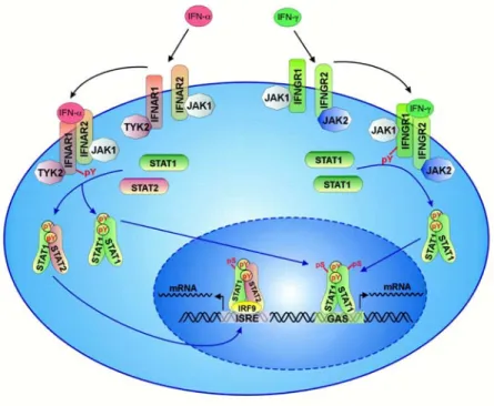
Type I and type II interferons are recognized by different receptors (Figure I1).
Biologically active IFN-γ is a homodimer and binds to the heterodimeric IFN-γ receptor (IFNGR1 and IFNGR2) expressed on all nucleated cells. Binding of IFN-γ to its receptor induces receptor dimerization and thereby a cascade of intracellular tyrosine phosphorylation events involving JAK1 and JAK2 ultimately leading to the activation and nuclear translocation of a STAT1 transcription factor homodimer called GAF (gamma activated factor) (33). In the nucleus GAF binds to gamma-interferon activated sequence (GAS) elements found at the promoter of many interferon responsive genes leading to their activation.
Figure I1: Simplified scheme of the type I and Type II signal transduction pathways taken from (34).
Binding of type I interferon (IFN-α) to its receptor induces dimerization of the IFNAR1 and IFNAR2 receptor subunits, activation of the associated JAK1 and TYK2 proteins subsequent phosphorylation of STAT1 and STAT2 and thereby their dimerization. The STAT1/STAT2 dimer translocates into the nucleus and forms a trimeric complex with IRF9 which binds to ISRE elements at the promoter of IFN stimulated genes and promotes their transcription. Binding of type II IFN (IFN-γ) to its receptor induces its dimerization, activation of JAK1 and JAK2 which is followed by phosphorylation of STAT1 and its homodimerization. The STAT1 homodimer binds to GAS elements at the promoter of IFN-γ stimulated genes after translocation into the nucleus.
Among the primary response genes are further transcription factors such as IRF-1 leading to a second wave of gene expression (18).
The signal transduction pathway of type I IFN is very similar to that of IFN-γ. Binding of IFN-α or IFN-β to its heterodimeric receptor (IFNAR1 and IFNAR2) induces receptor dimerization, activation of JAK1 and TYK2 and subsequent phosphorylation of
STAT1 and STAT2. The STAT1/STAT2 heterodimer associates with IRF-9 to form a heterotrimeric complex called ISGF3. ISGF3 binds to ISRE (interferon stimulated response elements) near the promoter of IFN activated genes. Type I and type II IFNs activate distinct but overlapping sets of genes (19, 35-37).
Experiments with mice carrying targeted deletions of components of the IFN-γ and IFN- α/β signal transduction pathways respectively have shown that IFN-γ is essential for resistance against bacterial and protozoan pathogens having a rather mild effect on certain viral infections. The opposite effect was observed for IFN-α/β which is central to combat viral infections but has a comparably moderate effect on bacterial and protozoan infections (38, 39).
Due to its complexity the function of IFN regulated genes is hard to assess not least because the function of many inducible genes is unknown. However, in general IFN inducible genes play a role in one or more of the following cellular programs (18, 19).
This includes the regulation of immune cell function, regulation of proliferation, enhancement and modulation of antigen-presentation and direct anti-microbial effects.
Many of the direct anti-microbial effects are mediated in a cell-autonomous manner.
1.4 Cell-autonomous immunity
It is becoming increasingly clear that cell-autonomous immunity plays a major role in resistance to intracellular pathogens of all classes. Many of the molecular players of intracellular resistance are inducible by interferons. The main focus has been on factors restricting viral growth (12) but it is now appreciated that also bacteria and protozoa are restricted by cell-autonomous resistance mechanisms (40). By definition cell- autonomous immunity is mediated by a cell for itself without the requirement for other specialized cell types. However, the induction of cell-autonomous resistance in a particular cell might be dependent on specialized cells as exemplified by the production of IFN-γ. Indeed it turned out that production of IFN-γ is used by activated T-cells to induce cell-autonomous immunity in the target cell and thereby to clear viral infections (16, 15).
The list of proteins implicated in cell-autonomous immunity is long and still growing (41). The following table includes an incomplete overview of factors known to play an important role in cell-autonomous immunity (Table 1). Many of the listed proteins have
been shown to be central for signal transduction in a variety of cellular pathways (11) (42) or to mediate general functions in membrane traffic like Rab5a (43, 44). However many of the included proteins have no known function besides pathogen resistance.
Table 1
NAME SPECIES TARGET LOCATION REFERENCE
Mx vertebrates Bunyaviridae, Orthomyxoviridae
nucleus, cytoplasm (smooth ER)
(45-48)
2´-5´ OAS, RNaseL mouse, human Picornaviridae nucleus, cytoplasm (12, 49) PKR mouse, human EMCV, Vaccinia
Virus, VSV
cytoplasm (50-53)
ADAR1 human Hepatitis delta virus nucleus, cytoplasm (54, 55) ISG20 human VSV, Influenza virus,
EMCV
nucleus (56, 57)
p65 GTPases vertebrates VSV, EMCV cytoplasm (58-60) PML mouse, human VSV, influenza virus,
human foamy virus, HSV1
nucleus (61-63)
ZAP rat Murine leukaemia
virus (MLV)
ND (64)
CEM15/APOBEC3G mammals Hepatitis B Virus and retroviruses including HIV, SIV, MLV, EIAV
cytoplasm (65-70)
TRIM5α primates HIV, SIV cytoplasmic bodies (41) RNAi machinery eukaryotes dsRNA viruses
including HIV-1, HCV, Poliovirus, Hepatitis delta virus
cytoplasm (71-75)
FV1, Ref1, Lv1 rodents, primates retroviruses including FV1, MLV, HIV-1,
HIV-2, EIAV
Golgi, ER (76-79),
IDO mouse, human viruses, bacteria and protozoa including cytomegalovirus, Clamydia, Toxoplasma gondii
cytosol (80-83)
iNOS vertebrates over 80 pathogens of all classes
cytoplasm (84-87)
phox complex vertebrates pathogens of viral, bacterial and protozoan origin
phagosomal membrane (88)
NRAMP1 mouse, human Salmonella, Leishmania, Mycobacterium
phagosomal membrane (89-91)
p47 GTPases mouse bacterial and protozoan pathogens
ER, Golgi, cytosol (40, 92-96), this study
Rab5a eukaryotes Listeria early endosomes (43)
The diversity of factors included in the table (Table 1) reflects the diverse and complex intracellular behaviours of the respective pathogens.
It is conspicuous that many of the proteins targeting bacterial and protozoan pathogens are membrane-bound. Intracellular membranes are central for the survival of many pathogens and therefore it is not surprising that pathogen and host attempt to take over control of cellular membrane dynamics (97). In eukaryotic cells many of the essential steps involving membrane dynamics are mediated by GTP binding proteins (10, 44, 98).
1.5 GTP binding proteins and membrane dynamics
GTP binding proteins are central to a plethora of cellular functions including protein biosynthesis, transport across the nuclear envelope, signal transduction and membrane traffic. Despite such diverse functions the underlying mechanism is the same for all GTPases involving a conformational change upon binding of GTP and subsequent hydrolysis of the nucleotide to GDP and/or GMP (99-102). The energetically favourable reaction is able to create order or force. Guanine nucleotide binding is essential for the function of GTPases and involves 5 motifs called G1-G5 among which G1, G3 and G4 are universally conserved. The consensus sequence of the G1 motif is GX4GKS contacting the α-, β- and γ-phosphate of the bound nucleotide. The G3 motif makes contact to the γ-phosphate and the G4 motif confers specificity by contacting the base of the guanine nucleotide (99-101).
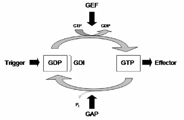
For the switch GTPases the GTP bound form is considered active and interacts with other molecules termed GTPase effectors. This interaction is responsible for the downstream effects of the GTP bound GTPase. Inactivation is achieved by hydrolysis of GTP and dissociation of the γ phosphate. Many GTPases have a low intrinsic activity and inactivation is mediated by the action of GTPase activating proteins (GAPs) which accelerate hydrolysis. The resulting GDP bound form is considered inactive. In many cases GTPases become activated upon interaction with guanine nucleotide exchange factors (GEFs). GEFs mediate dissociation of GDP from the GTPase. Due to the 3 fold higher concentration of GTP within the cell (103) GTPases become thereby activated despite often similar affinities for GTP and GDP (100). Some GTPases like the Rab and Rho are additionally regulated by guanine nucleotide dissociation inhibitors (GDIs)
which bind to the GDP bound form and prevent nucleotide dissociation (104). A simplified cartoon summarizing the GTPase cycle is shown in Figure I2.
Figure I2: Simplified scheme of the GTPase cycle.
Activation of the GDP bound GTPase by a trigger is achieved by exchange of GDP for GTP. This exchange is often mediated by the activation of exchange factors (GEF). The GTP bound GTPase interacts with molecules termed effectors mediating its downstream effects. GTP hydrolysis and dissociation of the γ-phosphate leads to the inactivation of the GTPase. The often low intrinsic hydrolytic activity can be accelerated by activating proteins (GAP). Some GDP bound GTPases are kept inactive by the action of guanine nucleotide dissociation inhibitors (GDI) (99-101).
Due to their ability to be regulated in multiple ways and to give complex reactions a direction, GTPases are central regulators and mediators of membrane traffic and membrane association is essential for their function.
Arf GTPases for example regulate COP mediated vesicular budding (105). The inactive GDP bound form is cytosolic but upon nucleotide exchange from GDP to GTP Arf exposes an N-terminal myristoyl-group and an amphipathic helix hidden within the molecule anchoring the protein in the lipid bilayer (106, 107). At the membrane Arf interacts with other molecules to initiate vesicular budding (105).
Rab GTPases in contrast are isoprenylated at their C-terminus and are kept soluble in the cytosol by the interaction with RabGDI burying the large C-terminal isoprenyl-group in a hydrophobic pocket (44, 105). Activation of Rab by nucleotide exchange from GDP to GTP leads to dissociation of Rab from RabGDI and subsequent membrane association.
At the membrane Rab GTPases interact with various effectors thereby determining specificity of vesicular transport and organelle identity (44).
Rho GTPases are yet another family of GTP binding proteins belonging, like Arf and Rab, to the p21 Ras superfamily of GTPases. Rho GTPases such as RhoA, CDC42 and Rac1 regulate actin dynamics (108-110) and function in phagosome formation (111- 113) and maturation by recruiting the phagosome oxidase complex to the phagosomal membrane (114). In addition they have various other cellular functions (108). Activation of Rho GTPases results in membrane association which is, as in the case of Rab, mediated by a C-terminal isoprenyl-group (115). The GDP bound form of Rho GTPases is found in the cytosol in a complex with RhoGDI (116). Upon exchange of GDP for GTP Rho GTPases bind to intracellular membranes collectively called endomembranes and the plasma membrane (117, 118). In all cases nucleotide exchange and membrane association are strictly coupled.
1.6 Dynamin
Dynamins are GTPases which mediate scission of vesicles budding from the donor membrane. Overexpression of dominant negative mutants of dynamin interferes with the formation of clathrin-coated vesicles, budding from caveolae and phagosome formation.
Dynamins differ in several key aspects from the p21 Ras superfamily of GTPases (98, 102, 119).
Dynamins are with a molecular weight of about 100 kDa much larger than Arf, Rab and Rho GTPases which are 20-30 kDa in size. Since their discovery it is under debate whether dynamins function by generating force or are molecular switches analogous to the members of the Ras superfamily of GTPases, or both (98, 102, 120-122).
The N-terminal nucleotide binding domain of dynamin is followed by the middle domain implicated in self-assembly, a pleckstrin homology domain (PH domain) involved in membrane targeting, a GTPase effector domain accelerating GTP hydrolysis and a C-terminal proline-rich domain interacting with other proteins (98). Although many proteins have been shown to interact with dynamin no proteins, apart from dynamin itself, could be identified so far interacting with dynamin in a nucleotide dependent manner (98, 102). Compared to Ras-like GTPases dynamins bind guanine nucleotides with a rather low affinity in the µM range and have high turn over rates of hydrolysis. Dynamin self associates in a GTP dependent manner which increases the
is cooperative. Dynamin associates with negatively charged liposomes in vitro and this association accelerates hydrolysis about 100 fold (123, 102).
In the presence of GTP-analogues dynamin tubulates lipids in vitro by forming ring-like structures around liposomes (124). Upon hydrolysis the diameter of the tubulated liposomes decreases (122). In vivo dynamin was observed to form spiral-like structures around the neck of budding vesicles (124). The presence of lipids massively accelerates GTP hydrolysis and enhances nucleotide dependent oligomerization and self-assembly (102, 123).
Membrane binding is essential for the function of dynamin itself and probably also for the other members of the dynamin superfamily of which the mammalian dynamin 1, 2 and 3 are the prototypes (98). The PH domain of dynamin is involved in targeting of dynamin to membranes mediated by its low affinity for the lipid head group inositol 1,4,5-triphosphate. Mutations in the PH domain of dynamin have a dominant negative effect on endocytosis (125-127). However other members of the dynamin superfamily lack a PH domain but are still capable of lipid binding as for example the Mx proteins (128, 45).
1.7 The antiviral Mx proteins
Mouse Mx was the first member of the dynamin superfamily discovered and was initially identified as a dominant locus in A2G mice conferring resistance to infections by orthomyxoviridea (129). Mapping and subsequent cloning of the gene led to the identification of mouse Mx1 (48, 130) and the human homologue MxA (131).
Surprisingly, in contrast to most out bred mice, most laboratory mouse strains do not carry a functional allele of Mx (132, 133).
Mx proteins exhibit nucleotide dependent oligomerization and cooperative hydrolysis (47, 134, 135). A direct interaction of Mx with viral particles has been shown and is proposed to be important for its antiviral activity (47). However a mutant Mx protein, defective in GTP hydrolysis and oligomerization still shows antiviral activity questioning the role of self assembly for its antiviral function (136).
Recently human MxA has been reported to localize to the smooth ER and to tubulate lipids in a nucleotide dependent manner (45). The meaning of membrane deformation by MxA for its biological function however is unclear recalling that dynamin has been first isolated as a protein assembling around microtubules (137) but there is still no in vivo function described giving this finding significance. A hallmark of the Mx proteins
is their exclusive inducibility by type I IFN (138) and low or absent level in resting cells.
1.8 Other Interferon inducible GTPases
The induction of high molecular weight GTPases by IFNs turns out to be an important component of their biological function. There are at least three more families of GTP binding proteins which are massively induced by IFN and whose functions are dedicated to host resistance.
The p65 family of GTPases has 5 members (GBP1-5) in mouse and human (139).
Homologues are found in all vertebrates analyzed so far (60). The p65 GTPases are abundantly induced by type I and type II IFN from low resting levels (60). When expressed in Hela cells human GBP1 (hGBP1) shows a cell-autonomous antiviral effect against VSV and EMCV (58). Besides its antiviral activity members of the p65 family have been implicated in the regulation of cell proliferation (140-142). The crystal structure of hGBP1 reveals a three domain protein having an N-terminal GTP binding domain, followed by a helical middle domain and a C-terminal GTPase effector domain (143). Some family members are isoprenylated mediated by a C-terminal CAAX motif (C, cysteine; A, large hydrophobic residues; X, any residue) and this isoprenylation appears to be responsible for the localization in enigmatic cytoplasmic dots (59) (H.
Kashkar, unpublished results). The structure of hGBP1 has attracted much attention because it is generally believed that its overall structural organization is shared with dynamin (143).
Recently a family of gigantic GTP binding proteins with a molecular weight of 280 kDa has been published (144). This very large inducible GTPases (VLIGs) are massively induced by type I and type II IFNs, at least in the mouse. No anti-microbial effect for the VLIGs has been shown so far but VLIG-1, the prototype of the VLIG family, shows highest homology to GTPases mediating cell-autonomous resistance within the GTPase superfamily (144). This suggests, in addition to their IFN inducibility, a role in intracellular defence.
The third family of IFN inducible GTP binding proteins are the p47 GTPases.
1.9 The p47 GTPases
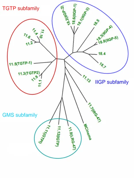
There is now compelling evidence that the p47 GTPases are an essential component of the immune response against intracellular pathogens in the mouse (40). So far 6 p47 GTPases have been published, namely TGTP (145), IRG-47 (146), IIGP1 (60), IGTP (147), GTPI (60) and LRG-47 (148) but the total number of p47 genes in the Mus musculus domesticus genome is 23 of which 4 are pseudogenes by one or another criterion (Julia Hunn, Cemali Bekpen and Jonathan C. Howard, personal communication). Figure I3 shows a phylogeny of the p47 GTPases of Mus musculus domesticus.
Figure I3: Phylogenetic Tree based on the protein sequence of the GTPase domain of the mouse p47 GTPases (courtesy of Cemali Bekpen) showing 22 out of the 23 members. The neighbour joining tree is based on the distance method using the MEGA2 program.
The phylogeny reveals a complex protein family which can be subdivided into several subgroups namely the TGTP, IIGP and GMS subfamily. In addition, the phylogeny
TGTP subfamily
IIGP subfamily
GMS subfamily
includes p47 GTPases which do not belong to any of this subgroup like IRG-47 and mCinema. The GMS GTPases carry an unusual methionine instead of the universally conserved lysine in the G1 motif changing its sequence from GX4GKS to GX4GMS (60). In human the p47 GTPase family is far less complex including only a very close homologue of mCINEMA called hCINEMA and a small fragment showing homology to the mouse GMS GTPases carrying the same unusual lysine to methionine substitution in the G1 motif (Cemali Bekpen and Jonathan C. Howard, personal communication).
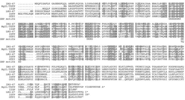
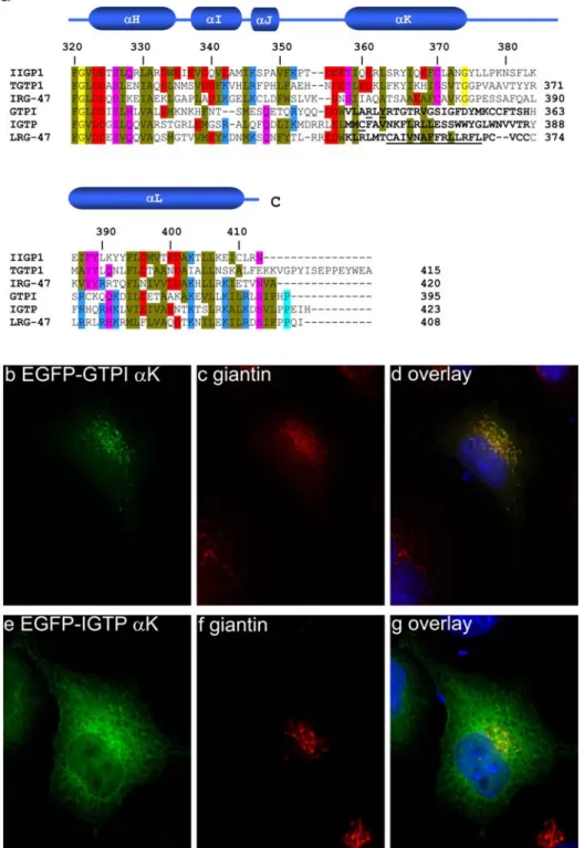
Figure I4: Amino acid alignment of the 6 published p47 GTPases (60).
The N- and C-terminal domains show low homology between the family members containing unique sequence patches. The core of the proteins is conserved showing stretches of identical sequence. The conserved GTP binding motifs are shown below the sequence.
The 6 published p47 GTPases show an identity between 25% and 55% which is clustered in the core of the proteins (Figure I4). The N-terminal and the C-terminal domains in contrast are rather divergent containing unique sequence patches with no similarity to other family members (60).
Nearly all family members are inducible by type II and to a minor extend by type I IFN in all cell-types tested so far including immune and non-immune cells (60, 92) (Cemali Bekpen, personal communication). Cinema appears to be an exception in this respect since it was not found to be IFN inducible in any cell-type tested so far (Christoph Rohde, personal communication) In general the basal expression of the p47 GTPases is low although exceptions to this rule are emerging. IIGP1 for example shows a high type I and type II IFN receptor independent basal expression level in liver which is rising
even further after infection with Listeria monocytogenes (Jia Zheng, personal communication).
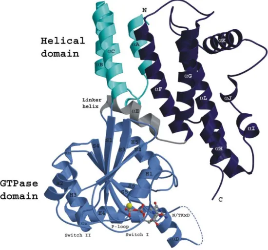
Recently the crystal structure of IIGP1 has been determined (149) (Figure I5).
Figure I5: Crystal structure of IIGP1 in the GDP bound form (149).
The first 13 amino acids of IIGP1 are not resolved in the structure and therefore not shown. The N- terminal domain (cyan) is composed of three αhelices and is followed by the GTP binding domain (light blue) which is similar to the G-domain of Ras. The helical C-terminal domain (dark blue) is connected to the G-domain by the linker helix αE (grey)
The GTP binding domain which is structurally similar to the canonical GTPase domain of Ras is preceded by an N-terminal domain composed of 3 αhelices. The first 13 amino acids are not resolved and therefore missing in the structure. The GTPase domain is connected with the C-terminal helical domain by the linker helix αE. The C-terminal domain folds back to come into close proximity to the GTP binding domain. The N- and C-terminal helical domains correspond to the regions of low sequence similarity shown in the alignment. In particular the region around αK shows virtually no sequence similarity between the published p47 GTPases. However patches of sequence identity throughout the whole sequence and secondary structure predictions suggest that the overall structure of IIGP1 is representative for the whole family (149). IIGP1
crystallized as dimer with the two molecules making contact at the N-terminal and GTPase domain involving αB and H3. The dimer appears to be relevant for the properties of IIGP1 as mutations designed to disrupt dimer formation alter the enzymatic properties of IIGP1 (149).
IIGP1 behaves strikingly similar to dynamins regarding several key features of its enzymatic behaviour (150). IIGP1 binds nucleotides with a rather low affinity in the µM range, shows cooperative activity and nucleotide dependent oligomerization. Upon hydrolysis the oligomers dissolve showing that multimerization is a reversible process (150).
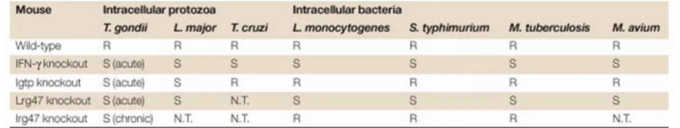
Experiments with mice carrying targeted deletions of single genes of the p47 GTPase family have shown that the p47s are essential, non-redundant resistance factors against intracellular pathogens in the mouse (40, 94-95). Table 2 summarizes the results obtained with IGTP, IRG-47 and LRG-47 knock out mice (40).
Table 2: The table, taken from a review by Taylor et al. (40), summarizes the phenotypes obtained with the shown knock out mice (S, sensitive; R; resistant).
LRG-47 and IGTP are required in the acute phase of infection. Mice carrying a targeted deletion of the LRG-47 gene die within 5 days after infection with Listeria monocytogenes (96) which is to fast for the adaptive immune system to mount an effective immune response. In view of the fact that IFN-γ induces at least several hundreds if not thousands of genes the profound loss of host resistance after deletion of single p47 GTPases is remarkable.
The resistance conferred by the p47 GTPases appears to be at least in some cases cell- autonomous. Astrocytes isolated from IGTP-/- mice show no IFN-γ mediated growth inhibition of Toxoplasma gondii (151). The finding of Carlow et al. reporting that TGTP expressing Hela cells are less susceptible to the cytopathic effects caused by VSV supports (152) the hypothesis of the p47 GTPases functioning in a cell-autonomous manner. In addition a small antiviral effect of IGTP against Coxsackievirus B3 when overexpressed in Hela cells has been observed (69). However both antiviral effects were
The cell-biology and thus the mode of function of the p47 GTPases is totally unknown although recent experiments begin to shed light on their mechanism of resistance. IIGP1 has been reported to localize to the ER and Golgi complex in bone marrow derived macrophages (92) and recently a physical interaction of IIGP1 with Hook3, a Golgi localized microtubule binding protein, has been published but the biological significance of this interaction remains to be established (225). IGTP was shown to localize to the ER in mammary gland derived C127 cells in a GTP independent manner (93). The nature of their membrane attachment is unexplored but none of the p47 GTPases contains a signal peptide for co-translational translocation into the ER or any other predictable transmembrane segment. LRG-47 was co-purified with phagosomes from Mycobacterium tuberculosis infected macrophages (95). This co-purification was sensitive to BrefeldinA treatment and targeted deletion of the LRG-47 gene resulted in a reduced acidification and thus maturation of the Mycobacterium containing phagosome (95). Where LRG-47 localizes, how it interacts with membrane and how it is recruited to the phagosome is totally unknown. The initial observation that LRG-47 functions in phagosome maturation however might provide the key for the understanding how the p47 GTPases and in particular LRG-47 mediate resistance against such a variety of pathogens with completely different intracellular life styles (40).
1.10 Phagocytosis of microbes
Phagocytosis describes the process of uptake of large particles by cells (10, 153).
Phagocytosis is of central importance for the function of innate immune cells such as neutrophils and macrophages but also non-immune cells are able to take up large particles as for example in the case of collagen-phagocytosis in fibroblasts (154).
Phagocytosis is an extraordinarily complex process whereby upon particle recognition, cytoskeletal dynamics and membrane traffic are co-ordinately orchestrated involving several hundreds of proteins (10, 114, 155). The recognition and engulfment of the phagocytosed particle is accompanied by the activation of several signal transduction pathways (10).
The initial recognition of a microbe or particle is followed by the formation of a so called phagocytic cup marked with actin filaments and subsequent closure of the phagocytic cup to form the phagosome (112). Recently the ER has been reported to supply membranes for the formation of the vacuole at least in macrophages (156). The initial steps of phagosome formation involve the generation, modification and cleavage
of phosphoinositites by the action of enzymes such as PI3K and PLC (10, 114). After closure of the phagocytic cup the resulting phagosome matures by sequential interactions with the endosomal compartment of the cell to form the phagolysosome (157). Maturation includes the recruitment of the phagosome oxidase complex to the phagosomal membrane by Rac2 (113), acidification and activation of hydrolytic enzymes, recruitment of iNOS and other antimicrobial proteins such as NRAMP1 to the phagosomal membrane (10, 158-159). Nearly all steps of phagosome formation and maturation involve the action of GTP binding proteins of the Rho, Arf, Rab and dynamin family of GTPases (43, 157, 160-165).
During maturation the phagosomal lumen becomes very soon uninhabitable for the internalized pathogen. It is therefore not surprising that pathogens attempt to control phagosomal maturation and vesicular traffic by secretion of virulence factors interfering with host cell processes promoting maturation of the phagosome (97). The host in contrast tries to accelerate phagosomal maturation before the pathogen takes over control. IFN-γ strongly increases the speed with which phagosomes mature (166) for example by the induction of Rab5a (43, 167).
Not all intracellular pathogens are passively taken up by phagocytosis like Mycobacterium tuberculosis (168, 169) but actively induce their uptake (97, 170).
Common to all mechanisms of entry into the host cell however is that the pathogen has to cross the plasma membrane and to control the fate of the resulting membrane bound compartment regardless of whether they reside in an intracellular vacuole (169, 171) or escape into the cytosol soon after uptake (172, 173).
1.11 The aim of this study
The introductory remarks outline above intended to emphasize the intimate relationship of intracellular pathogens and host cell membranes. The p47 are a battery of diverse cell-autonomous resistance factors and association with cellular membranes has been described for some members. The mechanism and dynamics of membrane association however is totally unexplored and is the subject of investigation in this work. The assumption is that information about the membrane association properties of the p47 GTPases reveals an essential part of their cell-biology in order to get further insights how the p47 GTPases contribute to host resistance.
Therefore the p47 GTPases LRG-47 and IIGP1 were analyzed in detail regarding their
factor among the p47 GTPases whereas the biochemical characteristics of IIGP1 have been thoroughly investigated. This information is used to design and interpret the experiments shown in this work. The results obtained for LRG-47 and IIGP1 are extrapolated to other family members and similarities and differences are discussed in the context of intracellular defence.
2. Material and Methods
2.1 Reagents and Cells
2.1.1 Chemicals, Reagents and Accessories
All chemicals were purchased from Aldrich (Steinheim), Amersham-Pharmacia (Freiburg), Applichem (Darmstadt), Baker (Deventer, Netherlands), Boehringer Mannheim (Mannheim), Fluka (Neu-Ulm), GERBU (Gaiberg), Merck (Darmstadt), Pharma-Waldhof (Düsseldorf), Qiagen (Hilden), Riedel de Haen (Seelze), Roth (Karlsruhe), Serva (Heidelberg), Sigma-Aldrich (Deisenhofen) or ICN biochemicals, Oxoid, (Hampshire UK). DNA size standards from Gibco-BRL (Eggenstein), electrophoresis chambers from FMC Bioproducts (Rockland Maine US), developing and fixing solutions for Western Blot detection from Amersham Pharmacia (Freiburg), Luminol from Sigma Aldrich (Deisenhofen), Coumaric acid from Fluka (Neu-Ulm) and
“Complete Mini” protease inhibitor cocktail from Boehringer (Ingelheim). Deionised and sterile water (Seral TM) was used for all the buffers and solutions, Ultra pure water derived from Beta 75/delta UV/UF from USF Seral Reinstwassersysteme GmbH, (Baumbach) equipped with UV (185/254nm) and ultrafiltration (5000 kd cut off), or from Milli-Q-Synthesis (Millipore).
2.1.2 Equipment
Centrifuges used were: Biofuge 13, Heraeus; Sigma 204; Sigma 3K10; Labofuge 400R, Heraeus; Sorvall RC-5B, Du Pont instruments; Optima TLX Ultracentrifuge, Beckmann. BioRAD Gel dryer, Model-583; BioRad Power pack 300 or 3000; Gel Electrophoresis Chamber, Cambridge electrophoresis; Biorad Mini Protean II; PTC-100, MJ Research Inc.; Centrifuge tubes 15ml, TPP Switzerland; 50ml Falcon, Becton Dickenson; Zeiss Axioplan II fluorescence microscope equipped with a Quantix cooled CCD camera and a Z-stepping device; Odyssey confocal laser scanning device.
2.1.3 Materials
Sterile filters FP 030/3 0,2 µm and ME 24 0,2 µm (Schleicher und Schüll, Dassel);
Whatmann Paper (purchased via LaboMedic); 100 Sterican 0,50 x 16mm hypodermic needles (Braun AG, Melsungen); 0.2µm and 0.45µm sterile filters (Schleicher und Schuell, Dassel); X-OMAT LS and AR X-ray films, Kodak.
All plastic ware for cell culture was from Sarstedt (Nümbrecht) or Greiner (Solingen)
2.1.4 Enzymes/Proteins
Restriction Enzymes were purchased from New England Biolabs (Bad Schwalbach);
“Complete Mini” protease inhibitor cocktail from Boehringer (Ingelheim); Pyrococcus furiosus (Pfu) DNA Polymerase (Promega, Mannheim); T4 DNA ligase (New England Biolabs); RNase A (Sigma); shrimp alkaline phosphatase (SAP) (USB, Amersham);
1Kb ladder for agarose gels (Gibco); rainbow molecular weight marker precision protein standardsTM (Biorad); wide range protein standard marker (Sigma, Deisenhofen)
2.1.5 Kits
Plasmid Maxi and Midi kit (Qiagen, Hilden), Terminator-cycle Sequencing kit version 3 (ABI),
QuikChange TM Site directed mutagenesis kit (Stratagen), Rapid PCR product purification Kit (Boehringer, Ingelheim),
2.1.6 Vectors
pGW1H (British Biotech, Oxford, England), pGEX-4T-2 (Amersham Pharmacia, Freiburg), pEGFP-C3 (Clontech),
pEGFP-N3 (Clontech),
2.1.7 Cell lines
RAW 264.7 (ATCC TIB-71), L929 mouse fibroblasts (CCL-1), mouse hepatocytes derived TIB-75 cells (ATCC TIB-75) (174)) and B6m29 cells (175) derived from a p53- /- C57BL/6J mouse were cultured in IMDM supplemented with 10% FCS (Sigma, Deisenhofen), 2 mM L-Glutamine (Gibco BRL, Eggenstein), 1 mM sodium pyruvate (ICN, Eschwege), 100 U/ml penicillin (Gibco BRL) and 100 µg/ml streptomycin (Gibco
BRL). Peritoneal macrophages were isolated from the peritoneal cavity of CB20 mice and cultured in 6-well plates with coverslips. Cells were grown in IMDM and washed several times to remove non-adherent cells over a period of 36-48h before experiments.
2.1.8 Media
Luria Bertani (LB) medium
10g bacto tryptone, 5g yeast extract, 10g NaCl, distilled water 1l LB plate medium
10g bacto tryptone, 5g yeast extract, 10g NaCl, 15g agar, distilled water 1l
IMDM (Iscove’s Modified Dulbecco’s Medium) Gibco BRL, Eggelstein supplemented with: 10% FCS, 2 mM 1-glutamine, 1 mM sodium pyruvate, 100 U/ml penicillin, 100 µg/ml streptomycin, 1x non-essential amino acids (Gibco BRL).
DMEM (Dulbecoo’s Modified Eagle Medium), Gibco BRL,Eggenstein supplemented with:10% FCS, 2 mM 1-glutamine, 1 mM sodium pyruvate, 100 U/ml penicillin, 100 µg/ml streptomycin, 1x non-essential amino acids (Gibco BRL).
2.1.9 Bacterial strains
Escherichia coli XL1-Blue: recA1, end A1, gyrA96, thi-1, hsdR17, supE44, relA1, lac, [F’, pro AB, lacIqZ∆M15, Tn10 (Tetr)]
Escherichia coli DH5α: 80dlacZ∆Μ15, recA1, endA1, gyrA96, thi-1, hsdR17 (rB-, mB+), supE44, relA1, deoR, ∆(lacZYA-argF)U169
Escherichia coli BL-21: E. coli B, F-, omp T, hsd S (rB- mB-), gal, dcm Salmonella typhimurium strain SL1344 (Hoiseth and Stocker 1981)
2.1.10 Serological reagents Primary antibodies and antisera:
NAME IMMUNOGEN SPECIES CONCENTRATION DILUTION ORIGIN 1D4B mouse LAMP-1 rat monoclonal IB: 1:100
IF: 1:1000
DSHB, University of Iowa αgiantin human giantin mouse
monoclonal
IF: 1:1000 Hans Peter Hauri, Basel (176) αIGTP clone 7 mouse IGTP aa
283-423
mouse monoclonal
0.25µg/µl IB: 1:5000 IF: 1:250
BD Transduction Laboratories
αΜΙΙ mouse
αmannosidase II
rabbit polyclonal IF: 1:100 Paul Slusarewicz via Albert Haas, Bonn
αCI-M6PR CI-M6PR rabbit polyclonal IF: 1:100 Gus Lienhard via Albert Haas, Bonn (177) Μ2 Flag-epitope
DTKDDDDK
mouse monoclonal
4.9mg/ml IB 1:1000
IF 1:4000
Sigma Aldrich
2078 mouse IRG-47 peptides CKTPYQHPKY PKVIF and CDAKHLLRKI ETVNVA
rabbit polyclonal IB: 1:1000 Eurogentec double X programme
N20 (sc-894) N-terminal peptide of human Caveolin- 1
rabbit-polyclonal 0.2µg/µl IB: 1:1000 IF: 1:100
Santa Cruz
L115 mouse LRG-47
peptides QTGSSRLPEVS RSTE and NESLKNSLGV RDDD
rabbit-polyclonal IB: 1:1000 Eurogentec double X programme
A19 (sc-11075) N-terminal peptide of mouse LRG-47
goat-polyclonal 0.2µg/µl IB: 1:200 IF: 1:100
Santa Cruz
A20 (sc-11079) N-terminal peptide of mouse TGTP
goat-polyclonal 0.2µg/µl IB: 1:500 IF: 1:100
Santa Cruz
M14 (sc-11088) C-terminal peptide of mouse GTPI
goat-polyclonal 0.2µg/µl IB: 1:100 Santa Cruz
V9 pig eye lens vimentin
mouse monoclonal
IF: 1:40 Sigma
SPA-865 N-terminal peptide of canine calnexin
rabbit-polyclonal IB: 1:10000 IF: 1:200
StessGen
αERP60 ERP60 rabbit-polyclonal IF: 1:1000 Tom Wileman, Pirbright
αRab6 human Rab6 rabbit-polyclonal IF: 1:100 Bruno Goud, Paris G65120 C-terminus of
Rat-GM130
mouse monoclonal
IB: 1:100
IF 1:1000
BD Transduction Laboratories 165 recombinant
mouse IIGP1
rabbit polyclonal IB: 1:25000 IF: 1:8000 10E7 recombinant
mouse IIGP1
mouse- monoclonal
1-3µg/µl IB: 1:1000
IF: 1:200
Jens Zerrahn, Berlin 10D7 recombinant
mouse IIGP1
mouse- monoclonal
1-3µg/µl IB: 1:1000 Jens Zerrahn, Berlin C15 (sc-6414) peptide mapping
to the C- terminus of human EEA1
goat-polyclonal 0.2µg/µl IB: 1:100 Santa Cruz
Secondary antibodies and antisera
goat anti-mouse Alexa 546/488, goat anti-rabbit Alexa 546/488, donkey anti-goat Alexa 546/488, donkey anti-mouse Alexa 488, donkey anti-rabbit Alexa 488, donkey anti-rat Alexa 488, goat anti-mouse Alexa 680, goat anti-rabbit Alexa 680, goat anti-rat Alexa 680, donkey anti-goat Alexa 680 (all Molecular Probes), donkey anti-rabbit HRP (Amersham), donkey anti-goat HRP (Santa Cruz), goat anti-mouse HRP (Amersham), goat anti-mouse IRDye 800, goat anti-rabbit IRDye 800, goat anti-rat IRDye 800 (all Rockland Immunochemicals Inc.).
2.2 Molecular Biology
All plasmids and constructs were amplified, cloned or propagated using protocols adapted from Sambrook, J., Fritsch, E.F., and Maniatis, T., Vol. 1, 2, 3 (1989), or from the cited references.
2.2.1 Agarose gel electrophoresis
DNA was analyzed by agarose gel electrophoresis (1x TAE; 0.04 M Tris, 0.5 mM EDTA, pH adjusted to 7.5 with acetic acid) The DNA was stained with ethidium bromide (0.3 µg/ml), a fluorescent dye which intercalates between nucleotide bases, and the migration of the DNA molecules was visualized by using bromophenol blue.
2.2.2 Generation of p47 GTPase expression constructs
The coding regions of p47 GTPases were amplified either by PCR from full length cDNAs described in (60) from IFN-γ (1000U/ml) stimulated mouse embryonic fibroblasts (MEFs) according to standard procedures using Pfu-polymerase (Promega) and primers ordered from Invitrogen. Restriction enzymes were ordered from “New England Biolabs”.
Primers used were
IIGP1 forward 5'-CCCCCCCCGTCGACCACCATGGGTCAGCTGTTCTCTTC-3', IIGP1 reverse 5'-CCCCCCCCGTCGACCTAGTTTCTTAAACATATCTCTTTAAG-3',
IIGP1 aa 1-287 reverse 5´-CCCCCCCCCGTCGACCTAAAATCCTTCCAGCCAAATCCTC-3´,
LRG-47 forward 5'-CCCCCCCCGTCGACCACCATGAAACCATCACACAGTTCC-3', LRG-47 reverse 5'-CCCCCCCCGTCGACCACCTAGATCTGCGGAGGGAAG-3',
LRG-47 aa 1-285 reverse 5´-CCCCCCCCCGTCGACCTACAGGATTTTCTCTAGGATG-3´
introducing a SalI site (underlined) at both ends. The PCR products were cloned into the SalI site of pGW1H (British Biotech).
IGTP forward: 5´-CCCCCCCCGTCGACCACCATGGATTTAGTCACAAAGTTGCC-3´, IGTP reverse: 3´-CCCCCCCCGTCGACTCAGTGAATTTCGGGAGGGAG-3´,
The PCR fragments were cut with SalI and cloned into pEGFP-C3 (Clontech) expression vector.
Primers to generate a C-terminal Flag tagged version of TGTP were TGTP-Flag 5 5´-CCCCCCCCGTCGACCTAACTACTTAGTGAGC-3´,
TGTP-Flag 3´ 5´-
CCCCCCCCGAATTCCTACTTGTCATCGTCGTCCTTGTAATCACCGGATCCAGCTTCCCAGTACTCGGGGGG-3´,
The PCR products were SalI and EcoRI cut and cloned into the appropriately cut pGW1H expression vector.
Primers used to clone N-terminal fragments of IIGP1 and LRG-47 into pEGFP-N3 (Clontech) were
pGW1H 5´ 5´-CTTTCCATGGGTCTTTTCTG-3´,
IIGP1 aa 1-69 reverse 5´-CCCCCCCCCGTCGACCACACTACTATCGATTTCTTTTAATG-3´,
LRG-47 aa 1-76 reverse 5´-CCCCCCCCCGTCGACTGGAATCTGGGACAATGTTGC-3´.
Fragments were amplified from the respective pGW1H constructs, cut with SalI, gel purified and cloned into pEGFP-N3.
Primers used to clone the G-domain, the C-terminal domain and C-terminal fragments of LRG-47 and IIGP1 into pGW1H were
IIGP1 G-domain 5´: 5´-CCCCCCCCCGTCGACCACCATGAGTGTGCTCAATGTTGCTGTC-3´, IIGP1 G-Domain 3´ Flag:
5´-CCCCCCCCCGTCGACCTAACCCTTATCGTCATCGTCCTTGTAATCAGGGAGGTCACTTATCAGCTTG-3´,
IIGP C-domain 5´: 5´-CCCCCCCCCGTCGACCACCATGGCTGACCTAGTGAATATCATC-3´,
LRG-47 G-domain 5´: 5´-CCCCCCCCCGTCGACCACCATGATTCCAGTGAGCATCTTTGTG-3´,
LRG-47 G-domain 3´: 5`-
CCCCCCCCCGTCGACTAGACCCTTATCGTCATCGTCCTTGTAATCGGAGAGATCTTTATGAAGTGTG-3´,
LRG-47 C-domain 5´: 5´-CCCCCCCCCGTCGACCACCATGAAGAATTCTCTCGGTGTCAG-3´,
LRG-47 alphaH 5´: 5´-CCCCCCCCCGTCGACCACCATGTTTGGTGTAGATGACGAATCAG-3´,
LRG-47 alphaI 5´: 5´-CCCCCCCCCGTCGACCACCATGGGGACAGTAGTCATGGAG-3´,
LRG-47 alphaJ 5´: 5´-CCCCCCCCCGTCGACCACCATGAAGTCCCAAAACTTTTATAC-3´,
LRG-47 alphaK 5´: 5´-CCCCCCCCCGTCGACCACCATGAAACTGAGGCTATGACATGTG-3´,
LRG-47 alphaL 5´: 5´-CCCCCCCCCGTCGACCACCATGTGTTTAAGACGCTTGAGACATAAAC-3´,
PCR products were cut with SalI and ligated to SalI cut pGW1H in case of the G- domain constructs and into pEGFP-C3 in case of the C-terminal domain constructs.
Primers used to clone αK fragments of LRG-47, GTPI and IGTP into pEGFP-N3 (Clontech) were
LRG-47 aK 5´: 5´-CCCCCCCCCGTCGACCACCATGAACTGAAGGCTGATGACATG-3´, LRG-47 aK 3´: 5´-CCCCCCCCCGTCGACCTAGCAGCATACGCATGGGAGAAATC-3´, IGTP aK 5´: 5´-CCCCCCCCCGTCGACCACCATGGATGTGTTTTGCCGTGAAC-3´, IGTP aK 3´: 5´-CCCCCCCCCGTCGACCTAGCGGGTGACGACGTTCCACAAG-3´, GTPI aK 5´: 5´-CCCCCCCCCGTCGACCACCATGGTGCTGGCTCGGTTGATACG-3´, GTPI aK 3´: 5´-CCCCCCCCCGTCGACCTAATGAGAGGTAAAGCAGCACTTC-3´,
Fragments were amplified from the respective pGW1H constructs, cut with SalI, gel purified and cloned into pEGFP-C3.
Mutation into the ORFs of LRG-47 and IIGP1 were introduced according to the
“QuikChange” site directed mutagenesis kit (Stratagene) protocol using the respective pGW1H and pEGFP-C3 constructs as template. Primers used were
LRG-47 S90N forward: 5´-GGGACTCTGGCAATGGCATGAATTCTTTCATCAATGCACTTCG-3´,
LRG-47 S90N reverse:5´-CGAAGTGCATTGATGAAAGAATTCATGCCATTGCCAGAGTCCC-3´,
LRG-47 ins 359E forward: 5´-ATGACATGTGCAATTGAAGTGAATGCATTCTTC-3´,
LRG-47 ins 359E reverse: 5´-GAAGAATCGATTCACTTCAATTGCACATGTCAT-3´,
LRG-47 ins 362E forward: 5´-GCAATTGTGAATGCAGAATTCTTCCGTTTGTTG-3´, LRG-47 ins 362E reverse: 5´-CAACAAACGGAAGAATTCTGCATTCACAATTGC-3´,