Original article:
TOXOPLASMA GONDII : PREVENTIVE AND THERAPEUTIC EFFECTS OF MORPHINE AND EVALUATION OF TREATMENT PARAMETERS OF TACHYZOITES AND INFECTED MACROPHAGES
IN VITRO AND IN A MURINE MODEL
Leila Zaki
1, Fatemeh Ghaffarifar
1*, Zohreh Sharifi
2, John Horton
3, Javid Sadraei
11 Department of Parasitology, Faculty of Medical Sciences, Tarbiat Modares University, Tehran, Iran
2 Blood Transfusion Research Center, High Institute for Research and Education in Transfusion Medicine, Tehran, Iran
3 Tropical Projects, Hitchin, United Kingdom
* Corresponding author: Fatemeh Ghaffarifar, Ph.D., Department of Parasitology, Faculty of Medical Sciences, Tarbiat Modares University, Tehran, P.O. Box 14115-111, Iran, Tel: +98-21-82884553, Fax: +98-21-82884555, E-mail: ghafarif@modares.ac.ir
http://dx.doi.org/10.17179/excli2019-1961
This is an Open Access article distributed under the terms of the Creative Commons Attribution License (http://creativecommons.org/licenses/by/4.0/).
ABSTRACT
Common medicines for the treatment of toxoplasmosis have limited efficacy and unwanted side effects. Opiates can effect both innate and cell-mediated immunity and stimulate the immune responses in different parasitic in- fections. In this work, preventive and therapeutic effects of morphine were evaluated on the tachyzoites of Toxo- plasma gondii and infected macrophages in vitro and in a murine model. Different concentrations of morphine (0.1 and 0.01 μg/ml) were evaluated on mortality rate of T. gondii by direct counting after 3 and 24 hours. The cytotoxic and apoptotic effects of these drugs were measured by the MTT assays and flow cytometry analysis, respectively. The same procedures were assessed in T. gondii-infected macrophages. The parasite loads were de- termined using quantitative polymerase chain reaction (qPCR). For in vivo assessment, BALB/c mice treated with morphine before or after infection with tachyzoites. The survival rate of animals, parasite load in the spleen, and the IFN-γ and IL-4 cytokines levels were measured. Morphine was effective on tachyzoites of T. gondii and had a reverse relationship with its concentration. The results of flow cytometry showed that the toxic effects of morphine on tachyzoites after 3 hours was not statistically significant (p<0.05). Also, apoptosis in infected MQs rose with a decreasing concentration of morphine. The parasitic load in MQs treated with morphine before infec- tion was lower than that in cells treated after infection and the differences were statistically significant (p<0.01).
In mice that received morphine before infection, survival rate, parasite load and the IFN-γ level were significant- ly lower than in mice treated after infection (p<0.01). The results of this study have shown that morphine in the pre-treatment group had higher anti-Toxoplasma activity than morphine in post-treatment in vitro and in murine model.
Keywords: Toxoplasma gondii, morphine, In vivo, BALB/c mice Abbreviations:
T. gondii Toxoplasma gondii MQs macrophages
S sulfadiazine
PYR pyrimethamine
M morphine
IFN-γ interferon-γ IL-4 interleukin-4
INTRODUCTION
Toxoplasmosis is a common parasite dis- ease, caused by a ubiquitous intracellular protozoan parasite known as Toxoplasma gondii (T. gondii) that infects up to one third of the people around the globe (Dubey, 2008; Montoya and Liesenfeld, 2004). This parasite is transmitted through consumption of T. gondii tissue cysts contained in raw/undercooked meats, by ingestion of food or water that is contaminated by mature oo- cysts and congenitally from pregnant moth- ers to the fetus. Occasionally it occurs in in- dividuals who received blood transfusions and organ transplantation (Fallahi et al., 2018; Gharavi et al., 2011). It is likely that chronic toxoplasmosis has a relationship with some diseases such as type 2 diabetes mellitus, mental illnesses, schizophrenia and bipolar disorder (Foroutan et al., 2019). In addition, seronegative pregnant women and patients suffering from immune deficiency disorders such as immunodeficiency virus‒
positive individuals and with some types of malignancy appear to be most susceptible to congenital and fatal toxoplasmosis if not treated (Fishman, 2013; Wang et al., 2017).
Despite of great advances, establishing an effective therapeutic scheme for preven- tion and control of toxoplasmosis in both humans and animals still remains a great challenge for researchers to select new anti- Toxoplasma drugs with specific activity against the parasite. The most common drugs for treatment of toxoplasmosis are combina- tions of pyrimethamine (PYR) and sulfadia- zine (S) that have limited efficacy and un- wanted side effects (Montoya and Gomez, 2016) Additionally, these drugs are unable to eliminate the encysted parasites within in- fected hosts. Hence, identification of appro- priate and effective drugs that can act as ad- junctive therapy are of great priority and are urgently required to enhance efficacy and re- duce toxicity (Antczak et al., 2016; Foroutan et al., 2018).
Although morphine and other opioids have critical roles as antifibrotic, anti- inflammatory and antitumor agents, antimi-
crobial activities are also important effects of these drugs (Dinda et al., 2005).
Opioid medicines can effect ordinary functions of immune cells such as macro- phages and polymorphonuclear leukocytes, which produce a variety of factors necessary for a functional immune response in humans and experimental animals (Fuggetta et al., 2005; Singal and Singh, 2005). On the other hand, the ability of morphine to provoke the immune system is relied up on in the ob- served dose-dependent impacts of this drug (Fuggetta et al., 2005). Administration of high doses of morphine trigger the immune response to defense against infectious dis- ease (Wang et al., 2005), but administration of low doses of morphine suppresses im- mune responses to increase susceptibility to infection (Jabari et al., 2019; Singal et al., 2002; Singh et al., 1994). It is well known that an extracellular agonist binds to G- protein coupled receptors to cause cellular hyperpolarization (Sharp et al., 1998).
Until now, no previous studies have been conducted to investigate preventive and ther- apeutic effects of morphine on the tachyzo- ites and experimental infected macrophages by Toxoplasma gondii. Hence, in this work, we determine in vitro and in vivo activities of the morphine against Toxoplasma gondii.
MATERIALS AND METHODS In vitro assay
Parasite culture and harvesting
RH strains of T. gondii were obtained from the Parasitology Department of Tarbiat Modares University. In order to increase the number of parasites, intraperitoneal passages in mice were carried out with 1×10
6para- sites, and then the ascitic fluid collected 72 h after infection. Tachyzoites were washed with cold phosphate-buffered saline (PBS) and centrifuged at 2000 RPM for 10 min at 4 °C and their concentration was determined by trypan blue exclusion in hemocytometric chamber.
Preparation of drugs
In this work, 1 mg of morphine sulfate
powder (Temad Co, Teheran, Iran) was dis-
solved in 1 ml normal saline 0.9 % and then various concentrations (100, 10, 1, 0.1 and 0.01 μg/ml) of morphine were prepared from the stock solution with 1000 μg/ml concen- tration. Sulfadiazine and pyrimethamine (Sigma Aldrich, St. Louis, USA) were used at concentrations of 40 and 1 μg/ml, respec- tively.
Cytotoxicity assay on tachyzoites
In order to evaluate the parasite survival, tachyzoites of T. gondii were seeded at 100 µL/well in 96-well plates (cell suspen- sions 2 × 10
6cells/mL in RPMI 1640 medi- um enriched with 10 % FBS), in the pres- ence of various concentrations (100, 10, 1, 0.1 and 0.01 μg/ml) of morphine in triplicate and were kept at a temperature of 37 °C for 6 and 24 hours separately (wells without drug were used as negative control groups).
Morever, S (40 µg/mL) plus PYR (1 µg/mL) were considered as positive control groups.
After incubation, the number of treated and untreated parasites at the different drug doses was estimated by direct counting in a hemo- cytometer (Neubauer chamber) using a phase contrast microscope and the data was ana- lyzed using Graph pad Prism version 8.0.1 software for determination of the parasite survival for each well with the results com- pared to the control groups.
Cultivation and collection of infected macrophage cells
Initially, RAW 264.7 macrophage cells (cell line from mouse BALB/c monocyte macrophage) were cultured in a 75 cm
2cell culture flask containing RPMI 1640 medium (Gibco, Germany) with 10 % heat inactivat- ed FCS, 100 U/ml penicillin, and 100 µg/ml streptomycin, and then incubated in a humid- ified atmosphere of 95 % air and 5 % CO
2at 37 °C.
For evaluation of therapeutic effects, these MQs were trypsinized and embedded to 24-well microplates by 1 ml per well at 1×10
5cells/well in 10 % FCS RPMI 1640 medium at a temperature of 37 °C with 5 % CO
2for 24 h. MQs were then infected with T. gondii at a final concentration of 2×10
5cells/ml and the plates maintained under the
previous conditions. Six hours after infec- tion, the cells were washed twice with cold phosphate buffer saline (PBS) to remove un- adhered MQs from the wells and fresh cul- ture medium was supplemented with the dif- ferent concentrations of drugs. Control wells contained parasite-infected MQs without drug. After 24 h incubation, the cells were collected and transferred into 1.5 mm DNase/RNase-free tubes and kept at a tem- perature of -70 °C until used.
For evaluation of preventive effects, the cultured MQs (1 ml, 1×10
5cell ml
−1) were added to 24-well microplates and incubated for a period of 24 h under the same condi- tions. Next, supernatant was discarded and unadhered cells were removed. For treat- ment, 1 ml culture media containing differ- ent concentrations of drug were added to the wells. Incubation of the treated MQs was carried out for 24 h again. The adhered MQs were infected using tachyzoites of T. gondii, at a ratio of 2:1 (parasite/MQs). The plate was then incubated in 37 °C with 5 % CO
2. The rest of the stages were done as men- tioned above.
Assessment of parasite load of infected macrophages
In both models (pre-treatment and post- treatment), the RNA extraction from MQs was accomplished by the Qiagen RNA isola- tion kit following the manufacturer’s proto- col (RNeasy Mini Kit, Qiagen Company).
The purity and quantity of the extracted
RNA was determined with Nanodrop device
(Roche-Germany). After that, cDNA was
synthesized using the cDNA Synthesis Kit
(Quanti Tect Reverse Transcription Kit, Qi-
agen Company) with 2 μg of total RNA. The
standard curve samples were obtained by 6
fold-dilutions ranging from 2×10
1- 2×10
6parasites (initial concentration of 10
7tachyzoites). The Threshold Cycle (Ct) was
calculated for these standard curves through
SYBR-green real-time PCR using RE gene
primers (5′-AGG GAC AGA AGT CGA
AGG GG -3′ for the forward and 5′-GCA
GCC AAG CCG GAA ACA TC -3′ for the
reverse), and then the parasite loads were
evaluated for all experimental samples. Am-
plification reactions were conducted in final volumes of 25 μL, comprising 12.5 μl of SYBR Green PCR Master Mix (QIAGEN), 2 μl of template cDNA, 1 μl of each primer and 8.5 μl of nuclease-free water. The quan- titative real-time PCR conditions consisted of an initial step at 95 °C for 15 min, fol- lowed by 40 cycles of 95 °C for 15 s, and 60 °C for 15 s, and a final extension step at 72 °C for 15 s. Eventually, the temperatures of the melt curves were adjusted from 72 to 95 °C to ensure the specificity of the ampli- fication products.
Toxic effects of morphine upon the uninfected macrophages
In the present study, MTT assay was tak- en for assessing uninfected MQs viability and cytotoxicity. In brief, MQs (2 × 10
4cells/well, 100 µl) were seeded in the 96- well microtiter plate that supplemented with complete RPMI 1640 medium as triplicates.
After adhering the cells to the bottom of the wells, the plate was washed twice and then 100 µl of fresh culture medium was added to each well, containing different concentra- tions of morphine (from 100 to 0.01 μg/ml).
After 24 h incubation (37 °C, 5 % CO
2), 20 μL of solution of MTT (0.5 µg/ml) was add- ed into each well and further incubated for 3- 4 h. The plate was centrifuged for 10 minutes at 3000 g and then the contents of each well was discharged slowly and re- placed with 100 μL of dimethyl sulfoxide (DMSO) to transform the yellow tetrazolium salt to insoluble blue formazan dye in healthy cells. Optical density (OD) of each well, using an ELISA reader was read within 30 minutes at 570 nm. The following formu- la was taken to assess the percentage of cell viability in comparison to controls.
% viable cells = (drug well absorption – blank well absorption / control well absorp- tion – blank well absorption)×100.
Apoptotic effects of morphine on the tachyzoites and infected macrophages by flow cytometry
Annexin V-FITC Apoptosis Detection Kit (Bio Vision, Palo Alto, USA) was used
to identify both necrosis and apoptosis in cells. In brief, in 12-well plates, 5 × 10
5tachyzoites of T. gondii were cultured in the presence of various concentrations of mor- phine and incubated for 6 h at a temperature of 37 °C in 5 % CO
2and 95 % humidity. Al- so, infected MQs exposed to different drug compounds were incubated for 24 h as de- scribed above. The cells were transferred to 1.5 ml microtubes and centrifuged at 3000 rpm for 5 min. The supernatant was then drained off and replaced with 500 μL bind- ing buffer. Afterwards, 5 μL of annexin V and 5 μL of propidium iodide (PI) were add- ed to cell pellets, and then incubated in the dark at a temperature of 24 ± 2 °C for 5 minutes. Using FACSCaliber (BD Biosci- ences), absorption of annexin-v was estimat- ed and finally analyzed by CellQuest soft- ware.
In vivo assay
Allocation and treatment
Sixty BALB/C mice (female, 6-8 weeks
old) with a mean weight of 18–20 g were
purchased from the Royan Institute (Tehran,
Iran) and housed under standard laboratory
conditions at an ambient temperature of 20
to 25 °C and relative humidity ranging from
55 ± 65 %. All mouse experiments were per-
formed in accordance with approved proto-
cols by the Medical Ethics Committee of
Tarbiat Modares University of Iran for the
care and use of laboratory animals. In the
present study, these mice were grouped into
6 sets of 10 animals in each cage. One group
before infection with 1 ×10
4T. gondii
tachyzoites was treated with M (1 mg/kg,
once a week) for 3 weeks. The rest of the
groups after infection with the same dose
were treated with M (1 mg/kg, once a week),
S (40 mg/kg/day) + PYR (1 mg/kg/day), S
(40 mg/kg/day) + PYR (1 mg/kg/day) + M (1
mg/kg, once a week). Healthy mice just re-
ceived PBS as a negative control group
while a positive control group was infected
with tachyzoites and was not treated.
Survival assay
To evaluate the survival rate of the mice, five mice in each group after infection with 10
4live tachyzoites of strain RH T. gondii, were monitored daily and the mortality rate was documented for each group until death or day 10 post-inoculation.
Evaluation of parasite load of spleen tissues by quantitative real time PCR
After treatment as above and as soon as death occurred, five animals from each of the study groups were humanely killed, the spleen tissues were carefully removed and immersed in 5 ml of sterile phosphate buffer solution (PBS; pH 7.4). In order to estimate the parasite load using quantitative real time PCR targeting the RE gene, approximately 30 mg of spleen tissue from each mouse were transferred into 1.5 mm DNase/RNase- free tubes and homogenized with a mortar and pestle. The remaining spleen tissues were used for cytokine evalution. Assess- ment of parasite load of spleen tissues was carried out according to the protocol de- scribed above.
Cytokine level measurement
Following destruction of the spleens, lymphocytes were extracted and cultured (1 ml, 5×10
6cell ml
−1) in 24-well plates in the RPMI 1640 containing 10 % FBS (Gib- co-BRL, France), penicillin (100 unit ml
−1), and streptomycin (100 μgml
−1) (Sigma, Germany). For preparing Toxoplasma Lyza- te Antigen, 2×10
9tachyzoites of RH strain of T. gondii were passed through filter mem- branes of 3 μm pore size and centrifuged at 4 °C, 3 times for 15 min. Then, the superna- tant was discarded without disturbing the pellet and debris cells were removed. Phe- nylmethylsulphonyl fluoride (PMSF) as a protease inhibitor was added to a final con- centration of 5 mM and the cell pellet was freeze-thawed 5 times alternatively in liquid nitrogen and water bath of 37 °C until no in- tact parasites were visible microscopically.
After that, the protein content of TLA was measured by Bradford method and kept at - 20 °C. To stimulate cells, 50 μg/ml Toxo- plasma Lyzate Antigen were added to wells
and incubated in a humidified atmosphere of 95 % air and 5 % CO
2at 37 °C for 3 days.
After incubation, the supernatant was har- vested and stored frozen at -80 °C until used for serologic evaluation. Assessment of IL4 and IFN-γ cytokines were performed accord- ing to manufacturer’s recommended proce- dure (MabTag, Friesoythe, Germany) using ELISA.
Data analysis and statistics
All statistical analyses were carried out using IBM SPSS Statistics Version 21 and Graph Pad Prism version 8.0.1. The results were recorded and described by mean, standard deviation (SD), and 95 % confi- dence interval (CI). Analysis of variance (one-way-ANOVA) was performed to de- termine statistical significance. Parametric tests such as unpaired samples t-test were used for the comparisons of the data between the treatment and control groups and the lev- el of significance was assessed at 5 % (p <
0.05).
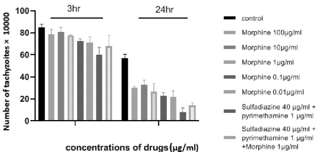
RESULTS Tachyzoite viability test
The number of tachyzoites of T. gondii was investigated in the presence or absence of various concentrations of morphine after 3 h and 24 h and a temperature of 24 °C (Sup- plementary Table 1). The results demonstrat- ed that tachyzoite proliferation decreased significantly in the presence of all concentra- tions of morphine compared to those ex- posed to no drugs after 24 h (P < 0.05) (Fig- ure 1). Furthermore, it was shown that expo- sure to different concentrations of morphine for 3 h was not effective and toxic when compared to the negative control. The growth inhibitory effects of the drugs were dose–dependent, such that 0.01 µg/ml and 100 µg/ml concentrations showed the most and the least efficacies, respectively, on in- hibiting the proliferation of tachyzoites of T.
gondii. The greatest differences were related
to morphine with concentration of
0.01 μg/ml and sulfadiazine plus py-
rimethamine (S+PYR) after 24 h.
Figure 1: Mean and standard deviation of the number of tachyzoites of T. gondii cultured in different concentrations of morphine in 3 h and 24 h compared to control groups (sulfadiazine plus pyrimetham- ine and untreated). Quantity is ×104.
Uninfected macrophage cells viability test Toxicity effects of morphine on uninfect- ed raw macrophage cells were explored by optical density (OD) following MTT assay to access the best concentration for further ex- periments. In this regard, the effects of 5 dif- ferent concentrations of morphine (100, 10, 1, 0.1, and 0.01 μg/ml) were measured after 24 h. According to the data, the percentage viability declined with increases in drug con- centrations. On the other hand, the concen- tration of 100 μg mL
−1had the maximum toxicity (Figure 2) at concentration of 0.01 μg/mL of morphine; the results were similar to untreated control group. In addition, the viability rate for both sulfadiazine plus py- rimethamine (S+PYR) alone and in combi- nation with morphine (S+PYR+M) were measured as 75.6 % and 73 %, respectively.
More details can be found in Supplementary Table 2.
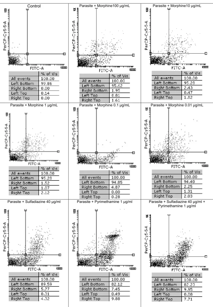
Flow cytometry analysis
In Figure 3 the results of flow cytometric analysis for tachyzoites with dissimilar con- centrations of morphine and with no drugs are shown. It was observed that the survival rates among five concentrations of morphine (100, 10, 1, 0.1 and 0.01 μg/ml) were
95.62 %, 95.28 %, 95.28 % , 94.85 %, and 94.40 %, respectively, after 3 h of treatment whereas the control group (tachyzoites) without treatment had 99.86 % viable para- sites. In groups treated with sulfadiazine (S), pyrimethamine (PYR) and sulfadiazine plus pyrimethamine (S+PYR) demonstrated more lethal effects on tachyzoites of T.
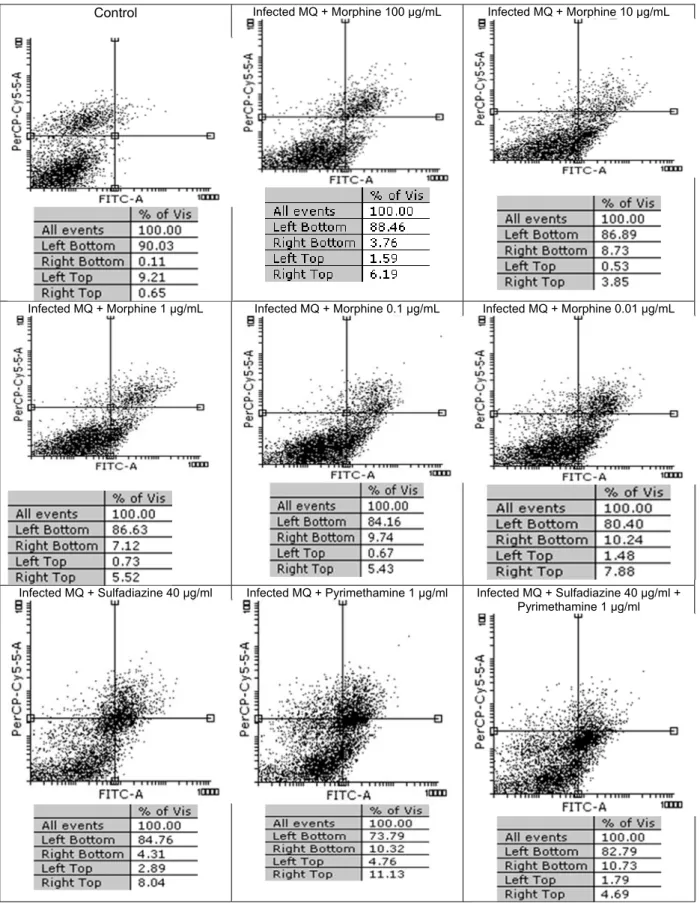
gondii rate than morphine group. The per- centage of apoptosis induced after 24 h in T.
gondii infected MQs were 5.35 %, 7.85 %, 9.26 %, 10.41 %, and 11.72 %, after being treated with 100, 10, 1, 0.1, and 0.01 µg/ml
Figure 2: Viability (%) and cytotoxicity of the un- infected RAW macrophages with dissimilar con- centrations of drugs and on control group (P <
0.05; ANOVA). M: Morphine, S: Sulfadiazine, PYR: Pyrimethamine
Control Parasite + Morphine100 μg/mL Parasite + Morphine10 μg/mL
Parasite + Morphine 1 μg/mL Parasite + Morphine 0.1 μg/mL Parasite + Morphine 0.01 μg/mL
Parasite + Sulfadiazine 40 µg/ml Parasite + Pyrimethamine 1 µg/ml Parasite + Sulfadiazine 40 µg/ml + Pyrimethamine 1 µg/ml
Figure 3: Flow cytometry results of the effect of morphine with dissimilar concentrations on tachyzoites of T. gondii viability and comparing them with the control group (untreated) after 3 h. Regions of quadrat show necrosis cells (propidium iodide positive) in left top, late apoptosis in right top, right bottom region belongs to apoptotic cells (annexin positive) and left bottom region be- longs to live cells.
of morphine, respectively, while in the nega- tive control group (macrophages) was 9.32 % (Figure 4). It was found that the max- imum toxicity was related to low concentra- tion (0.01 µg/ mL) of morphine. Additional- ly, the percentage of apoptosis in infected macrophages group treated with sulfadiazine (S), pyrimethamine (PYR) and sulfadiazine plus (S+PYR) pyrimethamine were 7.20 %, 15.08 % and 12.52 %, respectively. This shows that the drugs have no toxic effect on tachyzoites after 3 hours and were not signif- icantly different from control.
Quantification of parasite load of macrophage cells using quantitative real-time PCR (q-PCR)
The results demonstrate that parasite load was significantly reduced in MQs treated with drugs before infection compared with those in the MQs treated with drugs after in- fection (p<0.001). However, the parasite load decreased in both groups treated with drugs compared with control group without drugs. Low concentrations (e.g., 0.01 and 0.1 µg/ mL) of morphine were more effective in reducing parasite load than high concentra- tions (Table 1).
Quantification of parasite load of spleen tissues using quantitative real-time PCR (q-PCR)
The parasite load for all groups was measured by comparison with the standard curve and qPCR analysis. As represented in Table 2, compared to the untreated group (negative control), a remarkable decrease oc- curred in the parasite load in the drug groups (P < 0.001). The findings showed that the greatest reduction of parasite load was seen in the group that received morphine before being challenged with parasites.
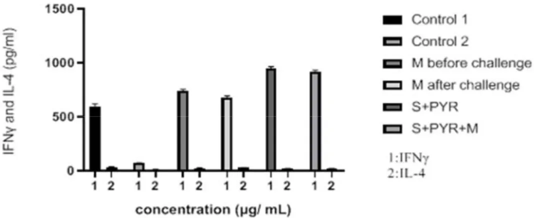
Cytokine assay
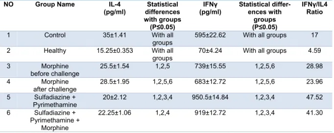
IFN-γ and IL-4 cytokine levels after 3 days of exposure in treated and untreated groups were estimated by ELISA. The mean IFN-γ level in the mice that received mor- phine before challenge was significantly higher than control group (P < 0.05) (Figure
5). Considerable differences in IFN-γ cyto- kine level were observed in the animals that received sulfadiazine plus pyrimethamine (SDZ+PYR) alone and in combination with morphine (M+S+PYR) in comparison with other groups (P < 0.05). It was found that there was no significant difference in IL4 measurements among all groups. On the oth- er hand, the mean IL4 level was similar in the treated and control groups (Table 3).
Survival rate measurement
For evaluation of survival rate, five mice in each group were treated, then followed for 10 days. Mortality in untreated infected groups started from day 5 after the infection and all mice died by the seventh day of the study. Mortality in the morphine before chal- lenge group started from day 7 after the in- fection and all mice died by the tenth day of the study. In the morphine after challenge group, the mortality of mice began from day 6 and all mice died by the eighth day. In ad- dition, there were no deaths in animals ex- posed to the sulfadiazine plus pyrimethamine (S+PYR) alone and in combination with morphine (M+S+PYR). The survival rates of the mice in the treatment and control groups are illustrated in Figure 6.
DISCUSSION
Despite that many effective prevention and treatment strategies exist, toxoplasmosis remains an enormous threat that unfortunate- ly lacks a global solution. Lack of sufficient budget, unacceptable toxicity, and several side effects of medicines have been identi- fied as major obstacles in achieving thera- peutic goals (Maubon et al., 2010; Pinzan et al., 2015). Potent new drugs are therefore re- quired to improve early treatment against toxoplasmosis.
In this study, we evaluated the preventive
and therapeutic effects of morphine on the
tachyzoites of Toxoplasma gondii in vitro
and in vivo. It is known that the use of opi-
ates such as morphine, apart from being an-
algesics, can have effects on both innate and
cell-mediated immunity and stimulate the
Figure 4: Flow cytometry results of the effect of morphine with 5 different concentrations (100, 10, 1, 0.1 and 0.01 μg/ml) on infected MQs viability and comparing them with the control group (untreated) after 24 h. Regions of quadrat show necrosis cells (propidium iodide positive) in left top, late apoptosis in right top, right bottom region belongs to apoptotic cells (annexin positive) and left bottom region be- longs to live cells.
Infected MQ + Morphine 10 μg/mL Infected MQ + Morphine 100 μg/mL
Control
Infected MQ + Morphine 0.01 μg/mL Infected MQ + Morphine 0.1 μg/mL
Infected MQ + Morphine 1 μg/mL
Infected MQ + Sulfadiazine 40 µg/ml + Pyrimethamine 1 µg/ml Infected MQ + Pyrimethamine 1 µg/ml
Infected MQ + Sulfadiazine 40 µg/ml
Table 1: Cycle of Threshold (CT) and parasite load test copy/reaction according to Real Time PCR method for macrophage cells before and after challenge with 1 × 10⁴ tachyzoite forms of T. gondii RH strain and control groups.
Groups parasite load of macrophage cells before treated with morphine
parasite load of macrophage cells after treated with morphine Cycle of
Threshold for Test
Parasite load Test Copy/Reaction
Cycle of Thresh- old for Test
Parasite load Test Copy/Reaction Control
(healthy macrophages)
30.50 0 30.50 0 Control
(infected macrophages)
11.71 376435 11.71 376435
Morphine 100 µg/ml 15.02 170715 20.65 350166 Morphine 10 µg/ml 15.51 153244 20.16 342787 Morphine 1 µg/ml 15.82 141479 17.55 289541 Morphine 0.1 µg/ml 16.23 104127 16.95 260122 Morphine 0.01 µg/ml 17.02 68848 16.12 239564 Sulfadiazine 40 µg/ml +
Pyrimethamine 1 µg/ml
20.70 7522 13.80 25443
Sulfadiazine 40 µg/ml + Pyrimethamine1 µg/ml
+ Morphine 1 µg/ml
19.71 13633 16.1 43076
Table 2: Cycle of Threshold (CT) and parasite load test copy/reaction according to Real Time PCR method for all treated and control groups
Number Test
Sample Type Cycle of Threshold for Test
Parasite load Test Copy/Reaction
1 Control
(healthy mice)
30.53 ND*
2 Control
(infected mice) 12.93 132545
3 Morphine before challenge 20.01 306
4 Morphine after challenge 14.67 29000
5 Sulfadiazine + Pyrimethamine 28.32 ND 6 Sulfadiazine + Pyrimethamine +
Morphine 28.16 ND
*ND = not detected
Table 3: Mean and standard deviation of interleukin 4 (IL-4) and interferon gamma (IFNγ) concentra- tions (pg/ml) and IFNγ/IL4 ratio in treated and control groups
NO Group Name IL-4
(pg/ml) Statistical differences with groups (P≤0.05)
IFNγ
(pg/ml) Statistical differ- ences with
groups (P≤0.05)
IFNγ/IL4 Ratio
1 Control 35±1.41 With all groups
595±22.62 With all groups 17 2 Healthy 15.25±0.353 With all
groups
70±4.24 With all groups 4.59 3 Morphine
before challenge
25.5±1.54 1,2,5 739±15.55 1,2,5,6 28.98 4 Morphine
after challenge 28.5±1.95 1,2,5,6 683±12.72 1,2,5,6 23.96 5 Sulfadiazine +
Pyrimethamine 20±2.12 1,2,3,4 950.5±14.84 1,2,3,4 47.52 6 Sulfadiazine +
Pyrimethamine + Morphine
22.25±1.06 1,2,4 919±12.72 1,2,3,4 41.30
Figure 5: Levels of IFNγ and IL-4 (pg/ml) cytokines secreted from spleen lymphocyte culture in test and control groups after 72 h stimulation with Toxoplasma Lyzate Antigen. S: Sulfadiazine (40 mg/kg/day), PYR: Pyrimethamine (1 mg/kg/day), M: Morphine (1 mg/kg, once a week).
Figure 6: Survival rates of treated and control BALB/c mice during 10 days after and before challenge with 1×10⁴ tachyzoite forms of T. gondii RH strain (5 mice per group). One group treated with mor- phine (1 mg/kg, once a week) for 3 weeks before challenge with 1×10⁴ tachyzoite forms of T. gondii RH strain. The rest of the groups after infection treated with morphine alone (1 mg/kg, once a week), sulfadiazine (40 mg/kg/day) pluse pyrimethamine (1 mg/kg/day), and sulfadiazine (40 mg/kg /day) plus pyrimethamine (1 mg/kg /day) in combination with morphine (1 mg/kg, once a week). We used healthy mice as a negative control group, which just received PBS whereas the positive control group was in- fected with tachyzoites.
immune responses on different parasitic in- fections (Liang et al., 2016; Sharp, 2004).
Consequently, the use of morphine, because of its therapeutic implications, has become
popular in recent years. Opiates have been shown to modify immune responses directly with the help of opioid receptors on immune cells, while they can act indirectly through
0 20 40 60 80 100 120
1 2 3 4 5 6 7 8 9 10 Healthy mice
Control
Morphine before challnge Morphine after challege Sulfadiazine + pyrimethamine Sulfadiazine + pyrimethamine + Morphine
opioid receptors in the central nervous sys- tem (CNS) (Salimi et al., 2013; Singal and Singh, 2005). It should be noted, following morphine binding to opioid receptors, with nitric oxide released and vasodilation pro- moted reduce the healing process in cutane- ous leishmaniasis (Alavi-Naini, 2008).
Nevertheless, several publications have confirmed that prolonged use of morphine attenuates the general functions of the im- mune cells including macrophages and pol- ymorphonuclear leukocytes resulting in sup- pression of the immune system and increased susceptibility to a variety of infectious dis- eases (Friedman et al., 2006; Risdahl et al., 1993; Wang et al., 2005).
The results of the current study revealed that morphine inhibited the proliferation of tachyzoites of T. gondii in a concentration- and time-dependent manner and provided the best result after 24 hours for all concentra- tions. Notably, a morphine concentration of 0.01 µg/mL was most effective in inhibiting tachyzoites growth after 24 h incubation. In a previous study of promastigotes of Leishma- nia major, the results were similar (Jabari et al., 2019).
Sulfadiazine plus pyrimethamine in combination with morphine was also effec- tive in reducing tachyzoites numbers like the reference drug (S+PYR). It was found that the viability of macrophage cells exposed to morphine was also to be based on a dose- dependent response. The results of MTT showed that as concentration of morphine was increased, uninfected macrophage cell viability was reduced. The demonstration of apoptosis and necrosis by flow cytometry has been previously verified in uninfected MQs with different morphine concentrations and the results showed low apoptosis and necrosis (Ebrahimisadr et al., 2018). Also, morphine was able to induce little apoptosis in parasite-infected MQs. Compared to the control group (MQs without treatment) in- duction of necrosis was reduced in infected MQs during treatment with morphine. More- over, apoptosis occurred in tachyzoites of T.
gondii after exposure with different mor- phine concentrations, although these apop-
totic effects were not significant. Morphine showed lower toxicity upon infected MQs and parasite compared with sulfadiazine (S), pyrimethamine (PYR) and sulfadiazine plus pyrimethamine (S+PYR) groups. Hence, it can be assumed that morphine may act as an appropriate anti-Toxoplasma agent through amplification of the immune response and expression of cell level receptors. With re- gard to the preventive and/or adjuvant role of morphine, when the MQs were treated be- fore tachyzoite infection in vitro, a signifi- cant reduction was observed in parasite bur- den. It was also shown that the antiparasite behavior of morphine was dose-dependent.
Interestingly, the lowest parasite burden was at a concentration of 0.01 μg/mL morphine.
MQs are essential for generation of numer- ous immune responses by enhanced phago- cytosis and secrete particular cytokines in combatting toxoplasmosis. Also, MQs are a significant site of action for opioid drugs (Cabral et al., 2018; Ebrahimisadr et al., 2018; Pacifici et al., 1994). Therefore, it can be concluded that morphine can induce a strong immune response with enhancing de- fensive function of the MQs as well as in- flammatory cytokines to confine this parasit- ic disease. The results obtained from the mu- rine model demonstrated that the parasitic burden was decreased significantly in mice treated with morphine but this reduction in those received morphine before challenge was more specific. Also, a considerable re- duction in the parasitic load was observed in groups treated with sulfadiazine plus py- rimethamine alone or in combination with morphine. There was significant relationship between survival rate and parasitic burden.
The mortality rate in groups that received
morphine before parasitic challenge was
lower than those received morphine after
parasitic challenge. In the context of the tox-
oplasmosis in mouse model, production of
IFN-γ from immune system cells as the
adaptive cellular immune response has a
substantial role in the controlling and re-
stricting growth of the parasite and also in-
duce a strong Th1 type immune response
with the help of MQs (Foroutan et al., 2019;
Krishnamurthy et al., 2017). Levels of inter- feron-gamma (IFNγ) production were high in mice treated with morphine before infec- tion (just like the sulfadiazine plus py- rimethamine) compared with those in the no drug group, whereas levels of interleukin 4 (IL-4) productions were low across all treat- ment groups. Hence, it can be inferred that the morphine through boosting the immune system reduces disease due to T. gondii in BALB/c mice.
However, the presented results demon- strate that morphine has an acceptable ability to treat infection due to Toxoplasma gondii under both in vitro and in vivo conditions.
CONCLUSION
Based on the results of in vitro and in vi- vo conditions, antiparasitic activity of mor- phine on tachyzoites of Toxoplasma gondii and infected macrophages was promising.
Also, morphine in pretreated mice showed higher anti-Toxoplasma behavior than mor- phine in posttreated mice. On the other hand, the preventive effects of morphine were stronger than the therapeutic effects and therefore, morphine can be used as an ad- junctive treatment at low doses for the treat- ment of toxoplasmosis. Furthermore, the ef- ficiency of morphine is increased when it is used in combination therapy with sulfadia- zine plus pyrimethamine (S+PYR). Howev- er, the results of the study suggest that mor- phine can be considered as an additional drug either alone or along with other anti- Toxoplasma products for treatment in future studies.
Acknowledgment
The authors would like to thank all staff of Department of Parasitology of Tarbiat Modares University, Iran. This paper is is- sued from thesis of Leila Zaki, Ph.D student of Medical Parasitology.
Ethical statement
This experiment was approved by the Research Department of Medical Sciences, Tarbiat Modares University, with issue num-
ber 52D/1883 and approved by the Ethical Committee with issue number 52D/1239.
Conflict of interest
The authors declare that they have no conflict of interest.
REFERENCES
Alavi-Naini R. Topical morphine for the treatment of cutaneous leishmaniasis. Med Hypotheses. 2008;70:
81-4.
Antczak M, Dzitko K, Dlugonska H. Human toxo- plasmosis - searching for novel chemotherapeutics.
Biomed Pharmacother. 2016;82:677-84.
Cabral GRA, Wang ZT, Sibley LD, DaMatta RA. In- hibition of nitric oxide production in activated macro- phages caused by Toxoplasma gondii infection occurs by distinct mechanisms in different mouse macro- phage cell lines. Front Microbiol. 2018;9:1936.
Dinda A, Gitman M, Singhal PC. Immunomodulatory effect of morphine: therapeutic implications. Expert Opin Drug Saf. 2005;4:669-75.
Dubey JP. The history of Toxoplasma gondii—the first 100 years. J Eukaryot Microbiol. 2008;55:467- 75.
Ebrahimisadr P, Ghaffarifar F, Horton J, Dalimi A, Sharifi Z. Apoptotic effect of morphine, imiquimod and nalmefene on promastigote, infected and unin- fected macrophages with amastigote of Leishmania major by flow cytometry. Iran J Pharm Res. 2018;17:
986-94.
Fallahi S, Rostami A, Nourollahpour Shiadeh M, Behniafar H, Paktinat S. An updated literature review on maternal-fetal and reproductive disorders of Toxo- plasma gondii infection. J Gynecol Obstet Hum Re- prod. 2018;47:133-40.
Fishman JA. Opportunistic infections—coming to the limits of immunosuppression? Cold Spring Harb Per- spect Med. 2013;3:a015669.
Foroutan M, Zaki L, Ghaffarifar F. Recent progress in microneme-based vaccines development against Tox- oplasma gondii. Clin Exp Vaccine Res. 2018;7:93- 103.
Foroutan M, Zaki L, Tavakoli S, Soltani S, Taghipour A, Ghaffarifar F. Rhomboid antigens are promising targets in the vaccine development against Toxoplas- ma gondii. EXCLI J. 2019;18:259-72.
Friedman H, Pross S, Klein TW. Addictive drugs and their relationship with infectious deseases. FEMS Immunol Med Microbiol. 2006;47:330-42.
Fuggetta M, Di Francesco P, Falchetti R, Cottarelli A, Rossi L, Tricarico M, et al. Effect of morphine on cell-mediated immune responses of human lympho- cytes against allogeneic malignant cells. J Exp Clin Cancer Res. 2005;24:255.
Gharavi MJ, Jalali S, Khademvatan S, Heydari S. De- tection of IgM and IgG anti-Toxoplasma antibodies in renal transplant recipients using ELFA, ELISA and ISAGA methods: comparison of pre- and post- transplantation status. Ann Trop Med Parasitol. 2011;
105:367-71.
Jabari J, Ghaffarifar F, Horton J, Dalimi A, Sharifi Z.
Evaluation of morphine with imiquimod as opioid growth factor receptor or nalmefene as opioid blocking drug on leishmaniasis caused by Leishmania major in vitro. Iran J Parasitol. 2019;14:394-403.
Krishnamurthy S, Konstantinou EK, Young LH, Gold DA, Saeij JP. The human immune response to Toxo- plasma: autophagy versus cell death. PLoS Pathog.
2017;13:e1006176.
Liang X, Liu R, Chen C, Ji F, Li T. Opioid system modulates the immune function: a review. Transl Perioper Pain Med. 2016;1:5-13.
Maubon D, Bougdour A, Wong Y-S, Brenier-Pinchart M-P, Curt A, Hakimi M-A, et al. Activity of the histone deacetylase inhibitor FR235222 on Toxo- plasma gondii: inhibition of stage conversion of the parasite cyst form and study of new derivative compounds. Antimicrob Agents Chemother. 2010;54:
4843-50.
Montoya JG, Gomez CA. Toxoplasmosis after solid organ transplantation. In: Ljungman P, Snydman D, Boeckh M (eds.): Transplant infections. 4th ed. (pp 781-93). Cham: Springer, 2016.
Montoya J, Liesenfeld O. Toxoplasmosis. Lancet.
2004;363:1965-76.
Pacifici R, Patrini G, Venier I, Parolaro D, Zuccaro P, Gori E. Effect of morphine and methadone acute treatment on immunological activity in mice: pharma- cokinetic and pharmacodynamic correlates. J Pharma- col Exp Ther. 1994;269:1112-6.
Pinzan CF, Sardinha-Silva A, Almeida F, Lai L, Lopes CD, Lourenço EV, et al. Vaccination with recombinant microneme proteins confers protection against experimental toxoplasmosis in mice. PloS One. 2015; 10:e0143087.
Risdahl JM, Peterson PK, Chao CC, Pijoan C, Molitor TW. Effects of morphine dependence on the patho- genesis of swine herpesvirus infection. J Infect Dis.
1993;167:1281-7.
Salimi V, Hennus MP, Mokhtari-Azad T, Shokri F, Janssen R, Hodemaekers HM, et al. Opioid receptors control viral replication in the airways. Crit Care Med. 2013;41:205-14.
Sharp BM. Opioid receptor expression and function. J Neuroimmunol. 2004;147:3-5.
Sharp BM, Roy S, Bidlack JM. Evidence for opioid receptors on cells involved in host defense and the immune system. J Neuroimmunol. 1998;83:45-56.
Singal P, Singh PP. Leishmania donovani amastigote component-induced colony-stimulating factor pro- duction by macrophages: modulation by morphine.
Microbes Infect. 2005;7:148-56.
Singal P, Kinhikar AG, Singh S, Singh PP. Neuro- immunomodulatory effects of morphine in Leish- mania donovani-infected hamsters. Neuroimmuno- modulation. 2002;10:261-9.
Singh PP, Singh S, Dutta G, Srimal R. Immuno- modulation by morphine in Plasmodium berghei- infected mice. Life Sci. 1994;54:331-9.
Wang J, Barke RA, Charboneau R, Roy S. Morphine impairs host innate immune response and increases susceptibility to Streptococcus pneumoniae lung infection. J Immunol. 2005;174:426-34.
Wang ZD, Liu HH, Ma ZX, Ma HY, Li ZY, Yang ZB, et al. Toxoplasma gondii infection in immuno- compromised patients: a systematic review and meta- analysis. Front Microbiol. 2017;8:389.