Characterization of the Differential Significance of Sugar Import in the Apicomplexan Parasites Toxoplasma gondii
and Plasmodium
Dissertation
zur Erlangung des akademischen Grades doctor rerum naturalium
(Dr. rer. nat.) im Fach (Biologie)
eingereicht an der Mathematisch-Naturwissenschaftlichen Fakultät I der Humboldt-Universität zu Berlin
von
Diplom Biophysiker Martin Blume
Präsident der Humboldt-Universität zu Berlin Prof. Dr. Jan-Hendrik Olbertz
Dekan der Mathematisch-Naturwissenschaftlichen Fakultät I Prof. Dr. Andreas Herrmann
Gutachter/innen: 1. Prof. Richard Lucius 2. Prof. Kai Matuschewski 3. Prof. Dominque Soldati-Favre
Tag der mündlichen Prüfung: 30.05.2011
I
Acknowledgements
I would like to thank my supervisor Nishith Gupta, who was always present and approachable during the last years for his demanding support.
I would also like to thank Richard Lucius for his support and the opportunity to work in his department.
I would like to thank Kai Matuschewski for the openness that founded a fruitful collaboration.
Also big thanks to the members of Kai’s lab: Diana, Manu, Caro, Georgina, Katja B., Elyzana, Alyssa, Caro, Katja M., Markus, Taco and Joana for the very friendly working at- mosphere and all the technical help.
I also would like to thank Dominique Soldati for letting me spend pivotal months in her lab and for the support afterwards.
Thanks also to Tobias and Paco for the funny and inspiring beer hours.
Special thanks to Frank Seeber for very useful advice and with many clarifying discussions.
I would like to thank our collaborators Scott Landfear, Dayana Rodriguez-Contreras and Marco Sanchez that contributed a lot of data and advice to this research. I also would like to thank David Roos and Dhanasekaran Shanmugam for perfoming the microarray experiments for us.
Big thanks to Christian, Veikko, Dingo and Martin and Gregor for being such good and un- complicated friends. Thank you, my dear parents, for your unconditional support.
Marion, thank you for taking care of me inside and outside of the lab.
I want to thank all the members of the Molecular Parasitology department for making this place a very friendly working environment. Special thanks to Grit for her incredible multi- tasking skills and for managing the lab and creating a very familiar atmosphere. I also thank to Jörg and Vera for “splitting cells” together and for discussions.
Finally, I would like to thank the organizers of the MDC PhD program. It is a pleasure to carry out a dissertation in such a terrific environment.
III
Abstract
Toxoplasma gondii and Plasmodium species are obligate intracellular pathogens that utilize host sugars for energy homeostasis, macro molecular synthesis and to generate glycoconju- gates, which are all important to their survival and/or virulence. Here, we report that expres- sion of the T. gondii glucose transporter, TgGT1, and of its homologs of P. falciparum and P.
berghei (PfHT1 and PbHT1) can restore the transport of glucose, mannose, galactose and fructose in a Leishmania mexicana null mutant. Besides TgGT1, Toxoplasma harbours three additional putative sugar transporters (TgST1-3), of which only TgGT1 and TgST2 localize to the parasite surface, while TgST1 and TgST3 remain intracellular. Surprisingly, TgGT1 and TgST2 are nonessential in T. gondii tachyzoites as their individual and collective ablation inflicts only a 30% or no defect in the parasite replication, respectively. The Δtggt1 mutant is unable to import glucose and consequently displays an attenuated glucose-dependent motility, which is completely rescued by glutamine. The lack of exogenous glucose in the parasite cul- ture prompts T. gondii to procure glutamine to sustain its metabolism. Unexpectedly, the in vivo virulence of the Δtggt1 in mice remains unchanged. Taken together, these data demon- strate that host-derived glucose is absolutely nonessential for T. gondii tachyzoites and under- score glutamine as a complement substrate. Furthermore, the Δtggt1 strain provides a model for further investigating metabolic interactions between T. gondii and its host cell, and high- lights its adaptation to disparate host cells.
In contrast to T. gondii, erythrocytic stages of Plasmodium species critically depend on glu- cose uptake, and the PfHT1 transporter is considered as a drug target against human malaria.
Here, we report that PbHT1 (a PfHT1 homolog) is also essential for blood stage development in the rodent malaria parasite P. berghei. PbHT1 is expressed throughout the life cycle. More- over, a PfHT1- and PbHT1-specific sugar analogue, compound 3361, can inhibit the hepatic development and ookinete formation in P. berghei. These results signify that PbHT1 and ex- ogenous glucose are also required during the ex-erythrocytic stages of P. berghei. To permit a high-throughput screening of selective PfHT1 inhibitors and their subsequent in vivo assess- ment, we have established a Saccharomyces cerevisiae mutant expressing codon-optimized PfHT1, and generated a PfHT1-dependent Δpbht1of P. berghei strain, respectively. This re- search provides a platform that would facilitate the development of drugs against human ma- laria, and suggests a disease control aspect by preventing the parasite transmission. Collec-
tively, this thesis underscores various previously unknown aspects of sugar metabolism in Toxoplasma and Plasmodium, and unravel their metabolic differences.
Keywords:
Toxoplasma gondii, Plasmodium, glucose, sugar, transporter, metabolism
V
Zusammenfassung
Toxoplasma gondii und Plasmodium Spezies sind obligat intrazelluläre Parasiten, die Zucker zur Energiehomöostase als auch für die Synthese lebenswichtiger und pathogener Glycokon- jugate und Makromoleküle verwenden. Die hier vorgestellten Daten zeigen, dass der in einer Leishmania mexicana Nullmutante exprimierte Glukosetransporter von T. gondii, TgGT1, und die homologen Transporter von P. falciparum und P. berghei, PfHT1 und PbHT1, neben Glukose auch Mannose, Fructose und Galactose transportieren. Toxoplasma Tachyzoiten exprimieren neben TgGT1 noch drei weitere putative Zuckertransporter (TgST1-3), von de- nen allerdings nur TgST2 an der Parasitenoberfläche lokalisiert. TgST1 und TgST3 verblei- ben intrazellulär. Erstaunlicher Weise sind TgGT1 und auch TgST2 nicht essentiell, wie durch ihre individuelle und gleichzeitige Gendeletion belegt wird. Die Gendeletion von TgST2 bewirkt keinen, und Deletion von TgGT1 lediglich einen 30%igen Wachstumsdefekt.
Die TgGT1 Mutante, Δtggt1, zeigt keine Glukoseaufnahmeaktivität und folglich eine vermin- derte glukoseabhängige Motilität. In Δtggt1 Parasiten wird ein verstärkter Glutaminstoff- wechsel nachgewiesen, der anscheinend ausreichend ist dessen Motilität und Replikation zu erhalten. Diese Tatsache wird durch die unverminderte Virulenz des Δtggt1 Stammes in Mäu- sen weiterhin bestätigt. Zusammenfassend zeigen diese Daten, dass die Glukoseaufnahme aus dem Wirt keine essentielle Rolle für Toxoplasma Tachyzoiten spielt und, dass sie komplett durch Glutamin ersetzt werden kann. Weiterhin erweist sich Δtggt1 als hervorragendes Mo- dell, um die Parasiten-Wirts-Wechselwirkung auf der Stoffwechselebene zu untersuchen. Es gewährt Einblick in die Anpassungsfähigkeit von Toxoplasma gondii an unterschiedliche Wirtszellen.
Im Gegensatz zu Toxoplasma benötigen erythrozytäre Plasmodien Glukose und der Transpor- ter PfHT1 wird derzeit als drug-target eingestuft. In dieser Arbeit wird gezeigt, dass das PfHT1-Homolog, PbHT1, essentiell in Blutstadien des Nagerparasiten Plasmodium berghei ist, jedoch auch während des gesamten Lebenszyklus des Parasiten exprimiert wird. Ein PfHT1- und PbHT1-spezifischer Inhibitor (Compound 3361) kann die Entwicklung von Le- berstadien und die Ookinetenbildung stark hemmen, was die Notwendigkeit der Zuckerauf- nahme in diesen exoerythrozytären Stadien bezeichnet. Um zukünftig PfHT1-Inhibitoren im Hochdurchsatzverfahren zu identifizieren und ihre in vivo Aktivität testen zu können, wurden auf Saccharomyces cerevisiae und P. berghei basierende Expressionssysteme für PfHT1 ent- wickelt. Diese Plattform sollte die Entwicklung von antiplasmodiellen Zuckeranaloga verein-
fachen. Unsere Daten zeigen weiterhin eine beeinträchtigte Parasitentransmission von C3361- behandelten Mäusen und deuten damit auf neue Gesichtspunkte hinsichtlich der Wirkung die- sen Verbindungen hin.
Abschließend stellt diese Arbeit die Unterschiedlichkeit des zentralen Kohlenstoffwechsels von Toxoplasma und Plasmodium Parasiten durch bisher unbekannte Aspekte heraus.
Schlagwörter:
Toxoplasma gondii, Plasmodium, Glukose, Zucker, Transporter, Metabolismus
VII
Table of Contents
Acknowledgements...I Abstract ... III Zusammenfassung... V Table of Contents... VII List of Figures ...XI List of Tables... XIII List of Abbreviations... XV
1 Introduction ... 1
1.1 Apicomplexan Parasites ... 1
1.1.1 The life cycle of Toxoplasma gondii... 2
1.1.2 The life cycle of Plasmodium... 3
1.2 Replication Compartments in Parasitized Host Cells ... 4
1.2.1 Toxoplasma gondii... 4
1.2.2 Plasmodium spp... 5
1.3 Central Carbon Metabolism of Hexoses ... 5
1.3.1 Glycolysis and apicoplast synthesis pathways... 6
1.3.2 TCA cycle ... 7
1.3.3 The pentose phosphate pathway (PPP) pathway ... 7
1.4 Protein and Lipid Synthesis Using Hexoses... 8
1.4.1 Lipid synthesis in Toxoplasma and Plasmodium... 8
1.4.2 Protein glycosylation in T. gondii and Plasmodium... 8
1.5 Sugar Import in Toxoplasma and Plasmodium... 9
1.5.1 Sugar transport proteins ... 9
1.5.2 Glucose uptake in Toxoplasma gondii... 10
1.5.3 Glucose uptake in Plasmodium... 10
1.6 Aim of This Study ... 11
2 Materials and Methods ... 13
2.1 Materials ... 13
2.1.1 Biological resources... 13
2.1.2 Chemical reagents... 14
2.1.3 Vectors ... 15
2.1.4 Antibodies and working dilutions... 16
2.1.5 Enzymes... 17
2.1.6 Instruments... 17
2.1.7 Plasticware and disposables... 17
2.1.8 Commercial kits... 18
2.1.9 Reagent preparations... 18
2.2 Methods - Culture and Transfection... 23
2.2.1 Propagation of mammalian cells... 23
2.2.2 Propagation of Toxoplasma gondii tachyzoites ... 23
2.2.3 Toxoplasma gondii transfection... 23
2.2.4 Giemsa staining of blood smears of P. berghei... 23
2.2.5 P. berghei propagation and transfection ... 23
2.2.6 Ookinete culture and purification ... 24
2.2.7 Propagation of Anopheles stephensi mosquitoes ... 25
2.2.8 Preparation of oocysts and salivary gland sporozoites of P. berghei... 25
2.2.9 Liver stages of P. berghei... 25
2.2.10 Making of Saccharomyces cerevisiae competent cells... 25
2.2.11 Transformation of S. cerevisiae competent cells ... 26
2.3 Methods - Molecular Cloning ... 26
2.3.1 PCR reactions... 26
2.3.2 Agarose gel-electrophoresis... 26
2.3.3 Vector preparation ... 27
2.3.4 Insert preparation ... 27
2.3.5 Ligation... 27
2.3.6 Preparation of competent Escherichia coli cells... 27
2.3.7 Transformation of Escherichia coli... 27
2.3.8 DNA preparations ... 28
2.3.9 RNA preparation and cDNA synthesis ... 28
2.4 Methods - Assays... 29
2.4.1 Indirect immuno-fluorescence assay (IFA) ... 29
2.4.2 Glucose and glutamine uptake assays with intracellular T. gondii... 29
2.4.3 Plaque and replication assays... 29
2.4.4 Motility assay... 30
2.4.5 Functional expression of PfHT1, PbHT1, TgST1-3 and TgGT1 in Leishmania mexicana and Xenopus laevis oocytes ... 30
3 Results... 35
3.1 Host-derived Glucose and Its Transporter in the Obligate Intracellular Parasite Toxoplasma gondii are Dispensable by Glutaminolysis... 35
3.1.1 TgGT1 is capable of transporting major sugars in L. mexicana null mutant... 35
3.1.2 TgGT1 is not essential to the survival of T. gondii... 37
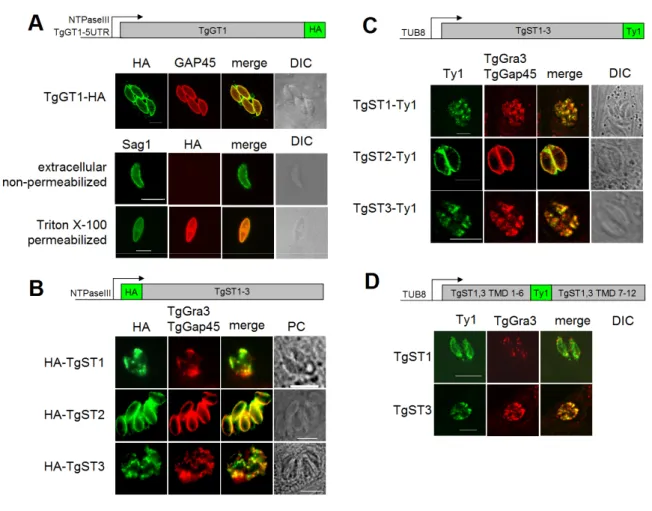
3.1.3 T. gondii tachyzoites express three additional novel sugar transporters... 38
3.1.4 TgGT1 and TgST2 localize to the surface of T. gondii, whereas TgST1 and TgST3 are intracellular. ... 39
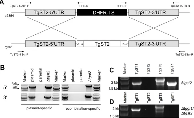
3.1.5 TgST2 is dispensable for T. gondii tachyzoite survival and is not redundant with TgGT1... 41
3.1.6 Only TgGT1 gene deletion confers a modest growth defect in T. gondii... 42
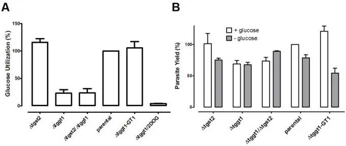
3.1.7 The ∆tggt1 but not Δtgst2 mutant display attenuated glucose metabolism ... 45
3.1.8 In vitro survival of T. gondii does not require host-derived glucose ... 46
3.1.9 In vivo virulence of T. gondii does not require host-derived glucose... 46
3.1.10 Glutamine and glucose are the major nutrients for Toxoplasma gondii... 47
3.2 A Constitutive Pan-Hexose Permease for the Plasmodium Life Cycle and Transgenic Models for Screening of Anti-Malarial Sugar Analogs... 50
3.2.1 PbHT1 and PfHT1 are functionally homologous pan-hexose permeases ... 50
3.2.2 PbHT1 is indispensable for the intra-erythrocytic stages of P. berghei... 55
3.2.3 Sexual development of P. berghei is inhibited by a glucose analog ... 56
3.2.4 Glucose is required for the hepatic development of P. berghei... 58
3.2.5 In vivo Δpbht1-PfHT1 mouse model for assessment of PfHT1 inhibitors ... 63
3.2.6 PfHT1-complemented S. cerevisiae for screening of PfHT1 inhibitors ... 65
4 Discussion ... 69
IX
4.1 Implications on the metabolism of Toxoplasma gondii... 69
4.1.1 Major nutrients of T. gondii... 69
4.1.2 Changes in gene expression of Δtggt1... 70
4.1.3 Novel aspects of bioenergetic metabolism of T. gondii... 72
4.2 Potential function of TgST1, TgST2, TgST3 ... 72
4.3 Metabolic Interactions Between Toxoplasma and Its Host Cell ... 73
4.3.1 The central carbon metabolism of the host cell and the parasitic requirements... 73
4.4 Outlook... 75
4.5 The Hexose Permeation Pathway of Plasmodium... 76
4.5.1 The pan-hexose nature of PfHT1 and PbHT1... 76
4.5.2 The essentiality for Plasmodium hexose transporters... 77
4.5.3 PfHT1 as a drug target ... 77
4.6 Comparative Discussion ... 78
4.6.1 Why is hexose import essential for Plasmodium but not for Toxoplasma?... 78
5 Conclusion ... 83
References ... 85
List of Publications... 95
Appendix A ... 97
Appendix B... 98
Appendix C ... 99
Eidesstattliche Erklärung ... 101
XI
List of Figures
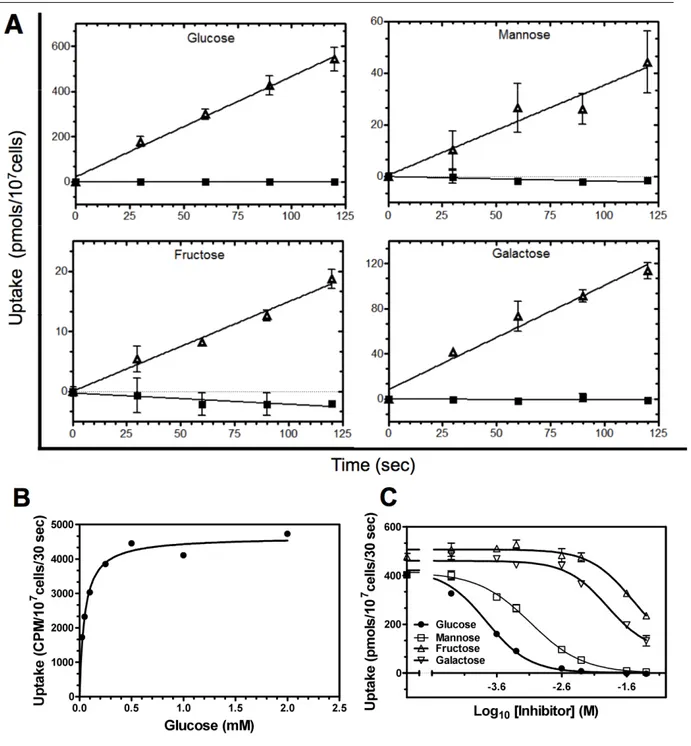
Figure 1: Canonical representation of an apicomplexan parasite. ... 1 Figure 2: Schematic life cycle of Toxoplasma gondii... 3 Figure 3: Life cycle of Plasmodium falciparum... 4 Figure 4: TgGT1 can mediate the import of glucose, mannose, fructose, and galactose in
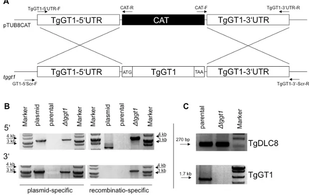
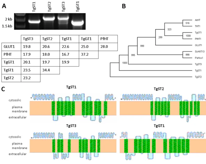
L. mexicana null mutant (Δlmgt)... 36 Figure 5: TgGT1 is not essential for in vitro survival of T. gondii. ... 37 Figure 6: Toxoplasma gondii expresses four sugar permeases. ... 39 Figure 7: Toxoplasma expresses three novel sugar permeases TgST1, TgST2 and
TgST3, of which only TgST2 resides in the plasma membrane. ... 40 Figure 8: TgST2 is expendable in parental and Δtggt1 parasites. ... 42 Figure 9: Δtggt1 but not Δtgst2 strain of T. gondii demonstrates a protracted in vitro
growth... 44 Figure 10: The Δtggt1 but not Δtgst2 strain of T. gondii is compromised in utilizing
host-derived glucose, and exhibits a delayed replication... 45 Figure 11: Virulence of T. gondii RH hxgprt- parasites in mice does not require glucose... 47 Figure 12: Glutamine fulfills the metabolic needs of the Δtggt1 mutant. Freshly-
harvested tachyzoites were used to perform the motility assays... 49 Figure 13: PfHT1 and PbHT1 mediate time-dependent uptake of four hexoses in
Leishmania mexicana and Xenopus laevis oocytes... 51 Figure 14: Glucose and mannose exhibit high-affinity towards PbHT1 and PfHT1,
where as fructose and galactose are low affinity ligands. ... 53 Figure 15: PbHT1, PfHT1, TgGT1 and EtHT1 are homologous proteins... 54 Figure 16: PbHT1 is essential in blood stage parasites... 56 Figure 17: A glucose analog, C3361, can inhibit glucose transport by PbHT1, and
reduces transmission and/or ookinete formation of P. berghei... 58 Figure 18: PbHT1 is expressed during liver stages, and C3361 attenuates the hepatic
development of P. berghei. ... 60 Figure 19: C3361 does not influence the replication of Toxoplasma gondii. ... 62 Figure 20: PfHT1 complementation-based PbHT1 deletion in P. berghei. ... 63 Figure 21: Transgenic P. berghei as a model for the in vivo assessment of PfHT1
inhibitors... 64 Figure 22: Yeast-optimized PfHT1 can rescue the growth of the S. cerevisiae mutant on
glucose and mannose, and confers a model for high-throughput screening of
analog-based PfHT1 inhibitors... 66 Figure 23: PfHT1 fusion transporters localize peripheral and confer growth rescue on
glucose... 67 Figure 24: C3361 inhibits PfHT1-mediated growth of Saccharomyces cerevisiae on
glucose... 68 Figure 25: Principal component analysis of Δtggt1 and ΔKU80 transcriptomes. ... 70
Figure 26: Toxoplasma-induced modification of the host metabolism in parasitized
cells... 75 Figure 27: The central carbon metabolism of Toxoplasma gondii and Plasmodium
species. ... 81 Figure 28: Alignment of TgST1, TgST2, TgST3, TgGT1 and HsGLUT1... 97 Figure 29: Mis-localization of TgGT1. ... 98
XIII
List of Tables
Table 1: Primer sequences for indicated purposes and used restriction site used for
cloning... 31 Table 2: Affinities of TgGT1 towards four hexoses. ... 35 Table 3: In vitro doubling rates of T. gondii tachyzoites in human foreskin fibroblasts. ... 44 Table 4: Genes of glutaminolysis and gluconeogenesis in Toxoplasma gondii and
Plasmodium... 79
XV
List of Abbreviations
ACT Artemisinin based combination therapies BCAA branched-chain amino acids
BSA bovine serum albumin fraction 5 CAT chloramphenicol acyltransferase cDNA complementary deoxyribonucleic acid cRNA coding ribonucleic acid
CSP circum sporozoite protein
DAPI 4',6-diamidino-2-phenylindole DEPC diethylpyrocarbonate
DMEM Dulbecco’s Modified Eagle Medium DMSO dimethyl sulfoxide
DNA deoxyribonucleic acid EDTA ethylendiamine-tetraacetate EGTA ethylene glycol tetraacetic acid
ER endoplasmatic reticulum
FAS 1/2 fatty acid synthesis type 1/2 pathway FBA flux-balance analysis
FCS fetal calf serum
G6Pase glucose-6-phosphatase
GPI glycosylphosphatidylinositol
H.O.S.T host organelle sequestering tubulo-structures HA Haemaglutinin
HBSS Hank’s Balanced Salt Solution HFF Human foreskin fibroblast HIF1 hypoxia-inducible factor 1
HPLC high performance liquid chromatography
HXGPRT hypoxanthine-xanthine-guanine phosphoribosyl transferase IPC inositol hosphoceramide
IPTG isopropyl-β-D-thiogalactopyranosid LDL low-density lipoprotein LiAc lithium acetate
MB megabases
MOPS morpholino propanesulfonic acid MTOC microtubule organizing center NMRI Naval Medical Research Institute PAS periodic acid-Schiff
PBS phosphate buffered saline PCR polymerase chain reaction PDH pyruvate dehydrogenase PEG polyethylenglycol
Pf/PbHT1 Plasmodium falciparum / berghei hexose transporter
Pfi/oTPT Plasmodium falciparum inner/outer triose phosphate transporter
PV parasitophorous vacuole
PVM parasitophorous vacuole membrane
RNA ribonucleic acid
ROP rhoptry secreted proteins ROS radical oxygen species rpm rotation per minute
rt-PCR reverse transcription polymerase chain reaction SAM significance analysis of microarray
SDS sodium dodecyl sulphate spp species
TCA pentose phosphate pathway
TE Tris EDTA
TFR transferrin receptor
TgAPT Toxoplasma apicoplast membrane-localized phosphate translocator TMHMM Transmembrane Hidden-Markov model
1 Introduction
1.1 Apicomplexan Parasites
Apicomplexan parasites constitute a phylum of diverse but evolutionarily related protists that follow an obligate intracellular life style. Two major classes of this phylum are the Coccidia (Toxoplasma gondii, Eimeria and Cryptosporidia) and the Hematosporidia (Plasmodia, Theil- eria and Babesia). The denominating structural feature is their apical complex that is involved in host cell invasion, and consists of the conoid (a conical tubular structure) and secretory organelles (rhoptries and micronemes). Invasive stages of apicomplexans share a similar cell architecture (Striepen et al., 2007). The cellular morphology (Fig. 1) is conserved by a micro- tubule network that underlies the inner membrane complex, which itself is firmly connected to the plasma membrane. All apicomplexans posses an apicoplast, a single golgi stack and an endoplasmatic reticulum network which surrounds the nucleus.
Figure 1: Canonical representation of an apicomplexan parasite.
AP apicoplast; AR apical rings; CC centrocone; CE centrosome; CO conoid; DG dense granule; ER endoplasmic reticulum; G Golgi; IMC inner membrane complex; MI mitochondrion; MN microneme; MT subpellicular microtubule; NU nucleus; RH rhoptry. Adopted from Striepen, et al., 2007.
The mitochondrion is present in all apicomplexan species except for Cryptosporidium, where it is present in a degenerated form known as a mitosome. The mitochondrion is found in close proximity with the apicoplast. This plastid-like organelle has been acquired by secondary en- dosymbiosis and is present in most apicomplexans. Together, apicomplexan parasites impose a major burden on human and animal health. Toxoplasma gondii is among the most successful
parasites and is able to infect most warm-blooded vertebrates. The global prevalence in hu- mans ranges from 0 to 100%, with 25% in the US (Jones et al., 2007) and 20-80% in Europe (Tenter et al., 2000). This parasite can invade and replicate in virtually all nucleated cells which demonstrates its ability to adapt to diverse nutritional environments (Ginger, 2006).
This enormous flexibility is also supported by its genome size (80 MB), which is the largest among the apicomplexans (Sibley and Boothroyd, 1992), and encodes ~8000 genes. Acute infections are usually asymptomatic in healthy individuals or cause mild flue-like symptoms, but may lead to chronic infection. T. gondii causes severe disease such as encephalites and systemic infections in immuno-compromised individuals and in newborns. Toxoplasma also serves as a research model organism for other apicomplexan parasites, due to relative ease of its in vitro culture and genetic manipulation (Kim and Weiss, 2004).
Plasmodium species cause malaria. In humans P. falciparum alone is responsible for more than 1 million human deaths every year, and inflicts a major socioeconomic burden in en- demic regions (Sachs and Malaney, 2002). In contrast to T. gondii, Plasmodium parasites are highly host-specific, and infect a particular cell type within a small range of host organisms.
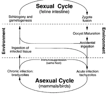
1.1.1 The life cycle of Toxoplasma gondii
The life cycle of Toxoplasma consists of the sexual and asexual phases (Fig. 2). The sexual cycle is confined exclusively to the intestine of feline species, whereas the asexual reproduc- tion can occur in most warm-blooded animals. Upon ingestion of tissue cysts by cats, the parasite invades, gut epithelium cells and differentiate into merozoites. These merozoites pro- duce progeny parasites and can also differentiate into "female" macrogametes or "male" mi- crogametes. After the intracellular mating process, unsporulated oocysts are released into the gut lumen and shed into the environment, where they undergo sporogony to form two sporo- blasts, each containing four sporozoites. Sporulated oocysts remain infectious for months and can initiate another sexual or asexual cycle through oral ingestion.
The asexual phase is initiated by oral infection of non-feline species with either oocyst- contaminated food or undercooked meat containing tissue cysts. Sporozoites exit the oocyst, penetrating the gut epithelia and differentiate into tachyzoites. Tissue cyst-release bradyzoites can also infect epithelial cells and form tachyzoites. These tachyzoites replicate quickly by endodyogeny and cause acute Toxoplasmosis hallmarked by tissue lysis. Their dissemination throughout the host organism and across the brain blood barrier occurs via infected macro- phages and dendritic cells (Lambert and Barragan, 2010; Lambert et al., 2010). The host im- mune response against tachyzoites induces their differentiation into encysted bradyzoites,
3
which cause a life-long chronic infection. Carnivorism or congenital transmission initiate an- other asexual cycle (Dubey 1998). Tachyzoites can also be maintained under in vitro condi- tions in a variety of cell types.
Figure 2: Schematic life cycle of Toxoplasma gondii.
Adapted from (Black and Boothroyd, 2000).
1.1.2 The life cycle of Plasmodium
The invertebrate vector of Plasmodium parasites are mosquitoes and the vertebrate hosts are mostly mammals including humans (Fig. 3) (Huff 1947). Infected mosquitoes inject sporo- zoites into the skin of the mammalian host during a blood meal. Sporozoites are motile cells and a fraction of them remains in the skin, while others enter the blood stream to reach the liver and infect hepatocytes. The schizogonic development into blood-stage merozoites mainly occurs in the liver, but both hepatic and cutaneous parasites can undergo this process and release merozoites within budding merosomes into the blood stream (Gueirard et al.).
Free merozoites invade erythrocytes and rapidly multiply asexually via schizogony and initi- ate the symptomatic phase of the Malaria disease. Simultaneously, a fraction of the intracellu- lar parasites differentiates into micro- and macro-gametocytes. These gametocytes are taken up by mosquitoes during a blood meal, and develop into gametes inside their midgut. After fertilization they form a zygote, and then motile ookinetes, which are able to escape the mid- gut by penetrating the peritrophic membrane and epithelial cells. A small fraction of the ooki- netes develops into oocysts outside of the midgut protected from the basal membrane. These oocysts eventually lyse and release sporozoites into the hemocoel, which mature and invade the salivary glands for another round of infection of intermediate hosts.
Figure 3: Life cycle of Plasmodium falciparum.
The right side shows the parasite development in the definite host, the mosquito. Developmental stages in the mammalian host are depicted on the left side. Hosts vary according to the Plasmodium species (Adapted from (Menard 2005).
1.2 Replication Compartments in Parasitized Host Cells
1.2.1 Toxoplasma gondii
The replicative stages of Toxoplasma include tachyzoite, bradyzoite and sporozoite. The tachyzoite stage is amicable for continuous in vitro culture and genetic manipulation. Its high replication rate demands import of large amounts of nutrients and presents a decent model for metabolic studies. Their in vitro culture is performed mainly in human foreskin fibroblasts (HFF) and in kidney epithelial cells (vero) cells from African green monkey.
Once the tachyzoite has actively invaded its host cell, it induces several modifications to the parasitized cell. The parasite forms a parasitophorous vacuole (PV) that confers a unique or- ganelle for its replication. It consists of host-derived lipids and selected proteins from the host cell (Sibley 1995) The selective exclusion of host proteins is fundamental to the non- fusogenic nature of the vacuole, which protects the parasite from lysosomal fusion and subse- quent degradation (Mordue et al., 1999). However, this non-fusogenic state of the PVM cre- ates an additional barrier for accessing to nutrients. To facilitate nutrient uptake, the PVM is
5
permeable to small compounds below 1.4 kDa including sugars and amino acids (Schwab et al., 1994).
The PV is further modified by rhoptry- and dense granule-secreted parasite proteins. The host endoplasmatic reticulum and mitochondria are also recruited by the parasite and establish an intimate contact with the PVM (Sinai et al., 1997) presumably to provide nutrients. Another significant ultrastructural feature of T. gondii-infected cells for nutrient acquisition is the re- cruitment of the host microtubules, which invaginate the PVM without penetrating it (Coppens et al., 2006). These host organelle sequestering tubulo-structures (H.O.S.T) are con- sidered to function as conduits for capturing endo-lysosomal nutrients, such as cholesterol.
This host LDL-derived cholesterol accumulates in these structures and released into the PV lumen in pinched-off vesicles. Another membranous structure, known as the nanotubular- network, fills the parasitophorous vacuole. It creates a dense network within the PV space, which is connected to the PVM (Sibley et al., 1995). As implied by its subcellular location, it is considered to be involved in nutrient transport to the parasite interior.
1.2.2 Plasmodium spp.
The parasite replication takes place extracellular within an oocyst in the mosquito host and primarily in two cell types within the mammalian host: hepatocytes and erythrocytes. The PV provides a replication niche in both cells. The PVM of intra-erythrocytic stages is permeable for small molecules including hexoses and amino acids (Desai et al., 1993), and also allows export of parasite proteins to the RBC. These secreted proteins extensively re-model the in- fected RBC (iRBC) (Maier et al., 2009). An exemplary host modification is the parasite- induced new permeation pathway (Merckx et al., 2009). These pore-like channels in the plasma membrane of the iRBC are permeable for small soluble molecules, and are assumed to support the parasite nutrition and disposal of metabolic waste. The PV forming during the hepatic development of the parasite also allows the diffusion of small molecules below 855 Da. Similar to T. gondii infected cells, the PVM, of Plasmodium associates with the host ER but not with mitochondria (Bano et al., 2007).
1.3 Central Carbon Metabolism of Hexoses
The purpose of the central carbon metabolism is to convert energy-rich molecules such as hexoses ways that support life. Hexoses are degraded in order to provide free energy, chemi- cal energy (ATP), reduction equivalents (NADH, NADPH) and biosynthetic precursors.
These products are used to synthesize and maintain the major constituents of a cell such as lipids, proteins and nucleotides. T. gondii (Polonais and Soldati-Favre et al., 2010) and Plas- modium spp. (Olszewski and Llinas 2010) harbour a full set of glycolytic enzymes (Fleige et al., 2007) (Roth et al., 1988), a complete tricarboxylic acid (TCA) cycle (Fleige et al., 2008;
Olszewski et al., 2010) and the pentose-phosphate pathway (PPP) (Bozdech and Ginsburg, 2005), to satisfy these cellular requirements. In addition, Toxoplasma features a gluconeo- genesis pathway (Fleige et al., 2008).
1.3.1 Glycolysis and apicoplast synthesis pathways
Both, Plasmodium and Toxoplasma parasites rely largely on glycolysis, and degrade glucose primarily to lactate (Fry et al., 1990; Ohsaka et al., 1982; van Dooren et al., 2006; Vander Jagt et al., 1990) (Scheibel and Miller 1969). Glycolytic enzymes are localized in the cytosol of Toxoplasma and are also expressed (Bozdech et al., 2003) and active (Olszewski et al., 2010; Scheibel and Miller, 1969) in intraerythrocytic Plasmodium parasites. Glucose is the main energy source for the gliding motility and parasitic invasion of the host cell of Toxoplasma (Pomel et al., 2008). It also maintains cytosolic ATP-levels and pH homeostasis in Plasmodium (Saliba et al., 2004). Pyruvate produced by glycolysis is mostly excreted as lactate or decarboxylated to acetyl-CoA by the pyruvate dehydrogenase (PDH) complex. This PDH complex localizes exclusively to the apicoplast in Plasmodium and Toxoplasma (instead of mitochondria like in many eukaryotes). Thus, the canonical connection of cytosolic glyco- lysis with the TCA cycle in mitochondria does not exist in these parasites (Fleige et al., 2007;
Foth et al., 2005; Olszewski et al., 2010). This further underlines the role of glycolysis for energy generation over oxidative phosphorylation.
Interestingly, several glycolytic steps can also take place in the apicoplast (Fleige et al., 2007;
Maeda et al., 2009; Saito et al., 2008). The apicoplast also harbours other essential pathways in both parasites. These include fatty acid synthesis type 2 (FAS2), the DOXP pathway for isoprenoid synthesis and heme biosynthesis (Seeber and Soldati-Favre, 2010). The apicoplast metabolism is connected to cytosolic and its own glycolysis to fuel its biosynthetic pathways with ATP, reduction equivalents, triose phosphates and acetyl-CoA. Plastid transporters, TgAPT in T. gondii (Brooks et al., 2010) and PfiTPT and PfoTPT in Plasmodium (Lim et al., 2010) import three-carbon phosphates. Toxoplasma can presumably, use these triose phos- phates to generate reduction equivalents and ATP by glycolytic enzymes within the apicoplast (Fleige et al., 2007). The energy source for the Plasmodium apicoplast is unknown (Polonais and Soldati-Favre, 2010).
7 1.3.2 TCA cycle
The cycle serves as a major hub of cellular metabolism and functions by integrating different carbon fluxes that originate from the degradation of amino acids, fatty acids and/or sugars.
The pathway directs these substrates towards biosynthetic pathways and/or oxidative phos- phorylation. The TCA localizes to the mitochondrion in Toxoplasma and Plasmodium (Fleige et al., 2008; Seeber et al., 2008; van Dooren et al., 2006). T. gondii tachyzoites do not seem to require a canonical flow of substrates through the TCA cycle (Fleige et al., 2008) as depletion of the succinyl-CoA synthetase causes only a mild replication defect in vitro. The TCA cycle functions in a bifurcated fashion during in vitro blood stages of P. falciparum. It is fuelled by glutamine to produce citrate and ultimately malate via the reductive branch, and generates malate via its oxidative branch (Olszewski et al., 2010). Its importance for the parasite sur- vival has not been reported yet.
1.3.3 The pentose phosphate pathway (PPP) pathway
The PPP directly connects the parasite replication to the available of hexoses. It uses glucose- 6-phosphate and generates precursors for the synthesis of nucleotides and aromatic amino acids. It also recycles NADP to NADPH. The PPP is well conserved and expressed in T.
gondii (Bahl et al., 2010). Also P. falciparum (Bozdech et al., 2003) and P. berghei (Hall et al., 2005) express most of the enzymes in their blood stages and generate up to 80 % of the nucleotides directly or indirectly using this pathway (Roth et al., 1986). Furthermore, the PPP-generated reduction power is needed to sustain the glutathione use of Plasmodium (Becker et al., 2003).
1.4 Protein and Lipid Synthesis Using Hexoses
Besides being substrate for above described metabolic pathways, hexoses are also critical for lipid synthesis and protein modification. Glycosylation of parasite protein and lipid synthesis are common and essential eukaryotic phenomena that are also conserved in Toxoplasma and Plasmodium.
1.4.1 Lipid synthesis in Toxoplasma and Plasmodium
Toxoplasma (Gupta et al., 2005) as well as Plasmodium (Dechamps et al., 2010a; Dechamps et al., 2010b; Dechamps et al., 2010c) can synthesize major phosholipids using precursors such as polar head groups (serine, choline, ethanolamine, inositol), fatty acids and glycerol-3- phosphate. The bulk synthesis of latter two molecules involves the degradation of hexoses via glycolysis and respective FAS pathways (Mazumdar and Striepen, 2007). Inositol, a close derivative of hexoses, is utilized for the synthesis of phosphatidyl inositol, inositol phospho- ceramide (IPC) and glycerol phosphatidylinositol (GPI) anchors in Toxoplasma (Azzouz et al., 2000; Azzouz et al., 2006; Sonda et al., 2005) as well as in Plasmodium (de Macedo et al., 2003; Kimmel et al., 2003; Naik et al., 2003). Furthermore, T. gondii tachyzoites and Plasmodium can incorporate galactose into plant-type lipids such as monogalactosylcere- brosides (MCDG), monogalactosyldiacylglycerol (MGDG) and digalactosyldiacylglycerol (DGDG) (Marechal et al., 2002). Finally, hexoses are also utilized to synthesize sphingolipids in both parasites (Azzouz et al., 2002; Couto et al., 2004; Landoni et al., 2007; Sonda and Hehl, 2006; Zhang et al., 2010).
1.4.2 Protein glycosylation in T. gondii and Plasmodium
The hexose-requiring N-glycosylation of proteins is essential in T. gondii, and its disruption by the inhibitor tunicamycin kills the parasites (Luk et al., 2008). Also, GPI-anchored pro- teins, whose synthesis requires hexoses and their close derivative inositol, fulfill essential functions in T. gondii such as motility, host cell adhesion (Lagal et al., 2010) and host interac- tion (Fauquenoy et al., 2008). Direct N- or O-glycosylation of proteins is less common in Plasmodium (Gowda and Davidson, 1999), however proteins are readily modified by GPI- anchorage (Gowda et al., 1997). GPI-anchored proteins are surface molecules, which are highly expressed throughout the life cycle of Plasmodium, and perform crucial functions. For example, the well known circum sporozoite protein (CSP) requires a GPI anchor for its func- tion (Dame et al., 1984). These proteins also include several merozoite surface proteins dur-
9
ing the blood stage (Debierre-Grockiego and Schwarz, 2010; Gilson et al., 2006) and P28 in ookinetes (Blanco et al., 1999; Martinez et al., 2000). An inhibitor, mannosamine that selec- tively disrupts GPI synthesis by preventing acetylation of the premature GPI anchor aborts in vitro blood cultures of P. falciparum (Gowda and Davidson, 1999; Naik et al., 2000; Naik et al., 2003). This illustrates the omnipresence and functional significance of GPI anchors for Plasmodium parasites.
1.5 Sugar Import in Toxoplasma and Plasmodium
1.5.1 Sugar transport proteins
A distinct class of trans-membrane proteins transports hexoses and inositol. These transporter proteins are categorized according the energy dependence of their transport reaction. Primary active permeases such as ATP-binding cassette (ABC-) transporters hydrolyze ATP to trans- locate their substrates. Secondary active transporters exploit an electrochemical gradient of mostly ions to energize otherwise unfavorable transport reactions. Facilitative transporters function passively and allow gradient-dependent diffusion of selected molecules across the membrane. Many proteins of the second and third group belong to the multi-facilitator super- family (MFS) of transporters.
Hexose-specific ABC-transporters require hexose-binding proteins that bind hexoses with an extremely high affinity. Their Km values are in the range of 1 μM or below (Verdon et al., 2003). They can, therefore, also access minute amounts of sugars. These hexose-binding pro- teins appear to be confined to bacteria and are not conserved in Toxoplasma and Plasmodium.
Secondary active sugar transporters form a subfamily within the MFS known as solute carrier family 2A (SLC5A). These transporters are present in bacteria and in mammalian intestine.
Other cells requiring sugars rely mostly on facilitative sugar transporters of the SLC2A sub- family and exploit an inward hexose gradient across their plasma membrane that is estab- lished by active catabolism of hexoses. Once inside hexoses are quickly phosphorylated in the cytosol to prevent their outward diffusion. These transporters contain 12 trans-membrane do- mains (TMDs) linked by short loop-regions and their N- and C-termini face the cytosol.
These proteins are characterized by well-conserved amino acids and motifs, which are also considered as their signature residues (Joost and Thorens, 2001).
1.5.2 Glucose uptake in Toxoplasma gondii
The first indirect evidence of sugar import by T. gondii comes from the detection of periodic acid-Schiff (PAS)-stained carbohydrate storage and by a prominent lactate dehydrogenase activity in Toxoplasma tissue cysts (Meingassner et al., 1977). The very low succinate dehy- drogenase activity in these cysts indicated glycolysis (and thus sugars) as being the main en- ergy source. Accordingly, isolated extracellular tachyzoites metabolized glucose to lactate and acetate, which accumulated in exogenous media (Ohsaka et al., 1982). Finally a sugar trans- porter (TgGT1) was cloned from tachyzoite cDNA that mediated uptake of glucose (Km ~30 µM) and fructose in Xenopus oocytes (Joet et al., 2002). TgGT1 localizes uniformly to the parasite plasma membrane (Pomel et al., 2008). The finding that gliding motility (and thus host cell invasion) of extracellular tachyzoites is also dependent on glucose (Pomel et al., 2008) led to the hypothesis that TgGT1 might be essential in Toxoplasma tachyzoites.
1.5.3 Glucose uptake in Plasmodium
P. knowlesi-infected erythrocytes metabolize glucose to lactate 25 to 75 times faster than un- infected cells (McKee et al., 1946) and glucose is vital for in vitro blood cultures of all treated Plasmodium species (Schuster, 2002). Glucose import in Plasmodium was initially characterized as an active energy-dependent and concentrative process (Izumo et al., 1989).
However, later on the accumulation of radiolabeled glucose inside infected red blood cells was attributed to glucose phosphorylation rather than its active transport, which was found to be entirely equilibrative (Kirk et al., 1996). Indeed, the P. falciparum hexose transporter, PfHT1, was found to be a high-affinity facilitative transporter for glucose (Km ~1 mM) and fructose (Km ~11.5 mM). It is localized at the parasite plasma membrane in blood stages and its mRNA was most abundant in asexual blood stages and least expressed in the gametocytes (Woodrow et al., 1999; Woodrow et al., 2000). Other species of Plasmodium such as P.
knowlesi and P. yoelii, that infect monkeys and rodents, respectively, also express similar transporters (Joet et al., 2002).
Interestingly, a glucose analog, compound 3361 (C3361), that carries an alkyl-substitution at its O3-position, selectively inhibits PfHT1 (Ki ~53 µM) and blood cultures at a very similar concentration (IC50 ~16 µM) (Saliba et al., 2004). Consistently, PfHT1 as well as its ho- molog in the murine malaria parasite Plasmodium berghei (PbHT1) are refractory to gene deletion (Slavic et al., 2010). Thus, PfHT1 is considered as a drug target for blood stages. In this regard a Leishmania mexicana null mutant model expressing PfHT1 has been developed for in vitro screening of selective PfHT1 inhibitors (Feistel et al., 2008). Besides the expres-
11
sion of PbHT1 during mosquito stages (Slavic et al., 2010), little is known about the signifi- cance of HT1 during other stages of Plasmodium.
1.6 Aim of This Study
Glucose is considered as a central nutrient for T. gondii and Plasmodium. However, prior to this work there has been no genetic evidence that glucose import is essential (or otherwise) in Toxoplasma and Plasmodium. One hexose transporter of T. gondii (TgGT1) has been bio- chemically characterized. Other potential sugar permeases shall be identified and functionally analyzed together with TgGT1 to evaluate their potential as a drug target or to uncover poten- tial mechanisms by which the parasites may circumvent the need for host-hexoses.
A single hexose transporter (HT1) from different Plasmodium spp. has also been character- ized. However, the importance of hexose transport during the ex-erythrocytic life cycle is not clear. This work has employed the rodent malaria parasite model (P. berghei) to assess the functional significance of sugar for Plasmodium. In addition, the drug development against human malaria requires heterologous models expressing the PfHT1 for screening and testing of inhibitors in vivo and in vitro.
2 Materials and Methods
2.1 Materials
2.1.1 Biological resources
Naval Medical Research Institute (NMRI) mice Charles River Laboratories, Germany
Anopheles stephensi Nijmegen, Netherlands
Human Foreskin Fibroblasts (HFF) Carsten Lüder, University of Göttingen, Germany
Huh7 human hepatoma cells Kai Matuschewski, Max-Planck-Institut für Infektionsbiologie, Berlin, Germany Toxoplasma gondii tachyzoites (RH hxgprt-) Dominique Soldati-Favre, University of
Geneva, Switzerland
Plasmodium berghei ANKA (-/+GFP) Max-Planck-Institut für Infektionsbiolo- gie, Berlin, Germany, (Janse et al., 2006)
E. coli XL-1blue Stratagene, Germany
Saccharomyces cerevisiae EBY.VW.4000 Eckhard Boles, University of Frankfurt /M, Germany
Genotype:
CEN.PK2-1C MAT α leu2-3,112 ura3-52 trp1-289 his-Δ1 MAL2-8c SUC2 hxt17 hxt13Δ::loxP hxt15Δ::loxP hxt16Δ::loxP hxt14Δ::loxP hxt12Δ::loxP hxt9Δ::loxP hxt11Δ::loxP hxt10Δ::loxP hxt8Δ::loxP hxt514Δ::loxP hxt2Δ::loxP hxt367Δ::loxP gal2Δ stl1Δ::loxP agt1Δ::loxP ydl247wΔ::loxP yjr160cΔ::loxP
2.1.2 Chemical reagents
Ampicillin Sigma, Germany
Bromophenol blue Merck, Germany
BSA fraction 5 Roth, Germany
Chloramphenicol Roth, Germany
Chloroform Roth, Germany
Deoxytriphospatenucleotides (dNTPs) Rapidozym, Germany
Diethylpyrocarbonate (DEPC) Sigma, Germany
Dimethyl sulfoxide (DMSO) Sigma, Germany
Dubecco’s Modified Eagle Media (DMEM)
w/o Na-pyruvate, w/o L-glutamine, 4.5 g/l D- glucose
Biochrom, Germany
EDTA Applichem, Germany
Ethidium bromide Applichem, Germany
Fetal Calf Serum Biochrom, Germany
Fluoromount G / DAPI SouthernBiotech, USA
Gentamycin Invitrogen, Germany
Giemsa solution BDH, Dubai
Glacial acetic acid (99 %) Applichem, Germany
Glutaraldehyde Roth, Germany
Glutathione Sigma, Germany
Glycerol Applichem, Germany
Heparin Braun, Germany
Hexoses Applichem, Germany
HPLC-purified water Roth, Germany
IPTG Applichem, Germany
Isofluoran Baxter, Germany
Ketamine (10 %) Bayer, VFW
L-glutamine (200 mM) Biochrom, Germany
Lithium acetate Applichem, Germany
Methanol Roth, Germany
Mycophenolic acid, Xanthine Applichem, Germany
Na-pyruvate (100 mM) Biochrom, Germany
Non-essential amino acids (100x) Biochrom, Germany
Nycodenz Axis Shield, Norway
Paraformaldehyde Roth, Germany
PBS Biochrom, Germany
Penicillin / Streptomycin (10000 U/ml and 10000 µg/ml) Biochrom, Germany Penicillin / Streptomycin (5000 U/ml and 5000 μg/ml) Invitrogen, Germany
Protease peptone Difco, US
15
Pyrimethamine Sigma, Germany
RPMI-Medium 1640 Invitrogen, Germany
Salmon sperm DNA (10 mg/ml) Invitrogen, Germany
Salts Roth, Applichem, Germany
Tris-HCl Applichem, Germany
Triton X-100 Applichem, Germany
Trypsin / EDTA Biochrom, Germany
Tryptone Applichem, Germany
X-Gal Applichem, Germany
Xylazin hydrochloride (2 %) Bayer, VFW, Rompun ®
Xylen Cynol FF Merck, Germany
Yeast extract Roth, Germany
Yeast nitrogen base (YNB) Sigma, Germany
[14C(U)]-D-glucose, [3,4-3H(N)]-L-glutamine Hartmann Analytic, Germany
Synthetic codon-optimized PfHT1 Genscript, USA
Primers (see Table 1) Invitrogen, Germany
Compound 3361 David H. Peyton, Portland
State University, US 2.1.3 Vectors
The vector for P. berghei transfection was originally created by A.P. Waters (University Lei- den, Netherlands) (pb3D-) and modified into pb3D+ by M. Ganter (Harvard University, Bos- ton, US). Both vectors are pyrimethamine-selectable in P. berghei. The drug resistance is con- ferred by a mutated dihydrofolate thymidylate synthase (DHFR-TS) gene of T. gondii that is regulated by untranslated regions (UTR) of P. berghei DHFR-TS. The pb3d+ vector also con- tains 3’UTR of DHFR-TS, which regulates the expression of a cloned cDNA. The pb3d- vec- tor was used for conventional gene replacement in P. berghei, whereas the pb3D+ vector was used for gene replacement by functional complementation.
All transfection vectors for T. gondii were obtained from Dominique Soldati-Favre (Univer- sity of Geneva, Switzerland), except for the pNTP3, which was provided by Isabelle Coppens (John Hopkins Bloomberg School of Public Health, Baltimore, US). A pTUB8-CAT knock- out vector was modified to construct a chloramphenicol-selectable TgGT1 knock-out vector.
An ApaI-excisable fragment was replaced with 3 kb long 5’UTR of TgGT1, and a XbaI- plus XhoI-excised fragment was replaced with 3 kb of the respective 3UTR. The p2854 (pyrimethamine-selectable) vector was used to construct a TgST2 knock-out vector by the introduction of 3 kb long 5’ and 3’ UTRs between the ApaI / HindIII and at the NotI sites respectively. For complementing TgGT1 knock-out parasites, the pNTP3 vector was altered
by replacing the TgNTP3 promoter between the ApaI and PacI sites by a 4.5 kb fragment con- taining the TgGT1 promoter and cDNA with a C-terminal HA-tag. Furthermore, a hypoxan- thine guanosine ribosyltransferase (HXGPRT) expression cassette was introduced between KpnI and ApaI sites as a selection marker in this vector. This expression cassette confers my- cophenolic acid resistance to HXGPRT knock-out (RH hx-) parasites. For N-terminal HA- tagged expression of TgST1-3, and C-terminal HA-tagged expression of TgGT1 the respec- tive cDNAs were inserted between NheI and PacI sites (TgST1-3) and NcoI and PacI site (TgGT1) of the pNTP3 vector. For the Ty1 epitope-tagged expression of all four transporters respective cDNAs were inserted between EcoRI and NsiI sites, except TgST2 which was cloned between the EcoRI and Sbf1 site into the pT8 vector.
The pESC-ura vector (Agilent Technologies, US) was modified by inserting PfHT1 and ScHXT9 genes into its NotI site for expression in S. cerevisiae. This vector was also modified by introducing cDNAs encoding for 80 or 55 N-terminal amino acids of ScITR1 (ScITR1- GM80) and ScHXT9 (ScHXT9-GM55) transporters between its EcoRI and NotI sites. The eventual vector was used to clone hybrid isoforms of PfHT1 at NotI site. A yeast expression plasmid (p426-HXT7-his) is designed to express N-terminal his-tagged proteins under the ScHXT7 promoter. In order to express PfHT1 and ScHXT9 as his-tagged fusion proteins ScITR1-GM80-PfHT1, ScHXT9-GM55-PfHT1, PfHT1 and ScHXT9 were amplified from pESC-ura templates and introduced into the SpeI site of p426-HXT7-his. Both yeast expres- sion vectors contain the S. cerevisiae ura3 gene that confers uracil prototrophy to mutant strains.
The pX63NeoRI vector was used as provided by Scott Landfear (Oregon Health University, Oregon, US) for expression in L. mexicana. All four T. gondii transporters as well as PfHT1 and PbHT1 were cloned at its EcoRV site. The pL2-5 vector was used for expression in Xenopus laevis oocyte. Both Plasmodium transporters were cloned between BglII and PacI sites. This vector contains β-globulin sequences for stabilizing of transcribed cRNA.
2.1.4 Antibodies and working dilutions
α-TgGAP45 (1:3000) (Plattner et al., 2008) α-TgGra3 (1:500) (Dubremetz et al., 1993) α-TgSag1 (1:1000) (Kim and Boothroyd, 1995) α-HSP70 (1:1000) (Tsuji et al., 1994)
α-P28 (for bead coating) (Siden-Kiamos et al., 2000) pro- vided by Robert Sinden (Impe- rial College, London, UK)
17
PbACP (1:500) (Friesen et al., 2010)
α-Ty (BB2 hybridoma cell culture supernatant 1:50) (Bastin et al., 1996) α-HA (rabbit, mouse) (1:1000) Invitrogen, Germany Alexa 594, Alexa 488 (anti mouse, anti rabbit) (1:3000) Invitrogen, Germany
2.1.5 Enzymes
Pfu Ultra II Fusion HS DNA polymerase Stratagene, Germany
Dream Taq polymerase Fermentas, Germany
Restriction endonucleases, Klenow enzyme NEB, Germany
T4 ligase Invitrogen, Germany
Antartic phosphatise, Klenow NEB, Germany
Proteinase K Sigma, Germany
Zymolase Zymo research, USA
2.1.6 Instruments
PCR Thermocycler (DNA engine PTC-200) Bio-Rad, Hercules, USA PCR Thermocycler (FlexCycler) JenaAnalytic, Germany PCR Thermocycler (Mastercycler Gradient) Eppendorf, Germany
Gel electrophoresis chamber and power supply Amersham Biosciences, USA Water purification systems (MilliQ PF, MilliPure RX20) MilliPore, USA
Microscope (Laborvert) Leitz / Leica, Germany
Microscope (Apotome Imager.Z2) Zeiss, Germany
Confocal Microscope Leica LSM-SP2 Leica Microsystems, Germany Stereo fluorescence microscope setup MZ10F Leica Microsystems, Germany Gel documentation & EASY Enhanced Analysis Herolab, Germany
BioPhotometer Eppendorf, Germany
Nanodrop ND 1000 Wilmington, USA
BTX square wave electroporator (ECM 830) BTX, USA
Amaxa Electroporator Amaxa, Germany
Scintillation counter (1450 MicroBeta TriLux) PerkinElmer, USA
2.1.7 Plasticware and disposables
Cryo tubes Biochrom, Nalgene, Germany
Syringes BD, Germany
Needles BD, Germany
Microscopy slides Menzel, Germany
Cover slips Roth, Germany
Improved Neubauer counting chamber Neubauer, Germany Tissue culture flasks, Petridishes, Multi-well plates Greiner Bio-One, Austria
LabTek chamber slides ThermoScientific, Germany
PCR tubes Rapidozym, Germany
Eppendorf tubes (1.5 ml, 2 ml) Greiner Bio-One, Austria
Pipette tips Greiner Bio-One, Austria
RNAase-free barrier tips Sorenson BioScience, USA
Filter sterilizer (0.22 µm) Schleicher Schuell, Germany Disposable pipettes (10 ml, 25 ml, 50ml) Greiner Bio-One, Austria Falcon tubes (15 ml, 50 ml) Greiner Bio-One, Austria Polypropylene tubes (12 ml) Greiner Bio-One, Austria Electroporation cuvettes (4mm gap) Eppendorf, Germany
Amaxa electroporation cuvettes Amaxa, Germany
Amaxa electroporator Amaxa, Germany
2.1.8 Commercial kits
DNA purification (plasmid preps) Jena Analytic, Invitek, Invitrogen, Qiagen, Germany
RNA isolation kit Qiagen
pDrive cloning kit Qiagen, Germany
Reverse transcription PCR (SuperScript III) Invitrogen, Germany
Trizol Invitrogen
RNA purification Qiagen
µMACS mRNA isolation Miltenyi Biotec, Germany
µMACS one-step cDNA synthesis Miltenyi Biotec, Germany
Yeast miniprep kit Zymo Research, US
2.1.9 Reagent preparations
D10
500 ml of DMEM media were supplemented with 5 ml of Penicillin / Streptomycin (10000 U/ml and 10000 µg/ml), 5 ml of sterile 200 mM L-glutamine, 50 ml FCS, 5 ml of 100 mM Na-pyruvate and 5 ml of 100x non-essential amino acids.
19 P. berghei freezing solution
This solution was prepared by mixing glycerol and Alsever’s solution at 1:9 ratio. 200 µl of isolated blood were diluted with 100 µl of freezing solution and directly frozen at -80°C.
P. berghei media for transfection
This media contained 160 ml of RPMI 1640 with 25 mM HEPES and L-Glutamine 2.05 mM (Gibco), 40 ml of heat-inactivated FCS and 50 μl gentamycin (50 mg/ml). The media was filtered sterile.
P. berghei ookinete (incomplete media)
0.425 g NaHCO3 and 2.5 ml of Penicillin / Streptomycin (5000 U/ml and 5000 μg/ml) were added to 500 ml of RPMI 1640, and the pH was adjusted to 8.0 and the solution was sterile filtered.
P. berghei ookinete (complete media)
Ookinete complete media was prepared from above-mentioned incomplete medium by addi- tion of 10 % heat inactivated FCS, 100 nM hypoxanthine and 50 µM xanthurenic acid. The pH was adjusted to 8.0 prior use.
Pyrimethamine stock solution (100x)
7 mg pyrimethamine was dissolved in 1 ml of DMSO and finally diluted in drinking water.
LB media
10 g of tryptone, 5 g of yeast extract and 10 g NaCl were dissolved in a final volume of 1 liter ddH2O. For LB plates, 15 g of agar-agar was also included. The LB media was sterilized by autoclaving. Ampicillin (100 µg/ml final concentration) or supplements such as IPTG (0.0012
%) and X-gal (0.004 %) were added prior to pouring plates or inoculation.
SOB media
20 g of tryptone, 5 g of yeast extract, 0.5 g NaCl and 186 mg KCl were dissolved in 1 liter ddH2O and autoclaved. The MgCl2 (2 M sterile stock solution) was added to a final concen- tration of 10 mM just before usage.
SOC-media
Deionised water containing 2 % of tryptone and 0.5 % of yeast extract (w/v), 10 mM NaCl and 2.5 mM KCl, was autoclaved, allowed to cool down before addition of filter-sterile glu- cose (20 mM).
YP-media
20 g peptone, 10 g yeast extract and 20 g agar-agar (optional) was solubilized in a final vol- ume of 950 ml ddH2O water, autoclaved and stored at room temperature.
Sugar stock solutions
D-Glucose, maltose, D-mannose or D-galactose were dissolved to make stocks of 40 % in ddH2O (v/w) filter sterilized and stored at 4°C.
Yeast-extract peptone dextrose / maltose (YPD / YPM) media
950 ml of YP media was supplemented with 50 ml of filter-sterile glucose stock (40%) for YPD media or with sterile maltose stock (40 %) for YPM media, and stored at 4°C. Final sugar concentration in media was 2 %.
10x amino acid mix
The following compounds were dissolved in a final volume of 500 ml ddH2O:
adenine hemisulfate (400 mg), L-arg (200 mg), L-asp (1000 mg), L-gln (1000 mg), L-his (200 mg), L-leu (600 mg), L-lys (300 mg), L-met (200 mg), L-phe (500 mg), L-ser (3750 mg), L- thr (2000 mg), L-try (400 mg), L-tyr (300 mg), L-val (1500 mg) and uracil (200 mg).
21
The solution was filter sterilized and stored in dark (wrapped in aluminium foil) at 4°C up to 4 months. Uracil was omitted for the preparation of uracil selection media.
Synthetic drop-out media
The basic media contained 1.7 g YNB (free of ammonium sulphate and amino acids) and 5 g ammonium sulphate in 500 ml ddH2O (filter-sterilized and stored at 4°C). The SD media was prepared by supplementing YNB with 10x amino acid mix (-ura) and appropriate sterile sugar stock solution (40 %) to final concentration of 2 %.
For SD plates, 20 g agar agar was also included in YNB, and the media was autoclaved and stored at room temperature. The media was warmed in microwave before adding supplements and pouring plates.
Cytomix for Toxoplasma gondii transfection
The solution contained 120 mM KCl, 0.15 mM CaCl2, 100 mM K2HPO4/KH2PO4, 500 mM HEPES, 100 mM EGTA and 100 mM MgCl2. It was filter sterilized and stored at 4°C.
Escherichia coli transformation buffers
Transformation buffer 1: 30 mM KOAc (pH 5.8), 50 mM MnCl2 x H2O, 10 mM CaCl2, 100 mM RbCl and 15 % glycerol.
Transformation buffer 2: 10 mM MOPS (pH 7), 75 mM CaCl2, 10 mM RbCl and 15% glyc- erol.
Both buffers were autoclaved before the addition of sterile glycerol.
S. cerevisiae transformation buffers
TE buffer
The 10x TE buffer contained 100 mM Tris-HCl plus 10 mM EDTA, and was adjusted to pH 7.5 before autoclaving. The 1x TE buffer was freshly prepared from 10x TE buffer.
LiAc / TE buffer
The 10x LiAc contained 1 M lithium acetate in ddH2O. The pH was adjusted with acetic acid to 7.5 and the solution was autoclaved. The 1x LiAc / TE buffer was prepared by diluting 10x LiAc solution in 10x TE buffer and water.
PEG3350 / LiAc / TE buffer
This buffer contained 40 % PEG3350 and was prepared fresh from 10x TE, 10x LiAc and PEG3350 (50 %). The autoclaved PEG 3350 stock solution was composed of 50 % (w/v) polyethylene glycol (M = 3350 g/mol) in ddH2O.
TAE buffer for agarose gel electrophoresis
The 1x TAE buffer was prepared from 50x buffer, which contained 242 g/l Tris base, 57.1 ml/l glacial acetic acid and 18.6 g/l of EDTA.
Lysis buffer for Toxoplasma DNA preparation
10 mM Tris-HCl, pH 8; 5 mM EDTA, 0.5% SDS, 200 mM NaCl were solved in water and autoclaved. 100 µg/ml proteinase K were added prior use.
Zymolase digestion buffer
This buffer contained 1.2 M sorbitol, 0.3 % β-mercaptoethanol and 16.7 U / ml zymolase en- zyme diluted in water.
Egression buffer
This buffer contained 10 μM A23187 in HBSS with Calcium and Magnesium. 25 mM glu- cose or 10 mM glutamine were also added for egression of radioactively labelled parasites.
23
2.2 Methods - Culture and Transfection
2.2.1 Propagation of mammalian cells
Human foreskin fibroblasts (HFF) and human hepatoma (Huh7) were maintained in T-300 or T-75 tissue culture flasks using D10 media and seeded as required into dishes (22 cm2), in multi-well plates or in chamber slides by trypsinization. Cells were maintained humidified incubator (10 % CO2, 37°C).
2.2.2 Propagation of Toxoplasma gondii tachyzoites
Tachyzoites were maintained using confluent monolayers of HFF cells. Transgenic parasites were selected in 25 μg/ml mycophenolic acid (MPA) with 50 μg/ml xanthine, 1 μM pyrimethamine or chloramphenicol (6.8 µg/ml). Limiting dilutions of parasites were used to infect confluent monolayers in 96-well plates and the parasite clones were scored one week after infection.
2.2.3 Toxoplasma gondii transfection
10 to 30*106 freshly-egressed or syringe-released parasites were centrifuged at 300 g for 10 min at room temperature, and the pellet was suspended in 700 µl of cytomix. This mixture was complemented with 5-50 µg of linearized or circular plasmid DNA, 30 µl ATP (sterile 100 mM stock) and 12 µl GSH (sterile 250 mM stock). Electroporation was done by two 1.7 kV pulses at an interval of 100 msec using a BTX square wave electroporator.
2.2.4 Giemsa staining of blood smears of P. berghei
In order to monitor parasitemia of infected mice, a drop of blood was collected from the tail and smeared on to a microscope slide. The smear was fixed in methanol for five minutes, stained for 20 min in 1:10 water-diluted Giemsa solution, washed in water and dried for mi- croscopic examination with an 100x oil-immersion objective.
2.2.5 P. berghei propagation and transfection
P. berghei ANKA strain parasites were propagated by intraperitoneal injection of fresh or stored blood cultures into about eight weeks old NMRI mice. For transfection, blood with a parasitemia of ~2 % was harvested by heart puncture from isofluoran-anesthetized mice using heparin-treated syringes. The blood was diluted with pre-warmed transfection media (1:2) and
red blood cells were isolated by centrifugation at 1000 rpm for 8 min. The pellet was sus- pended in 50 ml transfection media and mixed with another 100 ml of pre-warmed transfec- tion media. The culture was incubated for 17 hrs at 37°C with gentle shaking at 10 % O2, 5 % CO2, 85 % N2. To purify mature schizonts, 35 ml of culture media were layered onto 10 ml of 55% Nycodenz / PBS in 50 ml flacon tubes and centrifuged (no brake, 25 min, 1000 rpm) before harvesting the interphase with a Pasteur pipette. The schizonts were washed in 30 ml transfection media in two falcon tubes, and then suspended in 10-15 ml media (10 to 30*106/ ml schizonts). 1 ml of schizonts per transfection was sedimented and suspended in 100 µl human T-cell nucleofector solution supplemented with 2-5 µg (5-10 µl) of KpnI/NotI lin- earized DNA. The suspension was electroporated using the program U33 and the Amaxa elec- troporator. 50 µl of culture media was added to the parasites, and were then intravenously injected into animals (75µl / NMRI mouse). Pyrimethamine selection was applied to mice 24 hrs later. 100x pyrimethamine stock solution was used to supplement the drinking water and the pH was adjusted to 5 - 3.5 before feeding. A blood parasitemia could be detected five to eight days post-infection in Giemsa-stained blood smears. The blood was harvested at a para- sitemia of ∼2 % by heart puncture, and 200 µl were used to generate two freezer stocks. Re- sidual blood was used to isolate the parasite DNA for genotyping. The PCR-verified trans- genic parasite population was transferred into a NMRI mouse for further selection, genotyped and cryo-preserved at a parasitemia of about 2 %. Frozen stocks were injected into the perito- neum of NMRI mice for cloning and blood with a parasitemia below 1 % was harvested. The presence of 7*106 erythrocytes / µl of blood was assumed for absolute quantification of in- fected blood cells. The infected whole blood was diluted in RPMI 1640 media to obtain 1 iRBC / 100 µl. Five to ten mice were injected intravenously. Positive mice were then bled at a parasitemia of ∼2 %. The obtained parasites were genotyped to ensure clonality before mak- ing freezer stocks.
2.2.6 Ookinete culture and purification
1 ml of blood culture was harvested from infected NMRI mice three to four days post- infection, and analyzed for exflagellation. A blood drop was covered with a square cover slip on a microscopic slide and incubated for 10 min at room temperature. The temperature drop- induced exflagellation process could be microscopically monitored by counting areas of bouncing red blood cells that were hit by the released and highly mobile male gametes. Three exflagellation events per field using the 100x magnification objective were considered suffi- cient. 1 ml of the collected blood was incubated in 9 ml of complete ookinete media for 18 –
25
20 hrs at 20°C without shaking. The cell suspension was centrifuged at 1500 rpm for 8 min and suspended in 0.5 ml of ookinetes complete media containing 3 µl of P28-monoclonal an- tibody-coated (Siden-Kiamos et al., 2000) paramagnetic dynabeads. After inverting the sam- ple for 10 minutes in a 1.5 ml Eppendorf tube, the tube was placed into a magnetic rack. The media was removed and the beads were washed with PBS. After removal of the tube from the magnetic rack, the ookinetes were suspended in 100 µl deionized water and pipetted up and down to facilitate the detachment of ookinetes from the beads. The ookinetes were counted in a Neubauer counting chamber.
2.2.7 Propagation of Anopheles stephensi mosquitoes
The mosquitoes were raised in a 14 hrs light /10 hrs dark cycle, 75 % humidity and at 20°C.
2.2.8 Preparation of oocysts and salivary gland sporozoites of P. berghei
P. berghei-infected mice harbouring exflagellation-competent parasites were anesthetized with Ketamine / Xylazin hydrochloride by intraperitoneal injection and put on to a mosquito cage containing 12 hrs-starved mosquitoes for 30 min in dark. Ten mosquitoes were collected with a suction pump at 17 days post-infection, immobilized on ice, washed for 3 min in 70 % ethanol and put in RPMI 1640 supplemented with 3 % BSA for examination of infectivity by dissection under a binocular microscope. The midguts containing oocysts were collected in PBS on ice, 17 to 24 days post-infection. Salivary glands were dissected in RPIMI 1640 with 3 % BSA, stored on ice and disrupted in an 1.5 ml Eppendorf tube to release sporozoites for counting and assays. For infection of liver cells, mosquitoes were dissected directly in D10 media.
2.2.9 Liver stages of P. berghei
10.000 trypsinized Huh7 cells were seeded into one well of an eight-well LabTek chamber slide and grown in D10 media for one day. 30.000 sporozoites isolated from salivary glands were pipetted on to the cells. The chamber slide was centrifuged at 1000 rpm for 10 min at room temperature to enhance the infection rate. Infected cells were maintained for 68 hrs at 37°C with 10 % CO2, and washed three times daily with fresh media.
2.2.10 Making of Saccharomyces cerevisiae competent cells
4 ml of synthetic complete media containing 2 % maltose were inoculated with the EBY.VW.4000 strain picked from a master plate, and incubated with shaking at 30°C over