phosphatidylthreonine in Toxoplasma gondii
D i s s e r t a t i o n
eingereicht an der
Lebenswissenschaftliche Fakultät der Humboldt Universität zu Berlin
Zur Erlangung des Akademischen Grades Doctor rerum naturalium
(Dr. rer. nat.) im Fach Biologie
von MSc-Biologe Ruben D. Arroyo-Olarte
Präsidentin/Präsident der Humboldt Universität zu Berlin:
Prof. Dr. Jan-Hendrik Olbertz
Dekan der Lebenswissenschaftlichen Fakultät:
Prof. Dr. Richard Lucius
Gutachter/in: 1. Prof. Richard Lucius 2. Prof. Jos Brouwers 3. Prof. Frank Seeber Tag der mündlichen Prüfung: 04.11.2014
Acknowledgements
First, I would like to thank my supervisor Dr. Nishith Gupta, for his constant support.
I am grateful to Prof. Richard Lucius for his support and the opportunity to work in his department.
Moreover, I want to thank Prof. Frank Seeber, Prof. Jos Brouwers, and Prof. Richard Lucius for agreeing to review this thesis.
I would like to thank Prof. Bernd J. Helms and Prof. Jos Brouwers for the oportunity to establish a successful collaboration and introducing me into the the fascinating field of lipidomics. Also thanks to the members of Prof. Helms’s lab at the Institute of Biomembranes of Utrecht University for the friendly atmosphere and assistance.
To our collaborator Dr. Ildiko R. Dunay of the Department om medical microbiology of Magdeburg University, who contributed with the in vivo part of this research, also thanks to Aindrila Biswas for providing the brain histological data and to Friederike Hoffmann for her assistance with the Ca2+ measurements.
Special thanks to Boehringer-Ingelheim and Elsa-Neumann Stiftung des Landes Berlin for supporting my stay in Helm’s Lab and the final phase of my PhD, respectively.
I want to thank all the members of the department of Molecular Parasitology. Special thanks to Grit for her support and for managing the lab. I also thank all other PhD colleagues, Master and Bachelor students for their support and clarifications.
Also thanks to my friends for helping me out through the difficult times.
Finally, I would like to thank my family for their unconditional support from the distance.
Abstract
Toxoplasma gondii is arguably the most succesful parasite in Earth and a major cause of abortion and opportunistic infections in humans and livestock. Understanding the metabolic flexibility of this parasite is essential to comprehend its adaptability. Lipids are basic components of biological membranes. However, in the last decades non-customary roles for lipids beyond membrane biogenesis have been recognized. Here we report for the first time phosphatidylthreonine (PtdThr) as a major phospholipid in T. gondii and show its relationship with its archetypical analog, phosphatidylserine (PtdSer). We also identify a novel parasite enzyme (TgPTS), which diverged from the mainstream PtdSer-synthase family to produce this otherwise rare lipid. Genetic ablation of TgPTS not only abolished PtdThr synthesis, but also impaired severely the lytic cycle of T. gondii, particularly the egress but also the invasion into host cells without affecting the parasite replication. Our work indicates that, either a lower Ca2+ pool and/or a diminished Ca2+ mobilization from the parasite endoplasmic reticulum cause a reduced gliding motility, which underlies the observed phenotypes.
Moreover, the mutant parasites lacking PtdThr (Δtgpts) are avirulent and exert full protection against both, acute and chronic toxoplasmosis in a murine model. These results highlight the importance of parasite lipids as therapeutic targets and of genetically-attenuated strains in the development of effective vaccines against coccidian parasites.
Our work also identified the parasite enzyme that is responsible for PtdSer synthesis in T.
gondii (TgPSS). Contrary to TgPTS, we were unable to ablate TgPSS gene, suggesting its essentiality for T. gondii. By using a degradation-domain internal tagging approach, we were however able to downregulate TgPSS protein, which translated into a 60-70% reduction in PtdSer synthesis and content in T. gondii, without perturbing significantly the parasite fitness in both, wild-type and Δtgpts strains. Lipid analyses show that T. gondii can recompense for the reduction in PtdSer by increasing proportionally the levels of phosphatidylinositol, and for the loss of PtdThr by upregulating its PtdSer synthesis. Taken together, these results demonstrate the metabolic flexibility of T. gondii regarding its anionic phospholipid metabolism to maintain a normal membrane biogenesis and parasite replication. However, such compensatory mechanisms are unable to complement for the abscence of PtdThr concerning virulence, egress and invasion, demonstrating that T. gondii has evolved TgPTS and its product, PtdThr, as an adaptation to optimize its parasitic lifestyle.
Zusammenfassung
Toxoplasma gondii ist wohl der am besten an den Menschen und Tieren angepasste Parasit der Welt und eine der Hauptursachen für Abtreibungen und opportunistsiche Infektionen. Das Verständnis der Stoffwechselflexibilität dieses Parasiten ist essentiel um seine Anpassungsfähigkeit nachzuvollziehen. Lipide sind Grundbestandteile der biologischen Membranen. Doch in den letzten Jahrzehnten sind neben der Biogenese der Membranen noch weitere Funktionen entdeckt worden. Hier berichten wir zum ersten Mal über Phosphatidylthreonin (PtdThr) als Hauptphospholipid von T. gondii und zeigen seine Beziehung mit seinem archetypischen Analogon, Phosphatidylserin (PtdSer). Wir identifizieren auch ein neues Parasitenenzym (TgPTS), welches von der regulären PtdSer- Synthase-Familie abgewichen ist und dieses sonst selten vorkommende Lipid synthetisiert.
Die Gendeletion von TgPTS stoppte nicht nur die Synthese von PtdThr, sondern auch stark beeinträchtigt den lytischen Zyklus von T. gondii, insbesondere beim Aus- und Eintritt in die Wirtszellen, ohne dabei die Replikation des Parasiten zu verhindern. Unsere Arbeit zeigt, dass entweder ein niedriger Ca2+-Pool oder eine verminderte Ca2+-Mobilisierung aus dem endoplasmatischen Retikulum des Parasiten eine reduzierte Motilität verursachen, welche den beobachteten Phänotypen zugrunde liegt.
Darüber hinaus fehlt es den mutierten Parasiten an PtdThr (Δtgpts), sie sind avirulent und üben vollständigen Schutz gegen akute und chronische Toxoplasmose in einem Mausmodell aus. Diese Ergebnisse verdeutlichen die Bedeutung der Parasitenlipide als therapeutisches Ziel und von genetisch abgeschwächten Stämmen für die Entwicklung von wirksamen Impfstoffen gegen Kokzidien.
Unsere Arbeit konnte auch das Parasiten-Enzym identifizieren, welches verantwortlich für die Synthese von PtdSer in T. gondii (TgPSS) ist. Im Gegensatz zu TgPTS, waren wir nicht in der Lage das TgPSS Gen zu entfernen, das seine Essentialität für T. gondii andeutet. Durch die Verwendung einer gezielten Degradation-Domain-Tagging-Methode waren wir jedoch in der Lage das TgPSS Protein herunter zu regeln, was zu einer Reduzierung von 60 bis 70% in der PtdSer-Synthese und -Gehalt in T. gondii führte, ohne die Fitness im Wildtyp und ∆tgtps- Stämmen erheblich zu stören. Lipid-Analysen zeigen, dass T. gondii für die Reduzierung von PtdSer proportional Phosphatidylinositol erhöht und durch den Verlust an PtdThr die Synthese von PtdSer hochreguliert wird. Zusammengenommen zeigen diese Ergebnisse die
metabolische Flexibilität von T. gondii hinsichtlich seines anionischen Phospholipid- Stoffwechsels, um eine normale Membranbiogenese und Replikation der Parasiten beizubehalten. Allerdings sind solche Ausgleichsmechanismen nicht fähig die Abwesenheit an PtdThr bezüglich der Virulenz und dem Ein-und Austritt zu kompensieren. Wir können zusammenfassen, dass T. gondii TgPTS und seine Produkte (PtdThr) sich entwickelten, um seine Anpassung an den parasitären Lebensstil zu optimieren.
TABLE OF CONTENTS
ACKNOWLEDGEMENTS ... 1
ABSTRACT ... 2
ZUSAMMENFASSUNG ... 3
TABLE OF CONTENTS ... 5
LIST OF FIGURES ... 9
LIST OF APPENDICES ... 12
ABBREVIATIONS ... 13
1. INTRODUCTION ... 15
1.1. Toxoplasma gondii: Life cycle, distribution and pathogenesis ... 15
1.1. Regulation of the lytic cycle: Calcium signaling, motility and exocytosis in apicomplexan parasites ... 18
1.2. Genetic manipulation of T. gondii ... 20
1.3. Membrane and lipid biology of eukaryotes ... 21
1.3.1. Mammalian cells ... 24
1.3.2. Protozoan parasites ... 25
1.4. Objective of this study ... 27
2. MATERIALS AND METHODS ... 28
2.1. Materials ... 28
2.1.1. Biological resources ... 28
2.1.2. Chemical reagents ... 28
2.1.2. Primers ... 31
2.1.4. Vectors ... 35
2.1.5. Antibodies and working dilutions ... 36
2.1.6. Enzymes ... 37
2.1.7. Commercial kits ... 38
2.1.8. Plasticware and disposables ... 38
2.1.9. Instruments ... 40
2.1.10. Reagent preparations ... 41
2.2. Methods – Cell culture and transfection ... 42
2.2.1. Host cell culture ... 42
2.2.2. Parasite culture and selection ... 42
2.2.3. T. gondii transfection ... 42
2.2.4. Stable transfection of COS-7 cells ... 43
2.3. Methods – Molecular Cloning ... 43
2.3.1. PCR reactions ... 43
2.3.2. DNA ligation ... 43
2.3.3. Transformation of Escherichia coli ... 44
2.3.4. Transformation of Saccharomyces cerevisiae ... 44
2.3.5. Expression of recombinant proteins in E. coli ... 45
2.3.6. Nucleic acid preparation ... 45
2.4. Methods –Assays ... 46
2.4.1. Invasion and Egress assays ... 46
2.4.2. Motility assays ... 46
2.4.3. Evacuole assays... 47
2.4.4. Plaque and replication assays ... 47
2.4.5. Lipid Extraction ... 48
2.4.6. Thin-Layer Chromatography ... 48
2.4.7. Lipid phosphorus quantification ... 48
2.4.8. Lipidomics - Lipid extract fractionation ... 49
2.4.9. Lipidomics - HPLC and tandem mass spectrometry (MS/MS) analysis ... 49
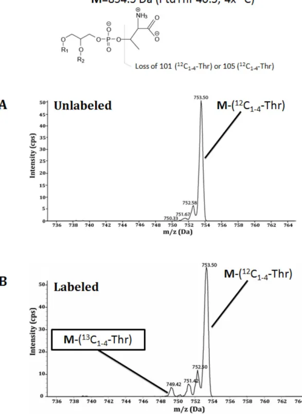
2.4.10. Metabolic labeling with radioactive precursors ... 50
2.4.11. Intracellular labeling with stable isotope precursors ... 50
2.4.12. Measurements of cytosolic calcium in intracellular parasites ... 50
2.4.13. Quantification of cerebral toxoplasmosis by PCR ... 51
2.4.14. Cerebral histopathology ... 52
2.4.15. Quantification of Toxoplasma cysts in the brain ... 53
3. RESULTS ... 54
3.1. The Lytic cycle and virulence of Toxoplasma gondii are regulated by a novel phosphatidylthreonine synthase ... 54
3.1.1. Phosphatidylthreonine is a natural-occurring major phospholipid in T. gondii .. 54
3.1.2. Phosphatidylthreonine is a parasite-exclusive phospholipid ... 56
3.1.3. Intracellular T. gondii can synthesize phosphatidylthreonine de novo from free threonine precursor... 57
3.1.4. Toxoplasma genome encodes 2 putative enzymes for the base-exchange
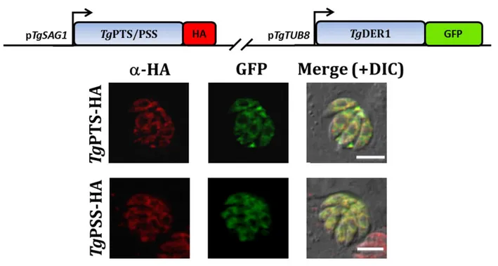
synthesis of phosphatidylserine and phosphatidylthreonine ... 59 3.1.5. Phosphatidylthreonine and phosphatidylserine syntheses occur in the parasite
endoplasmic-reticulum ... 62 3.1.6. TgPTS and TgPSS are expressed in the ER of transgenic COS-7 cells ... 63 3.1.7. The Δtgpts mutant lacks autonomous synthesis of PtdThr ... 63 3.1.8. Parasite Egress and Invasion but not intracellular replication are impaired by
genetic disruption of phoshatidylthreonine synthase ... 68 3.1.9. Evacuole formation is not affected by PtdThr depletion ... 70 3.1.10. A reduced parasite motility explains impaired lytic cycle of Δtgpts parasites .... 72 3.1.11. Phosphatidylthreonine is required for the virulence of T. gondii in a mouse
model and parasites lacking PtdThr exert protection against acute and chronic toxoplasmosis ... 73 3.1.12. Cytoplasmic calcium pool is downregulated during egress upon
phosphatidylthreonine depletion ... 75 3.1.13. Ionophore-induced calcium entry bypasses egress defect upon
phosphatidylthreonine depletion ... 78 3.1.14. Phosphatidylthreonine depletion induces higher endogenous PSS activity in T.
gondii ... 79 3.1.15. Gene expression analysis suggest product-inhibition/activation rather than a
transcriptional control of TgPTS and TgPSS regulating the phosphatidylserine/
phosphatidylthreonine balance in T. gondii ... 80 3.2. Phosphatidylserine synthesis in T. gondii ... 81
3.2.1. TgPSS and TgPTS can produce PtdSer, but only TgPTS utilizes threonine in E.
coli ... 81 3.2.2. TgPTS but not TgPSS has a growth-enhancement effect in PtdSer-defficient
yeast... 82 3.2.3. Conditional knockdown of TgPSS does not affect the parasite growth ... 85 3.2.4. The endogenous synthesis and the content of phosphatidylserine are decreased,
but not abolished upon conditional knockdown of TgPSS ... 91 3.2.5. Conditional degradation of TgPSS demonstrates its PtdSer synthase activity and
control over PtdSer content in T. gondii ... 94 3.3. Amplified PtdSer content is not involved in the phenotype of Δtgpts parasites ... 98
3.4. Distribution of PtdSer in T. gondii ... 101
4. DISCUSSION ... 104
4.1. PtdSer and PtdThr pathways of Toxoplasma gondii ... 104
4.2. Interregulation of the analog phospholipids, phosphatidylthreonine and phosphatidylserine in T. gondii ... 106
4.3. Role of phosphatidylthreonine for the lytic cycle of T. gondii ... 110
4.4. Phosphatidylthreonine as a parasite-adaptive trait in coccidians ... 113
4.5. Therapeutic potential of the Δtgpts strain ... 114
5. CONCLUSIONS... 116
APPENDICES……… …….117
REFERENCES ... 135
LIST OF PUBLICATIONS AND PRESENTATIONS ... 145
SELBSTSTÄNDIGKEITSERKLÄRUNG ... 146
LIST OF FIGURES
Fig. 1 Life cycle of Toxoplasma gondii ... 16
Fig. 2 Lytic cycle of T. gondii ... 17
Fig. 3 Schematic representation of endodyogeny and cell structure of T. gondii ... 18
Fig. 4 Microneme secretion and calcium-mediated signaling pathways in T. gondii ... 19
Fig. 5 Scheme depicting the fluid mosaic model of the plasma membrane ... 22
Fig. 6 Scheme showing the structure of the two major phospholipid groups ... 22
Fig. 7 Typical structures formed by phospholipids ... 23
Fig. 8 Inter-relationships among phospholipid biosynthetic pathways in mammalian Cells ... 24
Fig. 9 Biochemical pathways for phospholipid synthesis with experimental evidence in T. gondii ... 27
Fig. 10 Cartoon depicting the GCamp6s calcium measurement in T. gondii ... 51
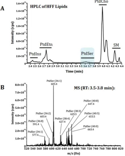
Fig. 11 Lipidomics of T. gondii tachyzoites identifies a novel parasite lipid, Phosphatidylthreonine ... 55
Fig. 12 Human foreskin fibroblast cells do not contain detectable amounts of phosphatidylthreonine ... 56
Fig. 13 Toxoplasma can incorporate free threonine into PtdThr during its intracellular replication ... 58
Fig. 14 PtdThr synthase from T. gondii harbors multiple substitutions in the conserved catalytic domain of an otherwise base-exchange PtdSer synthase... ...60
Fig. 15 Orthologs of PtdThr synthase are present in selected free-living and parasitic protists, but absent in most other organisms ... 61
Fig. 16 PtdThr and PtdSer are synthesized in the endoplasmic reticulum of T. gondii ... 62
Fig. 17 TgPTS and TgPSS are expressed in the ER in COS-7 cells ... 63
Fig. 18 Targeted gene disruption of TgPTS in T. gondii ... 64
Fig. 19 The Δtgpts strain is devoid of autonomous PtdThr synthesis ... 66
Fig. 20 The Δtgpts strain is deficient in PtdThr, and in lipid-derived threonine ... 67
Fig. 21 In vitro growth fitness defect of the ∆tgpts mutant... 69
Fig. 22 Δtgpts parasites have a defective exit and entrance into their host cells ... 70
Fig. 23 Evacuole formation is not altered upon PtdThr depletion ... 71
Fig. 24 ∆tgpts parasites display a reduced motility ... 72
Fig. 25 Lack of PtdThr impairs T. gondii virulence ... 73
Fig. 26 Δtgpts parasites protect against chronic toxoplasmosis ... 74
Fig. 27 The Δtgpts mutant has an impaired mobilization of ER-derived Ca2+ into its cytosol during natural egress ... 77
Fig. 28 Ionophore-induced influx of calcium can repair egress defect ... 78
Fig. 29 Loss of PtdThr upregulates PtdSer synthesis in T. gondii ... 79
Fig. 30 Relative abundance of the TgPSS and TgPTS transcripts of indicated strains as detected by qRT-PCR ... 80
Fig. 31 TgPSS and TgPTS can produce PtdSer, but only TgPTS makes PtdThr in E. coli . 81 Fig. 32 TgPTS enhances but does not complement the growth of a PtdSer-defficient yeast strain ... 83
Fig. 33 Growth enhancement of a PtdSer-defficient yeast strain granted by TgPTS is not due to higher mitochondrial stability ... 84
Fig. 34 Conditional mutagenesis of TgPSS ... 86
Fig. 35 Regulation of a tetracycline-inducible TgPSS knockdown in T. gondii ... 88
Fig. 36 In vitro growth phenotype of conditional knockdown of TgPSS ... 90
Fig. 37 Conditional knockdown of TgPSS downregulates PtdSer synthesis in T. gondii .... 92
Fig. 38 Conditional knockdown of TgPSS does not abolish PtdSer content of T. gondii .... 93
Fig. 39 Conditional destabilization of endogenous TgPSS does not alter significantly the parasite growth ... 95
Fig. 40 Conditional destabilization of TgPSS downregulates PtdSer synthesis in T. gondii 96 Fig. 41 Conditional degradation of TgPSS specifically downregulates PtdSer content in T. gondii ... 97
Fig. 42 Protein regulation of TgPSS by a conditional destabilization approach in Δtgpts background ... 98
Fig. 43 Conditional destabilization of TgPSS returns PtdSer synthesis and content to parental levels in PtdThr-deficient parasites ... 99
Fig. 44 Elevated PtdSer synthesis and content are not responsible for defective growth of Δtgpts strain ... 100
Fig. 45 Distribution of PtdSer pool is altered in ∆tgpts parasites ... 103
Fig. 46 Proposed model of metabolic pathways involved in the biogenesis of PtdSer and PtdThr in T. gondii ... 105
Fig. 47 Anionic phospholipid content is maintained despite PtdThr loss and PtdSer reduction in T. gondii ... 107 Fig. 48 Crucial amino acid residues for PtdSer-driven regulation of base-exchange activity
are missing in TgPSS and TgPTS ... 109 Fig. 49 Scheme illustrating the regulation of the lytic cycle of T. gondii by Ca2+ pathways
and steps in which PtdThr may be involved ... 113
LIST OF APPENDICES
Appendix 1 Human foreskin fibroblast cells do not contain detectable amounts of
phosphatidylthreonine...117
Appendix 2 TgPTS gene disruption does not affect transcription of neighboring genes...118
Appendix 3 Fatty acyl distribution of PtdCho, PtdEtn and PtdIns in parental and Δtgpts strains...119
Appendix 4 Catalytic activity of TgPTS is crucial for the lytic cycle of T. gondii...121
Appendix 5 Conventional gene replacement or disruption of TgPSS is not feasible...122
Appendix 6 Δtgpts parasites have normal organelle morphology...124
Appendix 7 Overexpression of TgPSS under pTetO7sag1 promoter causes ER- vacuolization...126
Appendix 8 Incorporation of 14C-serine into lipid fraction in presence of exogenously provided PtdSer and PtdThr is not significanlty altered...127
Appendix 9 Foreign complementation of the Δtgpts strain under the pTgGRA2 promoter causes a slight replication defect despite a normal localization...128
Appendix 10 Exogenously provided PtdThr does not recover the growth phenotype of the Δtgpts strain...129
Appendix 11 Stimulation of ryanodine-receptor (RyR)-type Ca2+channels recovers the motility of the Δtgpts strain...130
Appendix 12 Loss of PtdThr does not affect the microneme secretion of extracellular parasites...131
Appendix 13 T. gondii harbors a threonine biosynthetic pathway...132
Appendix 14 PtdThr is present in the coccidian parasite Eimeria tenella...133
ABBREVIATIONS
ATc Anhydro-tetracycline
ATP Adenosine triphosphate
CAT Chloramphenicol acetyltransferase
CCT Choline cytidylyltransferase
cDNA Complementary deoxyribonucleic acid
CHCl3 Chloroform
CK Choline kinase
CPT CDP-choline phosphotransferase
DAPI 4’,6-diamidino-2-phenylindole
DHFR-TS Dihydrofolate reductase thymidylate synthase
DMEM Dulbeccos’s modified Eagle medium
DNA Deoxyribonucleic acid
EDTA Ethylendiamine tetraacetate
EK Ethanolamine kinase
ER Endoplasmic reticulum
EtOH Ethanol
FAS I/II Fatty acid synthase type I/II
FCS Fetal calf serum
FUdR 5-Fluoro-2’-deoxyuridine
GSH Glutathione (reduced)
HFF Human foreskin fibroblast
HPLC High-Performance Liquid Chromatography
HXGPRT Hypoxanthine-xanthine-guanine phosphoribosyl transferase
IEM Immunoelectron microscopy
IFA Indirect immunofluorescence assay
IMC Inner membrane complex
IPTG Isopropyl-ß-D-1-thiogalactopyranoside
LDL Low-density lipoprotein
LiAc Lithium acetate
MeOH, (CH3OH) Methanol
MS Mass spectrometry
NADH Nicotinamide adenine dinucleotide
NBD 7-nitrobenz-2-oxa-1,3-diazol-4-yl
ORF Open reading frame
PBS Phosphate buffered saline
PCR Polymerase chain reaction
PEG Polyethylene glycol
PEMT Phosphatidylethanolamine methyltransferase
PSD Phosphatidylserine decarboxylase
Petn-Cer Phosphoethanolamine ceramide
PSS Phosphatidylserine synthase
PtdCho Phosphatidylcholine
PtdEtn Phopshatidylethanolamine
PtdGro Phosphatidylglycerol
PtdIns Phosphatidylinositol
PtdOH Phosphatidic acid
PtdSer Phosphatidylserine
PV Parasitophorous vacuole
PVM PV membrane
RNA Ribonucleic acid
RT-PCR Reverse-transcriptase PCR
SDS Sodium dodecyl sulfate
SM Sphyngomyelin
TaTi Trans-activator trap identified
TLC Thin layer chromatography
UPRT Uracil phosphoribosyl transferase
UTR Untranslated region
1. Introduction
1.1. Toxoplasma gondii: Life cycle, distribution and pathogenesis
T. gondii is an obligate intracellular parasite of cosmopolitan distribution. The life cycle of T.
gondii consist in two phases: one sexual phase which occurs only in the cat intestine, and an asexual phase which unlike most unicellular parasites, can occur in nearly all warm-blooded vertebrates, due in part to T. gondii special ability to differentiate into dormant stages or tissue cysts, where it remains largely protected from the host’s immune system (Black and Boothroyd, 2000) (Fig. 1).
The sexual phase of T. gondii life cycle starts in the feline intestine after ingestion of contaminated meat with tissue cysts that release bradyzoites which then differentiate into sexual stages. Gametes undergo fertilization and resulting zygotes differentiate into oocysts containing 4 sporozoites, which are released in the cat feces to the environment. During the asexual phase, intermediate hosts, including humans, can acquire the parasite after ingestion of contaminated food or water with tissue cysts or environment-resistant oocysts (Dubey, 1998). Recently, alternative routes of infection have been proposed, including toxoplasmosis as a potential sexually-transmited disease in both, animals (Lopes et al., 2013) and humans (Flegr et al., 2014), strengthening the evolutionary success of this cosmopolitan parasite.
Initially, either sporozoites (from oocysts) or bradyzoites (from tissue cysts) differentiate into the fast replicating stage or tachyzoite (Greek tachy: fast). Tachyzoites replicate within their host cell every 6-8 hours until reaching 64 to 128 parasites per host cell, which is then lysed releasing infective parasites (Radke and White, 1998). This lytic behavior is responsible for the acute phase of toxoplasmosis, which characterizes by tissue necrosis and inflammatory response (Mordue et al., 2001). Depending on genetic factors of both, the host and the parasite, acute infection either continues uncontrolled resulting in the death of the host or is put in check by the host’s immune system causing the differentiation of tachyzoites into slow- replicating bradyzoites inside tissue cysts (Behnke et al., 2012; Cavaillès et al., 2006; Jensen et al., 2013; Niedelman et al., 2012). These are found mainly in muscles and brain tissue, where they remain for the lifetime of the host. However, when the host immune system is immature (e.g. neonates (Torgerson and Mastroiacovo, 2013)) or compromised (e.g. AIDS (Navia et al., 1986), organ transplants (Wendum et al., 2002)), bradyzoites differentiate again
into fast growing tachyzoites, which lead to severe disease in different organs (brain, liver, eye, etc), with a potential lethal outcome.
Fig. 1. Life cycle of Toxoplasma gondii. Taken from (Black and Boothroyd, 2000).
During the acute phase of infection, the parasite undergoes the so-called lytic cycle (Fig. 2).
This begins when the extracellular parasite seeks for its host cell, attaches to it and actively invades it. Replication of T. gondii occurs exclusively within its host cell inside a non- fusogenic parasitophorous vacuole (PV), which the parasite secludes from the host lysosomal network. The PV is also actively modified by the parasite to enhance the nutrition acquisition from the host organelles, like mitochondria, Golgi and endoplasmic reticulum, which get in close contact with it and supply among others sugars, nucleotide precursors, cholesterol and phospholipids (Blume et al., 2009; Chaudhary et al., 2004; Coppens et al., 2000; Iltzsch,
1993; Romano et al., 2013a, 2013b). After several rounds of intracellular replication the parasites become motile and initiate the egress from its host cell, thereby causing its lysis.
Fig. 2. Lytic cycle of T. gondii. Adapted from (Arroyo-Olarte et al. 2014, under submission).
Intracellular replication has been the most commonly targeted phase of the lytic cycle by current drug treatments. In T. gondii it occurs through a special mode of cell division known as endodyogeny, in which the two daughter cells are formed within the scaffold of the mother cell, following a hierarchical sequence of organelle partioning and leaving behind a residual body (Fig. 3). The other two phases of the lylic cycle of T. gondii, however, invasion and egress, although also fundamental for the pathogenesis, have been disregarded as therapeutic targets.
Egress
Replication
Invasion
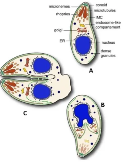
Fig 3. Schematic representation of endodyogeny and cell structure of T. gondii. (A) Mother cell. (B) developing daughter cells within mother cell, IMC (inner membrane complex) scaffolds are shown in green. (C) Cytokinesis. Adapted from (Agop-Nersesian et al., 2010).
1.1.1. Regulation of the lytic cycle: Calcium signaling, motility and exocytosis in apicomplexan parasites
Apicomplexan parasites are an ancient group of unicellular eukaryotic organisms, mostly related to other protozoans like dinoflagelates and ciliates (Baldauf, 2003). Apicomplexans often contain many plant-like genes probably resulting from their ancient origin pre-dating the plant-animal split, but mostly due to the acquisition of an algae-derived secondary plastid endosymbiont, called the apicoplast (Janouskovec et al., 2010; Oborník et al., 2009; Sato,
2011). Due to the particular origin and parasitic nature of apicomplexans, calcium signaling pathways regulating the lytic cycle of T. gondii and related parasites are unique and offer potential therapeutic targets (Nagamune and Sibley, 2006; Nagamune et al., 2008a; Plattner et al., 2012).
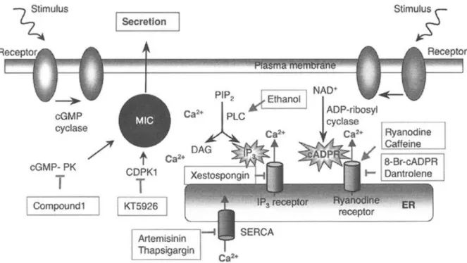
It has been shown that calcium plays a central role in the regulation of the invasion and egress stages of T. gondii lytic cycle by controlling parasite secretion and motility (Billker et al., 2009; Lourido et al., 2010; McCoy et al., 2012; Meissner et al., 2002). The rapid release from intracellular calcium storages into the cytosol where it is normally maintained at very low levels (ranging the nanomolar concentrations), is thus a potent trigger to initiate multitude of signaling cascades (Nagamune et al., 2008a). In the same way, released calcium must be quickly dampened to avoid toxic effects in the cells. Taken together, apicomplexans, and particularly T. gondii, has developed elaborated molecular mechanisms to control the calcium fluxes and the subsequent signaling pathways regulating its lytic cycle (Fig 4).
Fig 4. Microneme secretion and calcium-mediated signaling pathways in T. gondii. Chemical agonists ( ) or inhibitors (┬) are also shown. MIC, micronemal proteins; cGMP-PK, cGMP-dependent protein-kinase; CDPK1, calcium-dependent protein-kinase 1; PIP2, Phosphatidylinositol 4,5- bisphosphate; PLC, Phospholipase C; DAG, Diacylglycerol; cADPR, Cyclic ADP-ribose; SERCA, Sarcoplasmic reticulum calcium ATPase. Taken from (Nagamune et al., 2008a).
1.2. Genetic manipulation of T. gondii
T. gondii is the most experimentally-tractable Apicomplexan parasite due mainly to its promiscuous choice of host cells and its ability to be maintained in asexual cycle indefinitely in cell culture or murine systems. Initial forward genetic studies were performed by chemical mutagenesis and were fundamental for the establishment of basic cell culture protocols leading to the selection of clonal lines (Pfefferkorn and Pfefferkorn, 1976). Classical crossing experiments between different strains and analysis of the progeny have since then been made and allowed to tract loci controlling complex genetic traits, like virulence between different T.
gondii lineages and to analyze populations structure (Pfefferkorn and Kasper, 1983; Su et al., 2002). However, this approach is highly expensive and laborious, and also requires the definitive feline host.
It was not until the establishment of an efficient transfection protocol by electroporation that modern reverse genetic studies were feasible (Soldati and Boothroyd, 1993). Since then, vectors have been developed either for random integration or targeting of foreign genes to specific loci by homologous recombination. Initially, however, gene replacement was hampered by a high frequency of non-homologous recombination in T. gondii. To address this issue, Fox et al. (2009) developed a type I strain of T. gondii deficient in non-homologous end-joining (NHEJ) DNA repair pathway by deleting the parasite KU80 protein. The resulting mutant maintained most of the phenotypic features of the wild-type parasites, including virulence but had a high efficient rate of homologous recombination. Since then, the ∆ku80 strain has become the power horse to analyze the function of putative genes in T. gondii by targeted gene replacement with different selection markers. More recently, the same achievement was accomplished in a type II strain, which would facilitate studies on the genetic basis of bradyzoite differentiation (Fox et al., 2011).
Another breakthrough, especially for the analysis of essential genes in T. gondii was the tetracycline-inducible knockdown system (Meissner et al., 2002). This system utilizes a parasite line expressing a transactivator (TATi-1), which specifically activates a pTetO7 promoter to induce the expression of the gene of interest (G.O.I.). However, in presence of anhydrotetracycline (aTC), TATi-1 is blocked, silencing the pTetO7-driven expression. Once the inducible copy of the G.O.I. is introduced in T. gondii genome, the endogenous gene is deleted by double homologous recombination of its 5’ and 3’-UTRs, and replaced by a
resistance selection marker. This system is very useful in the analysis of genes thought to be essential and which remain refractory to direct deletion.
Other conditional systems include the targeting of the protein of interest to the proteasome where it is rapidly degraded, by adding a FKBP-derived destabilization-domain (DD) epitope.
The degradation of the chimeric protein, however can be overcome by a cell-permeable ligand (Shield1) in a dose- and time-dependent fashion (Banaszynski et al., 2006; Herm-Götz et al., 2007). This system has been successfully applied in other relevant human pathogens like Plasmodium falciparum (Armstrong and Goldberg, 2007) and Entamoeba histolytica (Liu and Singh, 2014).
More recently, a novel method to conditionally delete genes by a rapamycin-regulatable Cre- recombinase has been established (Andenmatten et al., 2013). The advantages of this system include a rapid generation of gene knockouts (including essential genes) by addition of the drug for their prompt analysis. In a first application of the technique, the authors have shown that genes encoding proteins considered to be essential for parasite invasion, e.g. myosin A and MIC2, are in fact dispensable. The disadvantages of the Cre-system, however are that the Cre-mediated recombination does not occur in 100% of parasites and therefore makes difficult the analysis of the knockout phenotype (Jiménez-Ruiz et al., 2014).
1.3. Membrane and lipid biology of eukaryotes
Lipids are the main components of biological membranes, which separate living cells from the surrounding medium. They also define the subcellular organelles and the vesicle trafficking of proteins between different cellular compartments (van Meer et al., 2008). The main organization of biological membranes was described by Singer and Nicolson (1972) as a fluid mosaic, or a two-dimensional oriented solution of integral proteins embedded in the viscous phospholipid bilayer solvent, where cholesterol (or ergosterol in fungi) intercalates and regulates the membrane fluidity (Fig. 5).
Fig 5. Scheme depicting the fluid mosaic model of the plasma membrane. Modified from http://www.biology.arizona.edu/cell_bio/problem_sets/membranes.
Phospholipids are divided in two major groups, glycerophospholipids, which are composed of a glycerol moiety attached to two fatty acyl chains and a hydrophilic polar head group linked by a phosphate group. On the other hand, sphingolipids consisting of head groups linked via phosphate to ceramide (Fig 6)
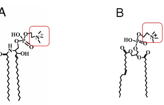
Fig 6. Scheme showing the structure of the two major phospholipid groups, sphingolipids (A) and glycerophospholipids (B), in particular, sphingomyeline and phosphatidylcholine, respectively. Polar head groups (red rectangle) may vary, including choline, serine, ethanolamine and inositol. Modified from (An et al., 2011).
The distribution and variety of phospholipids is responsible for the biophysical properties of biological membranes. For example, the length and degree of saturation of the fatty acyl chains is responsible for the thickness and ordering of the hydrophobic region of the membrane; while the electrostatic charge of their polar headgroups mediates interactions with charged proteins (Janmey and Kinnunen, 2006).
Each major phospholipid has intrinsical biophysical properties, which contribute to the stability and functions of biological membranes. For example, lysophospholipids and phosphoinositides have an inverted conical shape due to their relative bulky headgroups compared to their hydrophobic tails (Fig 7a), and tend to form structures with a positive membrane curvature like micelles. Cylindrical-shape phospholipids like PtdCho and sphyngomyelin, on the other hand, readily form flat bilayers and are responsible for the basic structure of the cell membranes (Fig. 7b). However, conical-shape lipids like PtdEtn, with small polar head group, tend to form structures with a negative curvature such as inverted hexagonal tubes with headgroups inside and hydrophobic tails facing out. (Fig 7c). Overall, the relative abundance and distribution of lipids will determine the shape and curvature of the membranes (Janmey and Kinnunen, 2006).
Fig 7. Typical structures formed by phospholipids (A) Conical lipids, like polyphosphoinositides and lysophospholipids usually self aggregate in micelles. (B) Cylindrical lipids like PtdCho and sphyngomyelin tend to form flat bilayers. (C) Inverted conical lipids, like PtdEtn forming inverted hexagonal tubes. Taken from (Janmey and Kinnunen, 2006).
1.3.1. Mammalian cells
In mammalian cells, the main phospholipid classes are phosphatidylcholine (PtdCho), phosphatidylethanolamine (PtdEtn), Phosphatidylserine (PtdSer) and Phosphatidylinositol (PtdIns) (van Meer and de Kroon, 2011; van Meer et al., 2008). The relative distribution of these and other lipids however, varies according to the subcellular organelle. In particular the plasma membrane is enriched in cholesterol to give more rigidity, compared to the endoplasmic reticulum or the Golgi complex, which have more PtdCho to give more fluidity (van Meer and de Kroon, 2011). Mitochondria, in accordance to their bacterial origin are enriched in PtdEtn, phosphatidylglycerol and cardiolipin, a tipical bacterial lipid (Daum, 1985). On the other hand, the endosomes on their way to maturation are enriched in bis(monoacylglycero)phosphate (Kobayashi et al., 2002), which participates in multivesicular body generation, fusion processes and sphingolipid hydrolysis (Kolter and Sandhoff, 2005;
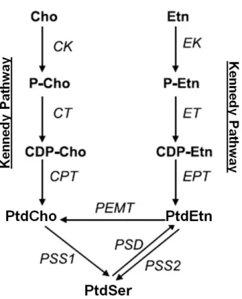
Matsuo et al., 2004). The main pathways for phospholipid synthesis in mammals, including humans, are shown in Figure 8.
Fig 8. Inter-relationships among phospholipid biosynthetic pathways in mammalian cells. Cho:
choline; CK: choline kinase; CPT: CDP-choline:1,2-diacylglycerol cholinephosphotransferase; CT:
CTP:phosphocholine citidytransferase; Etn: ethanolamine; EK: ethanolamine kinase; ET:
CTP:phosphoethanolamine cytidil-transferase; EPT: CDP-ethanolamine:1,2-diacylglycerol ethanolamine-phosphotransferase; PtdCho: phosphatidylcholine; PtdEtn: phosphatidylethanolamine;
PEMT: phosphatidylethanolamine N-methyltransferase; PtdSer: phosphatidylserine; PSD:
phosphatidylserine decarboxylase; PSS: phosphatidylserine synthase. Modified from (Vance and Vance, 2004).
Based on their charge, phospholipids are also actively segregated between the outer and inner leaflet of the plasma membrane of eukaryotes, with anionic phospholipids (PtdSer and PtdIns) at the inner leaflet, and cationic phospholipids like PtdCho and sphingomyelin predominantly at the outer/luminal face (Ikeda et al., 2006; Yamaji-Hasegawa and Tsujimoto, 2006). Among these, PtdSer is one of the most abundant an its roles extend far beyond the membrane biogenesis including apoptosis (Fadok et al. 1992, Li et al. 2003), membrane potential (Levenstein and Grinstein 2010), protein sorting and secretion (Uchida et al. 2011) and as a precursor of other phospholipids (Gupta et al., 2005, 2012; Vance, 2008; Voelker, 1984).
Many of the PtdSer functions depend on the acidic nature and negative charge of this phospholipid, which allows it to associate with calcium ions and cationic proteins, and to help in the maintenance of the membrane potential at the inner leaflet of the plasma membrane (Levenstein and Grinstein 2010). PtdSer has been shown to be essential in mammals (Kuge et al. 1997) and plants (Yamaoka et al. 2011), but not in yeast, for which it is dispensable but necessary for its optimal growth (Hikiji et al., 1988).
1.3.2. Protozoan parasites
Protozoan parasites are a very heterogeneous group comprising distantly related phyla. There are several specific lipid components and pathways with potential therapeutic applications.
Such differences arise not only from their parasitic nature, but also from their divergent phylogenetic origin (Vial et al., 2003). Most of the available research data come from the trypanosomatid and apicomplexan parasites, due to their medical and veterinary importance and also to their relative ease of culture (Ramakrishnan et al., 2013).
Apicomplexan parasites, including Plasmodium (Hsiao et al., 1991; Vial and Ancelin, 1992), Toxoplasma (Foussard et al., 1991; Gupta et al., 2005; Welti et al., 2007) and Babesia
(Florin-Christensen et al., 2000) are characterized by a higher amount of PtdCho and much less PtdSer as well as lower cholesterol/phospholipid ratio (implicating a higher degree of fluidity) in their membranes compared to their host mammalian cells (Ramakrishnan et al., 2013; Vial et al., 2003). In particular Plasmodium-infected erythrocytes contain almost no detectable cholesterol and less sphingomyelin (14.6% versus 28.0%), but contain more PtdCho (38.7% versus 31.7%) and PtdIns (2.1% versus 0.8%) (Hsiao et al., 1991).
In the case of Toxoplasma, PtdCho is the dominant phosholipid (60-75% of total phosphorus lipid) followed by PtdEtn (25-15%), PtdIns (10%), PtdSer (6%) and PtdOH (1.5%) (Foussard et al., 1991; Gupta et al., 2005; Hartmann et al., 2014). Lipidomic analysis using electrospray ionization mass spectrometry has also revealed small amounts of phosphoethanolamine- ceramide (PEtn-Cer), in addition to the above-mentioned phospholipid classes (Welti et al., 2007). The same report also showed that T. gondii phospholipids, particularly PtdCho, are enriched in shorter and saturated fatty acyl chains.
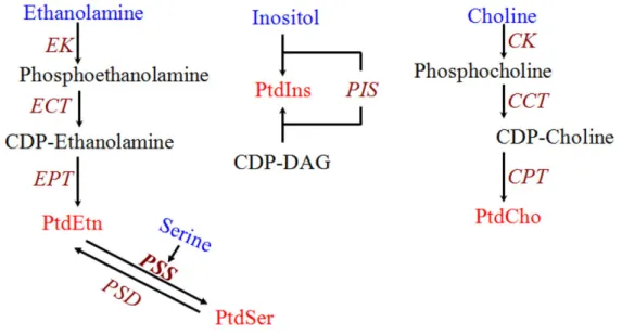
Figure 9 shows the main pathways for phospholipid synthesis documented so far in T. gondii, including the Kennedy pathways for PtdCho and PtdEtn synthesis (Gupta et al., 2005;
Sampels et al., 2012). As well as the decarboxylation of Ptdser as a source of PtdEtn (Hartmann et al., 2014). T. gondii also expresses a functional phosphatidylinositol synthase which catalyzes the synthesis of PtdIns from CDP-DAG and inositol (Séron et al., 2000).
PtdSer synthesis on the other hand has been shown to proceed by a base-exchange mechanism, most likely with PtdEtn (Gupta et al., 2005). However, contrary to PtdCho and PtdEtn, the genetic identity of the enzyme(s) responsible for PtdSer synthesis in T. gondii has not been described so far, and constitutes one of the main objectives of the present work.
Fig 9. Biochemical pathways for phospholipid synthesis with experimental evidence in T. gondii.
C/EK: choline/ethanolamine kinase; CCT: CTP:phosphocholine cytidylyltransferase; CPT: 1,2- diacylglycerol cholinephosphotransferase; ECT: CTP:phosphoethanolamine cytidylyltransferase; EPT:
1,2-diacylglycerol ethanolaminephosphotransferase; PIS: phoshatidylinositol synhthase; CDP-DAG:
CDP-diacylglycerol; PSS: base-exchange phosphatidylserine synthase; PtdCho: phosphatidylcholine;
PtdEtn: phosphatidylethanolamine; PtdIns: phosphatidylinositol; PtdSer: phosphatidylserine.
1.4. Objective of this study
Toxoplasma gondii is an obligate intracellular parasite, which is able to synthesize its major phospholipids to cover the membrane biogenesis needed for its rapid intracellular replication.
However, there is evidence that T. gondii is also able to scavenge host-derived lipids for their membrane biogenesis (Coppens et al., 2000; Hartmann et al., 2014). The physiological importance of these two mechanisms remains uncertain for most phosphoholipids, including phosphatidylserine. Additionally, other functions of phospholipids for the parasite biology beyond their traditional role as membrane building blocks remain underappreciated. The aim of the present work is to identify and describe the enzymes responsibles for the endogenous synthesis of phosphatidylserine and its novel analog phosphatidylthreonine in T. gondii, and to determine the role of both lipids not only for membrane biogenesis but also for the regulation of the parasite lytic cycle and virulence.
2. Materials and Methods
2.1. Materials
2.1.1. Biological resources
Cell line Source
Human Foreskin Fibroblasts Carsten Lüder, University of Göttingen, Germany
T. gondii tachyzoites (RH∆ku80-∆hxgprt- strain)
Vern Carruthers, University of Michigan, Ann Arbor, USA
T. gondii tachyzoites (∆ku80-TaTi strain) Boris Striepen, University of Georgia, USA
COS-7 cells Isabelle Coppens, Johns Hopkins University,
Baltimore, USA
E.coli (XL-1blue) Stratagene, Germany
E. coli (M15/pREP4) Qiagen, Germany
C57BL/6 mice Janvier Labs, France
2.1.2. Chemical reagents
Product Manufacturer
1,4-Dithiothreitol (DTT) Roth, Germany
4’,6’-Diamidino-2-phenylindol- dihydrochloride (DAPI)
Merck, Germany
5-Fluoro-2’-deoxyuridine (FudR) Sigma-Aldrich, Germany
Acetic acid Roth, Germany
Product Manufacturer
Adenosintriphosphate (ATP) Sigma-Aldrich, Germany
Ammonium molybdate Applichem, Germany
Ammonium persulfate Sigma-Aldrich, Germany
Ampicillin Applichem, Germany
Anhydrotetracycline hydrochloride (aTC) Sigma-Aldrich, Germany
Ascorbic acid Applichem, Germany
Bovine serum albumin fraction V (BSA) Applichem, Germany
Bromophenol blue Merck, Germany
Calcium chloride Applichem, Germany
Chloramphenicol Roth, Germany
Chloroform Roth, Germany
Crystal violet Sigma-Aldrich, Germany
Cytochalasin D Sigma-Aldrich, Germany
Deoxynucleotide-triphosphate (dNTPs) Rapidozym, Germany Dimethyl sulfoxide (DMSO) Sigma-Aldrich, Germany DNA marker (1kb ladder) Thermo Scientific, Germany Distilled water (HPLC-purified) Roth, Germany
Dulbecco’s Modified Eagle Medium (DMEM)
Biowest, Germany
Dulbecco’s phosphate buffered saline (PBS) Biowest, Germany
Product Manufacturer
Ethanol Roth, Germany
Fluoromount G / DAPI Southern Biothech, USA
Geneticin Life Technologies, USA
α-D(+)-Glucose monohydrate Applichem, Germany
Glycerol Applichem, Germany
Hank’s balanced salt solution (HBSS) PAA, Austria
Iodine (anhydrous beads) Sigma-Aldrich, Germany
Isopropanol Applichem, Germany
Isopropyl-beta-D-thiogalactopyranoside (IPTG)
Applichem, Germany
Kanamycin sulfate Applichem, Germany
L-glutamine Biowest, Germany
L-glutathione Applichem, Germany
Lipofectamine 2000 Life Technologies, USA
Methanol Roth, Germany
Mycophenolic acid Applichem, Germany
MEM essential amino acids (50X) Biowest, Germany MEM non-essential amino acids (100X) Biowest, Germany
Ninhydrin Spray solution Roth, Germany
Paraformaldehyde Roth, Germany
Product Manufacturer Penicillin/Sptreptomycin Biowest, Germany
Perchloric acid Applichem Germany
Phenylmethylsulfonyl fluoride (PMSF) Roth, Germany
Potassium acetate Roth, Germany
Potassium chloride Roth, Germany
Potassium hydroxide Merck, Germany
Rotiphorese gel 30 (Acrylamide) Roth, Germany
2.1.3. Primers
Primer Name (restriction site)
Nucleotide Sequence (restriction site underlined)
Cloning Vector (research objective) Annotation of TgPTS and TgPSS
TgPTS-F ATGCAACTCCCTTCAAGA pDrive (T/A-cloning
for testing and sequencing TgPTS (TGGT1_273540)
TgPTS-R TCACTGACTTCGTTCCATTTTCACG
TgPSS-F ATGTGTCGGGGACCGCCGCT pDrive (T/A-cloning
for testing and sequencing TgPSS (TGGT1_261480)
TgPSS-R TCACTCGTCTTTTTGGCCTTC
Expression and localization of TgPTS and TgPSS in T. gondii (Δku80-TaTi strain) TgPTS-F (EcoRV) CTCATCGATATCATGCAACTCCCTTCAAGA pTETO7SAG1-
UPKO (Ectopic
expression of TgPTS-HA at the TgUPRT locus)
TgPTS-HA-R (PacI) CTCATCTTAATTAATCAAGCGTAATCTGGA ACATCGTATGGGTACTGACTTCGTTCGATT
TgPSS-F (EcoRV) CTCGATATCATGTGTCGGGGACCGCCGCT pTETO7SAG1- UPKO (Ectopic
expression of TgPSS-HA at the TgUPRT locus)
TgPSS-HA-R (PacI) CTCTTAATTAATCAAGCGTAATCTGGAACA TCGTATGGGTACTCGTCTTTTTGGCCCTTCC
Making of ∆tgpts mutant in T. gondii (∆ku80-hxgprt- strain) 5'COS-TgPTS-F (NotI) CTCATCGCGGCCGCGTTCGCCTCGAGTGCT
TG
pTKO-HXGPRT
(Cloning of the TgPTS 5’ crossover sequence)
5'COS-TgPTS-R (EcoRI)
CTCATCGAATTCACGAGCCAGTGGAACGA C
3'COS-TgPTS-F (HpaI) CTCATCGTTAACAGCATCTTTATCGATGCG CT
pTKO-HXGPRT
(Cloning of the TgPTS 3’ crossover sequence)
3'COS-TgPTS-R (HpaI) CTCATCGTTAACTCACTGACTTCGTTCGAT TTTC
Screening for 5’ and 3’ recombination in the Δtgts mutant of T. gondii
5'Scr-TgPTS-KO-F CGATTCCTTGAGAGCAACTG pDrive (TA-cloning
of 5’ PCR product for sequencing) 5'Scr-TgPTS-KO-R GACGCAGATGTGCGTGTATC
3'Scr-TgPTS-KO-F ACTGCCGTGTGGTAAAATGAA pDrive (TA-cloning
of 3’ PCR product for sequencing) 3'Scr-TgPTS-KO-R GCCATAGAGTTCATTGCGGACTC
Genetic complementation of the Δtgts mutant of T. gondii
TgPTS-F (NsiI) CTCATCATGCATATGCAACTCCCTTCAAGA
AAGG
pTgGRA2-UPKO (Ectopic expression of TgPTS-HA at the TgUPRT locus) TgPTS-HA-R (PacI) CTCATCTTAATTAATCAAGCGTAATCTGGA
ACATCGTATGGGTACTGACTTCGTTCGATT TTCACG
TgPTS(ΔECWWD)-P1-F (NsiI)
CTCATCATGCATATGCAACTCCCTTCAAGA AAGG
pTgGRA2-UPKO (Ectopic expression of TgPTS(ΔECWWD)- myc at the TgUPRT locus)
TgPTS(ΔECWWD)-P1-R (SbfI)
CTCATCCCTGCAGGGCGCAGAGTTCGGGG ACGAG
TgPTS(ΔECWWD)-P2-F (NsiI)
CTCATCATGCATAGCATCTTTATCGATGCG CTG
TgPTS(ΔECWWD)-P2-myc- R (PacI)
CTCATCTTAATTAATCACAGATCTTCTTCA GAAATAAGTTTTTGTTCCTGACTTCGTTCG ATTTTCACGT
Expression of GCamp6s in T. gondii (∆ku80-hxgprt- and ∆tgpts strains) GCamp6s-F (NsiI) CTCATCATGCATTCTCATCATCATCATCAT
CATGG
pTgGRA2-UPKO (Ectopic expression of GCamp6s at the TgUPRT locus) GCamp6s-R (PacI) CTCATCTTAATTAATTACTTCGCTGTCATC
ATTTGTACA
Expression of genes flanking the TgPTS locus in T. gondii (∆ku80-hxgprt- , ∆tgpts and
∆tgpts/TgPTS-HA strains)
TGGT1_273550-F ATGCATTGTCAACTAGGAGGC ORF-specific PCR of
TGGT1_273550 TGGT1_273550-R TTACAGTGTCGAACTGGGGTC
TGGT1_273530-F ATGTTGAAGACACCAGTAACGGT ORF-specific PCR of
TGGT1_273530 TGGT1_273530-R TCAAGCGACAGATAGGTCGTC
Expression of TgPTS and TgPSS in E. coli (M15/pREP4) TgPTS-F (BglII) CTCATCAGATCTATGCAACTCCCTTCAAGA
AAGG
pQE60 (expression of TgPTS-6xHis) TgPTS-His-R (BglII) CTCATCAGATCTCTGACTTCGTTCGATTTTC
ACG
TgPSS-F (BglII) CTCATCAGATCTATGTCGGGGACTGCCGCT
pQE60 (expression of TgPSS-6xHis) TgPSS-His-R (BglII) CTCATCAGATCTCTCGTCTTTTTGGCCTTCC
AACA
Making of ∆tgss/TgPSS-HAi mutant in T. gondii (∆ku80-TaTi strain)
TgPSS-F (EcoRV) CTCATCGATATCATGCAACTCCCTTCAAGA pTetO7Sag1-UPKO (Ectopic expression of TgPSS-HAi at the TgUPRT locus) TgPSS-HA-R (PacI) CTCATCTTAATTAATCAAGCAACTCCCTTC
AAGA
5'UTR-TgPSS-F (AgeI) CTCATCCCACCGGTCACCTGGCTCGGCGAC A
pTKO-DHFR-TS (Cloning of the TgPSS 5’ UTR)
5'UTR-TgPTS-R (SpeI) CTCATCACTAGTCCACTGACCCATACGTTA
3'UTR-TgPTS-F (NheI) CTCATCGCTAGCACCGTCGAGTCTGGAAAT pTKO-DHFR-TS (Cloning of the TgPSS 3’ UTR)
3'UTR-TgPTS-R (ApaI) CTCATCGGGCCCGCATATCTGTACGTAAGC
Screening for 5’ and 3’ recombination in the Δtgpss mutant of T. gondii
5'Scr-TgPSS-KO-F CGTGCATGCAGAGGACATC pDrive (TA-cloning
of 5’ PCR product for sequencing) 5'Scr-TgPSS-KO-R CACAGTCTCACCTCGCCTTG
3'Scr-TgPSS-KO-F CTCGCGGCGTTGAATGTG pDrive (TA-cloning
of 3’ PCR product for sequencing) 3'Scr-TgPSS-KO-R GAGAAATCGTGCATGCGACC
C-terminal 2HA-DD-tagging of TgPSS gene locus of T. gondii TgPSS-3’IT-2HA-DD-F TACTTCCAATCCAATTTAATGCGACGGGGA
AGTCCTTTGG
pLIC-2HA-DD- DHFR (Endogenous C-terminal tagging of TgPSS with 2HA- DD)
TgPSS-3’IT-2HA-DD-R TCCTCCACTTCCAATTTTAGCCTCGTCTTTT TGGCCTTCC
Quantification of T. gondii infection in mouse brain tissue
TgB1-F TCCCCTCTGCTGGCGAAAAGT Quantification of
TgB1-R AGCGTTCGTGGTCAACTATCGATTG parasite load by qRT-PCR
MmASL-F TCTTCGTTAGCTGGCAACTCACCT Quantification of
mouse cells by qRT- PCR for
normalization of parasite load
MmASL-R ATGACCCAGCAGCTAAGCAGATCA
TgPTS and TgPSS expression in COS-7 cells TgPTS-F (XbaI) CTCATCTCTAGAATGCAACTCCCTTCAAGA
AAGG pcDNA 3.1+
(Expression and localization of TgPTS-v5 in COS-7 cells)
TgPTS-V5-R (XbaI) CTCATCTCTAGACTCACTTCGTTCGATTTTC ACG
TgPSS-F (HindIII) CTCATCAAGCTTATGTCGGGGACTGCCGCT pcDNA 3.1+
(Expression and localization of TgPSS-v5 in COS-7 cells)
TgPSS-V5-R (XbaI) CTCATCTCTAGACTCGTCTTTTTGGCCTTC
Quantitative Real-Time PCR of TgPSS and TgPTS
TgPTS-qPCR-F CTCTGCGAATGCTGGTGG qPCR of TgPTS
transcript TgPTS-qPCR-R AGAAGCTCCAGTCGGAAGCTT
TgPSS-qPCR-F GGTGACTTTGCTGGACCTGA
qPCR of TgPSS transcript
TgPSS-qPCR-R AGTGCCTCTGTGCTGACGAC
TgGT1-qPCR-F GGCTATTTTGGCACCTTTCA qPCR of TgPSS
transcript
(housekeeping gene)
TgGT1-qPCR-R AACGGGAAGACAAACCACAG
2.1.4. Vectors
Plasmid Source
pcDNA3.1+ Isabelle Coppens, John Hopkins University,
Baltimore, USA
pESC-Ura Stratagene, USA
pQE60 Qiagen, Germany
Plasmid Source
pNTP3 Isabelle Coppens, John Hopkins University,
Baltimore, USA
pNTP3TetO7Sag1 Modified pNTP3
pTetO7Sag1-NTP3-UPKO (pTetUPKO) modified pNTP3
pTKO John Boothroyd, Stanford University School
of Medicine, USA
pLIC-DHFR-2HA-DD Boris Striepen, University of Georgia, USA pTUB8-Der1 -GFP Boris Striepen, University of Georgia, USA
pS9-GFP Frank Seeber, Robert Koch Institute,
Germany
pGRA2-NTP3-UPKO (pGRA2-UPKO) Modified pNTP3
pGRA2-UPKO-GCamp6s Modified pGRA2-UPKO
pGRA2-UPKO-Lact-C2-GFP Modified pGRA2-UPKO
2.1.5. Antibodies and working dilutions
Antibody and dilution factor Source
Alexa 594, Alexa 488 (anti-mouse, antirabbit) (1:3000)
Life Technologies, Germany
α-HA (rabbit, mouse) (1:1500) Life Technologies, Germany Anti-6xHis-tag mAb IgG1 (mouse) Dianova, Germany
α-TgActin (mouse) (1:1000) Dominique Soldati, University of Geneva, Switzerland
Antibody and dilution factor Source
α-TgSag1 (mouse) (1:1000) (Kim and Boothroyd, 1995) α-TgGap45 (rabbit) (1:3000) (Plattner et al., 2008) α-TgHSP90 (rabbit) (1:1000) (Echeverria et al., 2005) α-TgROP2 (mouse) (1:1000) (Sadak et al., 1988)
α-TgMIC2 (mouse) (1:1000) Dominique Soldati, University of Geneva, Switzerland
α-KDEL (mouse) (1:1000) (Kaufusi et al., 2014)
α-v5 (mouse) (1:1000) Abcam
2.1.6. Enzymes
Enzyme Manufacturer
Antartic phosphatase NEB, Germany
Dream Taq polymerase Thermo Scientific, Germany Pfu Ultra II Fusion HS DNA polymerase Stratagene, Germany
Proteinase K Sigma, Germany
Restriction endonucleases, Klenow enzyme NEB, Germany
T4 ligase Invitrogen, Germany
2.1.7. Commercial kits
Product Manufacturer
Avansta western blotting analysis system Biozym, Germany DNA purification (plasmid preps) Analytik Jena, Germany InnuPREP DOUBLE
pure gel DNA extraction kit
Analytik Jena, Germany
pDrive cloning kit Qiagen, Germany
Platinum SYBR Green qPCR Superscript-
UDG Invitrogen, Germany
Pure Link RNA Mini Kit Ambion, Germany
2.1.8. Plasticware and disposables
Product Manufacturer
96, 24, 6-well plates Costar, USA
Cryo tubes for frozen stocks Nalgene, Germany Disposable pipettes (10 ml, 25 ml, 50 ml) Greiner Bio-One, Austria Eppendorf tubes (1.5 ml, 2 ml) Greiner Bio-One, Austria Electroporation cuvettes (4 mm gap) Eppendorf, Germany Falcon tubes (15 ml, 50 ml) Greiner Bio-One, Austria
Filters (5 μm) Millipore, Germany
Filter sterilizer (0.22 μm) Schleicher Schuell, Germany
Product Manufacturer Glass beads (0.45 – 0.6 mm) Sartorius, Germany
Glass Cover slips Roth, Germany
High performance chemiluminescence film GE Healthcare, Germany
Microscopy slides Menzel, Germany
Needles BD, Germany
Nitrocellulose transfer membrane Applichem, Germany Improved Neubauer counting chamber Neubauer, Germany
Parafilm Pechiney, USA
PCR tubes Rapidozym, Germany
Pasteur pipettes Hartenstein, Germany
Pipette tips Greiner Bio-One, Austria
Polypropylene tubes (12 ml) Greiner Bio-One, Austria RNAase-free barrier tips Sorenson BioScience, USA
Syringes BD, Germany
Tissue culture flasks, Petridishes, Multi-well plates
Greiner Bio-One, Austria
Whatman (3 MM) A. Hartenstein, Germany
X-ray film (FUJI Medical) A. Hartenstein, Germany
2.1.9. Instruments
Instrument Manufacturer
AMAXA Nucleofactor Lonza, Germany
BioPhotometer Eppendorf, Germany
BTX square wave electroporator (ECM 830) BTX, USA ELISA microplate reader Biotek, Germany Gel documentation & EASY Enhanced
Analysis
Herolab, Germany Gel electrophoresis chamber and power
supply
Gel electrophoresis chamber and power supply Amersham Biosciences, USA
Fluorescence microscope (Apotome Imager.Z2)
Zeiss, Germany
NanoDrop 2000 UV-Vis Spectrophotometer Thermo Scientific, Germany PCR Thermocycler (FlexCycler) JenaAnalytic, Germany
Safety work benches Heracell, Germany
Scintillation counter (1450 MicroBeta TriLux)
PerkinElmer, USA
TLC developing tank Roth, Germany
Western Blotting chamber Peqlab, Germany
2.1.10. Reagent preparations
Solution Composition
D10 medium DMEM (high glucose) supplemented with
10% FCS, 2 mM L-Glutamine, 1x NEAA, 1 mM Sodium pyruvate, 100 U/ml Penicillin and 100 μg/ml Streptomycin
Lysogeny Broth (LB) medium 10 g tryptone, 5 g yeast extract and 10 g NaCl in 1 liter deionized H2O (15 g of agar for plates)
Super Optimal Broth (SOB) medium 20 g tryptone, 5 g Yeast extract, 0.5 g NaCl, 0.186 g KCl and 10mM MgCl2 in 1 liter deionized H2O
Super Optimal broth with Catabolite repression (SOC) medium
20 g tryptone, 5 g Yeast extract, 0.5 g NaCl, 0.186 g KCl, 10 mM MgCl2 and 20 mM glucose in 1 liter deionized H2O
10x amino acid mix Adenine hemisulfate (400 mg), L-Arg (200 mg), L-Asp (1000 mg), L-Gln (1000 mg), L- His (200 mg), L-Leu (600 mg), L-Lys (300 mg), L-Met (200 mg), L-Phe (500 mg), L-Ser (3750 mg), L-Thr (2000 mg), L-Try (400 mg), L-Tyr (300 mg), L-Val (1500 mg) and Uracil (200 mg) in 500 ml deionized H2O.
Uracil was omitted for selective media.
Synthetic drop-out (SD) medium 1.7 g YNB (free of ammonium sulphate and amino acids) and 5 g ammonium sulphate in 500 ml deionized H2O. The 10x amino acid mix and 40% sugar (final 2 %) stocks were added to obtain synthetic drop-out media
2.2. Methods – Cell culture and transfection 2.2.1. Host cell culture
Human foreskin fibroblasts were cultured in Dulbecco’s modified Eagle Medium (DMEM) supplemented with 10% fetal calf serum (FCS), 1mM sodium pyruvate, 2 mM glutamine, 100 µM MEM-non-essential amino acids (glycine, alanine, asparagine, aspartic acid, glutamic acid, proline and serine), 100 units/ml penicillin and 100 μg/ml streptomycin in a humidified incubator (37°C, 5% CO2, pH 7.4). The cell monolayers were collected after trypsinization and seeded into flasks, plates and dishes as needed.
2.2.2. Parasite culture and selection
Tachyzoites of all T. gondii strains were routinely cultured on confluent HFF monolayers at a multiplicity of infection (M.O.I.) of 3 every 2-3 days unless stated otherwise in the same culture conditions as uninfected cells. Selection of transgenic parasites was done in 25 µg/ml mycophenolic acid (MPA) plus 50 µg/ml xanthine (XA), or 1 µM pyrimethamine or 5 µM fluoruracil deoxyribose (FudR) or 20 µM chloramphenicol. In the case of the selections with MPA+XA, pyrimethamine or chloramphenicol, the drug was added 8-24 hrs after transfection with the plasmid encoding the corresponding resistance cassettes. For selection with FudR, parasites were passaged for 2-3 cycles (4-6 days) after transfection under normal conditions before adding D10 medium with the drug. Infected monolayers were then left undisturbed until host cell lysis occurred, usually after 1-2 weeks of selection. Stable resistant parasites were cloned by limiting dilution in all cases.
2.2.3. T. gondii transfection
Freshly egressed or syringe-released parasites (10-20 x 106 tachyzoites) were pelleted (400xg, 10 min, RT) and resuspended in 700 μl cytomix. Parasite suspension was complemented with 1-50 μg plasmid DNA, 30 μl ATP (sterile 100 mM stock) and 12 µl GSH (sterile 250 mM stock). Electroporation was done by two 1.7 kV pulses at an interval of 100 msec using a BTX square wave electroporator or an AMAXA nuclefactor (program T16). For transfections done using AMAXA nucleofactor, reaction mixture was scaled down to 100 μl.
2.2.4. Stable transfection of COS-7 cells
The cDNAs of TgPTS and TgPSS were amplified from tachyzoite mRNA and cloned at the at HindIII and XbaI sites in the mammalian expression vector pcDNA3.1+. The cloning resulted in expression of C-terminally V5-tagged proteins under the pCMV promoter.
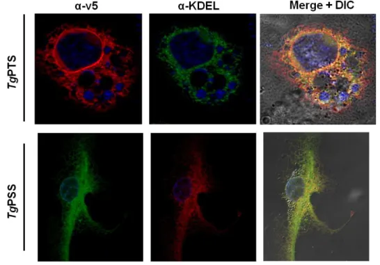
For transfection of COS-7 cells the BglII-linearized plasmid (4 µg) was diluted in 250 µl of Opti-MEM I (Reduced Serum) Medium. Another batch of 250 µl medium was mixed with 10 µl Lipofectamine 2000 and incubated for 5 min at room temperature. Both solutions (500 µl) were mixed and incubated for 20 min at room temperature. Meanwhile, a 6-well plate containing cultures of COS-7 cells was washed 3 times with PBS and prepared with antibiotic-free medium. The DNA-lipofectamine solution was added to the COS-7 cells and incubated at 37°C for 24 hrs. Cells were harvested by trypsine/EDTA treatment and diluted by at least 10-fold into T150 flasks. Selection of stable transgenic cells was started 48 hrs post-transfection using 800 µg/µl geneticin. Medium with fresh antibiotic was changed twice a week until cells were confluent and stable expression and subcellular localization of V5- tagged proteins was tested by immunofluorescence assay using anti-V5 and anti-KDEL (ER- marker; (Kaufusi et al., 2014)) antibodies.
2.3. Methods – Molecular Cloning 2.3.1. PCR reactions
Template DNA (10-500 ng) was used for PCR. For standard detection PCR, the Dream-Taq polymerase (Thermo Scientific) was utilized. For amplification of open reading frames to be used for expression cloning, the Pfu-Ultra FusionII high fidelity polymerase (Stratagene) was employed. Reaction mixtures and conditions were set according to primer sequences, type of polymerase and the lengths of amplification targets. PCR products were tested by gel electrophoresis in 1% agarose gels with 0.5% ethidium bromide or RotiSafe (Roth, Germany) ran in 1xTAE buffer at 100V. Visualization of DNA product was done under UV light.
2.3.2. DNA ligation
PCR products were purified either by gel extraction after evaluation of their expected size, or directly by column purification kit according to the manufacturer’s instructions (Analytic