Immunity-Related GTPases in
Cell-autonomous Resistance against Toxoplasma gondii
Inaugural-Dissertation zur
Erlangung des Doktorgrades
der Mathematisch-Naturwissenschaftlichen Fakultät der Universität zu Köln
vorgelegt von Yang Zhao aus Anqing, P.R. China
Köln 2008
Berichterstatter: Prof. Dr. Jonathan C. Howard
Prof. Dr. Thomas Langer
Tag der mündlichen Prüfung: 15th, April 2008
献给我的父母和爱妻刘导
1. Introduction ... 1
1.1 Pathogens and immunity ... 1
1.2 Cytokines and interferons... 3
1.3 Cell-autonomous immunity... 5
1.4 GTP binding proteins ... 5
1.5 Interferon-inducible GTPases and dynamins ... 7
1.6 Immunity-related GTPases... 8
1.6.1 IRG gene family ... 8
1.6.2 Induction and expression of the IRG proteins... 9
1.6.3 Structure and biochemical properties of the IRG proteins ... 9
1.6.4 Subcellular localization of IRG proteins ... 11
1.6.5 Involvement of the IRGs in resistance against intracellular pathogens in vivo and in vitro ... 12
1.7 Toxoplasma gondii infection as a model to study the functions of IRG proteins ... 14
1.8 Aims of this study ... 17
2. Material and Methods... 19
2.1 Reagents and Cells ... 19
2.1.1 Chemicals, Reagents and Accessories ... 19
2.1.2 Equipments... 19
2.1.3 Materials... 20
2.1.4 Enzymes and Proteins ... 20
2.1.5 Kits ... 20
2.1.6 Vectors and constructs used in the present study ... 20
2.1.7 Cell lines, bacterial and protozoan strains... 21
2.1.8 Media... 21
2.1.9 Serological reagents ... 22
2.1.10 Peptides ... 23
2.2 Molecular Biology... 23
2.2.1 Agarose gel electrophoresis ... 23
2.2.2 Generation of the expression constructs... 23
2.2.3 Cloning of PCR amplification products ... 25
2.2.4 Purification of DNA fragments from agarose gels... 25
2.2.5 Ligation ... 25
2.2.8 Plasmid isolation... 27
2.2.9 Determination of the concentration of DNA...27
2.2.10 Site directed mutagenesis...27
2.2.11 DNA Sequencing... 28
2.3 Cell biology...28
2.3.1 Transfection... 28
2.3.2 Transferrin uptaking experiments... 28
2.3.3 Lysotracker loading experiments... 29
2.3.4 Latex beads phagocytosis experiments... 29
2.3.5 Immunocytochemistry... 29
2.3.6 Peptide-streptavidin complex experiments... 30
2.3.7 In vitro passage and infection of Toxoplasma gondii... 30
2.3.8 Live cell imaging... 31
2.3.9 Quantification of IRG signals on T. gondii parasitophorous vacuoles... 31
2.3.10 Cell viability assay... 32
2.3.11 Western blotting... 32
3 Results-I... 33
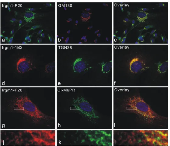
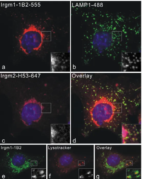
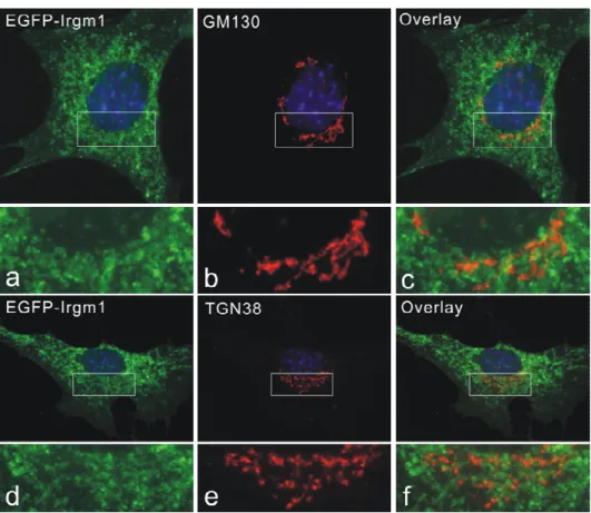
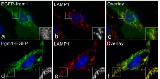
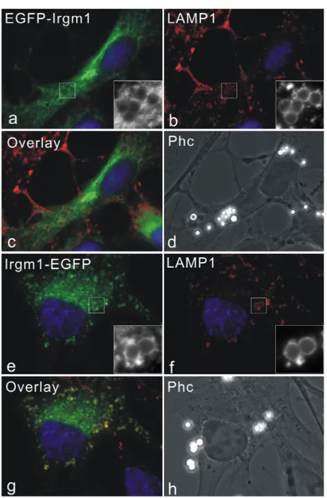
3.1 Irgm1 localizes to Golgi apparatus and late endocytic/lysosomal compartments... 33
3.2 Both N and C terminal EGFP-tag lead to the mislocalization of Irgm1... 36
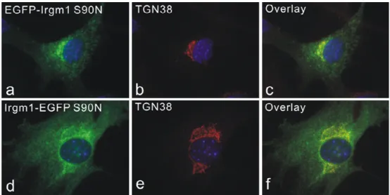
3.3 Mislocalization of Irgm1 by EGFP tagging is nucleotide dependent... 39
3.4 Phagosomal accumulation of Irgm1 is nucleotide dependent, but IFN-γ independent. ...40
3.5 Amphipathic helix near the C-terminus is responsible for both Golgi and lysosomal targeting of Irgm1... 44
3.6 Artificial αK amphipathic peptide mimics the localization of endogenous Irgm1... 50
3.7 Irgm1 is not recuited to Toxoplasma gondii ME49 strain vacuoles and is not absolutely necessary for IFN-γ-dependent cell-autonomous resistance against T. gondii ME49 strain...52
4. Results-II... 56
4.1 Dynamics of the Irga6-loading onto the T. gondii ME49 strain parasitophorous
vacuolar membrane... 56
4.3 EGFP-LC3 associates with a proportion of T. gondii ME49 strain vacuoles
independent of IFN-γ... 61
4.4 Intracellular T. gondii is killed after the disruption of parasitophorous vacuole... 64
4.5 Host cell death is a mechanism of IFN-γ-dependent cell-autonomous resistance against T. gondii avirulent strain...67
4.6 The property of host cell death induced by IFN-γ and T. gondii avirulent strain infection is rather necrosis than apoptosis... 70
4.7 Cathepsin B is not necessary for the IFN-γ-dependent T. gondii avirulent strain elicited host cell necrosis... 73
4.8 T. gondii avirulent strain induced IFN-γ-dependent necrosis dominates over the virulent strain resistance mechanisms against IFN-γ protection...74
5. Discussion... 76
5.1 Irgm1 localizes mainly to the Golgi apparatus and lysosomes, and mislocalizes as a result of EGFP-tagging at the N or C terminus...76
5.2 Both Golgi and lysosomal localization of Irgm1 is dependent on a predicted amphipathic helix near the C-terminus... 82
5.3 Irgm1 is not absolutely necessary for IFN-γ-dependent cell-autonomous resistance against T. gondii... 85
5.4 Loading of Irga6 onto the intracellular T. gondii vacuoles in real time...87
5.5 Intracellular T. gondii is killed after the disruption of the parasitophorous vacuole.. 89
5.6 IFN-γ-treated host cell undergoes necrotic-like death, which could serve as an IFN-γ- mediated cell-autonomous resistance mechanism against T. gondii...90
5.7 Autophagy in cell-autonomous resistance against T. gondii infection... 94
5.8 Virulent vs avirulent T. gondii strain infection... 97
5.9 Manipulation of the host cell death: a common theme in the host-pathogen interaction...98
6. References... 100
7. Summary... 113
8. Zusammenfassung...114
9. Acknowledgement... 116
10. Erklärung...117
11. Lebenslauf... 118
1. Introduction
1.1 Pathogens and immunity
Our earth is now 4.6 billion years’ old and the earliest life on our planet appeared approximately 4 billions years ago. Under the pressure of natural selection, life on earth evolved from single cell organisms (prokaryotes) to multicellular organisms (eukaryotes), as complicated as ourselves. During the evolution of life, some different organisms tend to live together to benefit each other (synbiosis). They either live in proximity or one lives inside of the other. One famous example is the adoption of cyanobacteria and proteobacteria by host cells which gave rise to chloroplasts and mitochondria, respectively (Margulis 1975; Sagan 1993). This co-adaptation can also been seen in our human beings that the beneficial bacteria flourish in the gastrointestinal tracts.
However, this is not always the case. Some microorganisms have evolved special mechanisms to invade, colonize and multiply in the host body at the expense of disturbing the normal physiology of the hosts. The disease-causing microorganisms are termed pathogens (from Greek pathos (suffering/emotion) and gene (to give birth to)). Traditionally, pathogens are divided into five main groups: viruses (including bacteriophages), bacteria, fungi, protozoa and worms. On the other hand, under the pressure of invading pathogens, the host organisms, especially the multicellular organisms such as vertebrates and plants, evolved sophisticated defense systems to resist the infection of pathogens. This defense mechanism in host organisms is therefore termed immunity (derived from Latin immunis, meaning exemption from public services).
The immune system protects the host organisms from being overrun by the invading pathogens with multiple layered defense mechanisms. Most simply, the physical barriers prevent the pathogens from entering the body. If a pathogen breaches these barriers, the innate immune system provides an immediate, but non-specific response. The innate system is thought to constitute an evolutionarily older defense strategy, and is the dominant immune system found in plants, fungi, insects, and primitive multicellular organisms (Janeway 2001).
Even simple unicellular organisms such as bacteria possess enzyme systems to protect against
the viral infections. Other innate immune mechanisms include antimicrobial peptides called defensins, phagocytosis, and the complement system. However, if the pathogens successfully evade the innate immune responses, the vertebrates possess a third layer of protection, the adaptive immune system, which is activated by the innate immune responses. This improved response is then retained after the pathogens have been eliminated, in the form of an immunological memory, and allows the adaptive immune system to mount faster and stronger attacks each time this specific pathogen is encountered again (Janeway 2001).
Recognition of the infectious microorganism is the first and crucial step in immunity. Both innate and adaptive immunity depend on the ability of the immune system to distinguish between self and non-self molecules. When the pathogens invade the host, they are first recognized by the host innate immune system. In the innate immune system, the immune response is initiated by cells and molecules recognizing conserved features of microorganisms and activated immediately on encounter with them. These conserved features of pathogens are referred to as pathogen-associated molecular patterns (PAMPs), which include cell wall components (lipopolysaccharide, lipoprotein, peptidoglycan, flagellin, etc.), double-stranded RNA from viruses, or CpG-containing DNA. The recognition of PAMPs is accomplished by innate immune receptors termed pattern recognition receptors (PRRs). The list of PRRs is constantly expanding. Perhaps the most prominent PRRs include Toll-like receptors, the mannose receptor, Nod like receptors, and Plant R Proteins (Janeway 2002). For many host organisms, the innate immune system is enough to effectively eliminate most of the pathogens.
For vertebrates, the innate immune system not only could directly eliminate the pathogens,
but also could provide signals to further activate the adaptive immune system. The adaptive
immune system, compared to innate immune system, is composed of highly specialized cells
and molecules, which can recognize essentially unlimited numbers of different targets. This
system is adaptive because it prepares itself for future challenges and it is highly adaptable
because of irreversible genetic recombination and somatic hypermutation in the antigen
receptor (TCRs and BCRs) gene segments (Janeway 2001).
1.2 Cytokines and interferons
Cytokines are a group of intercellular regulatory proteins and peptides with a mass of between 8 and 30 kDa, which are produced by a wide variety of cell types (both haemopoietic and non-haemopoietic) (DeFranco 2007). They play important roles in both innate and adaptive immunity, and some of them serve as bridges to transduce signals from innate immune system to activate the adaptive immune system. According to the structures, cytokines can be grouped into four families: the chemokines, the hematopoietins, the interferons (IFNs), and the tumor necrosis factor (TNF) family (Janeway 2001). The cytokines function in every aspects of immune response, including facilitating inflammation, activation of macrophages, T and B lymphocytes, chemotaxis, and also negative regulation of immune response (DeFranco 2007). Generally, cytokines are pleiotropic, multifunctional and sometimes redundant.
The interferons were originally discovered by their ability to induce resistance to viral infection in cells (Isaacs 1957). It was later appreciated that, in addition to regulating resistance to viral infection, interferons are widely functional in innate and adaptive immunity, including activation of macrophages and NK cells, enhancement of antigen presentation by induction of major histocompatibility complex (MHC) class I and II molecules, as well as modulation of normal and tumour cell survival and death (Boehm 1997; Schroder 2004;
Borden 2007). They are nowadays extensively used in clinical practice for treatment of viral diseases, cancer, as well as autoimmune disease multiple sclerosis (MS) (Borden 2007).
According to sequence homology and molecular structures, as well as their receptor
specificity, interferons are classified into three groups: Type I interferons, including IFN-α
(14-20 members, depending on species) (van Pesch 2004), IFN-β (Mogensen 1999), IFN-ω
(Hauptmann 1985), IFN-κ (LaFleur 2001), IFN-ε (Pestka 2004), IFN-δ (pigs) (Lefevre 1998)
and IFN-τ (ruminants) (Bazer 1997); Type II interferon (IFN-γ as sole member) (Bancroft
1993); Type III interferons, IFN-λs (3 members) (Kotenko 2003; Lasfar 2006). The type I
interferons are synthesized and secreted by almost all nucleated cells when confronted with
viral infection, while type II interferon (IFN-γ) is mainly secreted by activated T cells, NK
cells and macrophages (Schroder 2004; Borden 2007). Each type of interferons is recognized
by corresponding heterodimeric receptors, which signals mainly through JAK/STAT (Janus
kinase/Signal Transducers and Activators of Transcription) pathways (Figure 1.1). The activated STATs and other transcription factors (IRFs, interferon regulatory factors) are then translocated into the nucleus and drive the transcription and expression of interferon- stimulated genes (ISGs) promoted by IFN-γ-activated site (GAS) elements and IFN- stimulated response elements (ISREs) (Schroder 2004; Borden 2007).
Figure 1.1 Simplified scheme of interferon receptors and signaling pathways (from Borden 2007).
Type I interferons (IFNs) (α, β ω, κ, ε, δ (pigs), τ (ruminants)) interact with IFN (α, β and ω) receptor 1 (IFNAR1) and IFNAR2; type II IFN-γ with IFN-γ receptor 1 (IFNGR1) and IFNGR2; and type III IFN-λs with IFN-λ receptor 1 (IFNLR1;
also known as IL28RA) and interleukin 10 receptor 2 (IL10R2; also known as IL10RB). Type II IFN-γ is an antiparallel homodimer exhibiting a two-fold axis of symmetry. It binds two IFNGR1 receptor chains, assembling a complex that is stabilized by two IFNGR2 chains. These receptors are associated with two kinases from the JAK family: JAK1 and TYK2 for type I and III IFNs; JAK1 and JAK2 for type II IFN. GAS, IFN-γ-activated site; IRF9, IFN regulatory factor 9; ISGF3, IFN- stimulated gene factor 3, refers to the STAT1–STAT2–IRF9 complex; ISRE, IFN-stimulated response element; P, phosphate;
STAT1/2, signal transducers and activators of transcription 1/2.
The pivotal functions of interferons in immunity can be documented in the phenotype of
STAT1-deficient mice. STAT1 is essential in both type I and type II interferons signaling
pathways. The mice lacking STAT1 showed no overt developmental defects, and normal
numbers of T cells, B cells and macrophages in thymus and spleen. But these mice displayed
a complete lack of responsiveness to either IFN-α or IFN-γ and were highly sensitive to
infection by microbial pathogens and viruses (Durbin 1996; Meraz 1996). Similar phenotypes were also observed in IFN-γ deficient and IFN-γ receptor deficient mice (Huang 1993).
The pleiotropic functions of interferons are executed by interferon-stimulated genes.
Examples of these are class I and II MHC molecules. Up-regulation of MHC molecules can enhance the antigen presentation and promote the delivery of the signals from innate immune system to the adaptive immune system (Boehm 1997). Other molecules induced by interferons include adhesion molecules (ICAM1, VCAM1, et al) and chemokines which can recruit leukocytes and promote inflammation (Boehm 1997; Schroder 2004). Indeed, by inducing as many as one thousand genes, IFN-γ is extremely potent in activating the cytotoxic effector function among cells of innate and adaptive immunity including NK cells, dendritic cells, macrophages and T cells (Schroder 2004).
1.3 Cell-autonomous immunity
Cells, including immune and non-immune cells, can resist pathogen infection without the participation of other cells (neutrophils, cytotoxic T cells, NK cells, macrophages, et al) and molecules (antibodies, complements, et al) from innate and adaptive immune systems. This is termed cell-autonomous immunity, even though, in most cases, it is acquired upon induction of cytokines. IFN-γ is the most competent cytokine that can confer host cell autonomous resistance against viruses, pathogenic bacterium, and protozoa infection. Besides the IFN-γ- induced genes introduced in chapter 1.2, the molecules responsible for cell-autonomous immunity include PKR, iNOS/NOS2, phox complex, NRAMP1, IDO, Mx, GBPs, and IRGs (Boehm 1997; Schroder 2004). In addtition to the well-understood universal resistance mechanisms like ROS/NO production and tryptophan depletion, it is surprising, but also interesting, to notice that several of these abundantly induced proteins are GTP binding proteins (GTPases), whose functions are largely unknown (Martens 2006).
1.4 GTP binding proteins
GTP binding proteins (GTPases) are a large family of hydrolase enzymes that can bind and
hydrolyze GTP to GDP and/or GMP. These proteins participate in many cellular functions,
such as translation (initiation, elongation, and release factors), signal transduction, cell
motility, intracellular transport, membrane trafficking, as well as cell-autonomous resistance against intracellular pathogens (Leipe 2002; Martens 2006).
GTPases function as molecular switches, cycling between active GTP-bound form and inactive GDP-bound form (Figure 1.2)(Bourne 1990; Bourne 1991). Only GTP-bound form proteins are able to interact with a variety of effector proteins, by which cellular responses are executed. During the GTPase cycle, other proteins such as GEF, GAP and GDI can regulate the activity of the GTPases. GEFs (guanine-nucleotide exchange factors) induce the dissociation of GDP from GTPases and facilitate GTP-binding, and therefore activate GTPases. In contrast, GAPs (GTPase-activating proteins) accelerate GTP hydrolysis by GTPases, and therefore drive GTPases to the inactive GDP-bound form. GDIs (guanine- nucleotide dissociation inhibitors) prevent the dissociation of GDP from GTPases and keep the enzymes in the inactive form (Vetter 2001). The GTP binding and hydrolysis takes place in the highly conserved nucleotide binding domain (G-domain) common to all GTPases. The G1-G5 motifs within the G-domain with more or less conserved sequences are the structural elements required for the nucleotide binding and hydrolysis (Leipe 2002).
Figure 1.2 The GTPase cycle (from Martens 2006)
1.5 Interferon-inducible GTPases and dynamins
Four GTPase families were found to be strongly induced by interferons, including Mx GTPases, p65 guanylate-binding proteins (GBPs), very large inducible GTPases (VLIGs) and p47 immunity-related GTPases (IRGs). All these GTPases share biochemical characteristics with the dynamins, as these proteins show GTP-dependent self-oligomerization and accelerated GTP hydrolysis upon oligomerization (Martens 2006).
Dynamins are large GTPases with molecular weight of about 100 kDa. In addition to the conserved GTP-binding domain, classical dynamins have a pleckstrin homology (PH) domain involving phosphoinositide binding, a GTPase effector domain (GED) mediating self- oligomerization, a proline rich domain (PRD) interacting with SH3 domain-containing proteins, as well as a middle domain. The function of dynamins was originally discovered as proteins crucial in the scission process of clathrin-coated vesicles from plasma membrane.
Later investigations showed that dynamins are important in many cellular processes involving membranes such as vesicles trafficking, division of organelles and cytokinesis (Hinshaw 2000;
Praefcke 2004).
Among the interferon-inducible GTPases, the Mx family of dynamin homologs was the first to be found mediating cell-autonomous resistance against intracellular pathogens. The Mx1 gene was originally mapped in lab mouse A2G strain for its resistance against influenza virus infection (Lindenmann 1963; Lindenmann 1964). Isolated macrophages from A2G mice showed cell-autonomous resistance against viral infection (Lindenmann 1978). Mx genes are present in all vertebrates and two members are found in human (MxA and MxB) and mouse (Mx1 and Mx2), respectively (Staeheli 1985; Goetschy 1989; Simon 1991). The anti-viral function of Mx proteins is accomplished, at least in part, by binding to the viral nucleocapsids.
This interferes with intracellular trafficking and activity of viral polymerases, thus inhibiting replication of many RNA viruses including influenza and measles viruses (Haller 2007). Mx proteins are exclusively induced by type I interferons (Simon 1991).
During the cellular responses to type II interferon (IFN-γ), p65 GBPs and p47 IRGS are the
two dominant induced protein families (Boehm 1998). Type I interferons can also induce
GBP and IRG proteins to a less extent (Nantais 1996; Bekpen 2005). The GBP family is
conserved in verbebrates (Robertsen 2006). 10 members have been found in mice (mGBP1-
10)(Degrandi 2007) and 7 in human (hGBP1-7)(Cheng 1991; Olszewski 2006). Even though GBPs are massively induced by interferons, only a weak anti-virus effect was reported in cultured cells using an over-expression system (Anderson 1999; Carter 2005). Surprisingly, no genetic analysis using gene targeted mice has yet been reported to link the functions of GBPs to immunity against intracellular pathogens (viruses and microbes). In addition, roles of GBPs in regulation of vasculogenesis by proinflammatory cytokines and IFN-mediated cell growth were suggested (Guenzi 2001; Gorbacheva 2002; Guenzi 2003).
The third GTPase family induced by interferons is the VLIGs. These are the largest GTPases known so far, with molecular weight of about 280 kDa. The prototype of this family, VLIG-1, is massively induced by IFN-γ and to a less extent by IFN-β in vitro, and induced by Listeria monocytogenes infection in vivo. The G-domain of VLIGs is more closely related to IFN- inducible GTPases mediating cell-autonomous resistance, especially Mx and GBP families, than to other GTPase families. Therefore, it has been suggested that VLIGs are a new family of resistance GTPases although no direct test for this idea has been reported (Klamp 2003).
1.6 Immunity-related GTPases
Unlike the poorly confirmed functions of GBP family in intracellular pathogen resistance, extensive experimental analyses have been reported describing the indispensable role of IRG family (p47 GTPases) in anti-microbial resistance in mice. A detailed introduction of the IRG family is therefore given here.
1.6.1 IRG gene family
An extensive and detailed analysis of the IRG gene family has been reported (Bekpen 2005).
Homologous genes of IRGs are present in zebrafish, dogs, rats, mice and humans. In C57Bl/6 mice strain, 21 genes have been found expressing 25 coding units. 4 genes are probably expressed as tandem proteins. Based on phylogenetic principles, IRG genes are further divided into Irga, Irgb, Irgc, Irgd and Irgm subfamilies. In the Irgm subfamily, the lysine in the canonical G1 nucleotide binding motif GX
4GKS was replaced by methionine (GX
4GMS).
Most of the mouse genes have IRSE and/or GAS elements in their promoter regions. Irga6
has an additional liver specific promoter responsible for the expression of an alternative splicing form. The only exception in mice is Irgc, which lack both IRSE and GAS in its promoter. Instead a Sox-related element is present in the proximal promoter region. Irga subfamily is clustered on chromosome 18, whereas Irgb, Irgd and Irgm subfamilies are clustered on chromosome 11. Irgc is located alone in chromosome 7.
In humans, only two IRG gene homologs have been found. Human IRGC encodes a full- length IRG protein, which is highly homologous to mouse Irgc. They are more than 85%
identical at nucleotide level and 90% at the amino acid level. The proximal promoter region of human IRGC is also largely conserved with that of mouse Irgc. Human IRGM encodes a considerably truncated protein that lacks the G5 motif in the classical nucleotide-binding domain, and is probably controlled by the LTR of an integrated ERV-9 repetitive element.
IRGC and IRGM are located in human chromosome 19 and 5, respectively.
1.6.2 Induction and expression of the IRG proteins
Consistent with the promoter situations of IRG genes, most mouse IRG proteins are only expressed upon induction by interferons. Type II interferon (IFN-γ) is the most potent inducer of IRGs. Some reports describing IRGs induction in other circumstances, e.g. LPS, may reflect the secondary production of IFNs (Zerrahn 2002; Lapaque 2006). In addition to the massively-inductivity of Irga6 by IFNs, this member of IRGs has a high constitutive expression in the liver due to an alternative live specific promoter. The expression of Irgc and its human homologue IRGC are not IFN-regulatory and only constitutively expressed in the testis. Another member of IRGs in human, IRGM, was found to be expressed in cultured human cell lines Hela and GS293 and not subjected to interferons induction (Bekpen 2005;
Rohde 2007).
1.6.3 Structure and biochemical properties of the IRG proteins
Irga6 is the only member of IRG proteins that has been successfully purified and whose
biochemical properties have been systematically studies and crystal structure has been
resolved to date. Recombinant Irga6 purified from E. coli crystallized as a dimer in the
nucleotide-free and GDP-bound form (Uthaiah 2003). The Irga6 molecule is built from a Ras-
like G-domain and a unique helical domain composed of N and C terminal helices. Based on sequence comparison and secondary structure prediction, Irga6 structure serves as a prototype for other IRG proteins (Figure 1.3)(Ghosh 2004). Recombinant Irga6 hydrolyzes GTP to GDP and form GTP-dependent oligomers. The hydrolysis activity is accelerated upon oligomerization with a maximum rate of 2 per minute per molecule of Irga6. Irga6 has a low nucleotide binding affinity (15µM for GTP and 1µM for GDP) compared to Ras family GTPases. Considering the celluar concentration of GDP and GTP (120µM and 330µM, respectively (Kleinecke 1979)), Irga6 is presumably in GDP-bound form in cells. The low affinities for nucleotide and cooperative hydrolysis upon oligomerization are properties shared also by GBPs, Mx proteins and dynamins, although GBPs and Mx proteins have similar affinities for GTP and GDP, while dynamins have a higher affinity for GTP (Uthaiah 2003).
Figure 1.3 Structure of Irga6 (from Ghosh 2004)
Irga6 has an N-terminal myristoylation site MGxxxS and indeed, the myristoyl group contributes to the membrane association of Irga6 in cells (Martens 2004b). As a matter of fact, 11 members in IRG family carry the amino-terminal myristoylation signal (Bekpen 2005).
Recent data showed that myristoylated Irga6 purified from insect Sf9 cells hydrolyses GTP to
GDP and GMP (Papic 2007). This suggests that, in vivo, myristoylated IRG proteins may have strikingly different biochemical properties.
No exogenous proteins have been found as regulators (GEF, GAP and GDI) of IRG proteins.
As a self-activating GTPase, Irga6 functions as GAP (GTPase-activating protein) for itself.
Recent data suggested that IRGM proteins interact with Irga6 and Irgb6 to prevent inappropriate activation (Hunn 2007). In this sense, IRGM proteins function like GDI proteins for other members of IRGs. A microtubule-motor binding protein, Hook3, has been found to interact directly with Irga6, and is likely an effector protein of Irga6 rather than a regulator (Kaiser 2004).
Incomplete biochemical analyses of other IRG proteins were documented as well. Partially purified GST-Irgb6 and GST-Irgm3 fusion proteins have been shown to hydrolyze GTP to GDP (Taylor 1996; Carlow 1998). Weak GTPase activity was also reported for Irgm3 immunoprecipitates from IFN-γ treated macrophages (Taylor 1996), and more than 90% of the bound nucleotide in priecipitated Irgm3 was GTP, independent of IFN-γ (Taylor 1997).
This suggests that Irgm3 is mainly present in an active state in cells.
1.6.4 Subcellular localization of IRG proteins
IRG proteins associate with membranes to different degrees in IFN-γ induced cells. Irgm1 and Irgm3 have been found almost exclusively in a membrane compartment, while more than 90% of Irgd is in the aqueous phase. Irgm2 has more membrane-bound form than cytosolic pool. Irga6 and Irgb6 partitioned roughly equally between membrane and cytosolic fractions (Martens 2004b). By immunofluorescence analyses, Irgm1 and Irgm2 localize to Golgi apparatus and Irgm3 is an ER-associated protein (Taylor 1997; Martens 2004a; Butcher 2005).
Irga6 is targeted to ER, probably partially by a myristoyl group (Martens 2004b). IRGM
proteins are organelle-targeted by a predicted α helix near the C-terminus, which, in case of
Irgm1, shows amphipathic property (Martens 2004b; Martens 2006). The resting localizations
of IRG proteins are largely independent of GTPase activity as GTP-binding defective mutants
show similar subcellular distributions, wherever tested (Taylor 1997; Martens 2004b).
Irga6 and Irgb6 forms GTP-dependent aggregates when expressed in the absence of IFN-γ.
Co-expression with IRGM proteins can restore the proper localization of Irga6 and Irgb6.
Yeast 2-hybrid experiments also confirmed the direct interaction between different members of IRG family. Therefore, IRGM proteins can co-ordinate localization of other IRG protein family members (Martens 2006; Hunn 2007; Papic 2007).
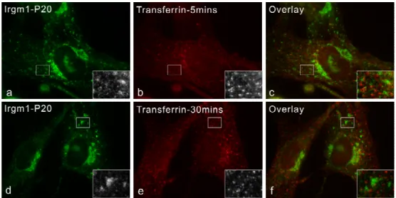
In cells involving pathogen infections or phagocytosis, IRG proteins are relocalized from resting compartments to the active membrane systems. Irgm1 is rapidly relocalized to F-actin- rich plasma membrane ruffles associated with phagocytic cups in fibroblasts and macrophages (Martens 2004b). Irgm1 and Irgm3 have been found to associate with latex beads phagosomes rapidly after uptaken (Butcher 2005). In addition, Irgm1 has been co- purified from mycobacterial phagosomes (MacMicking 2003). 5 members of IRGs have also been found on Toxoplasma gondii parasitophorous vacuole membrane shortly after the parasite invasion (Martens 2005). Compared to the nucleotide-binding independent of resting localization of IRG proteins, recruitment of IRGs to the T. gondii PVM requires the intact GTP binding motifs, suggesting a specific requirement for the nucleotide, presumably GTP, binding (Martens 2005; Hunn 2007).
Recent publications showed that Irgm1 colocalizes with MDC and LC3 positive compartments, suggesting an association between Irgm1 and autophagosome (Gutierrez 2004;
Singh 2006). The authors accordingly suggested that Irgm1 has a pro-autophagic function as a mechanism for mycobacteria elimination. However, in those publications, EGFP tagged Irgm1 was used, even though the influence on proper localization of Irgm1 by EGFP tagging was not investigated. In the present studies, we try to answer this question by in depth analyses.
1.6.5 Involvement of the IRGs in resistance against intracellular pathogens in vivo and in vitro
Studies of IRG-deficient mice and isolated cells have shown that members of IRG family are
indispensable, in most cases also non-redundant, resistance factors against a wide range of
intracellular bacteria and protozoal pathogens (Martens 2006; Taylor 2007). The published
results are summarized in table 1.1. Generally, Irgm1-deficient mice lose resistance to all
microbes tested so far; Irgm3-deficient mice show decreased resistance to a smaller group of protozoa and only one of several tested bacteria; and Irgd-deficient and Irga6-deficient mice only show weakly decreased or normal resistance to pathogens tested. The IRG-dependent resistances have also been documented on the cell-autonomous level: Irgm1 and Irgm3 are required for IFN-γ induced control of T. gondii growth in macrophages (Butcher 2005); Irga6 and Irgm3 in control of T. gondii in astrocytes (Halonen 2001; Martens 2005); Irgm1 in control of M. tuberculosis and T. cruzi in macrophages (MacMicking 2003; Santiago 2005);
Irgm1, Irgm3 and Irgb10 in control of C. trachomatis in fibroblasts (Bernstein-Hanley 2006;
Miyairi 2007; Coers Manuscripts in preparation). In these cases, there is a strong correlation between loss of resistance in mice and loss of IFN-γ-dependent control in cultured cells. This suggests that host cell-autonomous control of pathogens is a predominant function of IRG proteins. A recent report showed that a resistance/susceptibility polymorphism for C.
trachomatis had been mapped to the region on mice chromosome 11 encoding Irgm3 and Irgb10. And the resistance/susceptibility was phenocopied at cellular levels as well (Bernstein-Hanley 2006; Miyairi 2007).
The localization of IRG proteins to the pathogen-containing vacuoles in host cells suggests that they may execute their function by controlling vacuole processing. The proposed mechanisms include (i) lysosome fusion and/or acidification of the vacuole (MacMicking 2003), (ii) vesiculation of the vacuolar membranes (Martens 2005; Ling 2006) and (iii) autophagy of the vacuolar contents (Gutierrez 2004; Ling 2006). Concerning the last hypothesis, most of studies so far have been performed in vitro and it remains to be confirmed whether autophagy can restrict intracellular pathogens in vivo by studying mice with targeted autophagy genes.
As mentioned above, Mx and GBP GTPase families have potential anti-virus functions. A limited number of reports have addressed whether IRG family are also anti-viral. Ebola virus and mouse cytomegalovirus are controlled normally in Irgm3-deficient mouse (Taylor 2000).
In addition, Irgm1 and Irgd are not required for resistance against MCMV (Collazo 2001). In
vitro, a slightly elevated resistance to RNA virus VSV , but not DNA virus HSV, was found
in L cells stably expressing Irgb6 (Carlow 1998). An even weaker effect was documented for
Coxsackie virus in Hela cells expressing Irgm2 (Zhang 2003).
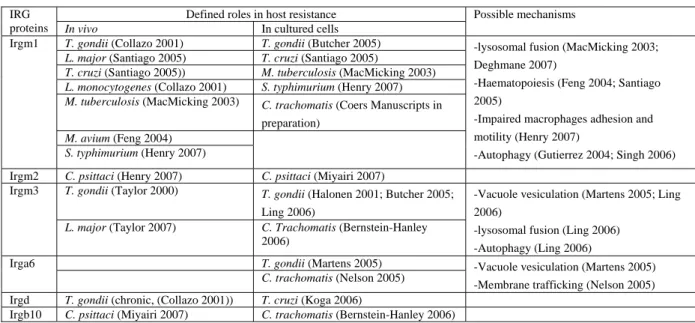
Table 1.1 Summary of evidences supporting roles of IRG proteins in host resistance (modified from Taylor 2007) Defined roles in host resistance
IRG
proteins In vivo In cultured cells
Possible mechanisms
T. gondii (Collazo 2001) T. gondii (Butcher 2005) L. major (Santiago 2005) T. cruzi (Santiago 2005)
T. cruzi (Santiago 2005)) M. tuberculosis (MacMicking 2003) L. monocytogenes (Collazo 2001) S. typhimurium (Henry 2007) M. tuberculosis (MacMicking 2003) C. trachomatis (Coers Manuscripts in
preparation) M. avium (Feng 2004)
Irgm1
S. typhimurium (Henry 2007)
-lysosomal fusion (MacMicking 2003;
Deghmane 2007)
-Haematopoiesis (Feng 2004; Santiago 2005)
-Impaired macrophages adhesion and motility (Henry 2007)
-Autophagy (Gutierrez 2004; Singh 2006) Irgm2 C. psittaci (Henry 2007) C. psittaci (Miyairi 2007)
T. gondii (Taylor 2000) T. gondii (Halonen 2001; Butcher 2005;
Ling 2006) Irgm3
L. major (Taylor 2007) C. Trachomatis (Bernstein-Hanley 2006)
-Vacuole vesiculation (Martens 2005; Ling 2006)
-lysosomal fusion (Ling 2006) -Autophagy (Ling 2006) T. gondii (Martens 2005)
Irga6
C. trachomatis (Nelson 2005) -Vacuole vesiculation (Martens 2005) -Membrane trafficking (Nelson 2005) Irgd T. gondii (chronic, (Collazo 2001)) T. cruzi (Koga 2006)
Irgb10 C. psittaci (Miyairi 2007) C. trachomatis (Bernstein-Hanley 2006)
1.7 Toxoplasma gondii infection as a model to study the functions of IRG proteins
Infections by the protozoan parasite Toxoplasma gondii are widely prevalent in humans and animals worldwide. The definitive host of T. gondii is the cat, but the parasite can be carried by a vast majority of warm-blooded animals. Toxoplasmosis, the disease caused by T. gondii, is usually minor and self-limiting but can have serious or even fatal effects on an immunocompromised host or on a fetus whose mother is infected during pregnancy. Infection with this parasite proceeds in two phases in immunocompetent hosts (Luft 1992). During the acute phase its rapidly proliferating form, the tachyzoite, disseminates throughout the host;
subsequently, during the chronic phase its dormant form, the bradyzoite, becomes established mainly in the central nervous system. This obligate parasite invades host cells, both haematopoietic and non-haematopoietic, by actively creating and entering into a unique non- fusogenic membrane-bounded cytoplasmic compartment, the parasitophorous vacuole (Sibley 2004).
Once inside of the host, T. gondii is a potent stimulus for cell-mediated immunity, and IL-12- dependent IFN-γ induction is vital for resistance to the parasite (Denkers 2003; Aliberti 2005).
IL-12 is produced mainly by dendritic cells, neutrophils and macrophages and is the main
cytokine to further stimulate the production of IFN-γ. T. gondii possesses two mechanisms for
triggering IL-12. One is dependent upon adaptor protein MyD88 and involves Toll-like
receptors (Scanga 2002). The other is a more unusual pathway that involves CCR5 by a parasite cyclophilin molecule (Aliberti 2000; Aliberti 2003). T. gondii has several mechanisms to down-regulate immunity. Intracellular infection causes a blockage STAT1 activation, therefore suppress the IFN-γ signaling (Luder 2001). In addition, intracellular parasite can subtlety modify host cell NFκB signaling pathway (Denkers 2003). NFκB is normally associated with transcription of proinflammatory mediators and resistance to infection. However, NFκB can also induce expression of several anti-apoptotic proteins, including inhibitor of apoptosis proteins (IAPs) and cFLIP, which behaves like a dominant- negative form of caspase-8 (Karin 2002; Hayden 2004). It is well-established that T. gondii triggers rapid phosphorylation and degradation of the NFκB inhibitor IκB on parasitophorous vacuole membrane, which depends on both the host IKK and a parasite kinase activity, TgIKK (Molestina 2005b; Molestina 2005a). However, there are seemingly controversial reports concerning the activity of NFκB triggered by the parasites. Some groups reported that T. gondii-infected cells exhibit increased NFκB activation and gene expression (Gazzinelli 1996; Blader 2001; Kim 2001), while several other groups reported that they are unable to observe NFκB activation despite infection-dependent increased IκB degradation (Butcher 2001; Goebel 2001; Shapira 2005). This may reflect the ability of T. gondii to establish a beneficial niche for their long-term survival, as the modification of host cell physiology may differ in different host cell types and in different stages of infection. For example, one interesting publication reported that, in macrophages, activated form of NFκB fails to tranlocate into the host nucleus despite of IκB degradation. However, after 24hours, the blockade in NFκB nuclear import is lifted and tranlocation of activated of NFκB is observed (Butcher 2002). Notably there is no dispute regarding the induction of NFκB once infection is established and parasite replication is initiated.
T. gondii infection can result in an anti-apoptotic state in host cells to facilitate the parasite
intracellular growth. T. gondii infected cells are resistant to numerous, both intrinsic and
extrinsic, apoptotic stimuli (Carmen 2007). In addition to the activation of NFκB-dependent
pro-survival mechanisms mentioned above, T. gondii has been suggested to inhibit caspase-9
activation through direct inhibition of apoptosome in in vitro reconstitution (Keller 2006) and
by inhibiting the release of cytochrome c (Goebel 2001; Carmen 2006). The activation of the
PI3-kinase pathway has also been suggested as a mechanism of induction of anti-apoptotic state during the early stage of infection (Kim 2006a), as the NFκB activation is blocked during that stage. Extracellular pathways of suppressing host immunity by T. gondii infection have been reported as well. These involve the infection-induced host production of IL-10 and lipoxins that have potent IL-12 down-regulatory effects (Figure 1.4)(Aliberti 2002a; Aliberti 2002b). Interestingly and surprisingly, no studies have been reported investigating how IFN-γ pretreatment influence the anti-apoptotic state conferred by T. gondii infection, considering IFN-γ is the most potent inducer for T. gondii resistance in vivo and in vitro.
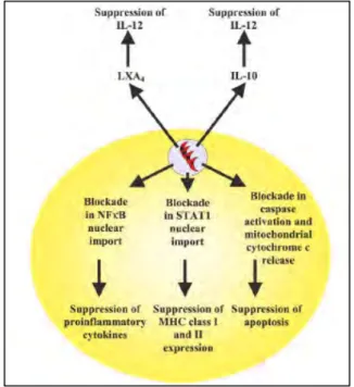
Figure 1.4 Intracellular and extracellular pathways of immunosuppression during T. gondii infection (from Denkers 2003).
Intracellular infection leads to a blockade in NFκB and STAT1 activation pathways. While IκB is phosphorylated and degraded, and STAT1 undergoes phosphorylation-dependent activation, neither NFκB nor STAT1 translocate into the nucleus during early infection. As a result, production of IL-12 and TNF-α is suppressed, and expression of MHC molecules is down-regulated. T.
gondii infection also renders cells resistant to apoptosis, as shown by reduced caspases proteolytic activation and mitochondrial cytochrome c release.
Extracellular pathways of immunosuppression involve Toxoplasma-induced host production of LXA4 and IL-10. These soluble mediators are potent down-regulators of IL-12 production that can act on infected and noninfected cells alike.
T. gondii is an excellent system to analyze the functions of IRG proteins. The host resistances for both phases of infection in vivo, as well as in cultured cells, are IFN-γ dependent (Suzuki 1988; Scharton-Kersten 1996). Either Irgm1 or Irgm3 deficient mice succumb to T. gondii avirulent strain infection within 9-11 days, which is comparable to the time frame as T. gondii infected mice lacking either IL12 or IFN-γ-signaling machinery (Taylor 2000; Collazo 2001).
In addition, Irgd-deficient mice can survive acute phase of T. gondii infection, but have
impaired resistance during chronic infection (Collazo 2001). On the cellular level, Irgm1 and
Irgm3 deficient macrophages and astrocytes lose resistance against T. gondii infection upon
IFN-γ induction, which correlates with loss of resistance in mice (Halonen 2001; Butcher
2005). At least five members of IRG proteins are concentrated on T. gondii PVM rapidly after
the parasite invasion in murine astrocytes and primary embryonic fibroblasts (Martens 2005).
Vesiculation and stripping of parasitophorous membrane have been observed in T. gondii infected astrocytes and macrophages (Martens 2005; Ling 2006). An autophagolysosomal process has been suggested in macrophages after stripping of T. gondii vacuolar membranes (Ling 2006). However, no lysosomal fusion has been observed in astrocytes (Martens 2005), and the question how naked parasites free in the cytosol are killed remains to be illustrated.
Furthermore, although Irgm1 is indispensable for resistance against T. gondii in vivo, this protein is absent from T. gondii PVM indicating that IRGs may function in multiple layers of resistance (Butcher 2005; Martens 2005).
1.8 Aims of this study
Immunity-related GTPases (IRG) are powerful, non-redundant resistance factors against a variety of intracellular pathogens in mice. This is manifested by well-documented analyses using gene-targeted mice and cells for individual members of IRGs. Cell-autonomous control of pathogens is a predominant function of IRG proteins, although the exact mechanisms are still not known. Irgm1 is the most prominent member in IRG family for resistance as mice lacking this protein are susceptible to all bacterial and protozoal pathogens tested so far.
Possible mechanisms have been suggested for functions of Irgm1 such as: (i) lysosome fusion and/or acidification of the vacuole (MacMicking 2003; Deghmane 2007), (ii) induction of autophagy (Gutierrez 2004; Singh 2006), (iii) regulation of the adhesion and motility of activated macrophages (Henry 2007), (iv) negatively regulation of TLR4-triggered proinflammatory cytokine production and prevention of endotoxemia (Bafica 2007) and (v) regulation of haematopoiesis during chronic infection (Feng 2004; Santiago 2005).
Irgm1 has been reported to localize to Golgi apparatus, ER and vesicular structures in resting
cells. During phagocytosis it is rapidly relocalized to F-actin-rich plasma membrane ruffles
associated with phagocytic cups in macrophages and fibroblasts (Martens 2004b). In the
present study subcellular localization of Irgm1 and corresponding mechanisms are further
investigated in depth. In addition, localization influenced by N and C terminal tag is analyzed,
as proposed pro-autophagic function of Irgm1 is based on experiments using EGFP-tagged
protein and correct subcellular localization was not confirmed. Furthermore, role of Irgm1 in
cell-autonomous resistance to T. gondii infection in fibroblasts is studied. Finally, live cell
imaging system is established and employed to investigate the involvement of IRG proteins in
resistance against T. gondii infection on a single cell level.
2. Material and Methods
2.1 Reagents and Cells
2.1.1 Chemicals, Reagents and Accessories
All chemicals were purchased from Aldrich (Steinheim), Amersham-Pharmacia (Freiburg), Applichem (Darmstadt), Baker (Deventer, Netherlands), Boehringer Mannheim (Mannheim), Fluka (Neu-Ulm), GERBU (Gaiberg), Merck (Darmstadt), Pharma-Waldhof (Düsseldorf), Qiagen (Hilden), Riedel de Haen (Seelze), Roth (Karlsruhe), Serva (Heidelberg), Sigma- Aldrich (Deisenhofen) or ICN biochemicals, Oxoid, (Hampshire UK). Developing and fixing solutions for Western Blot detection were from Amersham Pharmacia (Freiburg), Luminol from Sigma Aldrich (Deisenhofen), Coumaric acid from Fluka (Neu-Ulm). Deionised and sterile water (Seral TM) was used for all the buffers and solutions, Ultra pure water derived from Beta 75/delta UV/UF from USF Seral Reinstwassersysteme GmbH, (Baumbach) equipped with UV (185/254nm) and ultrafiltration (5000 kd cut off), or from Milli-Q- Synthesis (Millipore).
2.1.2 Equipments
Centrifuges used were: Biofuge 13, Heraeus; Sigma 204; Sigma 3K10; Labofuge 400R, Heraeus; Sorvall RC-5B, Du Pont instruments; Optima TLX Ultracentrifuge, Beckmann and Avanti J-20 XP, Beckman. BioRAD Gel dryer, Model-583; BioRad Power pack 300 or 3000;
electrophoresis chambers from FMC Bioproducts (Rockland Maine US); Gel Electrophoresis Chamber, Cambridge electrophoresis; Biorad Mini Protean II; PTC-100, MJ Research Inc.;
ÄKTA P-920, OPC-900, Frac-950, Amersham; Centrifuge tubes 15ml, TPP Switzerland;
50ml Falcon, BectonDickenson; Zeiss Axioplan II microscope equipped with AxioCam MRm
camera (Zeiss). Zeiss Axiovert 200M motorized microscope equipped with AxioCam MRm
camera (Zeiss). ELISA reader (Vmax, Molecular Devices)
2.1.3 Materials
Sterile filters FP 030/3 0,2 µm and ME 24 0,2 µm (Schleicher und Schüll, Dassel);
Nitrocellulose transfer membrane PROTRAN (Schleicher und Schüll, Dassel); 3MM Whatmann Paper (purchased via LaboMedic); 100 Sterican 0,50 x 16mm hypodermic needles (Braun AG, Melsungen); 0.2µm and 0.45µm sterile filters (Schleicher und Schuell, Dassel);
X-OMAT LS and AR X-ray films, Kodak. All plastic ware for cell culture was from Sarstedt (Nümbrecht) or Greiner (Solingen).
2.1.4 Enzymes and Proteins
Restriction enzymes (New England Biolabs); Pyrococcus furiosus (Pfu) DNA Polymerase (Promega, Mannheim); T4 DNA ligase (New England Biolabs); RNase A (Sigma); shrimp alkaline phosphatase (SAP) (USB, Amersham); PageRulerTM Prestained Protein Ladder (Fermentas); PageRulerTM Protein Ladder (Fermentas); SigmaMarkerTM Wide Range (Sigma); GeneRulerTM DNA Ladder Mix (Fermentas).
2.1.5 Kits
Plasmid Maxi and Midi kit (Qiagen, Hilden), Terminator-cycle Sequencing kit version 3 (ABI),
QuikChange TM Site directed mutagenesis kit (Stratagen), Rapid PCR product purification Kit (Roche, Mannheim), 2.1.6 Vectors and constructs used in the present study
pGW1H (British Biotech),
pEGFP-C3, pEGFP-N3 (Clontech),
pmDsRed-C3, pmDsRed-N3, pCherry-C3, pCherry-N3
pF25: gift from Dr. Gregory A. Taylor, Duke Univeristy, coding for the N-terminally EGFP- tagged Irgm1 (Irgm1-EGFP).
EGFP-Irgm1, EGFP-Irgm1 αK, EGFP-LC3, Irga6-ctag1-EGFP
2.1.7 Cell lines, bacterial and protozoan strains
Murine embryonic fibroblasts (MEFs) derived from C57/BL6 mice, L929 (CCL-1) and gs3T3 (Invitrogen) mouse fibroblasts were cultured in DMEM supplemented with 10% FCS (Biochrom AG, Berlin), 2 mM L-Glutamine, 1 mM sodium pyruvate, 100 U/ml penicillin and 100 µg/ml streptomycin, all from Gibco BRL. Human foreskin fibroblasts (Hs27, ATCC CRL-1634) were cultured in IMDM supplemented with 5% FCS and 2mM L-glutamine.
Sterile trypsin/EDTA solution in PBS (10x trypsin/EDTA solution: 0.05% (w/v) trypsin (1:250, Gibco BRL)/17 mM EDTA/145 mM NaCl)) was used to detach adherent cells from culture flasks.
Escherichia coli DH5α: 80dlacZ ∆Μ15, recA1, endA1, gyrA96, thi-1, hsdR17 (rB-, mB+), supE44, relA1, deoR, ∆(lacZYA-argF)U169
Toxoplasma gondii: Type II strain ME49, Type I strain RH-YFP
2.1.8 Media
Luria Bertani (LB) medium:
10 g bacto tryptone, 5 g yeast extract, 10 g NaCl, destilled water 1L
LB plate medium:
10 g bacto tryptone, 5 g yeast extract, 10 g NaCl, 15 g agar, destilled water 1L
IMDM (Iscove’s Modified Dulbecco’s Medium) supplemented with 10% FCS (Biochrom AG, Berlin), 2 mM L-glutamine, 1 mM sodium pyruvate, 1x non-essencial amino acids, 100 U/ml penicillin, 100 µg/ml streptomycin (all from Gibco BRL)
DMEM (Dulbecco’s Modified Eagle Medium) supplemented with 10% FCS (Biochrom AG,
Berlin), 2 mM L-glutamine, 1 mM sodium pyruvate, 1x non-essencial amino acids, 100 U/ml
penicillin, 100 µg/ml streptomycin (all from Gibco BRL)
2.1.9 Serological reagents Primary antibodies and antisera
Name Immunogen Species Concentration Dilution Origin
P20 N-terminus peptide
of Irgm1
Goat polyclonal
0.2 mg/ml IF: 1:100 Santa Cruz
Sc-11074
1B2 Irgm1 Mouse
monoclonal
IF: undiluted
supernatant from hybridoma cell culture
Dr. Gregory Taylor Duke University
165 Recombinant Irga6 Rabbit
polyclonal
1-3 mg/ml WB: 1:25000 IF: 1:8000
H53 N-terminus peptide
of Irgm2
Rabbit polyclonal
IF: 1:1000
αIGTP clone 7
aa 283-423 of Irgm3 Mouse monoclonal
0.25 mg/ml WB: 1:2000 BD Transduction
laboratories A20 N-terminal peptide of
Irgb6
Goat polyclonal
0.2 mg/ml WB: 1:500 IF: 1:100
Santa Cruz sc11079 GM130 C-terminus of rat
GM130
Mouse monoclonal IgG1
IF: 1:1000
WB: 1:250
BD transduction Lab.
610822 S-20 C-terminus peptide
from rat TGN38 origin
Goat polyclonal
0.2 mg/ml IF: 1:100 Santa Cruz
Sc-27681 CI-M6PR
antibody
CI-M6PR Rabbit polyclonal
IF: 1:100 Albert Hass, Bonn
1D4B Mouse LAMP1 Rat monoclonal IF: 1:1000 DSHB, Iowa
SPA-865 Peptide from canine calnexin aa 50-68
Rabbit polyclonal
WB: 1:10000
IF: 1:200
Stressgen
GRA7 GRA7 Mouse
monoclonal
IF: 1:1000 Gaby Reichmann
Düsseldorf Cytochrome
C antibody Rat
cytochrome c
Mouse monoclonal
0.5 mg/ml IF: 1:1000 BD PharMingen
556432 Clone: 6H2.B4 HMGB1
antibody
aa150 to the C- terminus of human HMGB1
Rabbit polyclonal
0.5 mg/ml WB: 2µg/ml Abcam
ab18256 Cleaved
Caspase-3 antibody
N-terminal peptide adjacent to Asp175 of human Caspase-3
Rabbit polyclonal
WB: 1:1000 Cell signaling Technology, Inc.
#9661 PARP
antibody
Peptide
corresponding to the caspase cleavage site
Rabbit polyclonal
WB: 1:1000 Cell signaling Technology, Inc.
#9542