Evolution and Cellular Resistance Mechanisms of the Immunity-Related GTPases
Inaugural-Dissertation zur Erlangung des Doktorgrades
der Mathematisch-Naturwissenschaftlichen Fakultät der Universität zu Köln
vorgelegt von Julia Hunn aus Singen (Htwl.)
Köln 2007
Berichterstatter: Prof. Dr. Jonathan C. Howard
Prof. Dr. Thomas Langer
Prof. Dr. Otto Haller
Tag der mündlichen Prüfung: 7. February 2008
Success is nothing more than going from failure to failure with undiminished optimism.
Winston Churchill
Table of contents
I. INTRODUCTION 1 I.1. IN F E C T I O N, I MM U N I T Y A ND C YT O K I NE S 1
I.2. IN T E R F E R O N S 1
I.3. IFN-M E D I ATE D C E L L A UT ON O M O U S R E S I ST A N C E 4 I.4. GUA NO S I N E T R I PH O S P H A T ASE S (GTPASE S) 4
I.4.1. Dynamin 6
I.4.2. Large, interferon-inducible GTPases 7
I.5. TOX OP LAS MA G ON D I I 19
I.6. THE A I M O F T H I S S T U DY 22
II. MATERIAL AND METHODS 24
II.1. MATE R I AL 24
II.1.1. Mammalian cells and media 24
II.1.2. Bacterial strains and media 24
II.1.3. Yeast strains and media 24
II.1.4. Toxoplasma gondii strains 25
II.1.5. Vectors 25
II.1.6. Generation of expression constructs 25
II.1.7. Primers 27
II.1.8. Primary immunoreagents 27
II.1.9. Secondary immunoreagents 28
II.2. METHODS: PH YL O G E N E T I C A N AL Y S I S 29
II.2.1. Use of database resources 29
II.2.2. Generation of multisequence alignment and phylogenetic trees 29 II.3. METHODS: MUTA G E NE S I S A N D C L O N I N G 29
II.3.1. Preparation of chemical competent bacteria 29
II.3.2. Transformation of competent bacteria 29
II.3.3. Plasmid DNA isolation 30
II.3.4. Agarose gel electrophoresis 30
II.3.5. Purification of DNA fragments from agarose gel 30
II.3.6. Restriction digest 30
II.3.7. Ligation 30
II.3.8. Sequencing 30
II.3.9. Site directed mutagenesis 31
II.4. METHODS: PR OT E I N BI O CH E M I ST R Y 31
II.4.1. Expression and purification of recombinant protein 31
II.4.2. Guanine nucleotide binding parameters 32
II.4.3. GTP hydrolysis assay 32
II.4.4. Analysis of protein oligomerisation by light scattering 32
II.5. METHODS: M AM M A L I A N C E L L S 33
II.5.1. Freezing and thawing of mammalian cells 33
II.5.2. Transfection of mammalian cells 33
II.5.3. Hormone-inducible mammalian expression system (GeneSwitch) 33
II.5.4. Generation of stable inducible cell lines 34
II.5.5. Induction with IFNγ and Mifepristone 34
II.5.6. Immunofluorescence 35
II.5.7. Cell proliferation assay 35
II.5.8. Cell cycle assay 35
II.6. METHODS: AN AL Y S I S O F C E L L UL A R PR O T E I N 35
II.6.1. Generation of cell lysates for SDS-PAGE 35
II.6.2. SDS-polyacrylamid gel electrophoresis (SDS-PAGE) 36
II.6.3. Western Blotting and Ponceau S staining 36
II.6.4. Sequential Triton X-114 partitioning assay 36
II.6.5. Co-Immunoprecipitation 37
II.6.6. Pull down 37
II.6.7. Coomassie staining 38
II.6.8. Analytical size exclusion chromatograpgy of cellular IRG GTPases 38
II.7. METHODS: IN FE C T I O N A S SA Y S 38
II.7.1. In vitro passage of Toxoplasma gondii 38
II.7.2. Infection of murine cells with Toxoplasma gondii 39
II.7.3. Quantication of IRG signals on T. gondii parasitophorous vacuoles 39
TABLE OF CONTENTS
II.8. METHODS: YE AS T 2 HYBRID 39
II.8.1. Lithium acetate transformation of Saccharomyces cerevisiae 39
II.8.2. Analysis of protein interaction by yeast two-hybrid (Y2H) 39 III. RESULTS 41 III.1. GE N O M I C O R G A N I S A T I O N, S Y N T E N Y A N D P H Y L O G E N E T I C R E L A T I O N S H I P
O F IRG GTPA S E S 41
III.1.1. Nomenclature 41
III.1.2. IRG genes of the C57BL/6 mouse 43
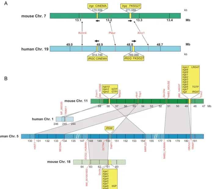
III.1.3. Human IRG genes and their synteny relationship to mouse IRGs 52
III.1.4. IRG homologues in rodents and lagomorphs (Glires) 59
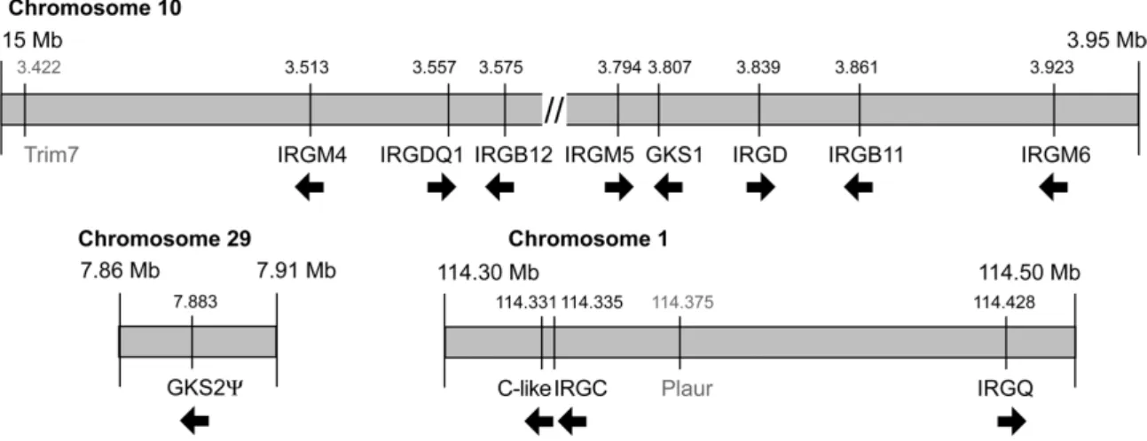
III.1.5. IRG homologues in the carnivore Canis familiaris 60
III.1.6. IRG homologues in other mammalian species 67
III.1.7. IRG homologues in non-human primates 69
III.1.8. IRG homologues outside the Eutheria 71
III.2. RE G U L A T O R Y I N T E R A C T I O N S B E T W E E N IRG GTPA S E S C O N T R O L L I N G
A C T I V AT I ON A N D F U NC T I O N 80
III.2.1. Generation and characterisation of stable cell lines inducibly expressing
single IRGs 80
III.2.2. Influence of IRG expression cell proliferation and survival 84 III.2.3. Influence of IFNγ on the subcellular localisation of Irgm1-3, Irgc, Irgd
and Irga6 86
III.2.4. Biochemical properties of Irga6 G1 mutants 94
III.2.5. Influence of nucleotide binding on the subcellular localisation of Irga6 96 III.2.6. Influence of IFNγ and nucleotide on the subcellular localisation of Irgb6 97 III.2.7. Behaviour of IRG proteins in size exclusion chromatography 99 III.2.8. Regulation of Irga6 and Irgb6 positioning by the GMS proteins 101 III.2.9. Nucleotide-dependent direct interactions of IRG proteins in Y2H 104 III.2.10. Direct GDP-dependent interaction of cellular Irgm3 with Irga6 106 III.2.11. Regulation of Irga6 in Toxoplasma-infected cells 109 III.2.12. Regulation of Irgb6, Irgd, Irgm1-3 and Irgc in Toxoplasma-infected cells113 III.2.13. Virulent T. gondii counteract IRG protein accumulation at the PVM 116 IV. DISCUSSION 118 IV.1. SI GN S F O R D I VE R GE N T E VO L U T I O N OF T H E IRG G E N E S I N R O D E NT S 118 IV.2. DI VE R GE N T IRG G E NE S AR E F O U N D T H R O U G H OU T T HE M AM M A L S 119 IV.3. LO SS O F IRG RE SI ST A N CE SY ST E M I N T H E A NT H RO PO I D S 121 IV.4. THE IRG F A M I L Y I S A N C I E N T A N D PR E SE N T I N E U C H O R D AT E S 123 IV.5. IRG P R O T E I N S I N C E L L PRO L I FE R AT I O N A N D S UR V I V A L 126 IV.6. COV A L E N T P R O T E I N M O DI F I C AT I O N O F IR G A6 126 IV.7. IRG L OC A LI SAT I O N I S REG U L AT E D BY IFN A N D N U C L E O T I DE 128 IV.8. IR G A6 A N D IR G B6 A G G R E G A T E S F O R M E D I N A B S E N C E O F IFNγ
R E PRE SE N T GTP-B O U ND H O M O ME R S 130
IV.9. GTP-B O U N D H O M O M E R I C IR G A6 A N D IR G B6 C O M PL E X E S
D I S S O C I AT E EX VIV O 131
IV.10. TH E T H R E E GMS P R O T E I N S N E G A T I V E L Y R E G U L A T E IR G A6 A N D IR G B6 131
IV.11. DIRE CT, N U C L E O T I DE-DEP E N D E NT I N T E R A C T I ON S O F IRG R O T E I N S 132 IV.12. DI R E C T, GDP-D E P E N D E N T I N T E R A C T I O N O F IR G A6 W I T H IR G M3
O C C U R S V IA T H E G-DO MA I N 134
IV.13. RE GU L A T O R Y IRG I NT E R AC T I O N S I N TO X OP LA S M A I N FE CT IO N 135 IV.14. IR GC – A PR OTE I N I N SE AR C H OF A FUNC T I ON 138 IV.15. VIRU L E NT TOXO P L AS MA GO N D I I I N HIB I T IRG P R O T E I N S 139 IV.16. MOD E L O F IRG F U N C T I ON I N U NI N FE C T E D A N D I N FE C T E D C E L L S 140 V. APPENDIX 142 V.1. PU R I F I C A T I O N OF R E C O M B I N A N T IR G A6(S83N) AN D (K82A) R OT E I N 142
V.2. EX P R E S S I O N VE C T O R S 143
V.3. DNA S E Q UE N C E O F T HE E XP R E S S E D MU R I N E IRGS 144 V.4. AB B R E V I AT I O N O F S P E C I E S N A ME S 145 V.5. SE QU E N C E S O UR C E S O F MO U S E A N D H U M A N IRG G E NE S 146 V.6. NU C L E OT I D E AL I G N ME NT O F M O U SE IR G A6 A N D IR G A6* C57BL/6) 148
V.7. PR OT E I N A N D NU C L E O T I DE AL I G N ME NT O F M O U SE IR G B3 A ND IR GB4 149 V.8. AL I GN M E N T O F M A M M A L I AN IRGC P R O T E I N S 150 V.9. AL I GN M E N T O F M A M M A L I AN IRGQ P R O T E I N S 151 V.10. AL I GN M E N T O F O R A N G-U T A N IRGM SE Q U E N C E S (PO N G O PY G M A E U S) 152 V.11. AL I GN M E N T O F TA K I F U G U R U B R I P E S IRG PROTEINS 152 V.12. AL I GN M E N T O F TA K I F U G U R U B R I P E S IRG PROTEINS 153
V.13. IRG P R O T E I N SEQ U E N C E S 153
VI. REFERENCES 174 VII. SUMMARY 192 VIII. ZUSAMMENFASSUNG 193 IX. ACKNOWLEDGEMENTS 194 X. KOLLABORATIONEN 195 XI. ERKLÄRUNG 196 XII. LEBENSLAUF 197
ABBREVIATIONS
Abbreviations
AD activation domain GPI glycosyl-phosphatidylinositol ADAR adenosine deaminase acting on RNA GppNHp 5’-guanylylimidodiphosphate
GRA dense granule protein ADRP adipocyte differentiation-related
protein GST glutathione-S-transferase GTP guanosine triphosphate APOBEC3G apolipoprotein B mRNA-editing
enzyme, catalytic polypeptide-like 3G GTPase guanosine triphosphatase
APS ammonium peroxide sulfate GTPγS guanosine 5'-(3-O-thio)triphosphate ATP adenosine triphosphate GTPI GTPase IFN-induced
ATPase adenosine triphosphatase HRP horse radisch peroxidase BAC bacterial artificial chromosome HSV-1 herpes simplex virus type 1 BD DNA binding domain IBDV infectious bursal disease virus BLAST basic local alignment search tool IDO indoleamine 2,3-dioxygenase BSA bovine serum albumin IF immunofluorescence CARD caspase recruitment domain IFN interferon
CID central interacting domain IFNAR IFNα receptor CIITA MHC class II transactivator (CIITA) IFNGR IFNγ receptor IFNLR IFNλ receptor DAPI 4',6-Diamidine-2'-phenylindole
dihydrochloride IGTP inducibly expressed GTPase DC dentritic cell IIGP IFN-inducible GTPase DBD DNA binding domain IL interleukin
DMEM Dulbecco’s modified Eagle’s medium IL10R2 interleukin 10 receptor 2 DMSO dimethylsulfoxid iNOS inducible nitric oxide synthase Ds double stranded IPTG isopropyl-β-D thiogalactoside DTT dithiothreitol IRF IFN regulatory factor ECMV encephalomyocarditis virus IRG immunity-related GTPases EDTA ethylen diamine tetraacetate IRG-47 IFN-regulated GTPase 47 kDa EF-G elongation factor G ISGF3 IFN-stimulated gene factor 3 EF-Tu elongation factor thermal unstable ISG15 IFN-stimulated gene 15 EGFP enhanced green fluorescent protein
ER endoplasmic reticulum
ISG20 IFN-stimulated 3’-5’ exonuclease gene 20
eRF1 elongation release factor 1 ISRE IFN-stimulated response element ERV9 endogenous retrovirus 9 Jak Janus kinase EST expressed sequence tag LD lethal dose
EtOH ethanol LPS lipopolysaccharide
FAT10 HLA-F adjacent transcript 10 LRG-47 LPS-regulated GTPase 47 kDa FCS fetal calf serum LTR long terminal repeat
FLUAV influenza A virus LZ leucine zipper
FTase farnesyl transferase MEC mouse oviduct epithelial cell FPLC fast protein liquid chromatography MEF mouse embryonic fibroblast G domain GTPase domain mGDP mant guanosine diphosphat GAF gamma activated factor mGTPγS mant GTPγS
GAL4 galactose induced gene 4 MHC major histocompatibility complex GAS gamma-activated sequence MIC microneme protein
GAP GTPase activating protein Mif Mifepristone
GBP guanylate binding protein MOI multiplicity of infection GDI guanine nucleotide dissociation
inhibitor
MW molecular weight
GDP guanosine diphosphate MxA Myxovirus resistance protein A GDPβS guanosine-[β-thio]-diphosphate
GED GTPase effector domain
NEDD8 neural precursor cell expressed, developmentally down-regulated 8 GEF guanin nucleotide exchange factor NK cell natural killer cell
GFP green fluorescent protein NMT N-myristoyl transferase GGTase geranylgeranyl transferase NOS2 nitric oxide synthase 2
NRAMP1 natural resistance-associated macrophage protein 1
Sox Sry-type high-mobility-group domain box
2’-5’ OAS 2’-5’-oligo-adenylate synthetase SRP signal recognition particle o.n. over night
OD optical density
SSAHA sequence search and alignment by Hashing algorithm
ORF open reading frame
PAGE polyacrylamind gel electrophoresis
STAT signal transducer and activator of transcription
PBS phosphate buffered saline SUMO small ubiquitin-related modifier PH pleckstrin homology TGTP T cell specific GTPase
phox phagocyte oxidase TIM triose-phosphate isomerase PKR dsRNA-dependent protein kinase TK HSV thymidine kinase PML promyelocytic leukemia TNFα tumor necrosis factor α PMSF phenylmethylsulphonyl fluoride TRIM5α tripartite motif protein 5α PRD proline-rich domain
PV parasitophorous vacuole
tsvGORASP testis specific splice variant Golgi reassembly and stacking protein 2 PVM parasitophorous vacuolar membrane Tyk2 tyrosine kinase 2
Rab Ras genes from rat brain U units
Ran Ras related nuclear protein UAS upstream activating sequence Ras rat sarcoma URG4 up-regulated gene 5
Rho rat sarcoma homologue UTR untranslated region
RNAi RNA interference VLIG very large inducible GTPase RON rhoptry neck protein VSV vesicular stomatitis virus ROP rhoptry protein WB Western blot
ROS reactive oxygen sspecies Y2H yeast two-hybrid
RT room temperature YFP yellow fluorescent protein SD synthetic defined YPD yeast-peptone-dextrose medium SDS sodium dodecylsulfate ZAP zinc-finger antiviral protein
I. Introduction
I.1. Infection, immunity and cytokines
Multicellular organisms are constantly threatened by the invasion of microorganisms.
The arms race between the host trying to prevent infection and eliminate the invaders and the pathogen seeking to exploit the host and to counteract its defence mechanisms led to the evolution of a complex, multilayered immune defence. The immune system has traditionally been divided into innate and adaptive components. The innate immune system predates the adaptive immune response evolutionary (Medzhitov 1997). Even single-cell organisms have heritable defence mechanisms, and every multicellular organism appears to have a complex innate immune system, whereas adaptive immunity is found only in vertebrates (Beutler 2004; Medzhitov 1997). The innate response includes both constitutive and inducible mediators and produces generic receptors that recognise conserved pathogen-associated molecular patterns (PAMPs) to trigger an inflammatory response that limits pathogen invasion (Janeway 2002). Specific adaptive immunity, by contrast, depends upon somatic diversification of antigen-receptor genes to generate a vast repertoire of lymphocytes, each expressing a different antigen receptor.
Recognition of specific antigenic pathogen compounds by these cell-surface receptors triggers clonal amplification, cellular differentiation and production of secreted receptors with the same antigen binding specificity (Burnet 1959). In the vertebrate immune system innate and adaptive components synergise in the clearance of pathogens. The innate response is crucial in limiting the early replication and spread of infectious agents.
By contrast, the generation of an adaptive immune response involves considerable lag time but culminates in the production of specialised effector mechanisms that are highly efficient in eliminating the pathogen and in the formation of an immunological memory (Le Bon 2002). In addition to these complementary activities, there is a fundamental connection and extensive crosstalk between innate and adaptive immunity. Namely, the magnitude and quality of the adaptive immune response is dependent on signals derived from the innate response to infection (Medzhitov 1997). Besides cell-cell interactions a central mode of communication within the immune system is via cytokines produced by hematopoietic and non-hematopoietic cells. Among the cytokines, interferons play a complex and central immunomodulatory role mediating host resistance to pathogens (reviewed in (Boehm 1997; Stark 1998; Young 2007)).
I.2. Interferons
Interferons (IFNs) were originally discovered as agents that interfere with viral replication (Isaacs 1957a; Isaacs 1957b). Beside their antiviral activity, IFNs exhibit growth-inhibitory effects and have important roles in immunosurveillance for malignant cells and in immunomodulation. The importance of interferons is apparent from mice that lack expression of IFNγ or its receptor due to targeted deletions and consequently are susceptible to many infectious agents (Dalton 1993; Huang 1993; Janssen 2002;
Jouanguy 1999a; Jouanguy 1999b; Orange 1995; Ottenhoff 2002; Scharton-Kersten
1996) (see Table 1). Humans with naturally occurring mutations in their IFNγ receptor
genes, however, exhibit a severe, profound and selective susceptibility to weakly virulent
INTRODUCTION
mycobacteria only (Doffinger 2002; Jouanguy 1997). IFNs are induced either directly by infection and tissue damage or by immune and inflammatory stimuli. IFNs are classified into type I, type II and type III according to sequence homology, cellular sources and receptor specificity.
Figure 1 Interferon signalling pathways. Type I and type II interferons bind to their respective receptors activating related, partially overlapping but distinct signalling pathways of the Janus kinase family (Jak)–
signal transducer and activator of transcription (STAT) type. These signalling cascades activate the expression of genes containing GAS (gamma-activated sequence) and ISRE (IFN-stimulated response element) elements in their promoter. Type III IFNs (λ-IFNs) signal through the same Jak/STAT pathway as type I IFNs driving expression of a common set of genes but engage a distinct heterodimeric receptor (IFNLR1/IL10R2) (not shown). The transcriptional response to IFNs includes numerous factors participating in the host immune response to viral and microbial pathogens. Protein kinase R (PKR), 2‘-5‘- oligoadenylate synthetase (2‘-5‘ OAS), adenosine deaminase acting on RNA (ADAR), Mx and guanylate- binding proteins (GBPs) have been implicated in viral resistance, whereas indoleamine 2,3-dioxygenase (IDO), nitric oxide synthase 2 (NOS2), phagocyte oxidase (phox), natural resistance-associated macrophage protein 1 (NRAMP1) and immunity-related GTPases (IRGs, p47 GTPases) all have been reported to inhibit the replication of bacteria, protozoa and viruses. Other proteins that are inducible by IFNs include MHC proteins and transcription factors IRF-1, IRF-9 and CIITA (modified from (Shtrichman 2001)).
Type I interferons are comprised of multiple IFNα subtypes (14-20 depending on
species) (Pestka 1987; van Pesch 2004), IFNβ (Mogensen 1999), IFNδ (Lefevre 1998),
IFNε (Conklin 2002; Pestka 2004), IFNκ (LaFleur 2001), IFNτ (Bazer 1997; Martal
1998), IFNυ (Samarajiwa 2006; Uze 2007), IFNω (Hauptmann 1985) and limitin/IFNζ
(Oritani 2000). IFNα and IFNβ are secreted by almost all cell types upon virus infection
and by activated immune cells such as dendritic cells and macrophages. The other type I
IFNs are not necessarily present in all mammalian species or expressed upon viral
infection. For example, mice have no orthologues of IFNυ, IFNκ is constitutively
expressed by keratinocytes, secretion of IFNτ has only been reported in ungulate
ruminants and has a specific function in maternal recognition of pregnancy (reviewed in
(Schroder 2004)). Type I IFN family members bind a common heterodimeric cell-surface
receptor, the IFNα receptor (IFNRA), which consists of IFNAR1 and IFNAR2 chains and is activated by ligand-induced dimerisation (Darnell 1994; Stark 1998) (Figure 1).
Each receptor subunit binds constitutively to a single specific member of the Janus kinase (Jak) family: IFNAR1 to tyrosine kinase 2 (Tyk2) and IFNAR2 to Jak1. Ligand binding induces the phosphorylation of Jak1, Tyk2, intracellular tyrosine residues of the IFNA receptor chains and the signal transducers and activators of transcription 1 and 2 (STAT1 and STAT2). Together with IFN-regulatory factor 9 (IRF9) phosphorylated STAT1 and STAT2 form a transcriptional complex called IFN-stimulated gene factor 3 (ISGF3), which translocates into the nucleus and binds to IFN-stimulated response elements (ISREs) present in the promoters of many IFN-regulated genes (Stark 1998).
Although the three type III IFNs or λ-IFNs (IFNλ1-3 or IL-28A/IL-28A/IL-29) differ genetically from type I IFNs, they exhibit similar biological antiviral, antitumour and antiproliferative activity and their expression is regulated in a similar fashion (Kotenko 2003; Osterlund 2007; Sheppard 2003; Uze 2007). Furthermore, they signal through the same Jak/STAT signalling pathway driving expression of a common set of genes. Importantly, however, λ-IFNs bind to a distinct membrane receptor composed of the IFNLR1 and IL10R2 receptor chains. This specific receptor usage suggests that this cytokine family does not merely replicate the type I IFN system and justifies its designation as type III IFN.
Ιnterferon gamma (IFNγ) is the sole type II IFN and is synthesised only by certain activated effector cells of the innate and adaptive immune systems including natural killer (NK) cells (Bancroft 1993), T lymphocytes (Mosmann 1989), macrophages (Gessani 1998; Munder 1998) and dendritic cells (DCs) (Ohteki 1999). Although originally defined as an agent with direct antiviral activity, the properties of IFNγ include regulation of several aspects of the immune response, stimulation of bactericidal activity of phagocytes, stimulation of antigen presentation through class I and class II major histocompatibility complex (MHC) molecules, orchestration of leukocyte-endothelium interactions, effects on cell proliferation and apoptosis, as well as the stimulation and repression of a variety of genes. The two chains of the IFNγ receptor, IFNGR1 and IFNGR2, heterodimerise upon binding of an IFNγ homodimer leading to the activation of the respective receptor associated Janus kinases, Jak1 and Jak2 (Figure 1) (Darnell 1994;
Stark 1998). The subsequent tyrosine phosphorylation cascade results in phosphorylated and activated STAT1. Upon phosphorylation STAT1 forms a homodimer termed gamma activated factor (GAF), translocates into the nucleus and initiates transcription by binding to gamma activated sequences (GAS) in the promoters of IFNγ inducible genes (Stark 1998).
Thus, binding of IFNs to their specific cell surface receptors leads to the
activation of distinct but related components of the signal transduction and
transcriptional activation machinery, resulting in the stimulation of the transcription of
more than thousand genes belonging to partially overlapping sets (Boehm 1997; Darnell
1994; Der 1998; Ehrt 2001; Stark 1998; Takaoka 2000; Valente 1992) (Figure 1). Among
these are a wide range of mediators that undermine the ability of pathogens to survive in
host cells, contributing to organismal and cellular resistance involving both the innate
and adaptive immune system (Boehm 1997).
INTRODUCTION
I.3. IFN-mediated cell autonomous resistance
It has become clear over the last decades that the resistance against pathogens does not solely rely on specialised immune cells like lymphocytes or macrophages that patrol through the body. To counteract intracellular pathogens every single body cell is equipped with multiple defence mechanisms that are independent of specialised immune cells. Many of the molecular players of this so-called cell autonomous resistance are induced by interferons and therefore strictly speaking not completely independent of other cells. Still, apart from the need of a ‘danger signal’ to induce cell autonomous immunity, the process of pathogen counteraction and clearance is independent from the world outside the cell boundaries.
The list of proteins implicated in cell autonomous immunity is long and still growing and includes factors active against all kinds of pathogens. The antiviral factors include double stranded (ds)RNA-dependent protein kinase (PKR) (Garcia 2007; Lee 1993; Meurs 1992; Stojdl 2000; Yang 1995), 2’-5’ oligoadenylate synthetase (2’-5’
OAS)/RNase L (Chebath 1987; Chebath 1983; Samuel 2001), dsRNA adenosine deaminase (ADAR1) (Jayan 2002; Wong 2002), ISG20 (interferon stimulated 3'-5' exonuclease gene 20kDa) (Degols 2007; Espert 2003), promyelocytic leukemia protein (PML/TRIM19) (Chee 2003; Chelbi-Alix 1998; Everett 2007; Regad 2001), zinc-finger antiviral protein (ZAP) (Gao 2002; Guo 2007), APOBEC3G/CEM15 (apolipoprotein B mRNA-editing enzyme, catalytic polypeptide-like 3G) (Harris 2003; Harris 2004;
Mangeat 2003; Sheehy 2002; Turelli 2004) and tripartite motif protein 5 alpha (TRIM5α) (Nisole 2005; Stremlau 2004; Stremlau 2006). Natural resistance-associated macrophage protein 1 (NRAMP1) (Atkinson 1997; Barton 1999; Bradley 1979; Gros 1981; Hackam 1998; Plant 1976; Vidal 1993) mediates resistance against bacterial and protozoal pathogens. A number of resistance molecules have been reported to counteract bacterial and protozoal as well as viral pathogens. Indoleamine 2,3-dioxygenase (IDO) (Bodaghi 1999; Murray 1989; Pantoja 2000; Pfefferkorn 1984; Pfefferkorn 1986) and the phagocyte oxidase (phox) complex (Jackson 1995; Vazquez-Torres 2001) belong to this group. Inducible nitric oxide synthase (iNOS/NOS2) has an even broader field of action, displaying cytostatic or cytotoxic activity against viruses, bacteria, fungi, protozoa, helminths and tumour cells (Bogdan 2001; Kapur 1999; MacMicking 1997; Turco 1986;
Vazquez-Torres 2001). The underlying resistance mechanisms range from inhibition of mRNA translation (PKR), degradation of RNA (OAS, ISG20, ZAP), RNA deamination (ADAR1, APOBEC3G) and binding and premature disassembly of viral capsids (TRIM5α) to depletion of divalent cations, tryptophan and arginine (NRAMP1, IDO and iNOS respectively) and cytostatic or cytotoxic effects mediated by nitric oxide (iNOS) and reactive oxygen species (ROS) (phox). Among this ever growing group of cell autonomous resistance factors are numerous members of the superfamily of large, interferon-inducible GTPases.
I.4. Guanosine triphosphatases (GTPases)
Guanosine triphosphatases (GTPases) are a diverse family of proteins that carry out
various cellular functions, including membrane trafficking (Rab, dynamin), cell
signalling and migration (Ras, Rho, Gα), nuclear transport (Ran), translation and protein
translocation (EF-Tu, EF-G, signal recognition particle (SRP)) (Leipe 2002) and cell autonomous resistance against intracellular pathogens (Mx, IRGs) (Martens 2006).
Guanine nucleotide binding is essential for protein function and is mediated by five motifs termed G1–G5, of which the G1 (GX
4GKS/T), G3 (DXXG), and G4 (N/TQ/KXD) are more or less universally conserved (Bourne 1991; Dever 1987). The G1 motif or P-loop interacts with the phosphate groups of the nucleotide, the G3 motif binds the magnesium ion and makes contact to the γ-phosphate and the G4 motif confers specificity by contacting the base of the guanine nucleotide (Bourne 1990; Bourne 1991).
Figure 2 The GTPase cycle. Simplified depiction of a model GTPase cycle. The GDP-bound form of the GTP-binding protein is considered inactive, whereas the GTP-bound form represents the active form mediating effector functions. For many GTPases the transition between the GDP- and GTP-bound states is regulated by other proteins. Guanine nucleotide dissociation inhibitors (GDIs) prevent dissociation of GDP, keeping the GTPase in the inactive form. Guanine nucleotide exchange factors (GEFs) release the bound GDP from the GTPase enabling GTPase reactivation by GTP-binding. GTPase activating proteins (GAPs), in contrast, trigger GTP hydrolysis, restoring the inactive GDP-bound form.
All GTPases analysed to date share a nucleotide-binding domain with a common structural fold (Leipe 2002) and cycle between two alternative conformations induced by binding of guanosine diphosphate (GDP) and triphosphate (GTP), respectively, often functioning as molecular switches (Bourne 1990; Bourne 1991) (Figure 2). The GDP- bound form of the GTP-binding protein is considered inactive, whereas the GTP-bound form represents the active form mediating effector functions. GTPases associate with different regulator and effector molecules depending on their position in the nucleotide hydrolysis and exchange cycle. For many GTPases the transition between the GDP- and GTP-bound states is regulated by other proteins. Three classes of regulatory proteins are distinguished. Guanine nucleotide dissociation inhibitors (GDIs) prevent dissociation of GDP, keeping the GTPase in the inactive form. Guanine nucleotide exchange factors (GEFs) release the bound GDP from the GTPase. This enables GTPase reactivation by GTP-binding due to the higher intracellular concentrations of GTP than GDP and the higher affinity for GTP of most GTPases. GTPase activating proteins (GAPs), in contrast, trigger GTP hydrolysis, restoring the inactive GDP-bound form (Vetter 2001).
The GAP activity essential for GTP-hydrolysis in many GTPases can be provided from a
separate GAP protein, as known from the GAP proteins of H-Ras that insert a catalytic
arginine side chain (the so called arginine finger) into the active site of the GTPase
INTRODUCTION
(Sprang 1997) and from the exceptional Rap1Gap that provides a catalytic asparagine (Daumke 2004). Alternatively, GTPases can provide their own GAP activity, either in cis from a separate protein domain as it is the case with Gα proteins (Sprang 1997) or in trans by self-association as seen in dynamins (Tuma 1994) and large IFN-inducible GTPases (Irga6 (Uthaiah 2003) and Pawlowski unpublished results; hGBP1 (Ghosh 2006)).
I.4.1. Dynamin
Dynamins are GTPases of about 100 kDa molecular weight found in animals, plants and yeast that exert various functions including vesicle formation, vesicle transport, organelle division, and cytokinesis (Praefcke 2004b). Dynamins also regulate membrane dynamics in the context of cell motility (Kruchten 2006) and have been shown to interact with the actin cytoskeleton via a large number of actin binding proteins (Schafer 2002; Schafer 2004). Dynamin-related proteins have also been reported in bacteria (Low 2006; van der Bliek 1999). Mammals possess three dynamin genes each coding for various alternative splice forms (Cao 1998; Urrutia 1997). Dynamin I is neuronal specific, dynamin II ubiquitously expressed and dynamin III is restricted to lung, brain and testis.
Dynamins contain a large N-terminal nucleotide binding domain (~300 aa) followed by the middle domain, a pleckstrin homology (PH) domain, a GTPase effector domain (GED) and a C-terminal proline-rich domain (PRD) mediating self-assembly, membrane targeting, GAP activity and interaction with other proteins, respectively (Praefcke 2004b; Urrutia 1997). In addition to these structural features, dynamin and dynamin-related proteins are distinguished from small GTPases by their oligomerisation- dependent GTPase activation and cooperative GTP hydrolysis, their low nucleotide- binding affinities in the micromolar range and the ability of many family members to interact with and tubulate lipid membranes (reviewed in (Hinshaw 2000; Praefcke 2004b;
Song 2003).
Under physiological salt conditions in absence of nucleotide dynamin is in a monomer-tetramer equilibrium in vitro (Binns 1999) showing rather high basal GTP hydrolysis (Praefcke 2004b) that is further stimulated by self-assembly into ring-like oligomers in solution (Hinshaw 1995; Tuma 1994; Tuma 1993) and into ring- and spiral- like structures on lipid membranes (Marks 2001; Stowell 1999). Within these oligomers, dynamin functions as its own GAP (Muhlberg 1997). The presence of lipids massively accelerates GTP hydrolysis and enhances nucleotide-dependent oligomerisation and self- assembly (Song 2003; Tuma 1994).
Dynamin tubulates membranes in the presence of GTP-analogues in vitro
(Hinshaw 1995) and in vivo (Marks 2001) whereby vesicle scission requires GTP
hydrolysis-dependent conformational changes (Marks 2001). The mechanism by which
dynamin distorts membranes is under debate (Praefcke 2004b; Song 2003). Dynamin
could either act as a mechanochemical enzyme pinching off vesicles from donor
membranes (Hinshaw 1995; Marks 2001) or, alternatively, mediating vesicle scission by
GTP-dependent recruitment of effector proteins to the neck of budding vesicles (Sever
1999; Stowell 1999; Sweitzer 1998) where dynamin assembles in spiral-like structures in
vivo (Hinshaw 1995). Although the classical dynamins do not seem to play roles in IFN- mediated immune resistance, all known IFN-inducible GTPase families share characteristics with the dynamins.
I.4.2. Large, interferon-inducible GTPases
The expression of four described families of large GTPases is regulated by IFNs: The Mx GTPases (Haller 2002), the 65 kDa (p65) guanylate binding proteins (GBPs) (Cheng 1986; Cheng 1983), the very large inducible GTPases (VLIGs) (Klamp 2003) and the immunity-related or 47 kDa (p47) GTPases (IRGs) (Bekpen 2005b; Boehm 1998). The large, IFN-inducible GTPases – despite negligible conservation outside the GTPase domain - share numerous dynamin-like features that clearly distinguish them from small, Ras-like GTPases (Martens 2006; Praefcke 2004b). Among these features are the possession of one or more domains in addition to the conserved G-domain, low micromolar nucleotide affinities, GTP-dependent oligomerisation leading to cooperative GTP hydrolysis, GAP activity provided by self-interaction, binding – and in some cases also tubulation – of lipid vesicles in vitro. In addition, these GTPases are all strongly induced by interferons and many have been shown to mediate cell autonomous resistance against intracellular pathogens. Two families of large, IFN-inducible GTPases, Mx proteins and the guanylate binding proteins (GBPs), share conserved domains with and belong to the dynamin superfamily of GTPases (Praefcke 2004b).
I.4.2.1.
The Mx GTPasesThe 70–80-kD Mx GTPases are strongly induced exclusively by type I (IFNα/β) and type III IFNs (IFNλ) (Aebi 1989; Haller 1980; Holzinger 2007; Kotenko 2003; Simon 1991; Staeheli 1986a). They were initially discovered in an inbred mouse strain (A2G) that showed an exceptionally high degree of resistance against infection with influenza A viruses (FLUAV) (Lindenmann 1962; Lindenmann 1963). The resistance phenotype was inherited as a single autosomal dominant trait, was specific for members of the orthomyxovirus family and was dependent on type I IFN (Haller 1979; Lindenmann 1963; Lindenmann 1964). The single gene responsible for the resistance phenotype was termed orthomyxovirus resistance gene 1 (Mx1) (Lindenmann 1963). The Mx1 antiviral effect was shown to be cell autonomous as macrophages from A2G mice were resistant to FLUAV infection in vitro (Lindenmann 1978) and to be independent of other IFN- induced factors (Arnheiter 1990; Staeheli 1986b). In contrast to wild mouse species, most laboratory inbred mouse strains carry non-functional Mx1 alleles and are highly susceptible to mouse adapted FLUAV strains (Haller 1987; Jin 1998; Staeheli 1988a).
The second IFN-regulated mouse Mx gene, Mx2, (Reeves 1988; Staeheli 1986c; Staeheli 1988b) was found to be disrupted in all laboratory strains examined (Staeheli 1988b).
Functional Mx2 was discovered later in Mus musculus musculus and Mus spretus derived
mouse strains of feral origin (Jin 1999). The widespread absence of functional Mx genes
in inbred mice strains kept under pathogen free conditions and the fact that Mx1
-alleles
are frequent in wild mice may indicate that possession of functional Mx alleles is linked
to high evolutionary costs. However, the absence of functional Mx genes from inbred
mice might be due to a founder effect (Haller 2007b).
INTRODUCTION
The mouse Mx1 and Mx2 proteins localise to the nucleus (Dreiding 1985) partially colocalising with PML bodies (Engelhardt 2004; Engelhardt 2001) and the cytoplasm (Meier 1988) respectively, and display antiviral activities against RNA viruses that replicate in these particular subcellular compartments. Thus, Mx1 confers resistance to orthomyxoviruses (e.g. FLUAV) and Thogoto viruses, whereas Mx2 provides resistance to bunyaviruses (e.g. La Crosse virus) and rhabdoviruses (e.g. vesicular stomatitis virus (VSV)) (Haller 1998; Jin 1999; Zurcher 1992b). Humans possess two Mx homologues, MxA and MxB (Aebi 1989; Staeheli 1985). MxA was shown to be partially soluble and partially associated with the smooth ER in punctate granula (Accola 2002; Staeheli 1985; Stertz 2006). It displays antiviral activity against a large range of RNA viruses in vivo and in vitro, including bunyaviruses, orthomyxoviruses, paramyxoviruses, rhabdoviruses, togaviruses, picornaviruses, and hepatitis B virus, a DNA virus with an RNA intermediate (Chieux 2001; Haller 1998; Hefti 1999; Landis 1998; Melen 1996). MxA can function in mice independent of other IFN-inducible factors (Hefti 1999) and is active even in mosquito cells (Miura 2001). In contrast, no antiviral function could be detected for MxB (Melen 1996; Pavlovic 1990). MxB contains a functional NLS at the N-terminus and mainly localises to the nuclear envelope in a nucleotide-dependent manner (King 2004; Melen 1996). Recently, MxB has been implicated in the regulation of nuclear transport and cell-cycle progression (King 2004).
As other large GTPases, Mx proteins have a relatively high molecular mass, a low affinity for nucleotides, and a high intrinsic rate of GTP hydrolysis (Haller 2007b;
Richter 1995; Staeheli 1993). Furthermore they show cooperativity in GTP hydrolysis and self-assemble into highly ordered homo-oligomers forming ring-like and helical structures in vitro in presence of non-hydrolysable GDP and GTP analogues respectively (Accola 2002; Kochs 2002a; Melen 1992; Nakayama 1993). Self-assembly seems to be critical for GTPase activity, protein stability (Janzen 2000; Schumacher 1998), and recognition of viral target structures (Johannes 1997; Kochs 2002b; Zurcher 1992a).
MxA was shown to bind and tubulate phosphatidyl serine vesicles in vitro in a nucleotide-independent manner (Accola 2002). Mx proteins contain an N-terminal GTPase (G) domain, a central-interacting domain (CID) in the middle and a C-terminal GTPase effector domain (GED) with leucine-zipper (LZ) motifs (Haller 2002; Haller 2007b; Melen 1992). Interaction of the LZ region of MxA with the G-domain increases the GTPase activity (Schwemmle 1995). The GED can function even when supplied in trans but the catalytic mechanism remains elusive (Schwemmle 1995). Further intra- or intermolecular interactions of Mx domains including interaction of the GED with the CID have been proposed (Di Paolo 1999; Janzen 2000; Ponten 1997; Schumacher 1998;
Schwemmle 1995). Homo-oligomerisation was proposed to result from binding of the LZ region of one molecule to the CID of a second neighbouring molecule leading to an enhancement of GTPase activity (Janzen 2000; Schumacher 1998). Homomeric MxA and MxB interactions were shown to occur in vivo (Melen 1997; Ponten 1997).
Mx proteins were found to bind to essential viral components and to interfere with viral trafficking, assembly, and replication. A physical interaction of MxA with viral nucleocapsid proteins was demonstrated for Thogoto virus, La Crosse virus and – weakly – for influenza viruses (Kochs 1999a; Kochs 1999b; Kochs 2002b; Kochs 1998;
Turan 2004). MxA blocked the transport of viral nucleocapsids into the nucleus in case
of Thogoto virus and depleted nucleocapsid proteins from the viral replication sites in the case of La Crosse virus (Kochs 1999b; Kochs 2002b; Reichelt 2004). Models of MxA action propose that the protein forms two types of assemblies in the cell: the first is a resting oligomeric pool of MxA occurring in the absence of viral infection, and the second is a co-polymer formed from monomeric MxA that breaks away from the oligomeric pools to complex with target viral proteins, effectively sequestering and inactivating them (Haller 2002; Haller 2007b). Mouse Mx1 inhibits nuclear primary transcription of the influenza virus RNA (Pavlovic 1992), possibly via the viral RNA- dependent RNA polymerase (Huang 1992; Stranden 1993). Cytoplasmic human MxA normally inhibits influenza virus replication at a later step, while primary viral transcription is unaffected (Pavlovic 1992). However, when artificially targeted to the nucleus, MxA also inhibits primary viral transcription (Zurcher 1992b), whereas mouse Mx1 has no activity against influenza virus in the cytoplasm (Zurcher 1992c). GTP binding is necessary for the antiviral effect of Mx proteins but, surprisingly, oligomerisation and GTP hydrolysis seem to be dispensable (Janzen 2000; Pitossi 1993;
Ponten 1997). The C-terminal domain shows the highest sequence divergence between different Mx proteins and is required for virus inhibition, thus, it may determine the specificity of the antiviral activity (Johannes 1997; Ko 2002; Kochs 2002b; Ponten 1997;
Zurcher 1992a).
IFN-inducible, antivirally active Mx proteins are found in most vertebrate species, including pigs, cows, birds and fish (Leong 1998; Watanabe 2007). However, several Mx proteins, such as human MxB and rat Mx3, appear devoid of antiviral activity (Meier 1990; Pavlovic 1990). Mx genes are polymorphic in most species and the resulting amino acid differences were show to account for variations in the antiviral activities of the allelic gene products in many cases (Haller 1987; Jin 1999; Nakajima 2007; Palm 2007; Seyama 2006). Recently, the first invertebrate Mx protein was identified in disk abalone (Haliotis discus discus). It shares certain features with fish and mammalian Mx proteins and is induced by PolyI:C injection at the message level (De Zoysa 2007).
I.4.2.2.
The Guanylate Binding Proteins (GBPs)The 65-67 kDa guanylate-binding proteins (GBPs) are among the most abundant proteins that accumulate in response to IFNγ stimulation (Boehm 1998; Cheng 1985; Cheng 1986; Cheng 1983; Nguyen 2002). Induction of GBPs, though somewhat weaker, has also been reported by type I IFN, interleukin-1 alpha and beta (IL1α/β), tumour necrosis factor alpha (TNFα), and lipopolysaccharide (LPS) (Cheng 1986; Guenzi 2001; Guenzi 2003; Lubeseder-Martellato 2002; Nantais 1996; Tripal 2007; Vestal 1996).
Furthermore, Listeria monocytogenes infection resulted in IFNγ-dependent induction of mGBP2 and mGBP4 expression in mouse liver (Boehm 1998). The GBP family is represented by seven genes in human (hGBP1-7) and ten genes in mouse (mGBP1-10) (Boehm 1998; Cheng 1991; Degrandi 2007; Fellenberg 2004; Han 1998; Luan 2002;
Olszewski 2006; Wynn 1991), and is well conserved throughout the vertebrates
(Robertsen 2006). ISRE and GAS elements have been detected in the putative promoters
of most GBP genes (Olszewski 2006).
INTRODUCTION
Most family members harbour an unusual G4 motif, T(L/V)RD, instead of the canonical (N/T)(K/Q)XD (Praefcke 1999), a property shared with the IFN-inducible giant GTPase, VLIG-1 (see below) (Klamp 2003). GBPs bind nucleotides with low micromolar affinity (Praefcke 1999; Praefcke 2004a; Schwemmle 1994), display GTP- dependent oligomerisation (Praefcke 1999; Prakash 2000a) and hydrolyse GTP cooperatively (Praefcke 2004a; Prakash 2000a). All GBPs analysed to date bind GTP, GDP and GMP with similar micromolar affinity (Cheng 1985; Cheng 1983; Praefcke 1999; Praefcke 2004a; Staeheli 1984). GBPs, in contrast to other GTPases, hydrolyse GTP to GDP and GMP in two successive cleavages of orthophosphate rather than by pyrophosphate generation (Neun 1996; Praefcke 1999; Schwemmle 1994). Hydrolysis of GDP to GMP involves the same catalytic machinery as GTP hydrolysis, whereby the β- phosphate of the GDP product of the first round of hydrolysis is brought into the position of the former γ-phosphate by movement of the phosphate cap (see below; (Ghosh 2006)).
GMP is the predominant product for hGBP1 at any time of the reaction, whereby the product ratio of GDP and GMP for hGBP1 is temperature dependent (Kunzelmann 2006;
Schwemmle 1994). GDP can be bound from solution but cannot serve as substrate for hydrolysis (Neun 1996; Schwemmle 1994). For hGBP2, the main product of hydrolysis is GDP (Neun 1996).
The structure of human GBP1 has been determined by X-ray crystallography (Prakash 2000a; Prakash 2000b). The N-terminal GTP-binding domain (G domain), associated with an elongated C-terminal helical domain, has several insertions relative to the canonical Ras domain (Pai 1989). The insertions include the unique guanine and phosphate caps that shield the nucleotide from the solvent (Prakash 2000b). The helical domain consists of 5 α-helices in two three helix bundles, representing the middle and the GED domain respectively, followed by a long penultimate helix that reaches back to and contacts the G-domain and a short C-terminal helix (Prakash 2000a). Recombinant human GBP1 is monomeric in the nucleotide free, GMP- and GDP-bound state but forms dimers in the GTP-bound state and tetramers in the GDP+AlFx-stabilised transition state (Ghosh 2006; Praefcke 2004a; Prakash 2000a; Prakash 2000b). The isolated nucleotide- binding domain of hGBP1 crystallised as G-domain–G domain dimer in the GTP- and GDP-bound forms (Ghosh 2006). No multimers containing more than four GBP molecules have been reported. GTP-dependent dimerisation increases the GTP hydrolysis rate probably by providing a catalytic arginine, R48, in cis (Ghosh 2006). The arginine residue in the P loop (R48) is conserved in most GBPs (GxxRxGKS) and mutation inhibits GTP-hydrolysis but not nucleotide binding (Praefcke 2004a).
Human and mouse GBP1, -2 and -5 contain a C-terminal CaaX motif (Olszewski
2006), defined by a cysteine (C) residue, followed by two small, generally aliphatic (a)
residues, and the X residue, which can be many amino acids (Clarke 1988). This
sequence enables posttranslational modification by covalent, irreversible attachment of
an isoprenoid lipid catalysed by one of three protein prenyltransferases: protein
farnesyltransferase (FTase), protein geranylgeranyltransferase type I (GGTase-I) and
type II (GGTase-II or Rab GGTase) (Lane 2006). FTase and GGTase-I transfer a
farnesyl (C15) or geranylgeranyl (C20) isoprenoid, respectively, to the cysteine of a C-
terminal CaaX motif. The specificity is determined by the X residue with a general
preference of the FTase for methionine, serine, glutamine, or alanine and of the GGTase-
I for leucine or phenylalanine in that position (Lane 2006). After covalent attachment of the isoprenoid in the cytoplasm, most CaaX proteins undergo two further prenylation- dependent processing steps at the endoplasmic reticulum: proteolytic removal of the aaX tripeptide by endopeptidases and carboxymethylation of the prenylcysteine residue by carboxyl methyltransferases (Clarke 1992; Schafer 1992). These modifications are thought to facilitate membrane binding and certain protein-protein interactions. Judging from the sequence of their CaaX motifs, hGBP1 and mGBP5 should be farnesylated whereas hGBP2, hGBP5, mGBP1 and mGBP2 are predicted to be geranylgeranylated (Olszewski 2006). Prenylation in vivo has been demonstrated for rat GBP (Vestal 1996), mGBP1 (Stickney 2000), mGBP2 (Vestal 1998) and hGBP1 (Nantais 1996).
In subcellular fractionations, hGBP1, mGBP1 and mGBP2 were largely cytosolic (Modiano 2005; Nantais 1996; Stickney 2000; Vestal 1998; Vestal 2000). A punctate cytoplasmic localisation in vesicle-like structures was observed for endogenous mGBP2 not colocalising with a variety of organellar markers (Gorbacheva 2002; Vestal 2000).
Neither GTP binding nor other IFNγ-induced factors but an intact CaaX box were required for this localisation (Gorbacheva 2002; Vestal 2000). In contrast, N-terminally tagged mGBP1 was diffusely distributed throughout the cytoplasm and not in puncta (Vestal 2000). GFP-tagged hGBP1, hGBP3, and hGBP5 were exclusively detected in the cytoplasm, whereas hGBP-2 and hGBP4 were also detected in the nucleus of endothelial cells (Tripal 2007). Treatment with aluminium fluoride, which can trap GTPases in the transition state (Combeau 1988), led to relocalisation of hGBP1 and hGBP2 but not of hGBP3 and hGBP4 to the Golgi apparatus (Modiano 2005; Tripal 2007). This required IFN induction of the cells and for hGBP1 also GTP binding and a functional isoprenylation motif. Human GBP5 was detected at the Golgi independent of IFN and AlFx treatment (Tripal 2007).
Several distinct, apparently unrelated functions have been reported for GBPs. For
hGBP1 and -2 a weak antiviral effect against VSV and encephalomyocarditis virus
(ECMV) has been reported in cultured cells and hGBP1 also mediated a similar effect on
hepatitis C virus (HCV) (Anderson 1999; Carter 2005; Itsui 2006). Furthermore, mGBP1
and hGBP1 have been implicated in the regulation of angiogenesis by proinflammatory
cytokines (Guenzi 2001; Guenzi 2003). Hereby hGBP1 is thought to inhibit angiogenesis
by downregulating the expression of the matrix metalloproteinase-1 in vascular
endothelia cells (Guenzi 2003). In addition, the C-terminal helical domain of hGBP1 was
reported to mediate anti-proliferative effects of proinflammatory cytokines on vascular
endothelial cells independent of the GTPase activity and isoprenylation of the molecule
(Guenzi 2001). In contrast, mGBP2 was reported to mediate IFNγ-induced enhanced
proliferation of murine fibroblasts in a nucleotide-binding dependent manner even in
absence of IFNγ (Gorbacheva 2002). Thus, the current picture of GBP function is
incomplete and controversial. Furthermore, none of the reported effects provides a
conclusive explanation for the striking dependence of the GBPs on IFNγ induction in all
cell types, their high intracellular concentrations, and their unique mechanism of GTP
hydrolysis. Only very recently a study reported the involvment of several murine GBPs
in the response to Toxoplasma gondii (Degrandi 2007) (see discussion for details).
INTRODUCTION
I.4.2.3.
Very large inducible GTPases (VLIGs)The very large inducible GTPases (VLIGs) are a family of giant GTP binding proteins with a molecular weight of approximately 280 kDa. The family is represented in mouse by 6 members on chromosome 7 (Klamp 2003) and in humans by a single conserved homologue encoded on chromosome 11. Mouse VLIG-1 was massively induced by IFNγ and less strongly by IFNβ in cultured cells and by infection with Listeria monocytogenes in different mouse strains. VLIG-1 is a soluble protein found in the cytosol and nucleus.
The open reading frame of VLIG-1 is encoded on a single very large exon and there is evidence that VLIG-1 is polymorphic in mice of different genetic backgrounds. The greatest part of the protein sequence does not show significant relationship to other known protein families. However, VLIG-1 possesses canonical G1 and G3 GTP-binding motifs embedded in a local sequence environment that resembles the nucleotide binding domains of the other IFN-inducible GTPase families, especially of the GBPs. The relation of VLIG-1 to other GTPase superfamily members is more distant. VLIG-1 is indeed a guanine nucleotide-binding protein as it was shown to bind strongly to GDP- agarose and very weakly, if at all, to GTP- and GMP-agarose. Recently a VLIG-1 related sequence was reported in a proteomic analysis of rat microglia cells (Zhou 2005) and infectious bursal disease virus (IBDV) was shown to induce a VLIG-1 homologue in chicken fibroblasts (Wong 2007). Sequence similarity of the central part of VLIG-1 with both caspase recruitment domain protein 6 (CARD6) and up-regulated gene 4 (URG4) has been reported in a region that is predicted to be an inosine 5’-monophosphate dehydrogenase/GMP reductase domain in those proteins (Dufner 2006). This domain family forms a triose-phosphate isomerase (TIM) barrel structure and is involved in biosynthesis of guanosine nucleotide (Andrews 1988; Collart 1988; Sintchak 2000).
CARD6 has been reported to associate with microtubules and to modulate NFκB activity while URG4 enhances cell proliferation and both proteins contain a caspase recruitment domain (Dufner 2006). The relevance of these observations remains to be determined experimentally. VLIG function remains elusive but the IFN-inducibility of VLIG-1 in mice and the sequence relationship to other large, IFN-inducible GTPases suggest a role in cell autonomous resistance to intracellular pathogens.
I.4.2.4.
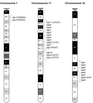
The immunity-related GTPases (IRGs)The immunity-related GTPases (IRGs) or p47 GTPases are guanine nucleotide binding
proteins with a molecular weight of approximately 47 kDa but no sequence homology to
any other known GTPase family outside the G-domain (Boehm 1998) (Figure 3). At the
beginning of this study six mouse IRG genes had been described, namely Irgd (IRG-47)
(Gilly 1992), Irgm1 (LRG-47) (Sorace 1995), Irgb6 (TGTP/Mg21) (Carlow 1995; Carlow
1998; Lafuse 1995), Irgm3 (IGTP) (Taylor 1996; Taylor 1997), Irga6 (IIGP) and Irgm2
(GTPI) (Boehm 1998) and no homologues from other species had been reported (for
details about the nomenclature of IRG genes and the newly identified genes see results
section (III.1.1) and (Bekpen 2005b)). Three of the mouse IRG proteins, Irgm1-3, have
the non-canonical sequence GX
4GMS in place of the otherwise universally conserved
GX
4GKS in the first nucleotide-binding motif (G1) correlating with other sequence
features to define the GMS (IRGM) and GKS subfamilies respectively (Bekpen 2005b;
Boehm 1998) (Figure 3). In the course of this study the number of IRG genes in Mus musculus domesticus was extended to a total of 21 genes falling into 25 coding units (see results section (III.1.2) and (Bekpen 2005b).
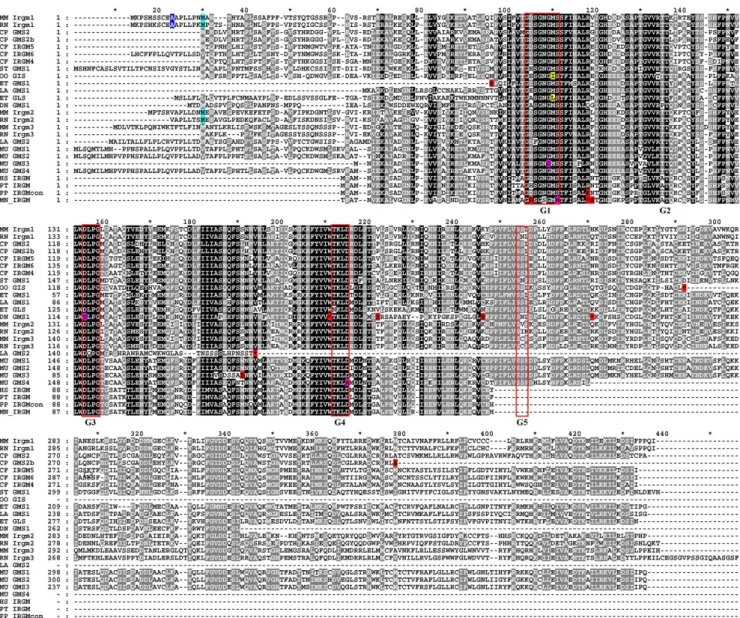
Figure 3 Amino acid alignment of six IRG proteins. Manual alignment of amino acid sequences of Irgd (IRG-47), Irgb6 (Mg21/TGTP), Irga6 (IIGP), Irgm1 (LRG-47), Irgm3 (IGTP) and Irgm2 (GTPI). Positions where at least four sequences share the same residue are shaded. The N- and C-terminal domains show low sequence homology between the family members whereas the core G-domain of the proteins is conserved.
The consensus sequences of the conserved GTP binding motifs are shown below the sequence. Three of the proteins contain an unusual methionine instead of the otherwise universally conserved lysine in the first nucleotide binding motif (G1) (from (Boehm 1998).
I.4.2.4.1. Expression of the IRG proteins
The IRG proteins are typically encoded on a single long exon and the promoters contain GAS and ISRE motifs mediating IFN-responsiveness but no other recurrent promoter motifs (Bekpen 2005b; Gilly 1996). In the mouse, 14 IRG genes have been shown to be inducible by IFNγ in fibroblasts. The mouse Irgc is a notable exception in that it lacks IFN regulation but is expressed exclusively in haploid spermatids in the mature testis (Bekpen 2005b; Rhode 2007). IRG protein expression is rapidly and strongly induced by IFNγ in a STAT1-dependent manner in vivo and in vitro in all cell types analysed (Boehm 1998; Collazo 2002; Gavrilescu 2004; Gilly 1992; Lafuse 1995; MacMicking 2003; Sorace 1995; Taylor 1996). IFNα/β also triggers expression, albeit to a lesser extent, as does lipopolysaccharide indirectly by stimulating type I IFN (Bafica 2007;
Carlow 1998; Lafuse 1995; Lapaque 2006; Sorace 1995; Taylor 1996; Zerrahn 2002).
Other cytokines tested have little effect on expression (Boehm 1998; Lafuse 1995;
Sorace 1995). In mice, IRG proteins are expressed at modest levels in absence of infection in tissues with a significant leukocyte contribution such as thymus and spleen (Collazo 2001; Taylor 1996) and in bone marrow derived haematopoietic stem cells (Advani 2004; Terskikh 2001; Venezia 2004). A developmentally regulated burst of IFN synthesis may be responsible for this expression. Infection induces strong IRG expression in most tissues by triggering IFN production (Boehm 1998; Collazo 2001;
Feng 2004; MacMicking 2003; Taylor 2000; Zerrahn 2002). IRG protein expression in
INTRODUCTION
the documented absence of IFN signalling has only been reported for Irga6, which shows a high constitutive expression in hepatic parenchymal cells (Zeng 2007, Parvanova 2005). This expression persists in mice deficient in STAT1 and both the type I and II IFN receptors and is driven by a second independent promoter containing numerous binding sites for liver-enriched transcription factors of the hepatocyte nuclear factor group (HNFs) (Bekpen 2005b, Zeng 2007, Parvanova 2005) known to mediate liver specific expression (Costa 2003; Schrem 2002).
I.4.2.4.2. Biochemical properties of the IRG GTPases
GTPase activity was first documented in vitro for Irgm3 immunoprecitated from IFNγ- induced cells and for recombinant Irgm3 (Taylor 1996). Furthermore, radiolabelled GTP rather than GDP co-immunoprecipitated with Irgm3 from cells (Taylor 1997). As neither the affinities of the protein for the different nucleotides nor the hydrolysis rate are known and the assay does not detect nucleotide free protein, one can conclude only that Irgm3 is capable of binding GTP in IFNγ-induced cells. Subsequently, also recombinant Irgb6 was shown to hydrolyse GTP to GDP in vitro (Carlow 1998). To date, only Irga6 has been systematically characterised biochemically (Ghosh 2004; Uthaiah 2003). Irga6 hydrolyses GTP to GDP cooperatively with a maximum rate of 2 per minute, oligomerises in the presence of GTP, and the oligomers resolve upon GTP hydrolysis (Uthaiah 2003). The oligomer is probably the site of rapid hydrolysis as the addition of aluminium fluoride to trap the transition state stabilises the oligomers (Ghosh 2004;
Uthaiah 2003). Thus, Irga6 is a self-activating GTPase, and to date no exogenous regulators of its activity have been described. Irga6 has a low nucleotide affinity, which is higher for GDP (1 µM) than for GTP (15 µM). Therefore, Irga6 should be predominantly GDP bound at cellular concentrations of 300 µM GTP and 100 µM GDP (Kleineke 1979) though GTP may be captured in oligomeric complexes in vivo (Pawlowski, unpublished data). Irga6 apoprotein and GDP-bound forms both crystallised as rotationally symmetrical dimers with identical contact interfaces (Ghosh 2004) (Figure 4). The G-domain is essentially Ras-like (Pai 1989), and lies between a three-helix bundle in the N-terminus, and a complex series of helices and loops in the C-terminus (Ghosh 2004). A destabilising mutation in the crystal dimer interface (M173A) yielded a monomeric Irga6 crystal with the non-hydrolysable GTP analogue GppNHp (Ghosh 2004) displaying only small structural changes relative to the GDP-bound structure. It is not clear whether the monomeric Irga6(M173A):GppNHp structure accurately reflects the actual GTP structure. Mutations in the dimer interface reduced oligomerisation and cooperative GTP hydrolysis but none eliminate the activity completely ((Ghosh 2004);
Pawlowski unpublished data). This and the lack of strong conservation of the dimer contact residues between IRG family members (Bekpen 2005b; Ghosh 2004) suggests that the crystal dimer interface is not identical to one of the two interfaces needed for oligomerisation. Recent mutational analysis defined the primary interface as a G-domain- G domain interaction involving bound nucleotides (Pawlowski unpublished data).
Formation of this contact is thought to induce large conformational changes exposing the
putative second interface.
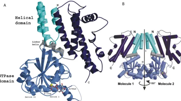
Figure 4 Crystal structure of Irga6 in the GDP bound form. (A) One molecule of the Irga6-GDP dimer is shown in ribbon presentation. The N-terminal domain (cyan) is composed of three α-helices and is followed by the Ras-like GTP binding domain (light blue). The helical C-terminal domain (dark blue) is connected to the G-domain by the linker helix αE (grey). The helices in Irgm1, Irgm2 and Irgm3 correlating to αK mediate membrane targeting of these proteins (see main text). GDP and Mg2+ are shown as atomic stick figure and yellow sphere respectively. The first 13 amino acids of Irga6 are not resolved in the structure. (B) Structure of the Irga6-GDP dimer. The subdomains are colour coded as in A. Secondary structure elements involved in the dimer interfaces I and II are labelled. The two-fold noncrystallographic symmetry axis is shown (from (Ghosh 2004).
I.4.2.4.3. Intracellular localisation of IRG proteins
IRG proteins associate with distinct subcellular membrane compartments by different targeting mechanisms. Irga6 concentrates at the endoplasmic reticulum (ER) (Martens 2004a) and at least in some cells also at the Golgi apparatus (Kaiser 2004; Zerrahn 2002) and contains a myristoylation sequence at the N-terminus (MGQLFSS) (Martens 2004a;
Uthaiah 2003). The consensus sequence for this lipid modification is
MG{EDRHPFYW}X
2[STAGCNDEF]{P} (Maurer-Stroh 2002), whereby the curly
brackets indicate non-permissive amino acids, the square brackets essential amino acids,
X any amino acid. Serine is favoured at position five of the motif, thus, most
myristoylation motifs can be simplified as MGXXXS. In the myristoylation process a
methionyl aminopeptidase first removes the initiator methionine (Farazi 2001) before the
N-myristoyl transferase (NMT) catalyses the covalent attachment of the 14-carbon fatty
acid myristate to the now N-terminal glycine residue (Casey 1995; Farazi 2001; Johnson
1994; Rajala 2000). This cotranslational modification is generally regarded as a
constitutive process, has only been observed in eukaryotes and appears to be irreversible
(Johnson 1994) though removal of the lipid from mature protein has been reported in
Dictyostelium (da Silva 1990). Mutations that convert the glycine at position two to
alanine (G2A) completely abolish myristoylation (Rajala 2000). Myristoylation is critical
for mediating protein-protein and/or protein-membrane interactions for some proteins
(Johnson 1994). For others, interactions with cell membranes require accessory factors or
other covalent modification. In addition, an attached myristoyl can also serve a structural
role in proteins (Ames 1994; Zheng 1993). An N-terminal peptide of mouse Irga2 (in that
INTRODUCTION
study misleadingly called human homolog of IIGP) was myristoylated in an in vitro assay (Maurer-Stroh 2004) and Irga6 is efficiently N-terminally myristoylated in vivo (Martens 2004b). Furthermore, the N-terminal 68 amino acids of Irga6 targeted EGFP to endomembranes in a myristoylation dependent manner (Martens 2004b). In contrast, lipid modification is largely dispensable for the membrane association of full length Irga6. Thus, Irga6 must employ other, yet unclarified, mechanisms for membrane association in uninfected cells. Ten other mouse IRG proteins also contain myristoylation sequences (see Table 7 in result section) but their subcellular localisation and myristoylation in vivo have not yet been explored. Irgm1 localises to the Golgi as a consequence of an amphipathic helix in its C-terminus (αK in the Irga6 crystal structure, Figure 4) (Martens 2004b) and Irgm2 and Irgm3 to the Golgi and ER, respectively, through the analogous helix, which in these cases lacks amphipathic character (Martens 2006; Martens 2004b; Taylor 1997). Irgm3 further localised to circular, vesicle-like cytoplasmic structures of unknown origin (Taylor 1997). The degree of membrane association varies among the IRG proteins. Irgm1 and Irgm3 are both >90% membrane bound, while Irga6, Irgb6 and Irgd are 60–70%, 20–30% and <10% membrane bound, respectively (Martens 2004b). The precise subcellular localisation of the non-cytosolic fraction of Irgb6 and Irgd remains to be determined.
I.4.2.4.4. Function of the IRG proteins
Only two interaction partners of IRG proteins have been reported to date. First, Irga6 interaction with the microtubule motor–binding protein, Hook-3, was detected in a yeast two hybrid (Y2H) screen and in cell lysates (Kaiser 2004). The interaction was abolished with the Irga6(S83N) mutant, which was assumed to be nucleotide-binding deficient by analogy to Irgm3(S98N) (Taylor 1997). Thus, the Irga6-Hook3 interaction is nucleotide dependent. The abrogation of the co-immunoprecipitation of Irga6 with Hook3 from IFNγ-induced cells by GDPβS (Kaiser 2004) strongly argues for GTP as the relevant nucleotide. It was proposed that Irga6 might influence cytoskeleton-based membrane trafficking via this route. Second, the rat homologue of Irgd was shown to interact with adipocyte differentiation-related protein (ADRP) in Y2H, a fatty acid binding protein that coats lipid droplets and is involved in their formation and in lipid uptake (Yamaguchi 2006). The interaction was confirmed in glutathione-S-transferase
(GST)-pulldown and mapped to the C-terminal half of Irgd (aa 246-420). Thus, Irgd might be involved in cellular lipid homeostasis, though the interaction remains to be confirmed in mammalian cells.
Over the last years a number of IRG proteins have been implicated in resistance
to intracellular protozoal and bacterial pathogens mainly by studying mice with targeted
deletions of single family members (Table 1). Irgm1-deficient mice were highly
susceptible to infection with Toxoplasma gondii (Collazo 2001), Leishmania major
(Taylor 2004), Trypanosomas cruzi (Santiago 2005), Chlamydia trachomatis (Coers
manuscript in preperation), Listeria monocytogenes (Collazo 2001), Salmonella
typhimurium (Taylor 2007; Taylor 2004), Mycobacterium tuberculosis (MacMicking
2003) and M. avium (Feng 2004). In most cases, Irgm1
-/-mice succumbed to infection
with kinetics similar to that of IFNγ-deficient mice. The other IRG proteins tested so far
have a narrower pathogen specificity. Irgm3-deficient mice lost resistance against Leishmania major (Taylor 2004), Toxoplasma gondii (Taylor 2000), Chlamydia trachomatis (Coers manuscript in preparation) and C. psittaci (Miyairi 2007). Irgm3 was shown to be required in hematopoietic as well as non-hematopoietic cells for resistance to Toxoplasma in vivo (Collazo 2002). Irgd, in contrast, displayed only a partial loss of resistance to T. gondii in the chronic phase of the infection (Collazo 2001). IRG function in pathogen resistance is non redundant since Irgm3, Irgd and Irgm1 all independently contribute to the resistance of mice to the protozoan parasite Toxoplasma gondii.
Table 1 IFNγ and IRG proteins in pathogen resistance. Summary of the susceptibility phenotypes of mice or cells (indicated in subscript) genetically (ko) or experimentally deficient for the indicated genes.
ND: not determined; S: susceptible; R: resistant; strain: susceptible mouse strains; MΦ: macrophages;
MEFs: mouse embryonic fibroblasts; astroc: astrocytes; MECs: mouse oviduct epithelial cells; RNAi:
expression suppressed by RNA interference; * in the chronic phase only; # in hematopoietic and non- hematopoietic cells in vivo: Note: weak antiviral effects observed in cellular overexpression studies for Irgm3 and Irgb6 in cellular are not listed here. See main text for more information and for references.