The Impact of Myeloid Cell-restricted Insulin Receptor Deficiency on Obesity-
induced Insulin Resistance
Inaugural-Dissertation zur
Erlangung des Doktorgrades
der Mathematisch-Naturwissenschaftlichen Fakultät der Universität zu Köln
vorgelegt von Jan Mauer
aus Köln
Köln 2008
Berichterstatter: Prof. Jens C. Brüning Prof. Thomas Langer
Tag der mündlichen Prüfung: 17.02.2009
Figure Index _____________________________________________________________ III Table Index _______________________________________________________________ V Abbreviations _____________________________________________________________ VI 1 Introduction __________________________________________________________ 1 1.1 Obesity _________________________________________________________________ 1 1.2 Diabetes Mellitus _________________________________________________________ 3
1.2.1 Systemic and Molecular Effects of Insulin Signaling __________________________________ 41.3 Obesity, Inflammation and Insulin Resistance ________________________________ 6 1.4 Macrophages ____________________________________________________________ 9
1.4.1 The Role of Macrophages in Obesity-induced Insulin Resistance _______________________ 10 1.4.2 Adipose Tissue Macrophage Heterogeneity ________________________________________ 111.5 Macrophages and Insulin _________________________________________________ 12 1.6 Objectives _____________________________________________________________ 13 2 Materials and Methods ________________________________________________ 14
2.1 Chemicals ______________________________________________________________ 14 2.2 Molecular Biology _______________________________________________________ 16
2.2.1 Isolation of Genomic DNA _____________________________________________________ 16 2.2.2 Southern Blot Analysis ________________________________________________________ 17 2.2.3 Quantification of Nucleic Acids _________________________________________________ 18 2.2.4 Polymerase Chain Reaction (PCR) _______________________________________________ 18 2.2.5 RNA Extraction, RT-PCR and Quantitative Realtime PCR ____________________________ 19 2.2.6 Protein Extraction ____________________________________________________________ 20 2.2.7 SAPK/JNK Kinase Assay ______________________________________________________ 20 2.2.8 Western Blot Analysis _________________________________________________________ 21 2.2.9 Gelatin Zymography __________________________________________________________ 21 2.2.10 ELISA _____________________________________________________________________ 222.3 Cell Culture and Tissue Analysis __________________________________________ 23
2.3.1 Preparation of L-cell Conditioned Medium _________________________________________ 23 2.3.2 Preparation of Palmitic Acid Media ______________________________________________ 23 2.3.3 Differentiation of Murine Bone Marrow to Macrophages ______________________________ 24 2.3.4 Culture of Primary Murine Macrophages __________________________________________ 24 2.3.5 Detection of Apoptotic Cells by TUNEL Assay _____________________________________ 24 2.3.6 Boyden Chamber Analysis _____________________________________________________ 25 2.3.7 Histological Analysis and Immunohistochemistry ___________________________________ 262.4 Mouse Experiments _____________________________________________________ 27
2.4.1 Animals ____________________________________________________________________ 27 2.4.2 IR∆myel mice _________________________________________________________________ 27 2.4.3 Body Weight and Blood Glucose Levels ___________________________________________ 28 2.4.4 Glucose and Insulin Tolerance Test_______________________________________________ 28 2.4.5 Isolation of Adipocytes and Stromal Vascular Fraction _______________________________ 28 2.4.6 Isolation of Primary Peritoneal Macrophages _______________________________________ 29 2.4.7 Isolation of Murine Bone Marrow ________________________________________________ 29 2.4.8 Glucose Transport ____________________________________________________________ 29 2.4.9 Hyperinsulinemic-euglycemic Clamp Studies _______________________________________ 302.5 Computer Analysis ______________________________________________________ 32
2.5.1 Densitometrical Analysis _______________________________________________________ 32 2.5.2 Statistical Methods ___________________________________________________________ 323 Results ______________________________________________________________ 33
3.1 Myeloid Cell-specific Disruption of the Insulin Receptor _______________________ 33
3.2 The Effect of Myeloid Cell-restricted Insulin Receptor Deficiency on Diet-induced Obesity ______________________________________________________________________ 35 3.3 The Effect of Myeloid Cell-restricted Insulin Receptor Deficiency on Obesity- induced Insulin Resistance ______________________________________________________ 37 3.4 The Effect of Myeloid Cell-restricted Insulin Receptor Deficiency on Obesity- induced Inflammation __________________________________________________________ 43 3.5 The Effect of Myeloid Cell-restricted Insulin Receptor Deficiency on Adipose Tissue Inflammation and Macrophage Accumulation ________________________________ 46 3.6 Cell-Autonomous Effects of Insulin Receptor Deficiency on Macrophages ________ 51
3.6.1 The Effect of Insulin on Macrophage Apoptosis _____________________________________ 51 3.6.2 The Effect of Insulin on Pro-inflammatory Gene Expression ___________________________ 54 3.6.3 The Effect of Insulin on Macrophage Migration _____________________________________ 564 Discussion ___________________________________________________________ 59 4.1 Recombination Efficiency of the LysMCre Transgene _________________________ 59 4.2 Myeloid Cell-specific Disruption of the Insulin Receptor protects against Obesity- induced Insulin Resistance ______________________________________________________ 61 4.3 Myeloid Cell-specific Disruption of the Insulin Receptor modulates the Obesity- associated Pro-inflammatory Tone _______________________________________________ 62 4.4 Myeloid Cell-specific Disruption of the Insulin Receptor blunts the Inflammatory Infiltration of Adipose Tissue by Macrophages _____________________________________ 63 4.5 The Effect of Metabolic Stress on Macrophages ______________________________ 65
4.5.1 Insulin as a Survival Signal for Macrophages _______________________________________ 65 4.5.2 Pro-inflammatory Effects of Insulin in Macrophages _________________________________ 67 4.5.3 The Effects of Insulin on Macrophage Migration ____________________________________ 684.6 Conclusions ____________________________________________________________ 70
5 Summary ____________________________________________________________ 71
6 Zusammenfassung ____________________________________________________ 72
7 References___________________________________________________________ 73
8 Acknowledgements ____________________________________________________ 95
9 Erklärung ___________________________________________________________ 96
10 Curriculum Vitae _____________________________________________________ 97
Figure Index
Fig. 1: Prevalence of overweight in the United States from 1991-2001. ... 1
Fig. 2: Prevalence of diabetes in the United States from 1991-2001. ... 3
Fig. 3: Insulin signal transduction pathway. ... 5
Fig. 4: Potential mechanisms for the inhibition of insulin signal transduction in obesity. ... 8
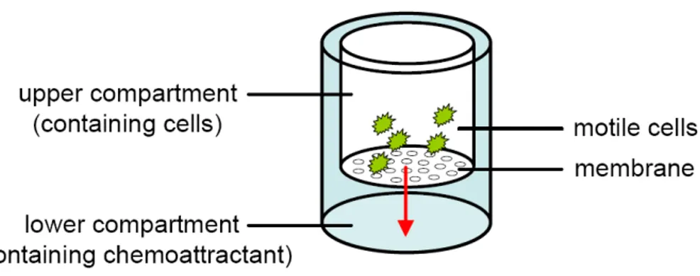
Fig. 5: Experimental design of the Boyden Chamber Analysis. ... 25
Fig. 6: Southern blot analysis of the insulin receptor allele in macrophages. ... 33
Fig. 7: Insulin receptor mRNA expression in macrophages. ... 34
Fig. 8: Insulin receptor protein expression in macrophages. ... 34
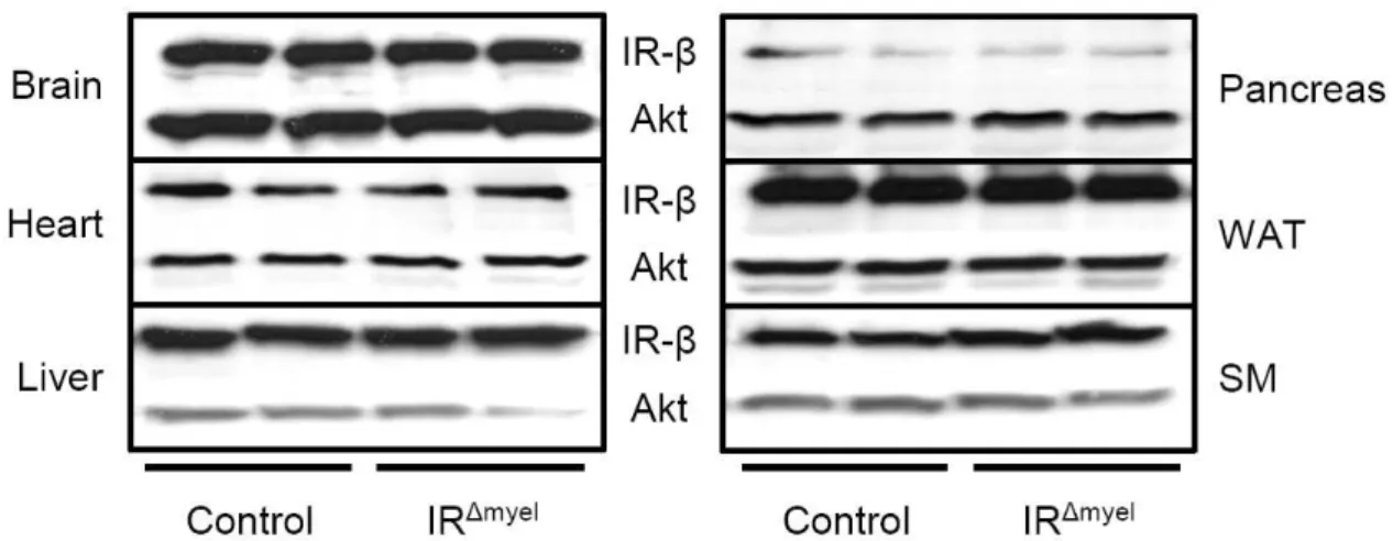
Fig. 9: Insulin receptor protein expression in insulin target tissues is unchanged in IR
∆myelmice. .. 35
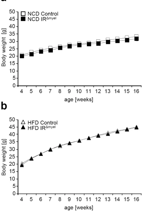
Fig. 10: IR
∆myelmice exhibit normal weight gain upon normal chow diet and high fat feeding. ... 36
Fig. 11: IR
∆myelmice show normal high fat diet-induced increase in epididymal fat pad mass and circulating leptin concentration. ... 37
Fig. 12: Diet-induced obese IR
∆myelmice exhibit significantly reduced fasted blood glucose and insulin levels. ... 38
Fig. 13: Obese IR
∆myelmice exhibit increased glucose tolerance and insulin sensitivity. ... 39
Fig. 14: Obese IR
∆myelmice exhibit decreased hepatic glucose production. ... 40
Fig. 15: Obese IR
∆myelmice show enhanced insulin-stimulated glucose disposal in skeletal muscle. ... 41
Fig. 16: Low dose insulin-stimulated glucose uptake is enhanced in adipocytes of obese IR
∆myelmice. ... 42
Fig. 17: The obesity-associated change of serum TNF-α and adiponectin concentration is abolished in IR
∆myelmice. ... 43
Fig. 18: Obese IR
∆myelmice show reduced JNK activity in skeletal muscle. ... 44
Fig. 19: Obesity-induced change of adipokine gene expression is blunted in IR
∆myelmice. ... 45
Fig. 20: Reduced macrophage surface marker expression in adipose tissue of obese IR
∆myelmice. 46 Fig. 21: Adipose tissue morphology of IR
∆myelmice and control mice exposed to HFD. ... 47
Fig. 22: Quantification of adipocyte size and size distribution. ... 47
Fig. 23: Obese adipose tissue of IR
∆myelmice shows decreased formation of crown-like structures. ... 48
Fig. 24: Reduced TNF-α expression in adipose tissue-derived stromal vascular cells of IR
∆myelmice. ... 49
Fig. 25: Enhanced apoptosis in insulin receptor-deficient macrophages. ... 52
Fig. 26: Insulin enhances expression of Bcl-2 mRNA in bone marrow-derived macrophages. ... 53
Fig. 27: Insulin augments pro-inflammatory gene expression in macrophages. ... 55
Fig. 28: Insulin enhances secretion of TNF-α in macrophages. ... 56
Fig. 29: Insulin receptor deficiency impairs matrix metalloproteinase 9 expression in macrophages.
... 57
Fig. 30: Insulin receptor-deficient macrophages show impaired chemotactic abbilities. ... 58
Table Index
Table 1: Chemicals ... 16
Table 2: Oligonucleotides used to amplify the southern blot probe ... 17
Table 3: Oligonucleotides used for genotyping ... 18
Table 4: Taqman Gene Expression Assays ... 19
Table 5: Primary antibodies used for western blot analysis ... 21
Abbreviations
A adenosine
aa amino acid
Akt proteinkinase B
Avertin tribromoethyl alcohol and tert-amyl alcohol β-me β-mercaptoethanol
BMDM bone marrow-derived macrophages
bp base pair
c DNA concentration
C cytosine
°C degrees Celsius
CAP cbl-associated protein
cDNA complementary DNA
cpm counts per minute
Cre site-specific recombinase (causes recombination)
Ctrl Control
ddH
2O double destilled water
DEPC diethylpyrocarbonate
dNTP desoxynucleotide-triphosphate
DMSO dimethylsulfoxide
DNA desoxyribonucleic acid
DTT 1,4-Dithio-DL-threitol
ECL enhanced chemiluminescence
EDTA ethylene-diaminetetraacetic acid
ELISA enzyme-linked immunosorbent assay
ERK extracellular signal-regulated kinase
EtBr ethidium bromide
FOXO forkhead transcription factor
G guanine
Glut glucose transporter
Grb growth factor receptor binding protein
GSK glycogen synthase kinase
GTT glucose tolerance test
h hour
HEPES N-2-hydroxyethylpiperazine-N’-2-ethansulfonic acid
i.p. intraperitoneal
IL interleukin
IR insulin receptor
IRS insulin receptor substrate
ITT insulin tolerance test
kDa kilodalton
loxP locus of x (crossing) over of P1
MCP-1 macrophage chemoattractant protein 1
MIP-1α macrophage inflammatory protein 1 α
min minute
mTOR mammalian target of rapamycin
NaCl sodium chloride
NaOH sodium hydroxide
NLS nuclear localization sequence
OD optical density
p70S6K p70 S6 kinase
PBS phosphate buffered saline
PCR polymerase chain reaction
PDK phosphoinositide-dependent kinase
PH pleckstrin homology
PI3K phosphatidylinositol-3 kinase
PIP
2phosphatidylinositol-4,5-biphosphate PIP
3phosphatidylinositol-3,4,5-triphosphate
PKA proteinkinase A
PKC proteinkinase C
PTB phosphotyrosine binding
Ras rat sarcoma
Raf v-raf-leukemia viral oncogene
RNA ribonucleic acid
rpm rounds per minute
RT room temperature
sec second
SDS sodium dodecyl sulfate
SH src homology
SOS son of sevenless
SSC sodium chloride/ sodium citrate buffer
TAE Tris-acetic acid-EDTA buffer
Taq Pol polymerase from Thermus aquaticus
TE Tris-EDTA buffer
TNF tumor necrosis factor
Tris 2-amino-2-(hydroxymethyl-)1,3-propandiole
TWEEN polyoxethylene-sorbitan-monolaureate
U units
v/v volume per volume
WAT white adipose tissue
w/v weight per volume
wt wildtype
5’ five prime end of DNA sequences
3’ three prime end of DNA sequences
1 Introduction
1.1 Obesity
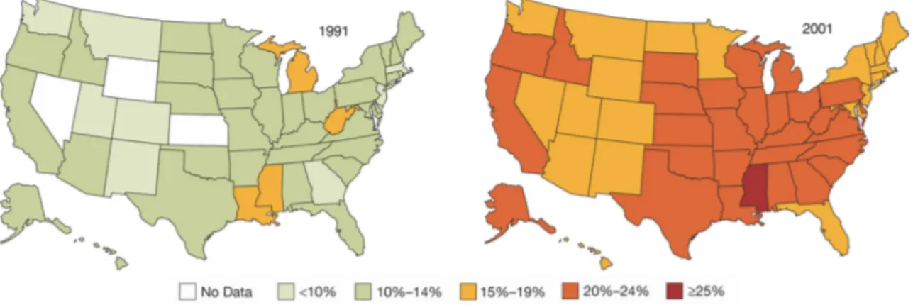
Obesity represents a steadily increasing health threat to our society and is a major cause of morbidity and mortality. Over the last 10 years, the percentage of overweight adults in the United States has increased from 45 to 58% (Fig. 1) (1). This increment in the prevalence of overweight and obesity, a trend found not only in adults but also in children, has been observed in many other countries of the world (2-4). According to latest projections of the World Health Organisation (WHO) 1.6 billion adults are overweight and at least 400 million are obese worldwide. These numbers will presumably rise to 2.3 billion overweight and 700 million obese individuals until the year 2015 (5).
The underlying cause of excessive weight gain is a continuous imbalance between energy intake on one hand, and energy expenditure on the other (6). Enhanced susceptibility to obesity is not only attributable to genetic variation but also to a number of other factors including a global shift in diet composition towards higher amounts of fat and carbohydrates and lower amounts of vitamins, minerals and other micronutrients (7).
Furthermore, decreased physical activity and a shift towards a sedentary lifestyle contribute to the obesity epidemic (8, 9).
Fig. 1: Prevalence of overweight in the United States from 1991-2001.
Since 1991, the percentage of overweight adults has risen from 45 to 58%. Of those overweight in 2001, 56%
were men and 44% were women. Mokdad et al, 2003 (1).
The most common measure for defining overweight and obesity is the body mass index (BMI) which is assessed by the ratio of body weight in kilogramm (W) and height in meters (H) multiplied by itself (W/H
2) (10). Individuals with a BMI of 25 to 29.9 kg/m
2are defined as overweight while obesity is characterized by a BMI of 30 kg/m
2or higher (5). Obesity predisposes to a variety of different diseases. It represents an independent risk factor for myocardial infarction, stroke, type 2 diabetes mellitus, and certain types of cancer (11-13). Adiposity, which is the fraction of total body mass comprised of neutral lipid stored in adipose tissue, is closely correlated with important physiological parameters such as blood pressure, systemic insulin sensitivity, serum triglyceride and leptin concentrations (14-16). Several obesity-related disorders including insulin resistance, glucose intolerance, dyslipidemia, hypertension, and coronary artery disease are positively correlated with adiposity (15, 17). It has been shown that visceral fat accumulation is more closely linked to obesity-associated pathologies than overall adiposity (18). Changes in adipose tissue mass are associated with changes in the endocrine and metabolic functions of adipose tissue that connect increased adiposity to alterations in systemic physiology. For example, the concentration of circulating leptin, the most prominent adipocyte-derived hormone, positively correlates with increased adipocyte volume and number (19). Leptin functions as an important regulator of energy intake and storage, insulin sensitivity, and metabolic rate (20-22). In contrast, adiposity is negatively correlated with plasma concentrations of adiponectin, an adipocyte-derived, insulin-sensitizing hormone that decreases hepatic gluconeogenesis and increases lipid oxidation in muscle (23-25).
Visceral fat accumulation belongs to a group of risk factors including high blood
pressure, high blood glucose, high levels of triglycerides and low levels of high density
lipoproteins which are subsumed under the term metabolic syndrome (26). These
conditions predispose for cardiovascular disease (CVD) and type 2 diabetes mellitus
(T2DM) (27-29).
1.2 Diabetes Mellitus
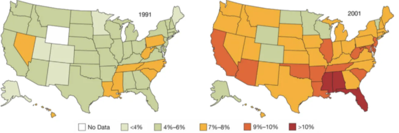
Since one of the major consequences of excessive weight gain is the development of insulin resistance, it is not surprising that an increase in number of overweight and obese individuals within a population (Fig. 1) is accompanied by a rise in the occurence of diabetes (Fig. 2). Diabetes mellitus is a chronic disease and the sixth leading cause of death in the United States. In 2007, a total of 23.6 million US citizens, representing 8% of the population, suffered from diabetes. Health care costs, whether assigned to diabetes directly or indirectly, were estimated at 174 billion USD for the year 2007 (30). According to the WHO 180 million people worldwide suffer from diabetes and these numbers are predicted to more than double by 2030. In 2005, 1.1 million people died from diabetes all over the world and an increase by more than 50% is projected for the next 10 years (31, 32).
Fig. 2: Prevalence of diabetes in the United States from 1991-2001.
The prevalence of people diagnosed with diabetes increased to 7.9% in 2001 from 4.9% in 1990, an increase of 61%. In 2001, 3.4% of US adults (2.9% men, 3.8% women) were both obese and had diabetes, an increase of 1.4% compared to 1991. Mokdad et al, 2003 (1).
In principal, there are two idiopathic forms of diabetes known as type 1 and 2. Type 1 diabetes mellitus (T1DM), also termed juvenile diabetes or insulin-dependent diabetes (IDDM), is characterized by the loss of insulin-producing β-cells in the Langerhans islets of the pancreas as a result of autoimmune reactions (33). T1DM can be treated and contained by an insulin replacement therapy and dietary management (34).
Type 2 diabetes mellitus, formerly known as adult-onset diabetes, is the non-
insulin-dependent (NIDDM) form of diabetes and accounts for 90-95% of all diagnosed
cases of diabetes (30). This disease develops when chronic overnutrition colludes with
genetic susceptibility to cause insulin resistance and a relative insulin deficiency of non-
autoimmune etiology (35, 36). When resistance to the metabolic effect of insulin occurs, the pancreas is able to compensate with expansion of β-cell mass and hypersecretion of the hormone (37). It is assumed that the ultimate stimulus is nutrient surplus in the blood, predominantly glucose and free fatty acids (FFA) (38-40). However, prolonged overproduction of insulin leads to progressive detoriation of β-cell function associated with apoptosis-driven loss of β-cell mass leading to subsequent progression to a type 2 diabetic state (41-43). T2DM is characterized by severe hyperglycemia and altered lipid homeostasis (44-46).
Treatment strategies for T2DM include oral agents such as sulfonylureas, biguanides and thiazolidinediones (TZD) which increase insulin secretion, insulin action and augment overall insulin sensitivity, respectively (47-50). Interestingly, anti- inflammatory drugs like salicylates can also improve insulin sensitivity (51, 52). In addition, lifestyle changes especially increased exercise and dietary adjustments including caloric restriction ameloriate T2DM morbidity (53, 54). In the long-term, diabetes is associated with complications such as atherosclerosis, retinopathy, nephropathy, neuropathy and impaired wound healing which all impose significant economical consequences (55-60).
1.2.1 Systemic and Molecular Effects of Insulin Signaling
Insulin has most potent anabolic effects and promotes the synthesis and storage of carbohydrates, lipids and proteins, while inhibiting their degradation and release into the circulation (44). It controls energy homeostasis by stimulating glucose uptake in peripheral tissues and suppressing the release of stored lipids from adipose tissue (61-64).
Furthermore, it increases glycolysis and inhibits gluconeogenesis in the liver (65-67).
The pleiotropic effects of insulin are mediated by binding of the hormone to its
membrane-bound receptor (68). The insulin receptor (IR) belongs to the family of receptor
tyrosine kinases and comprises four subunits of which two regulatory α-subunits inhibit the
two catalytic β-subunits. Upon binding of insulin, the α-subunits undergo a conformational
change that leads to derepression of the β-subunits which subsequently transphosphorylate
(69). This facilitates interaction with the phosphotyrosine binding (PTB) domains of
downstream signaling components (70-73) which localize to the plasma membrane by
interaction of their N-terminal Pleckstrin homology (PH) domain with lipid-bound inositol phosphates (74, 75).
There are at least nine known intracellular substrates of the insulin receptor. Four of these belong to the insulin receptor substrate (IRS) family (76-78). Others include Grb2- associated binding protein 1, p60
dok, Casitas B-lineage lymphoma (Cbl), adapter protein with a pleckstrin homology and a Src homology 2 domain (APS) and isoforms of Shc (79- 81). These molecules serve as docking platforms for Src-homology (SH)-2 domain- containing proteins such as the regulatory subunit of the phosphatidylinositol-3 kinase (PI3K) and the growth factor receptor binding protein (Grb)-2. Subsequent signal transduction results in the activation of the PI3K and the Ras/Raf Mitogen-activated protein (MAP)-kinase pathways (82-85).
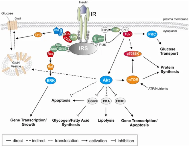
Fig. 3: Insulin signal transduction pathway.
Binding of insulin to the insulin receptor results in receptor trans-phosphorylation and activation leading to the recruitment and subsequent phosphorylation of insulin receptor substrates. This enables the binding of SH-2 domain containing proteins, which ultimately leads to the activation of downstream signaling pathways such as the PI3K or the Ras/Raf MAPK signaling pathway. (Abbreviations: Akt: Proteinkinase B, APS:
adapter protein with a pleckstrin homology and an Src homology 2 domain, CAP: cbl-associated protein, ERK: extracellular signal-regulated kinase, FOXO: Forkhead transcription factor , Glut4: glucose transporter 4, Grb2: growth factor receptor binding protein 2, Gsk3: glycogen synthase kinase 3, IR: insulin receptor, IRS: insulin receptor substrate, mTOR: mammalian target of rapamycin, p70S6K: p70 S6 kinase, PI3K:
phosphatidylinositol-3 kinase, PIP: phosphatidylinositol phosphate, Pdk: phosphoinositide-dependent kinase, PKA: Proteinkinase A, PKC: Proteinkinase C, SOS: son of sevenless, Raf: v-raf-leukemia viral oncogene, Ras: rat sarcoma)
Activation of the Ras/Raf MAP-kinase pathway leads to cellular proliferation and differentiation while no significant effect on the metabolic actions of insulin has been observed (86, 87). In contrast, insulin-induced PI3K activation mediates the vast majority of metabolic effects of the hormone including glycogen, protein and lipid synthesis, inhibition of apoptosis and stimulation of glucose transport. Most of these processes are mediated through activation of Protein kinase B/Akt (88-91). However, a PI3K- independent pathway for the regulation of glucose transport has also been identified (92).
In this pathway, the insulin receptor directly recruits the adaptor protein APS and the Cbl- cbl associated protein (93) complex. Tyrosine-phosphorylated Cbl then recruits the CrkII- C3G complex to lipid rafts, leading to downstream activation of TC10 and ultimately resulting in translocation of glucose transporter (GLUT) 4 to the plasma membrane (94-98) (Fig. 3).
1.3 Obesity, Inflammation and Insulin Resistance
The nutritional or metabolic state of an organism is closely linked to its immune system in a delicate balance. Malnutrition impairs immune processes thereby rendering an organism more susceptible to infectious diseases (99, 100). This arises from a reduced metabolic support under conditions of infection during which the immune system is highly dependent on the mobilization of nutrients and supply with energy to eliminate pathogens (101). In contrast, overnutrition i.e. obesity is associated with chronic, low-grade activation of the immune system and increased susceptibility to inflammatory diseases (102). This can lead to insulin resistance when inflammatory pathways interfere with insulin signaling cascades (103).
The first evidence for the importance of inflammatory signaling in obesity-induced
insulin resistance was the observation that the pro-inflammatory cytokine tumor necrosis
factor (TNF) α is elevated in adipose tissue of obese mice and humans and is capable of inducing insulin resistance in vitro (104-106). TNF-α acts via the TNF receptor and activates intracellular protein kinases e.g. the inhibitor of nuclear factor (NF) κB kinases (IKKs) and c-jun N-terminal kinases (JNKs) (107, 108). Subsequently, these kinases mediate inhibitory phosphorylation events on serine (S307) residues of IRS molecules (109, 110). S307 phosphorylation of IRS reduces both tyrosine phosphorylation of IRS in response to insulin and its ability to associate with the insulin receptor, thus inhibiting downstream signaling and insulin action (111, 112). Inhibition or inactivation of IKK or JNK in mice has been shown to protect from obesity-induced insulin resistance (51, 52, 113-115). Aside from TNF-α, a variety of other inflammation-associated factors including interleukin (IL) 6, IL-1, IL-8, c-reactive protein (CRP), monocyte chemoattractant protein 1 (MCP-1, also called CCL2), vascular cell adhesion molecules (VCAMs) and matrix metalloproteinases (MMPs) are upregulated in the adipose tissue and/or circulation of obese mice and humans and have been linked to obesity and insulin resistance (93, 116- 121).
In addition to these factors, the obesity-associated increase in circulating free fatty acids (FFAs) has been shown to interfere with insulin signaling in target tissues via activation of IKK, JNK and protein kinase C (PKC) (122-127). This interference is mediated via at least two mechanisms. On one hand, FFAs induce inflammatory signaling cascades by activation of toll-like receptors (TLRs). TLRs belong to a family of pattern- recognition receptors that play a crucial role in activating the pro-inflammatory response after contact with microbial pathogens (128). The best characterized member of this receptor family is TLR4 which recognizes lipopolysaccharides (LPS), components of the bacterial cell wall. Signal transduction after contact with LPS is mediated through intracellular binding of myeloid differentiation factor 88 (MyD88) to the Toll/IL-1 receptor (TIR) domain of the receptor and subsequent activation of the NFκB pathway. This leads to expression of pro-inflammatory cytokines and chemokines and other effectors of the innate immune response (129).
Besides LPS, whose lipid component is sufficient to mediate TLR signaling, saturated fatty acids have the full potential to promote TLR4 activation in vitro (130, 131).
Disruption of TLR4 in mice has been demonstrated to partially protect these animals from
obesity-induced insulin resistance and substantially reduce the negative effect of systemic
lipid infusion on glucose metabolism and muscle insulin action (132). In addition to TLR
signaling, FFA activate inflammatory serine kinases by intermediates of their intracellular
processing pathways such as β-oxidation in the mitochondria, storage to triglyceride depots and conversion into ceramides (133-136).
Another important aspect of obesity-induced insulin resistance is the development of endoplasmatic reticulum (ER) stress. Under normal conditions, the ER is the site of protein folding and assembly by chaperones (137). However, obesity generates conditions of glucose or nutrient deprivation, inundation with fatty acids or increased expression of secretory proteins that elevate the demand on the ER leading to accumulation of unfolded or misfolded proteins.
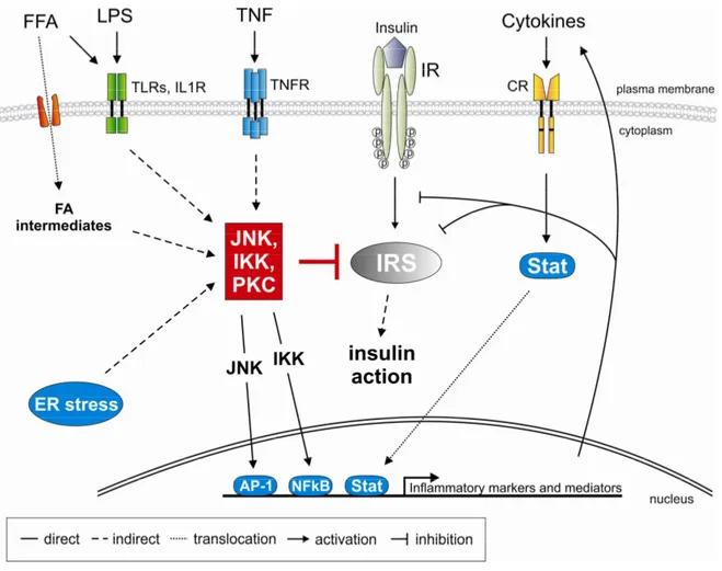
Fig. 4: Potential mechanisms for the inhibition of insulin signal transduction in obesity.
Obesity increases circulating inflammatory cytokine and free fatty acid concentration. This leads to activation of cell surface receptors which then induce serine kinases like c-jun N-terminal kinase (JNK), inhibitor of NFkB kinase (IKK) komplex and protein kinase c (PKC) isoforms. These kinases then mediate inhibitory serine (S307) phosphorylation events on insulin receptor substrates (IRS) thereby blocking insulin action. Additionally, transcription factors nuclear factor kB (NFkB), activator protein (AP) 1 and signal transducer and activator of transcription (Stat) activate inflammatory gene expression thereby enhancing production and secretion of inflammatory markers and mediators. Furthermore, endoplasmatic reticulum (ER) stress and intermediates of fatty acid metabolism may activate stress kinases. (Abbreviations: CR:
cytokine receptor, FFA: free fatty acid, FA: fatty acid, IL1R: interleukin-1 receptor, IR: insulin receptor, IRS:
insulin receptor substrate, LPS: lipopolysaccharide, TLR: toll-like receptor, TNF: tumor necrosis factor, TNFR: TNF receptor)
This in turn triggers the unfolded protein response (UPR) which enhances the transcription of genes involved in assembly, folding, modification and degradation of proteins to normalize ER function (138). It has been suggested that failure of this adaptive response leads to activation of various cell death effectors such as Bax, Bak and caspases (139-141). Nevertheless, the UPR not only affects gene transcription and pro-apoptotic factors but also activates stress kinases like JNK and expression of pro-inflammatory cytokines via the IKK/NFκB axis which ultimately induces insulin resistance (142-144). A recent study demonstrated that the genetic disruption of x-box protein (XBP) 1, a transcription factor that mediates expression of ER chaperones, leads to increased ER stress, activation of JNK and induction of insulin resistance via serine-phosphorylation of IRS in diet-induced obese mice (138). Furthermore, treatment of diet-induced obese mice with orally active chemical chaperones reverses these effects on ER stress and JNK and improves tissue insulin sensitivity (145). These findings underline the importance of ER stress and inflammatory signals in the development of obesity-induced insulin resistance.
1.4 Macrophages
In general, macrophages belong to the mononuclear phagocyte system which also includes monocytes and their lineage-committed precursors (146). The first step of macrophage development takes place in the bone marrow where myeloid progenitors differentiate into monocytes. Monocytes in turn enter the circulation and give rise to tissue-resident macrophage populations throughout the body (147). Recruitment of monocytes to peripheral tissues and differentiation into macrophages is enhanced by pro- inflammatory, metabolic and immune stimuli (148). Macrophages are classically defined to belong to the innate immune system and, due to their ubiquitous distribution, represent a first line of defense against invading pathogens (149). Their main functions are the maintenance of tissue homeostasis and initiation of the inflammatory response.
Furthermore, macrophages harbor pronounced chemotactic and phagocytotic abilities and
contribute to tissue remodeling and repair (150). Macrophage dysfunction is associated
with a variety of diabetes-associated diseases such as atherosclerosis, retinopathy, nephropathy, neuropathy and impaired wound healing (151, 152).
1.4.1 The Role of Macrophages in Obesity-induced Insulin Resistance
In obesity, the adipose tissue is an important initiator of the inflammatory response since it not only serves as a storage depot for excess calories, but is also capable of secreting fatty acids, hormones, cytokines and chemokines which then act in both endocrine and paracrine fashion (153). Although it mainly consists of adipocytes, the adipose tissue also contains preadipocytes, endothelial cells and immune cells which all reside in the stromal vascular fraction. During expansion of the adipose tissue in obesity, local hypoxia occurs due to hypoperfusion with blood vessels. This microhypoxia induces activation of JNK and NFκB signaling cascades and increases inflammatory gene expression, leading to secretion of chemokines and, ultimately, adipocyte death (154).
Chemokines that are released into the circulation during these events attract macrophages which then surround dead adipocytes for removal of cell debris and tissue remodeling (155). In addition, these macrophages secrete pro-inflammatory cytokines and chemokines thereby inducing insulin resistance in adjacent adipocytes and recruiting more macrophages from the vascularity to the fat tissue (156).
Accumulation of macrophages in adipose tissue is an important hallmark of
adiposity, a condition in which these cells represent 40% of the total adipose cell content in
contrast to only 10% in lean counterparts (157). Conditional disruption of pro-
inflammatory kinases JNK1 and IKKβ specifically in myeloid cells protected mice from
diet-induced insulin resistance, decreased the inflammatory tone and blocked accumulation
of macrophages in adipose tissue in different models of obesity (114, 158). Interestingly,
although JNK1 and IKKβ remained intact in all organs despite immune cells, no effect on
adiposity was observed in these studies. Furthermore, conventional disruption of MCP-1,
which is an important chemoattractant for immune cells, or its receptor CCR2, prevented
the accumulation of macrophages in adipose tissue of obese mice and improved overall
insulin sensitivity (159, 160). These studies clearly indicate that systemic inflammation
alone can affect insulin sensitivity and that the obesity-associated inflammatory state is
mainly mediated through the myeloid compartment of the immune system and infiltration
of immune cells into adipose tissue.
1.4.2 Adipose Tissue Macrophage Heterogeneity
Macrophages can be divided into at least two subgroups, M1 and M2 (161). M1- cells are defined as "classically activated" macrophages, which are induced by interferon (IFN) γ and LPS. These macrophages secrete pro-inflammatory cytokines (e.g. TNF-α, IL- 6, IL-12) and produce high levels of nitric oxide (NO) via inducible nitric oxide synthase (iNOS) expression (162). In addition, they show increased reactivity to fatty acids and LPS. M2-cells or "alternatively activated" macrophages are induced by IL-4 or IL-13 and display a more anti-inflammatory phenotype since they produce high levels of IL-10 and IL-1 decoy receptor and only secrete low levels of pro-inflammatory cytokines. M2 macrophages are generally involved in tissue repair and remodeling (163).
Recently, it has been demonstrated that macrophages invading the adipose tissue of
obese animals exhibit a different polarization compared to the cells identified in the
adipose tissue of lean animals. Freshly recruited macrophages are positive for surface
markers F4/80, CD11b and CD11c and have a pronounced pro-inflammatory profile
showing high reactivity to LPS and FFA allocating them to the M1 subgroup (164). In
contrast, resident adipose tissue macrophages in lean animals also express F4/80 and
CD11b surface markers, but lack CD11c expression almost completely. These cells exhibit
an M2/anti-inflammatory profile showing reduced expression of IL-6, iNOS and CCR2
(165). Notably, the obesity-induced switch in the adipose tissue macrophage activation
state is not dependent on the conversion of resident M2 macrophages to an M1 phenotype
but arises from a localized recruitment of inflammatory macrophage subtypes out of the
circulation (166). An important mediator of M1 to M2 polarization of macrophages is the
nuclear receptor peroxisome proliferator-activated receptor (PPAR) γ which acts as a lipid
sensor (167) and whose conditional disruption in myeloid cells has been shown to impair
alternative activation of macrophages and predisposes for obesity-induced insulin
resistance and glucose intolerance (168). Activation of PPARγ in macrophages e.g. by
treatment with TZDs polarizes towards an M2 phenotype, thus also providing an
explanation for the beneficial effect of these compounds on insulin sensitivity in humans
(169). Recently, the importance of M1 vs. M2 macrophages in obesity-induced insulin
resistance has been demonstrated by Patsouris and colleagues. In this study, ablation of
CD11c-positive cells in obese, insulin resistant mice lead to a drastic decrease in
inflammatory marker expression and normalized insulin sensitivity to that of lean controls
(170).
According to these findings, it is clear that activated macrophages infiltrate the adipose tissue in obesity. However, another important question asks which signals lead to activation of macrophages in this context. Obesity provides an extracellular environment that is enriched with lipids, pro-inflammatory cytokines, chemokines and even gut-derived bacterial compounds which can activate macrophages (171). Among the receptors activated by these signals, TLRs are the most intensively investigated family. Disruption of TLR2 or TLR4 or both prevent the activation of inflammation in macrophages by FFA (172, 173). In addition to FFA, cytokines derived from expanding adipose tissue, ER stress and also microhypoxia, whose central mediator hypoxia inducible factor (HIF) was shown to be crucial for the regulation of macrophage function (174), can activate inflammatory pathways in these cell types.
1.5 Macrophages and Insulin
Until now, only few studies have been performed to investigate the effect of insulin itself on macrophage activation and function. Primary monocytes and macrophages express insulin receptors as they were used to study insulin binding affinities and receptor turnover in the 1970s (175-177). The functionality of the insulin receptor on monocytes was demonstrated first when insulin was shown to modulate Fc receptor expression on these cells (178). Furthermore, insulin augments the bacteriocidal properties of macrophages against Salmonella typhimurium and enhances their chemotactic activity towards pancreatic β-cell islets in vitro (179, 180). Approximately 10 years later, Costa- Rosa and colleagues further investigated insulin's effect on macrophage key functions.
They demonstrated that insulin does not affect cell migration, at least in response to thioglycolate and Bacille Calmette-Guérin (BCG), but significantly enhances phagocytosis, H
2O
2production and glucose metabolism in macrophages (181).
More recent studies demonstrated that insulin induces TNF-α mRNA and protein
expression in an ERK-dependent manner and promotes survival in a human monocytic
cell-line (182, 183). Moreover, the same cell-line, when exposed to LPS followed by
insulin treatment, shows enhanced secretion of TNF-α and IL-1β compared to LPS alone
(184). The role of insulin in cell survival was also demonstrated in primary murine
macrophages. Treatment with insulin increased protein levels of anti-apoptotic B-cell
lymphoma (Bcl) 2 and induced XBP-1 and 78 kDa glucose response protein (GRP78), which are both involved in the UPR (185).
The concept of insulin's role in macrophage survival was further supported when Han et al. demonstrated that in a mouse model of atherosclerosis, transplantation of bone marrow from insulin receptor-deficient animals into a low density lipoprotein (LDL) receptor-deficient background, leads to advanced lesion formation (186). This was ascribed to increased ER stress and apoptosis in IR-deficient macrophages due to elevated scavenger receptor A (SRA) expression and enhanced uptake of oxidized LDL.
Interestingly, our lab demonstrated in a similar study that myeloid cell-autonomous IR- deficiency decreases formation of atherosclerotic plaques in mice (187). Here, IR-deficient primary macrophages displayed unaltered uptake and efflux of cholesterol. Furthermore, IR-deficient macrophages exhibited decreased expression and secretion of IL-6 after stimulation with LPS indicating a reduced inflammatory response in these cells.
Taken together, insulin exerts a variety of effects on macrophage function including survival, phagocytosis, migration and inflammatory gene expression. However, until today, the role of macrophage insulin receptor signaling in obesity-induced insulin resistance remains elusive.
1.6 Objectives
To study the role of insulin signal transduction in macrophages in obesity-induced
insulin resistance, mice with a conditional disruption of the insulin receptor specifically in
myeloid cells were generated. The aim of this study was to physiologically characterize
these animals under normal chow and high fat diet. In addition, we sought to study glucose
metabolism in insulin target tissues by euglycemic-hyperinsulinemic clamp analysis. Also,
the impact of myeloid cell insulin receptor deficiency on obesity-associated inflammation,
both locally and systemically, was to be investigated. Furthermore, we wanted to analyze
the action of insulin in primary macrophages in a cell-autonomous fashion in relation to
migration/chemotaxis, apoptosis and inflammation.
2 Materials and Methods
2.1 Chemicals
Size markers for agarose gel electrophoresis (Gene Ruler DNA Ladder Mix) and for SDS-PAGE (Prestained Protein Ladder Mix) were obtained from MBI Fermentas, St.
Leon-Rot, Germany. RedTaq DNA Polymerase and 10 x RedTaq buffer were purchased from Sigma-Aldrich, Seelze, Germany.
Chemical Supplier
ε-aminocaproic acid Sigma-Aldrich, Seelze, Germany 2-Deoxy-D-[1-14C]-Glucose Amersham, Freiburg, Germany
Acetone KMF Laborchemie, Lohmar, Germany
Acrylamide Roth, Karlsruhe, Germany
Agarose (Ultra Pure) Invitrogen, Karlsruhe, Germany
Amyloglucosidase Roche, Mannheim, Germany
Aprotinin Sigma-Aldrich, Seelze, Germany
Benzamidine Sigma-Aldrich, Seelze, Germany
β-Mercaptoethanol (β-ME) AppliChem, Darmstadt, Germany Bovine serum albumin (BSA) Sigma-Aldrich, Seelze, Germany BSA essentially fatty acid free Sigma-Aldrich, Seelze, Germany
Bradford reagent Bio-Rad, München, Germany
Bromphenol blue Merck, Darmstadt, Germany
Calcein Invitrogen, Karlsruhe, Germany
Chloroform Merck, Darmstadt, Germany
D-[3-3H]-Glucose Amersham, Freiburg, Germany
Desoxy-Ribonucleotid-Triphosphates (dNTPs) Amersham, Freiburg, Germany
Dextran sulfate AppliChem, Darmstadt, Germany
Dimethylsulfoxide (DMSO) Merck, Darmstadt, Germany Dithiothreitol (DTT) Boehringer, Mannheim, Germany Enhanced Chemiluminscence (ECL) Kit Perbio Science, Bonn, Germany
Ethanol, absolute AppliChem, Darmstadt, Germany
Ethidium bromide Sigma-Aldrich, Seelze, Germany
Ethylendiamine tetraacetate (EDTA) AppliChem, Darmstadt, Germany
Fetal Calf Serum (FCS) Gibco BRL, Eggenstein, Germany
Gelatine Sigma-Aldrich, Seelze, Germany
Glacial acetic acid Roth, Karlsruhe, Germany
Glucose DeltaSelect, Pfullingen, Germany
Glycerol Serva, Heidelberg, Germany
Hydrochloric acid (37 %) KMF Laborchemie, Lohmar, Germany
Insulin Novo Nordisk, Bagsværd, Denmark
Isopropanol Roth, Karlsruhe, Germany
LaddermanTM DNA Labeling Kit Cambrex Bio Science, Verviers, Belgium
Leptin Sigma-Aldrich, Seelze, Germany
Lipopolysaccharide (LPS) Sigma-Aldrich, Seelze, Germany
MCP-1 Sigma-Aldrich, Seelze, Germany
Methanol Roth, Karlsruhe, Germany
Non-essential amino acids Gibco BRL, Eggenstein, Germany
Palmitate Sigma-Aldrich, Seelze, Germany
Penicillin/Streptomycin Solution Gibco BRL, Eggenstein, Germany Phenol-Chloroform-Isoamyl alcohol AppliChem, Darmstadt, Germany Phenylmethylsulfonylfluoride (PMSF) Sigma-Aldrich, Seelze, Germany Phosphate buffered saline (PBS) Gibco BRL, Eggenstein, Germany
Potassium hydroxide Merck, Darmstadt, Germany
Proteinase K Roche, Mannheim, Germany
RPMI 1640 Gibco BRL, Eggenstein, Germany
Salmon sperm DNA Sigma-Aldrich, Seelze, Germany
Sodium acetate AppliChem, Darmstadt, Germany
Sodium chloride AppliChem, Darmstadt, Germany
Sodium dodecyl sulfate AppliChem, Darmstadt, Germany
Sodium hydroxide AppliChem, Darmstadt, Germany
Sodium fluoride Merck, Darmstadt, Germany
Sodium orthovanadate Sigma-Aldrich, Seelze, Germany
Sodium pyruvate Gibco BRL, Eggenstein, Germany
Sucrose AppliChem, Darmstadt, Germany
Tetramethylethylenediamine Sigma-Aldrich, Seelze, Germany
Thioglycollate Sigma-Aldrich, Seelze, Germany
Tramadolehydrochloride Grünenthal GmbH, Stolberg, Germany
Avertin Sigma-Aldrich, Seelze, Germany
Trishydroxymethylaminomethane AppliChem, Darmstadt, Germany
Triton X-100 Applichem, Darmstadt, Germany
Tween 20 Applichem, Darmstadt, Germany
Western Blocking Reagent Roche, Mannheim, Germany Table 1: Chemicals
2.2 Molecular Biology
Standard methods of molecular biology were performed according to Sambrook and Russell (188), unless stated otherwise.
2.2.1 Isolation of Genomic DNA
Mouse tail biopsies were incubated 2-3 hours (h) in lysis buffer (100 mM Tris-HCl (pH 8.5), 5 mM EDTA, 0.2% (w/v) SDS, 0.2 M NaCl, 500 µg/ml proteinase K) in a thermomixer (Eppendorf, Hamburg, Germany) at 56°C. Peritoneal macrophages were incubated in lysis buffer at 56°C overnight. Precipitation was performed by addition of one equivalent of isopropanol. After centrifugation and a single washing step with 70% (v/v) ethanol, the DNA pellet was dried at room temperature (RT) for 30 minutes and resuspended in double distilled water (ddH
2O).
For Southern blot analysis, 100 mg of murine tissue were digested in lysis buffer
containing 1 g/ml of proteinase K overnight in a thermomixer at 56°C. Samples were
centrifuged to discard debris and an equal volume of phenol-chloroform-isoamyl alcohol
mixture ((v/v/v) 25:24:1, saturated with 100 mM Tris (pH 8.0)) was added to the
supernatant. Following centrifugation, the aqueous phase was transferred to a fresh vial
and mixed with an equivalent of chloroform. After centrifugation, DNA was precipitated
from the supernatant as described above and resuspended in TE buffer (10 mM Tris-HCl
(pH 8), 1 mM EDTA) containing 50 µg/ml RNaseI.
2.2.2 Southern Blot Analysis
10 µg of phenol-chloroform-extracted DNA were digested overnight at 37°C, with 50 U of NcoI restriction enzyme (MBI Fermentas GmbH, St. Leon-Rot, Germany), and separated electrophoretically on a 0.8% (w/v) agarose gel at 60 V. The DNA was subsequently transferred to a Hybond
TM-N+ nylon membrane (Amersham, Braunschweig, Germany) by an alkaline capillary transfer (189) and crosslinked to the membrane by baking at 80°C for 20 min. A probe was PCR-amplified using customized primers (Table 2) and labelled with [
32P]-dCTP using the Ladderman
TMDNA Labeling Kit (TaKaRa;
Cambrex Bio Science, Verviers, Belgium). Membranes were equilibrated in 2 x SSC and then prehybridized at 65°C for 4 h in hybridization solution (1 M NaCl, 1% (w/v) SDS, 10% (w/v) dextran sulfate, 50 mM Tris-HCl (pH 7.5), 250 µg/ml sonicated salmon sperm DNA). The radioactively labelled probe was then added to the prehybridization solution.
Hybridization of the probe to its corresponding sequence on the nylon membrane was performed overnight at 68°C in a rotating cylinder. Unspecifically bound probe was removed by washing the membrane initially with 2 x SSC / 0.1 % (w/v) SDS, followed by 1 x SSC / 0.1% (w/v) SDS, if necessary. All washes were performed at 68°C under gentle shaking for 10-20 min. After each wash, the membrane was monitored with a Geiger counter and the washes were stopped when radioactivity reached 50 to 200 cps. The membrane was then sealed in a plastic bag and exposed to X-ray film (Kodak XAR-5 or BioMAX MS; Eastman Kodak) at -80°C. Films were developed in an automatic developer (Agfa, Köln, Germany).
Probe Primer Sequence (5’-3’) Orientation
IR NcoI5’ CCATGGGTCCATAACCTATC sense
IR NcoI3’ AGTGATGAGATGGCTCATTAG antisense
Table 2: Oligonucleotides used to amplify the southern blot probe
All primer sequences are displayed in 5’-3’ order. Primer orientation is designated “sense” when coinciding with transcriptional direction. All primers were purchased from Eurogentec, Cologne, Germany.
2.2.3 Quantification of Nucleic Acids
Nucleic acid concentration was assessed by measuring the sample absorption at 260 nm with a NanoDrop
®ND-1000 UV-Vis Spectrophotometer (Peqlab, Erlangen, Germany).
An optical density of 1 corresponds to approximately 50 µ g/ml of double-stranded DNA, 40 µg/ml of RNA and 33 µg/ml of ssDNA. The 260/280 nm absorbance ratio was used as a measure of purity for nucleic acid samples. A ratio of ~1,8 was accepted as pure DNA and a ratio of ~2,0 as pure RNA.
2.2.4 Polymerase Chain Reaction (PCR)
The PCR method (190, 191) was used to genotype mice for the presence of floxed alleles or transgenes with customized primers listed in Table 2. Reactions were performed in a Thermocycler iCycler PCR machine (Bio-Rad, München, Germany) or in a Peltier Thermal Cycler PTC-200 (MJ Research, Waltham, USA). All amplifications were performed in a total reaction volume of 25 µl, containing a minimum of 50 ng template DNA, 25 pmol of each primer, 25 µM dNTP Mix, 10 x RedTaq reaction buffer and 1 unit of RedTaq DNA Polymerase. Standard PCR programs started with 4 minutes (192) denaturation at 95°C, followed by 30 cycles consisting of denaturation at 95°C for 45 seconds (sec), annealing at oligonucleotide-specific temperatures for 30 sec and elongation at 72°C for 30 sec and a final elongation step at 72°C for 7 min. PCR-amplified DNA fragments were applied to 1% - 2% (w/v) agarose gels (1 x TAE, 0.5 mg/ml ethidium bromide) and electrophoresed at 120 V.
Primer Sequence (5’-3’) T
Annealing[°C] Orientation
LysMCre5’ CTC TAG TCA GCC AGC AGC TG 59 sense
LysMCre3’ ATG TTT AGC TGG CCC AAA TGT 59 antisense
IR5’ GAT GTG CAC CCC ATG TCT G 58 sense
IR3’ CTG AAT AGC TGA GAC CAC AG 58 antisense
IR∆ GGG TAG GAA ACA GGA TGG 58 sense
Table 3: Oligonucleotides used for genotyping
All primer sequences are displayed in 5’-3’ order. Primer orientation is designated “sense” when coinciding with transcriptional direction. All primers were purchased from Eurogentec, Cologne, Germany.
2.2.5 RNA Extraction, RT-PCR and Quantitative Realtime PCR
Total RNA from murine cells and tissues was extracted using the Qiagen RNeasy Kit (Qiagen, Hilden, Germany). 1 µ g of each RNA sample was reversely transcribed using the Eurogentec RT Kit (Eurogentec, Cologne, Germany) according to manufacturer’s instructions. The cDNA was subsequently amplified using an ABI Prism 7900HT Fast Real-time PCR System (Applied Biosystems, Foster City, USA).
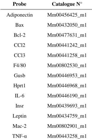
Probe Catalogue N°
Adiponectin Mm00456425_m1
Bax Mm00432050_m1
Bcl-2 Mm00477631_m1
CCl2 Mm00441242_m1
CCl3 Mm00441258_m1
F4/80 Mm00802530_m1
Gusb Mm00446953_m1
Hprt1 Mm00446968_m1
IL-6 Mm00446190_m1
Insr Mm00439693_m1
Leptin Mm00434759_m1
Mac-2 Mm00802901_m1
TNF-α Mm00443258_m1
Table 4: Taqman Gene Expression Assays
All assays were purchased from Applied Biosystems, Foster City, USA.
Relative expression of Adiponectin, CCl-2, CCl-3, F4/80, Leptin, Mac-1, Mac-2 and TNF- α mRNA was determined using standard curves based on white adipose tissue cDNA.
Samples were adjusted for total cDNA content by Glucuronidase beta (Gusb) and
hypoxanthine guanine phosphoribosyl transferase (Hprt-) 1 mRNA quantitative Realtime
PCR. Calculations were performed by a comparative method (2
-∆∆CT). In brief, the
amplification plot is the plot of fluorescence versus PCR number. The threshold cycle
value (Ct) is the fractional PCR cycle number at which the fluorescent signal reached the
detection threshold. Therefore, the input cDNA copy number and Ct are inversely related.
Data were analyzed with the Sequence Detector System (SDS) software version 2.1 (ABI) and Ct value was automatically converted to fold change RQ value ((RQ) = 2
− (∆∆CT)). The RQ values from each gene were then used to compare the gene expression across all groups.
2.2.6 Protein Extraction
Cell pellets or snap-frozen tissues were disrupted in lysis buffer (50 mM HEPES (pH 7.4), 1% (v/v) Triton X-100, 0.1 M sodium fluoride, 10 mM EDTA, 50 mM sodium chloride, 10 mM sodium orthovanadate, 0,1% (w/v) SDS, 10 µg/ml aprotinin, 2 mM benzamidine, 2 mM phenylmethylsulfonyl fluoride (PMSF)) by resuspension and gentle vortexing or by usage of a polytron homogenizer (IKA Werke, Staufen, Germany), respectively. Particulate matter was removed by centrifugation for 1 h at 4°C. The supernatant was transferred to a fresh vial and protein concentration was determined using a Bradford assay. Protein extracts were diluted to 5 mg/ml with lysis buffer and 4 x SDS sample buffer (125 mM Tris-HCl (pH 6.8), 5% (w/v) SDS, 43.5% (w/v) glycerol, 100 mM DTT, and 0.02% (v/v) bromophenol blue), incubated at 95°C over 5 min and stored at - 80°C.
2.2.7 SAPK/JNK Kinase Assay
A c-Jun fusion protein linked to agarose beads (SAPK/JNK Kinase assay #9810;
Cell Signaling, Danvers, MA, USA) was used to pull down SAPK/JNK enzyme from liver
and skeletal muscle protein extracts. Immunoprecipitation was performed by overnight
incubation at 4°C. After two washes with lysis buffer and kinase buffer, 200 µM ATP was
supplemented to the precipitate. The phosphorylation reaction was carried out at 30°C and
stopped after 30 min by addition of 4 x SDS sample buffer. Detection of phospho-c-Jun by
western blot analysis was used to measure SAPK activity.
2.2.8 Western Blot Analysis
Frozen protein extracts were thawed at 95°C for 5 min, then separated on 10-15%
(v/v) SDS polyacrylamide gels (193) and blotted onto PVDF membranes (Bio-Rad, München, Germany). Membranes were then incubated with 1% (v/v) blocking reagent (Roche, Mannheim, Germany) for 1 h at RT. Subsequently, primary antibodies (Table 5) diluted in 0.5% (v/v) blocking solution were applied overnight at 4°C. PVDF membranes were then washed four times for 5 min with 1 x TBS/0.01 (v/v) Tween. After 1 h incubation at RT with the respective secondary antibodies, membranes were washed 4 times for 10 min with 1 x TBS/0.01 (v/v) Tween, rinsed in 1 x TBS, incubated for 1 min in Pierce ECL Western Blotting Substrate (Perbio Science, Bonn, Germany), sealed in a plastic bag and exposed to chemiluminescence film (Amersham, Braunschweig, Germany). Films were developed in an automatic developer.
Antibody Catalogue N° Distributor Dilution
Actin A5441 Sigma Aldrich, Seelze, Germany 1:10000
Akt 9272 Cell Signaling, Danvers, MA, USA 1:1000
p-Akt (Ser473) 9271 Cell Signaling, Danvers, MA, USA 1:1000
p-c-jun (Ser63) 9810 (101) Cell Signaling, Danvers, MA, USA 1:1000
IRβ (C-19) sc-711 Santa Cruz, Heidelberg, Germany 1:200
SAPK/JNK 9252 Cell Signaling, Danvers, MA, USA 1:1000
Table 5: Primary antibodies used for western blot analysis
All respective secondary antibodies were purchased from Sigma Aldrich, Seelze, Germany, and used in a 1:1000 dilution.