Cancer Evolution
and the Emergence of Resistance to Targeted Cancer Therapy
Inaugural-Dissertation zur
Erlangung des Doktorgrades
der Mathematisch-Naturwissenschaftlichen Fakult¨at der Universit¨at zu K¨oln
vorgelegt von
Nina M ¨uller
aus Unna
K¨oln, 2018
Prof. Dr. Joachim Krug
Tag der m ¨undlichen Pr ¨ufung: 25.09.2018
A B S T R A C T
Targeted therapy to cancer acts on the molecular abnormalities driving a specific tumor. In clinical use, targeted therapies can lead to an impressive, but ultimately short-lived regression of solid tumors: In virtually all cases, a therapy-resistant tumor arises during targeted therapy, thus limiting its long-time efficacy. From a population dynamics perspective, the failure of targeted mono-therapy is in- evitable: a sufficiently large population of tumor cells contains therapy-resistant mutants as part of its standing genetic heterogeneity. Therapy then selects re- sistant mutants leading to a tumor consisting of resistant cells. As resistance to targeted therapy can be caused by diverse molecular mechanisms, targeting one particular resistance mechanism only leads to the emergence of resistance via an- other. If targeted therapy is to achieve a long-term tumor remission, it needs to address all resistance mechanisms present in a population of cancer cells.
As a proof of principle, we systematically derive cell lines resistant to combina- tions of targeted agents from PC9 cells, a well-studied lung cancer cell line. By characterizing the respective resistant lines, we show that several distinct resis- tance mechanisms exist simultaneously in the cell population prior to treatment.
We derive four cell lines driven by different resistance mechanisms. A drug com- bination targeting all these mechanisms prevents indefinitely the expansion of re- sistant cells. Our findings explain why, for solid tumors, long-term control of the disease with targeted therapy has proven elusive so far and point to a treatment strategy differing from current clinical practice: Instead of keeping the treatment fixed until a relapse occurs, tumor evolution has to be anticipated by targeting a broad spectrum of possible resistance mechanisms as early as possible. Our it- erative protocol offers a generic approach to explore the spectrum for resistance mutations and can be applied to cell lines driven by different molecular mecha- nisms.
However, large populations also contain non-dividing cells in a drug tolerant state, which a curative treatment would need to eradicate. To study this type of phenotypic heterogeneity, we develop a statistical model to infer growth rates in a population from single-cell lifetime measurements. Our method offers a way
clarify to what extent the drug tolerant state is heritable.
K U R Z Z U S A M M E N F A S S U N G
Zielgerichtete Krebstherapie wirkt auf die molekularen Anomalien, die das Wachs- tum eines bestimmten Tumors antreiben. Klinisch kann zielgerichtete Krebsthe- rapie zu einem beeindruckenden R ¨uckgang von Tumoren f ¨uhren, welcher aber nur von kurzer Dauer ist: In praktisch jedem Fall tritt w¨ahrend der Therapie Resistenz auf, was den langfristigen Nutzen der Therapie begrenzt. Aus Sicht der Populationsdynamik ist das Versagen von zielgerichteter Krebstherapie mit nur einem Medikament unausweichlich: Eine gen ¨ugend große Population an Tu- morzellen enth¨alt als Teil ihrer genetischen Heterogenit¨at Mutanten, welche auf das Medikament resistent sind. Die Therapie selektiert somit resistente Zellen, was zu einem neuen, resistenten Tumor f ¨uhrt. Weil Resistenz auf zielgerichtete Therapie normalerweise durch verschiedene molekulare Mechanismen auftreten kann, f ¨uhrt eine Therapie, welche auf nur einen bestimmten Resistenzmechanis- mus zugeschnitten ist, lediglich zum Auftreten von Resistenz ¨uber einen anderen Mechanismus. Daher m ¨ussten, wenn zielgerichtete Therapien zu einem langfristi- gen Tumorr ¨uckgang f ¨uhren sollen, alle Resistenzmechanismen, welche in einer Krebszellpopulation existieren, ber ¨ucksichtigt werden.
F ¨ur einen Grundsatzbeweis isolieren wir systematisch Zelllinien, welche resistent auf Kombinationen von zielgerichteten Medikamenten sind. Als Modellsystem nutzen wir PC9 Zellen, eine bekannte Lungenkrebszelllinie. Indem wir die je- weiligen resistenten Linien charakterisieren, zeigen wir, dass bereits vor Beginn der Therapie verschiedene Resistenzmechanismen gleichzeitig in einer Population existieren. Wir leiten vier resistente Linien ab, die alle einen unterschiedlichen Mechanismus aufweisen. Eine Medikamentenkombination, die auf all diese Me- chanismen abzielt, verhindert das Auftreten weiterer Resistenzen. Unsere Ergeb- nisse erkl¨aren, warum die langfristige Kontrolle von Tumoren mit zielgerichteter Therapie bislang nicht erfolgreich war und legen eine neue Behandlungsstrate- gie nah: Anstatt bei konstanter Therapie auf einen R ¨uckfall zu warten, sollte der Evolution des Tumors zuvorgekommen werden, indem die Therapie so fr ¨uh wie m¨oglich neben dem Wildtyp auch auf ein breites Spektrum an m¨oglichen Resistenzmechanismen abzielt. Unser iteratives Protokoll bietet eine generelle
llinien untersuchen kann.
Dennoch enthalten große Populationen neben resistenten Mutanten auch Zellen, die sich nicht oder nur sehr langsam teilen, aber die Therapie ¨uberleben. Diese
”schlafenden Zellen” m ¨ussten durch eine kurative Therapie ebenfalls beseitigt wer- den. Um diese Art der ph¨anotypischen Heterogenit¨at zu untersuchen, entwickeln wir ein statistisches Modell, mit dem wir Wachstumsraten aus der Messung von Lebenszeiten einzelner Zellen inferieren k¨onnen. Unsere Methode kann dazu genutzt werden, die Dynamik der schlafenden Zellen zu charakterisieren und festzulegen, ob dieser Zellzustand vererbt werden kann.
C O N T E N T S
1 a short introduction to the biology of cancer 1
1.1 Tumors in the human body . . . . 2
1.1.1 Cancer is a genetic disease . . . . 3
1.1.2 The hallmarks of cancer . . . . 4
1.1.3 Example: Overactive EGFR signaling . . . . 7
1.1.4 Standard treatment options . . . . 9
1.2 Targeted therapies: a novel treatment approach . . . . 10
1.2.1 Gleevec can cure chronic myelogenous leukemia (CML) . . . 11
1.2.2 The occurrence of resistance limits the long-term efficacy . . 13
1.3 Physics and theoretical models in cancer . . . . 14
1.4 Outline of this thesis . . . . 15
2 resistant mutants in evolving populations 17 2.1 Introduction . . . . 17
2.1.1 Growing cancer cells in cell culture . . . . 17
2.1.2 Handling cancer cell lines . . . . 19
2.1.3 Mathematical models of cancer resistance . . . . 21
2.1.4 Outline of this chapter . . . . 23
2.2 Semi-stochastic birth-death process with logistic growth . . . . 24
2.2.1 Growth dynamics of the sensitive cells . . . . 24
2.2.2 Stochastic emergence of resistant mutants . . . . 25
2.2.3 The number of resistant colonies . . . . 33
2.3 Splitting dynamics . . . . 39
2.3.1 Competition of two types of cells . . . . 39
2.3.2 Splitting under Luria-Delbr ¨uck dynamics . . . . 41
3 iterative exhaustion of resistance mechanisms to targeted ther- apy 45 3.1 Introduction . . . . 45
3.1.1 Can targeted therapy be curative? . . . . 45
3.1.2 Experimental protocol . . . . 49
. . Different resistance mechanisms pre-exist in a large cell pop-
ulation and respond to different compounds . . . . 52
3.2.2 Different resistance mechanisms show distinct disruptions in signaling pathways . . . . 53
3.2.3 No further resistance mechanisms arise under a combination of compounds targeting R1-R4 . . . . 57
3.2.4 Sleeper cells can survive the combination treatment . . . . 59
3.3 Discussion . . . . 62
4 inference of cellular states from single-cell lifetimes 67 4.1 Motivation: Persistence as a way to survive treatment . . . . 67
4.2 Lifetimes on a hidden Markov tree . . . . 68
4.3 The backward problem: Learning the parameters . . . . 71
4.3.1 The expectation maximization algorithm . . . . 71
4.3.2 Calculating the marginal probabilities using belief propagation 75 4.4 Results and discussion . . . . 77
5 conclusions and outlook 81
glossary 87
bibliography 91
list of figures 99
acknowledgements 101
1
A S H O R T I N T R O D U C T I O N T O T H E B I O L O G Y O F C A N C E RThe human body consists of around 1013 cells [1] that are organized in tissues which make up the different organs. Even though each cell stems from the same fertilized egg and thus (except for gametes) carries the same genetic information, cells specialize and form tissues that exhibit different functions. This is achieved via gene expression patterns that vary from cell type to cell type, meaning that not all genes that code for proteins are transcribed in all cells: Whereas a brain cell and a skin cell of the same organism are genetically nearly identical, very different proteins are made in either cell. Obviously, tissues in different parts of the body have distinct functions, but they all need to fulfill some basic requirements. Most importantly, they have to be stable and conserve their function during the lifetime of the organism.
Usually, the lifetime of a cell is smaller than the lifetime of the whole organism, which implies that all tissues have to be regularly renewed. This renewal happens at very different time scales: Whereas nerve cells last nearly a lifetime without replacement, cells lining the intestine live in a hostile environment and need to be replaced every few days. So in a living organism, cells divide and die all the time.
This process needs to be strictly regulated to preserve the organization and thus function of the tissue.
There are three major contributions how well-organized tissue renewal is achieved [2]. First, there exists acell memory. Each cell has stable patterns of gene expression characteristic for its specific type. This pattern has been inherited from ancestral cells and has already been established during embryonic development. This pre- serves the diversity of cell types in the tissue. Second, cells performselective cell- to-cell adhesion, meaning that cells do not stick equally well to all other cell types.
They rather adhere specifically to some types, which prevents chaotic mixture of cells in tissues. Finally, cell communicationis the last major contribution to orga- nized tissue renewal. Cells are covered with receptors that constantly sense the surroundings for proliferative or anti-proliferative signals: They only reproduce if
1
2 a short introduction to the biology of cancer
Genetic disease: mutations make normal cells „cancerous“
‣ allow uncontrolled growth
‣ avoid cell death
‣ grow blood vessels to supply nutrients
‣ allow for unlimited cell division
‣ invade other organs
Formation of a tumours consisting of up to 1011 cells.
What is cancer?
Normal cells Cancer cells
Figure 1:Each tumor is initiated from a single mutated and quickly dividing cell which is no longer subject to the strict controls of tissue formation and renewal. Over the time, a large amount of cancerous cells develops that disturb normal tissue function (sketch adapted from www.cancerresearchuk.org).
they receive the respective signals. They enter apoptosis if they receive signals to die. In this way, new cells are only created when and where they are needed.
If single cells occasionally disrespect the strict rules, the organism does not experience major problems. Only if cells systematically reproduce in places where they should not and the control mechanisms fail at stopping this behavior, first the initial tissue and later the whole organism can be affected. In that case the organism experiences a disease that is calledcancer.
1.1 tumors in the human body
Cancer is a term for a broad class of related diseases in which uncontrolled cell growth leads to the emergence of abnormal orneoplastictissue that forms tumors (see Fig. 1). Depending on where a tumor grows and how big it is, its mere physical presence is a threat to the organism. In the brain, for instance, tumors repress healthy tissue, which leads to neurological failures. In the lung, tumorous tissue disturbs oxygen intake. In the digestive system, tumors block routes for nutrient intake. In later stages, tumors even acquire the ability to invade into other tissues, a process calledmetastasis. The invasion leads to many tumors that are growing in the body at the same time. If metastatic cancers are left untreated, they usually lead to the death of the organism.
1.1 tumors in the human body 3
1.1.1 Cancer is a genetic disease
The reason for the abnormal and harmful behavior of cancer cells can be found in their genomes. Cancer cells accumulate thousands of mutations, some of which occur in genes that control cell proliferation. This leads to the transcription of malfunctioning proteins.
There are different ways in which mutations can make cells cancerous, and they often involve genes whose products participate in cellular signaling. A mutation can affect a cellular receptorsuch that it permanently folds into a state which sig- nals a growth signal, even in absence of an external stimulus from a ligand. The corresponding receptor is an example of anoncogene, and the mutation is termed again-of-functionmutation. Alternatively, a mutation can interfere with the proper function of a protein involved in the control of cellular proliferation. Such a gene is called atumor suppressorand the mutation is an example of aloss-of-functionmu- tation. In both cases it is mutations in specific genes that are key to the emergence of cancer.
Mutations or sets of mutations that make cells cancerous can be acquired in several ways. Sometimes, a predisposition to cancer is inherited: about 5−10%
of all human cancers are caused by germline mutations that are passed from one generation to the next [3]. This explains why in some families there is an accumu- lation of specific cancer types, such as breast or ovarian cancer. More frequently however, cancer is triggered bysomatic mutations, mutations that occur during the lifetime of an organism. Often, environmental factors increase the risk of cancer:
Carcinogenic substances such as tobacco smoke [4] or UV radiation [5] induce DNA damage leading to cancerous mutations, causing for instance lung or skin cancer. About12% of all human cancers are due tooncoviruses[6], viruses such as the Epstein–Barr (EBV) virus or the human papillomavirus (HPV) that can insert oncogenic genes into infected cells.
In all other cases, cancerous mutations occur just by chance due to unavoidable errors in DNA replication. The body usually has a very efficient machinery to check for errors and malfunctioning cells, but this mechanism does not work per- fectly. During the lifetime the human organism experiences approximately 1016 cell divisions [1]. Any error detection rate larger than10−16inevitably leads to the occurrence of mutations, some of which can cause cancer. Most unambiguously, pediatric cancers that occur very early in the life of the patients and that are not
number and thus maintenance of normal tissue architecture and function. Cancer cells, by deregulating these signals, become masters of their own destinies. The enabling signals are conveyed in large part by growth factors that bind cell-surface receptors, typically containing intracellular tyrosine kinase domains. The latter proceed to emit signals via branched intra- cellular signaling pathways that regulate progression through the cell cycle as well as cell growth (that is, increases in cell size); often these signals influence yet other cell-biological prop- erties, such as cell survival and energy metabolism.
Remarkably, the precise identities and sources of the prolifer- ative signals operating within normal tissues were poorly under- stood a decade ago and in general remain so. Moreover, we still know relatively little about the mechanisms controlling the release of these mitogenic signals. In part, the understanding of these mechanisms is complicated by the fact that the growth factor signals controlling cell number and position within tissues are thought to be transmitted in a temporally and spatially regu- lated fashion from one cell to its neighbors; such paracrine signaling is difficult to access experimentally. In addition, the bioavailability of growth factors is regulated by sequestration in the pericellular space and extracellular matrix, and by the actions of a complex network of proteases, sulfatases, and possibly other enzymes that liberate and activate them, apparently in a highly specific and localized fashion.
The mitogenic signaling in cancer cells is, in contrast, better understood (Lemmon and Schlessinger, 2010; Witsch et al., 2010; Hynes and MacDonald, 2009; Perona, 2006). Cancer cells can acquire the capability to sustain proliferative signaling in a number of alternative ways: They may produce growth factor ligands themselves, to which they can respond via the expres- sion of cognate receptors, resulting in autocrine proliferative stimulation. Alternatively, cancer cells may send signals to stim- ulate normal cells within the supporting tumor-associated stroma, which reciprocate by supplying the cancer cells with various growth factors (Cheng et al., 2008; Bhowmick et al., 2004). Receptor signaling can also be deregulated by elevating the levels of receptor proteins displayed at the cancer cell
Figure 1. The Hallmarks of Cancer
This illustration encompasses the six hallmark capabilities originally proposed in our 2000 per- spective. The past decade has witnessed remarkable progress toward understanding the mechanistic underpinnings of each hallmark.
surface, rendering such cells hyperre- sponsive to otherwise-limiting amounts of growth factor ligand; the same outcome can result from structural alter- ations in the receptor molecules that facilitate ligand-independent firing.
Growth factor independence may also derive from the constitutive activation of components of signaling pathways oper- ating downstream of these receptors, obviating the need to stimulate these pathways by ligand-mediated receptor activation. Given that a number of distinct downstream signaling pathways radiate from a ligand-stimulated receptor, the activa- tion of one or another of these downstream pathways, for example, the one responding to the Ras signal transducer, may only recapitulate a subset of the regulatory instructions transmitted by an activated receptor.
Somatic Mutations Activate Additional Downstream Pathways
High-throughput DNA sequencing analyses of cancer cell genomes have revealed somatic mutations in certain human tumors that predict constitutive activation of signaling circuits usually triggered by activated growth factor receptors. Thus, we now know that !40% of human melanomas contain activating mutations affecting the structure of the B-Raf protein, resulting in constitutive signaling through the Raf to mitogen- activated protein (MAP)-kinase pathway (Davies and Samuels 2010). Similarly, mutations in the catalytic subunit of phosphoi- nositide 3-kinase (PI3-kinase) isoforms are being detected in an array of tumor types, which serve to hyperactivate the PI3- kinase signaling circuitry, including its key Akt/PKB signal transducer (Jiang and Liu, 2009; Yuan and Cantley, 2008). The advantages to tumor cells of activating upstream (receptor) versus downstream (transducer) signaling remain obscure, as does the functional impact of crosstalk between the multiple pathways radiating from growth factor receptors.
Disruptions of Negative-Feedback Mechanisms that Attenuate Proliferative Signaling
Recent results have highlighted the importance of negative- feedback loops that normally operate to dampen various types of signaling and thereby ensure homeostatic regulation of the flux of signals coursing through the intracellular circuitry (Wertz and Dixit, 2010; Cabrita and Christofori, 2008; Amit et al., 2007; Mosesson et al., 2008). Defects in these feedback mech- anisms are capable of enhancing proliferative signaling. The prototype of this type of regulation involves the Ras oncoprotein:
the oncogenic effects of Ras do not result from a hyperactivation of its signaling powers; instead, the oncogenic mutations affecting ras genes compromise Ras GTPase activity, which
Cell144, March 4, 2011ª2011 Elsevier Inc. 647 Figure 2:With their hallmarks of cancer Hanahan and Weinberg identified general prin-
ciples necessary for tumor formation valid for all tumor types. Their analysis made clear that several genetic alterations have to occur before a cell becomes fully malignant (sketch taken from [7]).
driven by any germline mutations are caused by random mutations without any link to environmental influences.
1.1.2 The hallmarks of cancer
Over decades of cancer research, a broad and very detailed knowledge about dif- ferent cancer types has been accumulated. Tumorigenesis is a multi-step process, where several genetic alterations have to occur in a cell before it becomes fully malignant. In an attempt to simplify and structure the known facts, Hanahan and Weinberg [8] collected general observations that are true for all cancers, in- dependent of their tissue of origin (see Fig. 2). In their original formulation they identified six of such hallmarks of cancer, which were complemented by four ad- ditional characteristics in a second review article that was published eleven years later [7]. In the following, we briefly summarize these unifying features describ- ing the nature of cancer. A much more detailed discussion can be found in the original publications and the references therein.
1.1 tumors in the human body 5
sustaining proliferative signals. Healthy cells need external stimulus to divide, for instance induced by growth factors that bind to cell surface receptors.
Cancerous cells do not depend on external signals but permanently proliferate:
They are self-sufficient in their growth signals.
evading growth suppressors. Closely related to the first hallmark is the fact that cancerous cells also ignore anti-proliferative signals: Signaling circuits that stop healthy cells from excessive growth through binding of growth suppres- sor are modified in cancers such that the negative regulation of cell proliferation is disrupted.
resisting cell death. Usually, there exists a control machinery in cells sens- ing abnormalities, for example the tumor suppressor TP53. If errors such as DNA damage are detected, the cell is forced to enter a program of controlled death which is called apoptosis. Tumors can escape these mechanisms, for instance by the deletion or loss-of-function mutations in important tumor suppressors such as TP53.
inducing angiogenesis. All cells require an adequate supply of oxygen and of nutrients. Tumors that grow excessively cannot be sufficiently provided by the normal blood vessels of the tissue. Usually, angiogenesis (meaning blood vessel formation) extending this supply is strictly regulated. But tumors acquire the ability to actively induce angiogenesis to build their own nutrient supply and facilitate fast growth.
enabling replicative immortality. Normal cells cannot divide indefinitely.
During each cell division, nucleotide sequences protecting the ends of the chro- mosomes (the so-calledtelomeres) shorten. In this way, healthy cells age, a process that inhibits excessive growth independently of cell-to-cell signaling. Tumor cells on the other hand can be immortalized by activating mechanisms to maintain their telomeres above the critical length.
activating invasion and metastasis. Healthy cells stay in the tissue where they belong. Cancer cells by contrast have the ability to travel through the blood or lymph system to invade tissues all over the body. This process is calledmetastasis.
Apart from the six classical hallmarks, two further characteristics seem to be particularly important for tumor formation. Among the cellular alterations allow- ing for excessive growth, also adjustments in the energy metabolism are widely observed in cancer cells. Thismetabolic switchhas already been described in1923 by Otto Warburg [9] (and is for this reason termed Warburg effect). Cancer cells gain energy by processing glucose in a way healthy cells only do under anearobic (and thus stressful) conditions. As the altered metabolism is very inefficient, en- ergy metabolism was long thought to be a weakness of cancer cells. By contrast, more recent findings indicate that the altered metabolism facilitates the biosynthe- sis of components (such as nucleotides, amino acids, and lipids) that are necessary for cell replication [10] and thus actively contributes to tumorigenesis. The exact mechanism though remains elusive so far.
Another important issue is the interaction between tumors and the immune sys- tem. The immune system is able to recognize and destroy many cancer cells even before larger tumors form: Experiments in immunodeficient mice have shown that they develop more tumors than mice with an intact immune system when exposed to carcinogenic substances [11]. Consequently, solid tumors that actually form in hosts with a functioning immune system somehowevade immune destruction. The current understanding is that immunogenic tumors (tumors that provoke a strong immune response) can be attacked by the immune system and disappear before they are even diagnosed, whereas weakly immunogenic tumors are not detected by the immune system and can continue to grow. So it seems to be a necessary condition to efficiently hide from the immune system to enable tumor formation.
Altogether, many independent steps are involved in the formation of a tumor.
Healthy cells are usually well protected from major damage through a powerful genomic maintenance machinery and a surveillance system that detects malfunc- tioning behavior and forces affected cells to die. Therefore, it is surprising that cells can acquire all of the alterations that are necessary for tumor formation. The reason why it is still possible are twoenabling hallmarksthat simplify the required changes in tumorigenesis.
genome instability and mutation. Most importantly, tumors are genomi- cally unstable. This means that the rate at which point mutations and also major chromosomal rearrangements occur is much higher than in non-cancerous cells [12]. The loss of the tumor suppressor TP53, for instance, enables cells to resist cell death. Consequently, cells with compromised genomes are able to further
1.1 tumors in the human body 7
proliferate without intervening systems that monitor genomic integrity. Hence, defects in the genome maintenance and repair systems offer selective growth ad- vantages: The more severe the DNA supervision machinery is affected, the easier and faster mutated cells can expand.
tumor-promoting inflammation. Increasing evidence is also pointing to the important role of the tumor micro-environment during tumor formation. Vir- tual every neoplastic lesion is densely infiltrated with cells of the immune system, indicating an inflammation of the surrounding tissue. Historically, physicians and pathologists thought that this behavior was the attempt of the immune system to fight cancerous cells. Whereas it is indeed true that the immune system can detect tumor cells and trigger an anti-tumor defense, the immune response also contributes to the acquisition of the hallmark capabilities: It has been shown that the acute inflammation supplies bioactive molecules such as growth factors or pro-angiogenic factors to the tumor environment which is exploited by cancerous cells [13]. Moreover, chemicals that are actively mutagenic are released by inflam- matory cells [14], which further accelerates the rate at which new mutations are acquired. As such, tumor-promoting inflammation is another enabling hallmark.
1.1.3 Example: Overactive EGFR signaling
Whereas all of the different hallmarks of cancer are necessary for tumor formation and survival, they are acquired at different times and via different mechanisms in each tumor. To give a concrete example how cells can become self-sufficient in growth signaling, we briefly discuss the disruption of cell signaling by an overac- tive growth factor receptor called EGFR.
The epidermal growth factor receptor(EGFR or ERBB1) is part of the ERBB family of cell surface receptor tyrosine kinases. The ERBB receptors are trans-membrane molecules that mediate signals between theepithelium (the tissue lining the outer surface of organs and blood vessels) and the connective tissue. The receptors are part of the ERBB signaling network [15], which is a layered network: Receptor- ligand binding initiates cellular signaling cascades that produce a physiological outcome. It is structured as follows (see Fig. 3): Theinput layer consists of recep- tors and their ligands. Binding of a ligand to a receptor monomer induces receptor dimerization and phosphorylation of the receptor tail within the cell. The signal-
NATURE REVIEWS |MOLECULAR CELL BIOLOGY VOLUME 2 |FEBRUARY 2001 | 1 2 9
A network of networks?
The ErbB network might integrate not only its own inputs but also heterologous signals, including hor- mones, neurotransmitters, lymphokines and stress inducers29(FIG. 1). Many of these trans-regulatory inter- actions are mediated by protein kinases that directly phosphorylate ErbBs, thereby affecting their kinase activity or endocytic transport29. The most extensively studied mechanism involves activation of G-protein- coupled receptors (GPCRs) by agonists such as lysophosphatidic acid (LPA), carbachol (which specifi- cally activates muscarinic acetylcholine receptors) or thrombin (FIG. 2).
Experiments done with mutants and inhibitors of ErbBs imply that the mitogenic activity of some GPCR agonists requires transactivation of ErbB proteins.
These agents increase tyrosine phosphorylation of ErbB1 and ErbB2, either by increasing their intrinsic apoptosis (FIG. 1). Output depends on cellular context, as
well as the specific ligand and ErbB dimer. This has been best shown in terms of mitogenic and transform- ing responses: homodimeric receptor combinations are less mitogenic and transforming than the correspond- ing heterodimeric combinations, and ErbB2-containing heterodimers are the most potent complexes21–23(FIG. 3). Perhaps the best example of the ability of the ErbB module to tune mitogenic signalling is provided by the ErbB2–ErbB3 heterodimer: although neither ErbB2 nor ErbB3 alone can be activated by ligand, the het- erodimer is the most transforming24,25and mitogenic21 receptor complex. The ErbB2–ErbB3 heterodimer also increases cell motility on stimulation with a ligand26; but the other NRG receptor, ErbB4, which exists in sev- eral isoforms, has been associated with processes vary- ing from cellular chemotaxis27to proliferation and dif- ferentiation28.
Figure 1 | The ErbB signalling network. a| Ligands and the ten dimeric receptor combinations comprise the input layer.
Numbers in each ligand block indicate the respective high-affinity ErbB receptors8. For simplicity, specificities of receptor binding are shown only for epidermal growth factor (EGF) and neuregulin 4 (NRG4). ErbB2 binds no ligand with high affinity, and ErbB3 homodimers are catalytically inactive (crossed kinase domains). Trans-regulation by G-protein-coupled receptors (such as those for lysophosphatidic acid (LPA), thrombin and endothelin (ET)), and cytokine receptors is shown by wide arrows. b| Signalling to the adaptor/enzyme layer is shown only for two receptor dimers: the weakly mitogenic ErbB1 homodimer, and the relatively potent ErbB2–ErbB3 heterodimer. Only some of the pathways and transcription factors are represented in this layer. c| How they are translated to specific types of output is poorly understood at present. (Abl, a proto-oncogenic tyrosine kinase whose targets are poorly understood; Akt, a serine/threonine kinase that phosphorylates the anti-apoptotic protein Bad and the ribosomal S6 kinase (S6K); GAP, GTPase activating protein; HB-EGF, heparin-binding EGF; Jak, janus kinase; PKC, protein kinase C; PLCγ, phospholipase Cγ; Shp2, Src homology domain-2-containing protein tyrosine phosphatase 2; Stat, signal transducer and activator of transcription; RAF–MEK–MAPK and PAK–JNKK–JNK, two cascades of serine/threonine kinases that regulate the activity of a number of transcription factors.)
MAPK MEK
RAF JNK
JNKK PAK Shp2 GAP
Shc
Nck Vav Grb7
Rac
Crk Jak
Grb2 Sos PI(3)K
Src Cbl
PLCγ Ras–GDP
Ras–GTP
Bad Akt
S6K Abl
PKC
Stat Egr1 Elk
Fos
Sp1 Myc Jun
LPA, thrombin, ET, etc.
TGF-α (1)
EGF (1)
Epiregulin (1,4) β-cellulin
(1)
Amphi- regulin (1) HB-EGF (1,4)
NRG1 (3,4) α β
NRG2 (4) α β
NRG3
(4) Cytokines
NRG4 (4)
Apoptosis Migration Growth Adhesion Differentiation
Input layer
Output layer
Signal-processing layer
Ligands
Receptor dimers
Adaptors and enzymes
Cascades
Transcription factors
1 3 1 2 1 1 2 2 4 2 1 4 3 2
4 4
3 4
3 3
a
b
c
MESODERM
The middle germ layer of the developing embryo. It gives rise to the musculoskeletal, vascular and urinogenital systems, and to connective tissue (including that of the dermis).
ECTODERM
The outermost germ layer of the developing embryo. It gives rise to the epidermis and the nerves.
AKT PATHWAY Akt (or protein kinase B) is a serine/threonine protein kinase activated by the phosphatidylinositol-3-OH kinase pathway that activates survival responses.
© 2001 Macmillan Magazines Ltd
Figure 3:The ERBB signaling network is a layered network transmitting and translating extracellular signals to physiological cellular outcomes. Most important for tu- morigenesis are the RAS/RAF/MEK/MAPK and PI3K/AKT cascades, that lead to cell proliferation and migration (figure taken from [15]).
processing layerconsists of intracellular adapter molecules and enzymes that bind to the phosphorylated tail of the receptors triggering cellular signaling cascades such as the MAPK or AKT pathways. These signaling cascades translate the extra- cellular signal to specific transcriptional programs to the cell nucleus. Theoutput layerfinally produces a physiological outcome. Depending on which ligand was bound to which receptor, programs affecting apoptosis, adhesion or differentiation are started. Also cell division and migration can be triggered, which is important for tumorigenesis in some cancers.
If the receptor EGFR binds to a ligand, it specifically triggers the PI3K and MAPK pathways. Both lead to strong proliferative and anti-apoptotic signals. Con- sistent with this observation, overexpression and mutations of EGFR have been described in many cancers, mostly incarcinomas(tumors that arise from epithelial cells) [16]. Overexpression of EGFR leads to an increased amount of receptors that are made within a cell. As a result, small ligand concentrations can already lead to a strong cellular response [17]. Mutations, on the other hand, alter the struc- ture of the receptor: Large deletions in the extracellular domain of the receptor
1.1 tumors in the human body 9
are commonly observed [18], leading to ligand-independent kinase signaling – the receptor is stuck in its active state. Both mechanisms lead to constant proliferation and make cells insensitive to other extracellular signals.
1.1.4 Standard treatment options
The current treatment options that are routinely available for solid tumors are surgery, radiotherapy and chemotherapy. Clearly, surgeryis the oldest and most straightforward way of treating cancer. For the longest time in the history of cancer therapy, cutting out tumorous tissue was actually the only way of treating patients. Its rationale is based on two simple observation. First, small tumors even- tually develop into large tumors. Second, large tumors tend to metastasize into other parts of the body, thus exacerbating the tumor burden. In principle, surgery can be a curative treatment if all cancerous cells are removed. This requirement already shows its major limitation: Tumors tend to spread into surrounding tissue, the lymph and the blood system, which is why complete elimination is hard in practice. Nevertheless, still today it is the most effective treatment if tumors are locally constrained to a tissue that is surgically accessible.
The second main pillar of cancer management isradiotherapy[19]. Ionizing radi- ation causes DNA damage and by that leads to cell death. The field of radiation oncology was initiated after the discovery of X-rays by Wilhelm Conrad R¨ontgen in 1895[20] and the discovery of radium by Pierre and Marie Curie in 1898 [21].
Both forms of radiation were observed to cause strong skin burn, and first reports on successful medical use in cancer treatment followed around1900. At the time, the major limitations were severe radiation toxicities (radium) and extensive tissue damage of normal cells (X-rays) due to the limited depth of the low energy beams that were available. The invention of linear accelerators in the middle of the20th century made it possible to use high energy beams that could be confined to small areas, thus sparing healthy tissue from radiation. Today, advances in imaging and the possibility to control beam shapes facilitate treatment planning and minimize side effects. Radiation therapy is often used in combination with surgery to either shrink the tumor before the operation or decrease the risk of tumor recurrence afterwards. Nevertheless, side effects such as nausea, infertility or even secondary cancers due to radiation damage remain major limitations.
The termchemotherapyoriginally means the treatment of any disease with chemi- cal drugs. In the context of cancer, chemotherapy usually refers to cytotoxic drugs killing cells during cell division. The active development of chemotherapeutic drugs against cancer started after the observation that soldiers who have been ex- posed to sulfur mustard gas during World War I suffered from a depletion of the bone marrow and lymph nodes [22]. Subsequent experiments in mice with lym- phoid tumors showed that the delivery of mustard gas resulted in tumor regres- sion. Since then, different classes of chemotherapeutic drugs have been developed, for instance alkylating agents (such as mustard gas), antimetabolites or cytotoxic antibiotics. Some cancers such as acute childhood leukemia and Hodgkin’s dis- ease can be cured with chemotherapy [22]. As compared to surgery or radiation, chemotherapy has the big advantage of not acting on local parts of the body only:
Mostly delivered intravenously, it targets both the primary tumor and possible metastases. Today, chemotherapy is often used in combination with surgery or radiation as adjuvant chemotherapy to minimize the risk of recurrence from cells surviving the local treatment. Unfortunately, most chemotherapies have severe side effects ranging from immunosuppression, anemia and hair loss to major or- gan damages. This is why, even if the use of chemotherapeutic agents can be very effective, it often drastically reduces the quality of life [23].
1.2 targeted therapies: a novel treatment approach
Driven by more efficient and affordable sequencing methods, many mechanisms enabling cancer cells to proliferate are known today. The development was accom- panied by the discovery of oncogene addiction, a term that was first introduced by Weinstein [24,25]. Oncogene addiction means that cancer cells, even though they require several steps before becoming fully malignant, do depend on growth sig- nals transmitted by single oncogenes. Together with the finding that inactivation of those oncogenes in healthy cells is often well tolerated,molecular drivers(genetic alterations cancer cells depend on) were identified as possible vulnerabilities [26].
As advances in drug development allowed to design drugs targeting molecular drivers, in the last two decades the paradigm in cancer treatment shifted from cytotoxic chemotherapy to targeted therapy. Whereas classical chemotherapy in- terferes with all quickly dividing cells, targeted therapy kills cancerous cells of a specific molecular type only [27], implying significantly less side effects. Targeting
1.2 targeted therapies: a novel treatment approach 11
The Journal of Clinical Investigation http://www.jci.org Volume 117 Number 8 August 2007
first year of my pathology residency with Philip Custer in Philadel- phia (from 1953 to 1954), we had focused primarily on hematopoi- etic neoplasms. In the Pathology Department of the University of Pennsylvania School of Medicine, I used a short-term cell culture technique developed by Edwin Osgood and Marion Krippaehne (10) to study human leukemic cells. The cells were grown on small slides, and I rinsed the cells with tap water before staining them with Giemsa to visualize their chromosomes. This was an inad- vertent use of the “hypotonic technique” to disrupt the mitotic spindle and expand the cells, and it resulted in the presence of countable chromosomes in my metaphase chromosomal prepara- tions. I knew nothing about cytogenetics at this time but felt that the chromosomal preparations of the leukemic cells warranted investigation for any abnormalities. I found no one on our campus interested in human chromosomes but was eventually directed to a graduate student, David Hungerford, who was working at the Fox Chase Cancer Center and attempting to obtain material for a thesis on human chromosomes.
Hungerford and I, as well as other researchers around the world, began to use the new cytogenetic techniques to determine whether human leukemias could be characterized by specific chromosome abnormalities. Although we initially found no consistent genetic abnormalities in cells from individuals with acute myelogenous leu- kemia, Hungerford identified a characteristic small chromosome in the neoplastic cells of two patients with chronic myelogenous leu- kemia (CML) (Figure 1) (11). We then began, with the help of Paul Moorhead, a scientist at the Wistar Institute, to use an improved air drying technique for the cell preparations, which had been developed by Rothfels and Siminovitch (12), and were able to report that a series of seven patients that we had analyzed all had this minute chromo- some (13). Tough and colleagues (14), who were also studying human leukemias, designated this minute chromosome the “Philadelphia chromosome,” in accord with the Committee for the Standardiza- tion of Chromosomes, which had suggested that abnormal chromo- somes be named for the city in which they were discovered.
Our observation that all the neoplastic cells in nearly all cases of a specific human cancer contained a consistent somatic genetic
change provided strong evidence to support Boveri’s hypothesis (2) that a critical genetic alteration in a single cell, which provided the cell with a growth advantage, could give rise to a tumor. How- ever, in the years immediately before and after the identification of the Philadelphia chromosome, consistent chromosomal altera- tions were not found in other types of leukemia. The only other apparent consistent alteration, which was noted in a number of patients with chronic lymphocytic leukemia in New Zealand and designated the Christchurch chromosome (15), proved not to be a somatic alteration but rather a familial abnormality in one par- ticular family in that area.
During the 1960s, analysis of some human solid tumors revealed that in nearly all types of cancer the chromosome pattern was abnormal (16). In particular, extensive chromosome alterations, such as chromosome numbers in the hypotetraploid range and gross structural chromosome rearrangements, were observed in individuals with very advanced tumors and malignant effusions.
Furthermore, the extent of the cytogenetic changes often correlated directly with the extent to which the tumor had progressed clinical- ly (17) — as had been observed earlier in experimental tumors that caused the accumulation of tumor-derived fluid in the abdomen (ascites tumors) — with the tumor consisting of a single stem line of cells or several closely related sub-lines. However, the presence of a consistent chromosome abnormality in all neoplastic cells was somewhat less common in solid neoplasms than in the leukemias, and in fact, huge variety in the number and type of chromosome alterations in a given tumor were often observed (17). Most impor- tantly, when stem lines were present in solid tumors, they typically differed in their chromosome abnormality from one individual to another. Indeed, although a proportion of cases of some types of tumors, including tumors of the ovaries, testes, and meninges (18), show a characteristic chromosome abnormality, these abnor- malities are not found in sufficient individuals with a given type of tumor to be considered a marker chromosome for the neoplasm.
Thus, no chromosomal change comparable in consistency to the Philadelphia chromosome has been observed.
Part of the difficulty of identifying genetic abnormalities charac- teristic of specific neoplasms was methodological. Although dur- ing the 1960s technical advancements continued to be made — for example, Hungerford and coworkers developed more efficient hypo- tonic solution methods for generating chromosome preparations, and phytohemagglutin was used to stimulate mitotic cell division in lymphocyte cultures, thereby providing an easy source of mitotic chromosomes from non-neoplastic cells (19) — it was still impos- sible to individually identify human chromosomes. Furthermore, the often poor technical quality of metaphase chromosomal prepa- rations from tumor material, as opposed to normal cells, made even accurate counting of chromosomes sometimes difficult.
Despite the lack of progress in identifying genetic abnormalities characteristic of specific neoplasms during the 1960s, other areas of research provided evidence to support the concept that chro- mosome abnormalities are associated with cancer. An increased number of spontaneous chromosome breakages were observed to occur when chromosome preparations were made from circulating normal lymphocytes that had been isolated from individuals with inherited clinical disorders associated with an increased risk of leukemia and other malignancies such as Bloom syndrome, ataxia
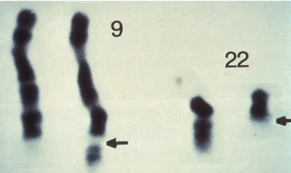
Figure 4:The unusual short Philadelphia chromosome was discovered in CML patients in 1960. The breakpoints of chromosome9 and 22are indicated by the black arrows (picture taken from [29]).
cancer at the molecular level also means a step towardspersonalized medicine: As tu- mors in different patients are not necessarily driven by the same genetic alteration (a fact that is usually referred to asintertumor heterogeneity), tumor classification needs to take into account thegenetic subtypeand not only the tissue of origin: For non-small cell lung cancer (NSCLC) for instance, more than 15 genetic subtypes have been identified [28] . Most targeted therapies approved today are either small molecules that can enter the cell membrane or monoclonal antibodies binding to cell surface proteins.
1.2.1 Gleevec can cure chronic myelogenous leukemia (CML)
The story of Gleevec is the biggest success of targeted therapies so far and was its major breakthrough: It transformed CML, which is a specific kind of blood cancer, from a deadly disease to a manageable condition such as diabetes.
Everything started with the discovery of an abnormally short chromosome in CML patients (see Fig.4) [30], which was later calledPhiladelphia chromosome, after the city where it was discovered. It became clear that the abnormal chromosome
was caused by the fusion of parts of chromosome9and chromosome22[31]. More specifically, the two genes BCR and ABL which are usually not located next to each other are connected by the fusion [32,33]. This leads to permanent kinase activity resulting in the accumulation of immature white blood cells [34]. The big hope at this point was that, if it is possible to inhibit the overactive signal, cells harboring the Philadelphia chromosome would die. Over the course of30 years of research from initial discovery to understanding the function of the BCR-ABL gene fusion, a very clear drug target had thus been identified: The overactive kinase domain in the BCR-ABL fusion gene.
The requirement to develop a kinase inhibitor was a completely new approach in drug discovery. So far, kinase inhibitors were considered to be inefficient due to the large number of different kinases existing in cells: An unspecific kinase in- hibitor would disrupt cell function in an uncontrolled way, and was thus expected to cause severe side effects in patients. Fortunately, enough variation in the ATP binding pockets of different kinases was found such that a compound specifically targeting the BCR-ABL kinase could be developed [35]. Imatinib, what the com- pound was called later, showed to be lethal for cells with the Philadelphia chromo- some and completely harmless in all others. Clinically, the relevant observable to quantify the efficacy of a new treatment is the fraction of patients that respond to therapy and experience a significant remission. This fraction is calledresponse rate.
The first clinical trials investigating the safety and tolerability of Imatinib in CML patients treated with Imatinib followed quickly and showed impressive response rates of close to100%, with very mild side effects [36]. In2001, Imatinib was ap- proved under the trade name Gleevec, and follow-up studies on patients treated with Gleevec showed an overall survival of89% after five years [37]. Today, most CML patients have a very good prognosis: After experiencing two years of remis- sion, patients have the same life expectancy as people who never had cancer.
Gleevec was the first targeted drug to be approved and used clinically; its over- whelming success promoted the complete field. Since then, hundreds of small molecules targeting different parts of cellular signaling pathways have been devel- oped and tested in patients. However, in all cases other than Gleevec, even if a subset of patients benefits greatly from specific therapies, the response is typically short-lived, as we will discuss below.
1.2 targeted therapies: a novel treatment approach 13
1.2.2 The occurrence of resistance limits the long-term efficacy
One challenge in delivering targeted therapies is the identification of patients that benefit from a specific drug. Actually, CML turned out to be the one example where the molecular mechanism causing continuous growth is always the overac- tive kinase in the Philadelphia chromosome. Most other cancers however, even if classified as the same disease, are genetically much more diverse. This explains the low response rates in clinical trials on various targeted drugs that followed after Gleevec: In lung cancer for instance, EGFR is often highly expressed. But the treatment with an EGFR kinase inhibitor only led to low response rates of ap- proximately10%. Only later, mutations in the EGFR kinase domain were found in the subset of patients responding most strongly to therapy, implying that not the high expression of EGFR but the specific activating mutations are driving the tumor [38,39].
Unfortunately, there is another problem limiting the efficacy of targeted thera- pies. Even after an initial response (which can be dramatic in some cases, with macroscopic tumors shrinking to the point where they can no longer be detected), patients in almost all cases experience a recurrence of the disease within one year:
Tumors that are resistant to therapy grow despite continued treatment. Sequenc- ing of biopsies from the relapsed tumor usually show genetic alterations such as point mutations or gene amplifications as compared to the genotype of the initial tumor which cause resistance [40].
There exist different ways how a tumor can become resistant to targeted therapy.
First, the drug target can be modified by a secondary mutation. In this case, the drug can no longer bind and the initial growth signal persists. Second, a mutation can permanently activate a part of the downstream signaling cascade. This means that the drug binds correctly, but has no growth suppressing effect. Finally, a bypass track can be activated. The resistant tumor does not depend on the initial growth signal anymore, which is why it does not respond to therapy. For a review on resistance mechanisms to targeted EGFR inhibition, for instance, see [41].
While the high specificity of targeted therapies accounts for manageable side ef- fects and is thus its biggest advantage, it is also the reason why resistance arises so frequently in practice. Often, a single nucleotide change is enough to confer resis- tance. Due to unavoidable errors in DNA replication, some genetic heterogeneity is found in large tumors. Cells carrying resistance mutations therefore inevitably exist in very low frequencies prior to any kind of treatment if the population size