Analysis of Rca1 function at the G1-S transition in Drosophila melanogaster
Inaugural-Dissertation
zur
Erlangung des Doktorgrades
der Mathematisch-Naturwissenschaftlichen Fakultät der Universität zu Köln
vorgelegt von Silvia Querings
aus Trier
Köln 2008
Berichterstatter: PD Dr. Frank Sprenger Prof. Dr. Maria Leptin
Tag der mündlichen Prüfung: 08. Februar 2008
Table of contents
Abstract... 1
1. Introduction... 2
1.1 The cell cycle... 2
1.2 Cell cycle modes in Drosophila melanogaster... 3
1.3 Regulation of the cell cycle ... 4
1.3.1 Cyclin-Cdk activity initiates mitosis ... 6
1.3.2 Cyclin degradation promotes mitotic exit ... 6
1.3.3 Establishment and maintenance of the G1 phase ... 10
1.3.4 Regulation of the G1-S transition... 10
1.3.5 Protein degradation by SCF complexes ... 12
1.3.6 Regulation of DNA replication ... 13
1.3.7 APC/C and endoreplication... 15
1.4 The cell cycle regulator Rca1 ... 16
1.4.1 The Rca1/Emi1 family ... 18
1.4.2 F-box dependent function of Rca1 at the G1-S ... 20
2. Aim... 21
3. Results... 22
3.1 Subcellular localisation studies of Rca1... 22
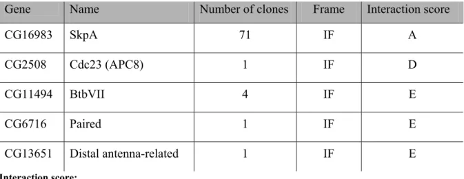
3.2 Yeast two-hybrid screen ... 28
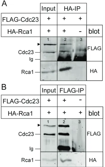
3.2.1 Interaction of Rca1 and Cdc23 ... 29
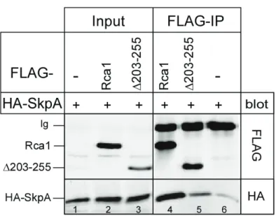
3.2.2 Interaction of Rca1 and SkpA depends on the F-box motif ... 31
3.3 Rca1 interacts with SCF subunits... 34
3.3.1 Endogenous Cul1 coprecipitates with Rca1 or SkpA... 35
3.3.2 Rca1 forms a complex with Cul1 and SkpA ... 36
3.3.3 Complex formation of Rca1-SkpA-Cul1 depends on the F-box ... 37
3.4 What are the targets of the putative SCF/Rca1 complex? ... 39
3.4.1 Fzr, Rux or Wee are not targets of the SCF/Rca1 ... 39
3.4.2 Peptide Mass Fingerprinting analysis (PMF) ... 42
3.5 Rca1 function during endoreplication ... 44
3.5.1 Gene expression analysis of endoreplicating cells ... 45
3.5.2 HA-Rca1 overexpression increases Cyclin E, Rnr2 and PCNA transcript levels in endoreplicating cells... 47
4. Discussion... 50
4.1 Localisation studies of Rca1... 50
4.2 Rca1 interacts with the APC/C subunit Cdc23... 53
4.3 Rca1 is part of an SCF complex ... 55
4.4 Substrates of the putative SCF/Rca1 ... 57
4.5 Gene expression in endoreplicating cells ... 59
4.6 Overexpression of HA-Rca1 increases Cyclin E and E2F1 target gene expression in endoreplicating salivary glands ... 62
5. Materials and Methods... 66
5.1 Materials ... 66
5.1.1 Chemicals ... 66
5.1.2 Special chemicals and kits... 66
5.1.3 Electronic equipment, computer and software ... 67
5.1.4 Media, solutions and buffers ... 67
5.1.5 Bacterial strains ... 70
5.1.6 Oligonucleotides... 71
5.1.7 Plasmids... 73
5.1.8 Fly stocks... 75
5.1.9 Antibodies... 76
5.2 Methods for molecular cloning... 77
5.2.1 Electrocompetent bacteria cells... 77
5.2.2 Transformation of electrocompetent bacterial cells ... 77
5.2.3 Transformation of chemical competent bacteria cells... 77
5.2.5 Dephosphorylation of DNA ends ... 78
5.2.6 Klenow fill-in of DNA ends ... 78
5.2.7 Agarose gel electrophoresis... 78
5.2.8 Isolation of DNA fragments ... 79
5.2.9 DNA ligation ... 79
5.2.10 DNA Mini-Preparation... 79
5.2.11 DNA Midi-Preparation... 79
5.2.12 DNA sequencing ... 80
5.2.13 Polymerase chain reaction (PCR)... 80
5.3 Drosophila Schneider (S2) cell culture ... 81
5.3.1 Maintenance of S2 cells... 81
5.3.2 Freezing and thawing of S2 cells... 81
5.3.3 Transient transfection of S2 cells ... 81
5.3.4 Coating coverslips with Poly-L-Lysine... 82
5.3.5 Immunostaining of S2 cells ... 82
5.3.6 Immunoprecipitation ... 83
5.3.7 SDS-Page, Western blot analysis and Coomassie staining ... 83
5.4 Drosophila techniques and qRT PCR ... 84
5.4.1 Maintainance of flies ... 84
5.4.2 Collection of embryos ... 84
5.4.3 Dissection of salivary glands overexpressing HA-Rca1 or CycE ... 85
5.4.4 mRNA isolation and cDNA synthesis... 85
5.4.5 Quantitative real-time PCR ... 86
5.5 Yeast two-hybrid screen ... 88
6. References... 89
Appendix ... 105
Abbreviations ... 108
Zusammenfassung ... 110
Erklärung ... 112
Lebenslauf ... 113
Danksagung ... 114
Abstract
Tight control of APC/C-Cdh1Fzr activity is essential for progression through mitosis and establishment of the G1 phase. Rca1 is a nuclear protein that inhibits the APC/C-Cdh1Fzr complex during G2 to allow cyclin accumulation and subsequent entry into mitosis. In this thesis, a localisation study of Rca1 was performed revealing that a nuclear localisation sequence (NLS) and other domains in the protein mediate efficient nuclear accumulation.
Besides its function in G2, Rca1 expression can promote S phase entry. Rca1 belongs to the family of F-box proteins that mediate substrate specificity in SCF (Skp-Cul1-F-box) ubiquitin ligases. Functional studies demonstrated that the F-box is crucial for S phase induction by Rca1, suggesting that Rca1 has a secondary function in an SCF complex.
A major part of this thesis was devoted to characterise the putative SCF/Rca1 complex and its target proteins. In a yeast two-hybrid screen, the SCF subunit SkpA was identified as a binding partner of Rca1. Coimmunoprecipitation studies confirmed this interaction and indicated moreover that SkpA binding relies on the F-box in Rca1. Furthermore, endogenous Cul1 coprecipitated with Rca1 and this also in an F-box dependent manner.
Altogether, these experiments demonstrated that Rca1 forms a complex with the SCF core subunits SkpA and Cul1. The SCF/Rca1 complex could promote S phase entry by degrading a negative regulator of the G1-S transition. Cyclin A/Cdk1 is a potent S phase inducer, but its activity is normally dampened by the CKI Rux, inhibitory phosphorylation by Wee and APC/C-Cdh1Fzr dependent degradation of Cyclin A. Protein levels of these S phase inhibitors were not altered by coexpression of Rca1 suggesting that these proteins are not targets of the SCF/Rca1 complex.
Overexpression of Rca1 in larval salivary glands results in impaired endoreplication and accumulation of Cyclin A, Cyclin E and Cdk1. Expression profiling revealed that mitotic genes (e.g. Cyclin A/B, Cdk1) are normally downregulated in salivary glands, but ectopic Rca1 expression promotes transcription of these genes. In addition, qRT-PCR analysis showed elevated transcript levels of the E2F1 targets Rnr2 and PCNA, suggesting that Rca1 overexpression leads to increased E2F1 activity. Cyclin E is also a known E2F1 target and hence, this might explain the marked increase in Cyclin E transcript levels.
Finally, the gene expression analysis indicated that components of the APC/C and its target Geminin were present in larval salivary glands, thus supporting the idea that rereplication
1. Introduction
1.1 The cell cycle
Cell proliferation is a fundamental process for the development of all organisms and function and maintenance of life. Cells arise by division of already existing cells and the resulting daughter cells contain the same genetic information as their progenitors. In unicellular organisms, cell division results in an entire new organism. However, multicellular organisms develop by countless divisions of a founder cell giving rise to a vast number of cells that make up the tissues and organs. Furthermore, cell proliferation enables self renewal of various tissues throughout the whole lifetime of an adult organism including the haematopoetic system or skin and intestinal epithelium. Cell reproduction occurs by a well defined series of events, a process that is also known as the cell cycle. All cell cycle events have to take place in correct order and timing to ensure precision and reliability over generations. These requirements are achieved by a complex regulatory network, the cell cycle control system. Different extrinsic and intrinsic factors like nutritional status, growth factors, cell density and developmental state promote or restrain cell division, cell cycle exit or apoptosis. Defects in the system can lead to abnormal, uncontrolled cell growth and cell proliferation during development or adulthood and eventually promote tumorigenisis.
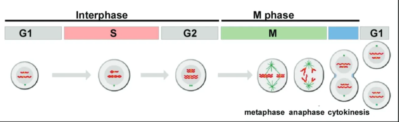
The cell cycle can roughly be divided into two broad periods: First, cells increase their cell mass and DNA is duplicated, whereas in the following step the cell splits itself into two daughter cells consisting of the same DNA content. In a standard cell cycle both processes are tightly coupled and consist of four distinct phases: G1 (Gap1) phase, S (Synthesis) phase, G2 (Gap2) phase and M (Mitotic) phase (Figure 1.1). The first three stages G1, S, G2 are collectively known as interphase. Usually these phases are morphologically indistinguishable, but each stage is determined by specific processes that prepare the cell for initiation of cell division. During G1, cells show a high rate of biosynthetic activities resulting in the synthesis of various proteins that are required in S phase. This phase is marked by DNA replication which leads to duplication of all chromosomes. After S phase completion, cells enter the G2 phase, in which additional proteins are synthesised for the following M phase. The M phase consists of two major events: the nuclear division
(mitosis) and the cytoplasmic division (cytokinesis). During mitosis, the duplicated DNA is divided equally into two daughter nuclei, a process which can be subdivided into different phases. First, the chromatin begins to condensate into highly ordered chromosome structures during prophase and this is followed by nuclear envelope break down in prometaphase. During metaphase, chromosomes are attached to spindle microtubules and align at the metaphase plate or equatorial plane. Chromosome segregation occurs in anaphase resulting in separated sister chromosomes that are pulled to opposite spindle poles. Mitosis ends with telophase when both chromosome sets are surrounded by a new nuclear envelope and DNA unfolds into chromatin. Cell division is completed after cytokinesis, a process in which the mother cell is eventually divided into two daughter cells both harbouring a complete copy of the parental genome. Some cells stop dividing at a certain developmental stage and remain in a quiescent G0 state for long periods of time, which is common for fully differentiated cells.
Figure 1.1 The eukaryotic standard cell cycle (adapted from Morgan, 2006).
The standard cell cycle consists of four phases: G1, S, G2 and M phase. During interphase, the DNA is replicated in S phase resulting in duplicated chromosomes. The Gap phases G1 and G2 provide time for cell growth and serve as important regulatory transitions for cell cycle progression. M phase is composed of two major events: mitosis and cytokinesis. During mitosis, chromosomes align at the metaphase plate and in anaphase sister chromatides are separated and distributed to opposite poles. In the following cytokinesis the cell divides into two new daughter cells that both exhibit a complete copy of the parental genome.
1.2 Cell cycle modes in Drosophila melanogaster
The fruit fly Drosophila melanogaster serves as a model organism to study cell proliferation and its coordination with cell growth and differentiation (for review see Edgar and Lehner, 1996). The standard cell cycle consisting of G1-S-G2-M is not the only cell cycle mode that is applied in Drosophila. The embryonic development of Drosophila
driven by maternal components and consist only of S-M phases. These rapid nuclear divisions occur in a shared cytoplasm that is named syncytium. During division 10-13, cellularisation of the embryo takes place resulting in the formation of a cell layer beneath the embryo’s surface which is called the cellular blastoderm. Gastrulation starts at cell cycle 14 transforming the simple blastoderm into a multilayered embryo, a process that is accompanied by cell movement and differentiation. The cell cycle lengthens and acquires for the first time a G2 phase. The postblastoderm divisions 14-16 are composed of S-G2-M phases and are driven by zygotic gene products since maternal supplies are almost depleted (Merrill et al., 1988; Wieschaus and Sweeton, 1988). Furthermore, nuclei start dividing asynchronously, but in programmed spatial and temporal patterns, the so-called mitotic domains (Foe, 1989). After mitosis 16, the first G1 phase is introduced in cycle 17 (Edgar and O'Farrell, 1990). Some epidermal cells exit the cycle after mitosis 16 and arrest in the terminal G1 phase of cell cycle 17 (Edgar and O'Farrell, 1990). These G1-arrested epithelial cells are programmed to become imaginal disc cells and reinitiate the standard cell cycle upon the onset of larval development. During Drosophila development, the imaginal discs give rise to adult structures like wing, notum and eyes. Cells of the gut, the fat body or the salivary glands enter endoreplication by the end of embryogenesis and continue throughout larval stages (Smith and Orr-Weaver, 1991). In addition, cells of the gut and the ovary undergo endoreplication during adulthood (Dej and Spradling, 1999;
Micchelli and Perrimon, 2006; Ohlstein and Spradling, 2006). Endoreplication or the endocyle is a cell cycle mode in which cells bypass mitosis resulting in G-S cycles (for review see Edgar and Orr-Weaver 2001; Lilly and Duronio, 2005). Cells undergo multiple rounds of initiation of DNA replication resulting in increased DNA content and polyploidy due to complete replication of the genome or amplification of specific chromosome regions (Calvi et al., 1998). This strategy of endoreplication helps to support the rapid growth after the larva hatches and enables an increased production of numerous gene products.
1.3 Regulation of the cell cycle
The eukaryotic cell cycle is a highly regulated series of events leading to cell reproduction.
These molecular events occur in a sequential fashion and are irreversible. In response to growth factors, the cell cycle is initiated first leading to duplication of cell organelles and increased protein synthesis. After this phase of massive cell growth, cells enter S phase and
duplicate their chromosomes. This process must be tightly regulated to ensure complete replication of the genome and only once per cycle. During M phase duplicated chromosomes and cell components must be distributed equally into individual daughter cells. Both processes must be achieved with extreme precision and in the correct order to guarantee accurate inheritance of the genetic information. Eucaryotic cells contain a complex regulatory network, the so-called cell cycle control system that regulates timing and coordination of each cell cycle event (for review see Morgan, 2006; Murray, 2004).
This system drives progression through cell cycle transitions called checkpoints and interrupts the cell cycle upon DNA damage or failures in mitosis, thereby preventing uncontrolled or defective cell division. Components of this regulatory network are highly conserved throughout all organisms providing a very robust and reliable control system.
The central components of the cell cycle control system are the cyclin-dependent kinases (Cdks) and their regulatory subunits called cyclins. Enzymatic activity of these kinases depends on changes in their phosphorylation state and tight binding of the cyclin which stimulates the catalytic activity. Cdk protein levels remain constant throughout the cell cycle, so that oscillations in Cdk activity depend primarily on fluctuations in the protein level of the corresponding cyclin. During the cell cycle, different types of cyclins and Cdks are synthesised resulting in the formation and activation of several cyclin-Cdk complexes at different time points. These cyclin-Cdk complexes control the progression through the three major cell cycle checkpoints: G1-S, G2-M and metaphase-anaphase transition. In mammalian cells, four different Cdk proteins (Cdk1, Cdk2, Cdk4 and Cdk6) are involved directly in cell cycle controls. In Drosophila, orthologues of Cdk1, Cdk2 and Cdk4 have been identified (for review see Edgar and Lehner, 1996; Lee and Orr-Weaver, 2003;
Swanhart et al., 2005), whereas in budding and fission yeast just one main Cdk, known as Cdc28 or Cdc2 has been identified (Beach et al., 1982). The cyclins can be divided into two main groups that oscillate during different cell cycle phases. Mitotic cyclins are responsible for the G2-M transition and mitosis, whereas the G1 cyclins drive progression from G1 into S phase and induce DNA replication. In Drosophila, the mitotic cyclins A, B and B3 and the G1 cyclins D and E are present.
1.3.1 Cyclin-Cdk activity initiates mitosis
Entry into mitosis is driven by the activity of cyclin-Cdk complexes. In Drosophila, mitotic cyclins A, B and B3 are expressed during S phase and accumulate in G2 (Sigrist et al., 1995). All mitotic cyclins can form a complex with Cdk1 and the activity of each cyclin-Cdk1 complex triggers a certain event during mitosis (Jacobs et al., 1998; Knoblich and Lehner, 1993; Lehner and O'Farrell, 1989; Lehner and O'Farrell, 1990b). Cyclin A is essential for chromatin condensation and nuclear envelop breackdown during prophase whereas Cyclin B is needed in metaphase and Cyclin B3 for events during anaphase (Ramachandran et al., 2007). Altough all three mitotic cyclins perform a certain task during mitosis, Cyclin A seems to be the most important one since Cyclin A mutants are embryonic lethal due to a cell cycle arrest in G2 of mitosis 16 (Jacobs et al., 1998;
Knoblich and Lehner, 1993; Lehner and O'Farrell, 1989). Cdk activity depends on phosphorylation by the Cdk-activating kinase (CAK) at threonine residue 161 (Morgan, 2006). In Drosophila, the CAK is a trimeric complex consisting of a Cdk-related protein Cdk7, Cyclin H and Mat1. Furthermore, Cdk activity is negatively regulated by the Wee1/Myt1 kinase that phosphorylates Cdk at threonine residue 14 and tyrosine residue 15 (Campbell et al., 1995; Morgan, 1997). The phosphatase Cdc25 removes these inhibitory phosphates and thereby allows entry into mitosis (Russell and Nurse, 1986). In Drosophila, string and twine are the Cdc25 homologues. At cell cycle 14, maternal string transcript supplies are exhausted which advances midblastula transition (Edgar and O'Farrell, 1989).
The embryo initiates zygotic string expression that regulates all mitotic divisions throughout development (Edgar and O'Farrell, 1990; Edgar et al., 1994a). In contrast, Twine activity is restricted to meiosis (Edgar and Datar, 1996).
1.3.2 Cyclin degradation promotes mitotic exit
Cell cycle progression must occur unidirectional and irreversible. These requirements can be achieved by proteolytic destructions of the regulatory proteins. Cdk activity is essential for mitotic entry, but for further progression and exit from mitosis Cdk activity must be restrained. Since Cdk levels remain stable throughout the cell cycle, Cdk activity is mainly regulated by oscillating cyclin levels. Cyclins are expressed before entry into mitosis and get degraded during mitosis resulting in decreased Cdk activity (Murray and
Kirschner, 1989). Destruction of the mitotic cyclins A, B and B3 occurs in a sequential fashion during mitosis (Sigrist et al., 1995). Expression of stable versions of Cyclin A, B or B3 results in a mitotic arrest in metaphase, early or late anaphase respectively, demonstrating that Cyclin A degradation is essential for sisterchromatide segregation, Cyclin B degradation for stable kinetochor-spindle attachments during anaphase and Cyclin B3 destruction for late mitotic events (Parry and O'Farrell, 2001; Ramachandran et al., 2007). Mitotic cyclins and other cell cycle regulators are targeted for ubiquitin- dependent degradation. Therefore, multiple ubiquitin molecules are attached to substrates resulting in polyubiquitinated proteins that are recognised and destroyed by the 26S proteasome (for review see Murray, 2004). Ubiquitination is a process carried out in a series of reactions including ubiquitin activation, conjugation and ligation to the substrate.
First, ubiquitin is covalently attached through its carboxyl terminus to the sulfhydryl group of a cysteine in the active site of the ubiquitin-activating enzyme (E1), a reaction that requires ATP hydrolysis. Subsequently, the activated ubiquitin is transferred to the ubiquitin-conjugating enzyme (E2). In the final step, the ubiquitin is transferred from the E2-ubiquitin conjugate to the amino group of a lysine residue in the target protein. This reaction is catalysed by the ubiquitin-protein ligase (E3) and occurs in a successive transfer of several ubiquitin molecules. Finally, the polyubiquitinated proteins are recognised and destroyed by the proteasome (Hochstrasser, 1996). The anaphase promoting complex/cyclosome (APC/C) is a large, multisubunit ubiquitin-protein ligase that mediates proteasomal destruction of mitotic cyclins and thereby progression through mitosis (for review see Peters, 2006; Pines, 2006; Yu, 2007; Zachariae and Nasmyth, 1999). The APC/C is a complex of 11-13 subunits including a cullin (APC2) and a RING subunit (APC11) that binds the E2-ubiquitin conjugate (Harper et al., 2002). APC/C activity depends on its phosphorylation state and association with the two WD40 activator proteins named Cdc20 and Cdh1 in yeast and mammals (Kramer et al., 2000; Visintin et al., 1997).
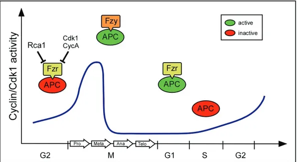
In Drosophila, the genes fizzy (fzy) and fizzy-related (fzr) encode for Cdc20 and Cdh1 (Dawson et al., 1993; Dawson et al., 1995; Sigrist et al., 1995). Binding and activation of the APC/C by Cdc20Fzy or Cdh1Fzr occurs at different phases of the cell cycle (Figure 1.2).
The APC/C-Cdc20Fzy complex is only active during early mitosis when Cdk activity is high resulting in phosphorylation of several APC/C subunits, a prerequisite for Cdc20Fzy binding to the APC/C (Kramer et al., 2000). It has been proposed that the early mitotic
phosphorylation by Polo-like kinase (Plk1) is essential for APC/C-Cdc20Fzy activation (Reimann et al., 2001a; Hsu et al., 2002). However, a more recent study demonstrated that Emi1 degradation is not needed for APC/C-Cdc20Fzy activation during early mitosis (Di Fiore and Pines, 2007). Moreover, APC/C-Cdc20Fzy activity is restrained by the spindle assembly checkpoint until metaphase. The spindle checkpoint proteins Bub1, BubR1 and Mad2 sense unattached kinetochors and prevent APC/C-Cdc20Fzy activity by binding and subsequent withdrawal of Cdc20 (Yu, 2006). As soon as all sister chromatides are attached to the spindle microtubules, the APC/C-Cdc20Fzy complex is fully active and triggers the metaphase/anaphase transition by degradation of mitotic cyclins. By the end of mitosis, Cdk activity drops resulting in dissociation and inactivation of APC/C-Cdc20Fzy until the cells enter the next mitosis. In addition to mitotic cyclins, the Separase inhibitor Securin is a major target of the APC/C-Cdc20Fzy (Hagting et al., 2002). Upon Securin destruction, Separase is released and cleaves the cohesin complex holding sister chromatides together and thereby enables chromosome segregation (Nasmyth, 2001). In Drosophila, the Securin homologue Pimples forms a complex with Three rows and Separase which is required for sister chromatide separation (Leismann et al., 2000). Embryos mutant for fizzy arrest in metaphase of mitosis 16 when maternal supplies of Fizzy are exhausted resulting in APC/C-Cdc20Fzy inactivation and subsequent accumulation of mitotic cyclins (Dawson et al., 1993; Dawson et al., 1995; Sigrist et al., 1995). Cdh1Fzr can only bind the APC/C during late mitosis and G1 phase, when Cdk activity is low and Cdh1Fzr is unphosphorylated (Zachariae et al., 1998; Kramer et al., 2000). Thereby, cyclin degradation is maintained throughout late mitosis and the G1 phase. In addition, APC/C- Cdh1Fzr mediates destruction of Cdc20Fzy as well as Geminin which is a DNA licensing inhibitor. At the G1-S transition, APC/C-Cdh1Fzr gets inhibited by vertebrate Emi1 or Drosophila Rca1, respectively (Hsu et al., 2002, Zielke et al., 2006). In the Drosophila embryo, epidermal cells mutant for fizzy-related fail to establish the terminal G1 and undergo an additional seventeenth mitosis (Sigrist and Lehner, 1997).
Figure 1.2 Regulation of the APC/C during the standard cell cycle (adapted from Yu, 2007).
The APC/C-Cdh1 mediates degradation of mitotic cyclins during late mitosis and G1. At the G1/S transition, the APC/C-Cdh1 is inhibited and subsequently Cyclin A is stabilised which activates Cdk2 and triggers S phase events. Active Cdk2 phosphorylates Cdh1 and further inhibits APC/C-Cdh1. In early mitosis, Cdk activity is high and APC/C subunits get phosphorylated which promotes Cdc20 binding. APC/C-Cdc20 activity is restrained by the spindle checkpoint. When all kinetochors are attached to spindle microtubules, inactivation of the spindle checkpoint results in APC/C-Cdc20 activity and Cyclin B and securin degradation.
In turn, separase is activated which cleaves the cohesin complexes and thereby enables sister chromatide separation and metaphase-anaphase transition. Degradation of mitotic cyclins reduces Cdk activity resulting in dephosphorylation of APC/C subunits and Cdh1. Cdh1 activates the APC/C during late mitosis and the G1 phase, when Cdk activity is low. At late G1, APC/C-Cdh1 activity gets inhibited by Emi1.
APC/C target proteins contain a conserved amino acid sequence that is important for substrate recognition. The most widespread motif is the destruction box (D-box) mediating degradation of B-type cyclins and securin (Glotzer et al., 1991; King et al., 1996). The D-box is recognised by both complexes, APC/C-Cdc20Fzy and APC/C-Cdh1Fzr. Mutations in the D-box result in stable Cyclin B or Cyclin B3 proteins that lead to an early or late anaphase arrest when overexpressed in the Drosophila embryo (Sigrist et al., 1995).
Degradation of Cyclin A is more complex and depends on other destruction elements in addition to the D-box (Ramachandran et al., 2007). Another important substrate recognition motif is the KEN-box which is mainly present in APC/C-Cdh1Fzr substrates (Burton and Solomon, 2001; Pfleger et al., 2001; Pfleger and Kirschner, 2000). Most APC/C substrates contain either or both destruction elements for their efficient ubiquitination. Both activator proteins Cdc20Fzy and Cdh1Fzr function as substrate receptors
APC core subunit Doc1 (APC10) contributes to D-box binding and is required for ubiquitination processivity in vitro (Carroll et al., 2005). Furthermore, it was demonstrated that the APC/C-Cdc20Fzy complex binds substrates with a higher affinity than the APC/C core does alone (Passmore et al., 2005).
1.3.3 Establishment and maintenance of the G1 phase
The first G1 phase is established after mitosis 16 during late Drosophila embryogenesis.
Most epidermal cells reside in the terminal G1 until they reinitiate the standard cell cycle upon larval hatching. During G1, reactivation of cyclin-Cdk complexes is prevented to allow controlled entry into the next cycle upon cell growth or other regulatory factors. Cdk inhibition in G1 is achieved by several mechanisms. Expression of mitotic cyclins is decreased and protein levels remain low due to ongoing cyclin degradation by the APC/C- Cdh1Fzr. In addition, Cdk activity is supressed by Cdk inhibitor proteins (CKIs) that bind and inactivate cyclin-Cdk complexes and thereby promote a G1 arrest (Peter and Herskowitz, 1994; Sherr and Roberts, 1999). In Drosophila, Dacapo (Dap) inhibits increasing Cyclin E/Cdk2 activity and enables a stable G1 state (de Nooij et al., 1996;
Lane et al., 1996). Furthermore, Roughex (Rux) is a specific inhibitor of Cdk1 and is also involved in mitotic exit (Foley et al., 1999; Foley und Sprenger, 2001; Thomas et al., 1994;
Thomas et al., 1997). Embryos mutant for rux exhibit a delay in the metaphase-anaphase transition resulting from prolonged Cyclin A/Cdk1 activity. In addition, the inhibitory effect of Rux is also essential for a stable G1. Loss of Rux leads to premature cyclin-Cdk1 activation which promotes S phase entry and results in a rough eye phenotype (Thomas et al., 1994; Thomas et al., 1997).
1.3.4 Regulation of the G1-S transition
In mammalian cells, Cyclin E/Cdk2 and Cyclin D/Cdk4/6 drive the G1-S transition (Matsushime et al., 1994; Sherr, 1993). Cyclin D expression is induced by growth factors thereby linking extrinsic growth signals and cell proliferation. Cyclin D/Cdk4/6 complexes phosphorylate and inactivate members of the Retinoblastoma (Rb) tumor supressor family.
Rb forms a heterodimer with the transcription factor E2F and represses E2F activity. E2F complexes are heterodimers consisting of two subunits, E2F subunit and one of the
Dp family. Phosphorylation of the Rb protein leads to dissociation from E2F (Attwooll et al., 2004; Blais and Dynlacht, 2004; Kato et al., 1993). The released E2F factor initiates the transcription of Cyclin E and Cyclin A (DeGregori et al., 1995; Pagano et al., 1992) and other genes involved in DNA replication such as ribonucleotide reductase (RNR), the DNA polymerase δ accessory subunit PCNA (DeGregori et al., 1995) and the vertebrate APC/C inhibitor Emi1 (Hsu et al., 2002). During late G1, the APC/C-Cdh1Fzr complex is inhibited by vertebrate Emi or Rca1 in Drososphila resulting in Cyclin A accumulation.
Simultaneously, CKIs normally restraining Cdk activity are degraded by the SCF (Skp-Cullin-F-box protein) complex (Cardozo and Pagano, 2004; Nakayama and Nakayama, 2006). As a result, Cyclin/Cdk activity increases thereby driving the G1-S transition and initiating DNA replication. Furthermore, Cyclin E/Cdk2 complexes enhance the transcription of Cyclin E by phosphorylating Rb and thereby promoting its own activity in a positive feedback loop. In Drosophila, orthologues of Cyclin D and Cdk4 have been identified, but Cyclin D/Cdk4 complexes are rather involved in regulating cell growth than in G1-S control (Datar et al., 2000; Meyer et al., 2000). S-phase induction mainly relies on Cyclin E/Cdk2 activity (Knoblich et al., 1994; Richardson et al., 1995). It is assumed that Cyclin E transcripts accumulate prior to the S phase in endocycling and mitotically dividing cells (Edgar and Nijhout, 2004). Cyclin E/Cdk2 phosphorylates the Rb-related proteins (Rbf1/Rbf2) and stimulates the transcription of E2F target genes like cyclinE, rnr and PCNA (Du et al., 1996; Duronio and O'Farrell, 1994; Duronio and O'Farrell, 1995;
Duronio et al., 1995). In Drosophila, a single DP and two E2F subunits (E2F1 and E2F2) do exist: E2F1 acts as a transcriptional activator and E2F2 as a repressor on the same promoters (Frolov et al., 2001). Thus, in mitotically dividing cells Cyclin E/Cdk2 activity is regulated on the transcriptional level by E2F1. In addition, the CKI Dacapo inhibits Cyclin E/Cdk2 complexes (de Nooij et al., 1996; Lane et al., 1996). In addition to Cyclin E/Cdk2 activity, overexpression of Cyclin A or loss of the Cyclin A/Cdk1 inhibitor Rux can induce S phases (Foley et al., 1999; Sprenger et al., 1997; Thomas et al., 1997).
Moreover, SCF complexes are also involved in G1-S regulation since several vertebrate CKIs are targeted for ubiquitin-dependent degradation by the proteasome (for review see Nakayama and Nakayama, 2006).
1.3.5 Protein degradation by SCF complexes
SCF complexes belong to a superfamily of Cullin-RING ubiquitin ligases (CRLS) that can be found throughout all eukaryotes. All CRLs share the catalytic core of Cullin-RING, but despite of that they show a great diversity in terms of composition and function, including cell cycle regulation, signalling pathways, circadian rhythms and apoptosis (Grima et al., 2002; Koepp et al., 1999; Maniatis, 1999; Nateri et al., 2004). Different CRLS are characterised by the Cullin isoform which is assembled into the complex. In humans, seven different Cullin proteins have been identified and each can nucleate into a multisubunit ubiquitin ligase. The archetypical CRL is the SCF ubiquitin ligase which is named by its core subunits Skp1, Cullin1 (Cul1) and F-box protein (for review see Nakayama and Nakayama, 2006; Petroski and Deshaies, 2005; Vodermaier, 2004). Cul1 acts as a scaffold protein binding to Skp1 and the RING-domain known as Roc1/Rbx1/Hrt1. The Roc1 subunit recruits a ubiquitin-conjugating enzyme (E2), while Skp1 acts as an adapter through binding the F-box motif of F-box proteins (Bai et al., 1996; Zheng et al., 2002).
Besides Cul1-Skp1 based SCF complexes, the family of CRLS also includes the classes of Cul2/5-Elongin B/C, Cul3-BTB and Cul4-DBB1 based ubiquitin ligases. Specificity of the SCF complex is determined by the F-box protein which binds the target via protein-protein interaction motifs such as WD40 or leucine-rich repeats (Kipreos and Pagano, 2000;
Skowyra et al., 1997). Many different F-box proteins exist, but not all of them are directly involved in SCF mediated protein degradation (Hermand, 2006). Phosphorylation of the target is a prerequisite for substrate recognition. In yeast, the most prominent F-box protein Cdc4 regulates the stability of Sic1, Far1, Cdc6 and the Cyclins Cln1/2 (Tyers and Jorgensen, 2000). In Drosophila, the F-box protein Slimb mediates destruction of the Dorsal /NFκB inhibitor Cactus/INFκB and of the transcription factors Cubitus interruptus (Ci) and Armadillo (Arm) which are involved in Hedgehog and Wingless signalling (Maniatis, 1999; Jiang and Struhl, 1998; Spencer et al., 1999). Furthermore, Cyclin E degradation depends on the SCF/Archipelago (Ago) and is essential for downregulation of Cyclin E/Cdk2 activity and exit from S phase (Koepp et al., 2001; Moberg et al., 2001;
Schwab and Tyers, 2001). In addition, Myc protein is also degraded by the SCF/Ago complex (Moberg et al., 2004). The Drosophila genome harbours six different Cullin (Cul1-6) isoforms and several Skp proteins (A-F), whereas in mammals and yeast just a single Skp1 protein exists (Nayak et al., 2002; Yamanaka et al., 2002). Furthermore, three
different Roc proteins (Roc1a, Roc1b and Roc2) have been identified in Drosophila, but so far there is only evidence for SkpA and Roc1a to be part of an SCF complex that regulates cell cycle progression (Murphy, 2003; Noureddine et al., 2002). Which members of the Cullin, Roc and Skp family preferentially associate and form an active SCF complex in vivo still remains unclear.
1.3.6 Regulation of DNA replication
During S phase, DNA replication takes place resulting in duplication of all chromosomes.
This process must be tightly regulated to prevent uncontrolled DNA synthesis and to ensure genome duplication once per cycle. Several factors are involved in initiation of replication and DNA synthesis (for review see Bell and Dutta, 2002; Machida et al., 2005;
Sivaprasad et al., 2007). High Cyclin E/Cdk2 activity drives initiation of DNA replication at discrete regions on the chromosomes called origins of replication. These origins are organised in clusters that are distributed throughout the chromosomes and are activated simultaneously in S phase. During late mitosis and G1, the pre-replicative complex (pre- RC) assembles at origins, a process also known as DNA licensing in which origions are prepared for activation by preloading replication factors and the DNA helicase. Pre-RC formation starts with a six-subunit complex (ORC1-6) called origin recognition complex (ORC) that is constantly bound to origins. The proteins Cdt1 and Cdc6 bind to the ORC complex followed by recruitment and loading of the minichromosome maintenance complex (MCM) onto the DNA. This hexameric MCM complex consists of the proteins MCM 2-7 and acts as a replicative helicase that unwinds the DNA helix using energy by ATP hydrolysis. Thereby, it enables other downstream factors to gain access to the DNA in order to initiate replication (Fletcher et al., 2003; Pape et al., 2003). After binding of the MCM complex, DNA licensing is completed and the pre-RC, now consisting of ORC 1-6, Cdt1, Cdc6 and MCM 2-7 is activated upon S phase entry. The components of the pre-RC are conserved throughout many organisms including Drosophila with the Cdt1 orthologue double-parked (dup) (Crevel et al., 2005; Thomer et al., 2004; Whittaker et al., 2000).
Origin firing requires Cdk activity resulting in phosphorylation of pre-RC components and recruitment of essential replication factors including Cdc45, Mcm10, RPA, proliferating cell nuclear antigen (PCNA) and DNA polymerases α and δ. After initiation of DNA
other pre-RC proteins remain at the origin and are destroyed or inhibited to ensure origin firing only once in the same cycle (for review see Bell and Dutta, 2002; Blow and Dutta, 2005). Thereby, cells proceed into mitosis after DNA replication. The pre-RC remains inactive until late mitosis and prevents MCM loading until formation of a novel pre-RC. In multicellular organisms, pre-RC assembly in G1 is promoted by the activity of the ubiquitin ligase APC/C-Cdh1Fzr which mediates the destruction of Cyclin A and Geminin.
Geminin inhibits pre-RC formation by association and inactivation of Cdt1 (Wohlschlegel and Dwyer, 2000; Tada, 2007). Vertebrate Emi1 has been proposed to link DNA replication and mitosis and to prevent rereplication (Di Fiore and Pines, 2007; Machida and Dutta, 2007). At late G1, the APC/C-Cdh1Fzr is inhibited by vertbrate Emi1 or Drosophila Rca1 resulting in Geminin and Cyclin A accumulation upon S phase entry (Machida and Dutta, 2007; N. Zielke, personal communication). Geminin binds and inhibits Cdt1 from S phase until late mitosis and prevents MCM loading after origin firing (McGarry and Kirschner, 1998; Tada, 2007; Wohlschlegel and Dwyer, 2000). Depletion of Geminin or overexpression of Cdt1 is sufficient to cause rereplication in mammals (Melixetian et al., 2004; Takeda et al., 2005; Zhu et al., 2004). In Drosophila, an orthologue for Geminin has been identified and also here depletion of Geminin or overexpression of Cdt1/Dup has been shown to induce rereplication (Mihaylov et al., 2002;
Quinn et al., 2001; Thomer et al., 2004). In yeast, rereplication is prevented by phosphorylation of Cdc6 and MCM proteins followed by nuclear export. In addition, phosphorylated Cdc6 is degraded by the SCFCdc4 complex (Perkins, et al., 2001). Besides Geminin, Cyclin/Cdk activity has been reported to prevent rereplication (Machida and Dutta, 2007; Mihaylov et al., 2002). While in yeast constitutive inactivation of Cdk is sufficient to cause rereplication (for review see Machida et al., 2005), the effect of cyclin/Cdk complexes on pre-RC formation are poorly understood in metazoa. In Drosophila, Geminin depletion or inactivation of Cyclin A/Cdk results in rereplication demonstrating that both mechanisms are nonredundant (Mihaylov et al., 2002). Cdk activity prevents pre-RC formation by phosphorylation of Cdc6 and Cdt1. While Cdc6 is exported from the nucleus, phosphorylated Cdt1 is a target of ubiquitin-dependent degradation. In vertebrates, the Cul4-DDB1CDT2 ubiquitin ligase and the SCFSkp2 complex have been reported to mediate Cdt1 degradation (Li et al., 2003; Liu et al., 2004; Nishitani et al., 2001; Sugimoto et al., 2004; Zhong et al., 2003). In Drosophila, the Cdt1 orthologue
Dup is also degraded throughout the cell cycle, but a potential SCF complex has not been identified so far (Thomer et al., 2004).
1.3.7 APC/C and endoreplication
In a standard cell cycle, pre-RC formation and subsequent DNA replication once per cell cycle depends on oscillating APC/C activity. During late mitosis and G1, the APC/C- Cdh1Fzr mediates degradation of Cyclin A and Geminin, thereby allowing pre-RC formation. At late G1, vertebrate Emi1 or Drosophila Rca1 inhibit the APC/C-Cdh1Fzr resulting in Geminin and Cyclin A accumulation that both prevent rereplication (Machida and Dutta, 2007; N. Zielke, personal communication). Emi1 or Rca1 depletion results in cells with giant nuclei which is due to rereplication (Grosskortenhaus and Sprenger, 2002;
Machida and Dutta, 2007). If the APC/C also regulates pre-RC formation during endoreplication still remains unclear. In Drososphila, APC/C-Cdh1Fzr activity is essential for downregulation of cyclin/Cdk1 activity and entry into the endocycle (Edgar and Orr- Weaver, 2001; Lilly and Duronio, 2005; Sigrist and Lehner, 1997). In epidermal cells of the Drosophila embryo, deletion of fzr triggers rereplication, but in endoreplicating cells it prevents entry into the endocycle program suggesting a cell type specific role of Fzr.
Mitotic cyclins and other mitotic regulators (e.g. Cdk1 and Cdc25/String) are assumed to be transcriptionally downregulated when cells switch from mitotic cycles to endocycles (Klebes et al., 2002; Smith and Orr-Weaver, 1991; Sauer et al., 1995; Schaeffer et al., 2004). So far, it has been assumed that APC/C activity is dispensable once cells have entered the endocycle (Edgar and Orr-Weaver, 2001; Lilly and Duronio, 2005). Also in mammals, a direct role of the APC/C during endoreplication has not yet been confirmed since trophoblast cells of Emi -/- embryos endoreplicate normally while other cells fail to proliferate (Lee et al., 2006). However, there are similarities in the regulation of G-S cycles in mitotic and endocycling cells (Edgar and Orr-Weaver 2001; Lee and Orr-Weaver 2003).
Endocycles are driven by oscillating waves of Cyclin E/Cdk2 activity (Lilly and Spradling 1996; Royzman et al., 1997; Weng et al., 2003) requiring regulated accumulation and destruction of Cyclin E. Continuous expression of Cyclin E interferes with the DNA licensing program and results in an endocycle block (Follette et al., 1998; Su and O'Farrell 1998; Weiss et al., 1998). It is proposed that Cyclin E/Cdk2 activity is low during the Gap
licensing in endoreplicating cells includes the same pre-RC components. Several mechanisms are assumed to influence the periodic expression of Cyclin E, but a final model of the Cyclin E oscillator has not been defined so far. It has been suggested that the SCF/Archipelago complex contributes to Cyclin E fluctuations by targeting phosphorylated Cyclin E for degradation (Koepp et al., 2001; Moberg et al., 2001; Schwab and Tyers, 2001). In addition, Cyclin E levels are regulated by E2F1 which activates transcription of S phase genes such as cyclin E, PCNA and Rnr2 (Duronio et al., 1995;
Royzman et al., 1997). The Cyclin E/Cdk2 specific inhibitor Dacapo (Dap) is also assumed to influence G-S kinetics during endoreplication by oscillating out of phase with Cyclin E.
Dacapo binds and inhibits Cyclin E/Cdk2 and thereby promotes entry into the Gap phase (de Nooij et al., 1996; Lane et al., 1996). A recent study has given evidence for Dacapo being essential during endocycles of Drosophila oocytes (Hong et al., 2007). Geminin prevents rereplication in mitotic cells, but to what extent Geminin contributes to endocycle regulation remains elusive.
1.4 The cell cycle regulator Rca1
The APC/C promotes cell cycle progression by triggering the periodic degradation of mitotic and other cell cycle substrates (for review see Nakayama and Nakayama, 2006;
Peters, 2006; Pines, 2006; Zachariae and Nasmyth, 1999). Activation of the APC/C requires association with the activator proteins Cdc20Fzy and Cdh1Fzr and depends on its phosphorylation state (see section 1.3.2). While the APC/C-Cdc20Fzy complex is active during early mitosis, degradation of cell cycle substrates is maintained during late mitosis and G1 by APC/C-Cdh1Fzr activity (Figure 1.3). APC/C activity can be restrained by different factors including components of the spindle checkpoint and members of the Emi family (for review see Schmidt et al., 2006). In Drosophila, the gene rca1 encodes for an APC/C inhibitor that belongs to the family of Emi proteins (Grosskortenhaus and Sprenger, 2002). Initially, Rca1has been identified in a screen for dominant suppressors of the roughex (rux) eye phenotype (Dong et al., 1997). The CKI Rux specifically inhibits Cyclin A/Cdk1 activity and thereby contributes to maintain a stable G1 state. Loss of rux results in a rough eye phenotype due to unrestrained Cyclin A/Cdk1 activity and premature S phase entry (Thomas et al., 1997). Overexpression of Rca1 or Cyclin A in the developing eye resembles the rough eye phenotype. Embryos mutant for rca1 arrest in G2 of cell cycle
16 resulting in a reduced number of epidermal cells compared to wild-type embryos (Dong et al., 1997). The rca1 mutant phenotype is similar to cyclin A mutants (Lehner and O'Farrell, 1989) indicating that Rca1 controls Cyclin A/Cdk1 activity and naming the gene regulator of cyclin A (rca1). The G2 arrest in rca1 mutant cells is due to premature cyclin degradation by the APC/C-Cdh1Fzr complex (Grosskortenhaus and Sprenger, 2002). Rca1 has a negative effect on Fzr. Embryos mutant for fzr fail to establish the terminal G1 and undergo an additional cell cycle 17 (Sigrist and Lehner, 1997). Since overexpression of Cyclin A results in the same phenotype, premature reaccumulation of Cyclin A seems to be responsible for mitosis 17 (Sigrist and Lehner, 1997). However, overexpression of Fzr prevents entry into mitosis 16 similar to rca1 mutants (Sigrist and Lehner, 1997).
Simultaneous overexpression of Rca1 and Fzr or double mutants for rca1 and fzr both show normal cyclin accumulation and mitosis 16 (Grosskortenhaus and Sprenger, 2002).
In addition, Rca1 and Fzr have been shown to interact physically (Grosskortenhaus and Sprenger, 2002). Rca1 is a nuclear protein, but Fzr and mitotic cyclins have been shown to localise in the cytoplasm during interphase (Dienemann and Sprenger, 2004;
Grosskortenhaus and Sprenger, 2002). It remains unclear in which compartment of the cell the active APC/C-Cdh1Fzr complex resides and how Rca1 can inhibit Fzr in G2 despite their spatial separation.
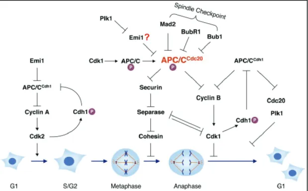
Figure 1.3 Rca1 restrains the APC/C-Cdh1Fzr activity in G2. APC/C promotes cell cycle progression by degradation of different substrates like mitotic cyclins. The activity of APC/C-Cdc20Fzy depends on high cyclin/Cdk activity and triggers the metaphase-anaphase transition. Cyclin/Cdk activity drops during late mitosis allowing Cdh1Fzr to bind and activate the APC/C. Cyclin degradation is maintained during late mitosis and G1 by APC/C-Cdh1Fzr activity. In G2, APC/C-Cdh1Fzr activity is inhibited by Rca1 to allow
Hence, these data show that Rca1 regulates Cyclin A/Cdk1 activity by inhibiting the APC/C-Cdh1Fzr during G2 of Drosophila development. Subsequently, APC/C-Cdh1Fzr mediated protein degradation is restrained resulting in the accumulation of mitotic cyclins and entry into mitosis. In addition, Cyclin A/Cdk1 and Cyclin E/Cdk2 activity contributes to restrain APC/C-Cdh1Fzr activity by phosphorylation of Cdh1fzr (Dienemann and Sprenger, 2004; Zielke, 2007). During G1, Cyclin A/Cdk1 activity is limited by the CKI Rux and the APC/C-Cdh1Fzr which targets Cyclin A for ubiquitin dependent degradation by the proteasome.
1.4.1 The Rca1/Emi1 family
The Emi protein family consists of several members including Drosophila Rca1 and vertebrate Emi1 and Emi2/XErp1 (Reimann et al., 2001a; Schmidt et al., 2005; Tung et al., 2005). Rca1 and Emi1 are both inhibitors of the APC/C-Cdh1Fzr complex and have been reported to be involved in the G1-S transition (Dong et al., 1997; Hsu et al., 2002;
Reimann et al., 2001a; Zielke et al., 2006; Zielke, 2007).
Sequence analysis of Rca1 and Emi1 revealed a limited sequence homology of 18%, but both proteins share a set of conserved functional domains (Figure 1.4). The carboxy- terminus contains a conserved zinc binding region (ZBR) which is known to mediate protein-protein interactions. A C-terminal fragment of Emi1 and Rca1 has been shown to be sufficient for its function to restrict APC/C-Cdh1Fzr activity in G2 (Reimann et al., 2001a; Zielke et al., 2006). Furthermore, a point mutation in the ZBR (C351S) abolishes the inhibitory effect of Rca1 and Emi1 (Reimann et al., 2001a; Reimann et al., 2001b;
Zielke et al., 2006). Emi1 harbours a D-box (RxxL) that has been shown to bind the APC/C core (Miller et al., 2006). While the D-box in Emi1 enables binding to the APC/C- Cdh1Fzr complex, the ZBR is assumed to prevent substrate access (Miller et al., 2006).
Mutation of the ZBR converts Emi1 to a D-box substrate suggesting that Emi acts as a pseudosubstrate inhibitor (Miller et al., 2006). Rca1 also contains a minimal D-box between amino acid 203 and 255. Although this region seems to harbour elements essential for Rca1 function (Radermacher, 2007; Zielke et al., 2006), there is so far no evidence for Rca1 acting as a pseudosubstrate inhibitor of the APC/C-Cdh1Fzr. In addition, Rca1 and Emi1 contain different putative localisation signals (NLS) and an F-box motif in the central region classifying both proteins to the family of F-box proteins (see section 1.3.5).
Finally, several putative Cdk1 phosphorylation sites, a DSGxxS degron and a KEN-box can be found in both proteins.
Figure 1.4 Schematic overview of Rca1 and Xenopus Emi1 protein structure. The C-terminal part of both proteins harbours a ZBR which is essential for APC/C inhibition. Furthermore, an F-box is located in the middle part as well as a KEN-box and a DSGxxS degron that is crucial for Emi1 degradation. Several putative Cdk1 phosphorylation sites (marked by an asteriks) and nuclear localisation sequences (NLS) are present in both proteins. All conserved domains described for xlEmi1 apply basically to all Emi1/Emi2 proteins.
In vertebrates, Emi1 degradation is initiated by Cdk1 phosphorylation that facilitates Plk1 binding and phosphorylation of the DSGxxS motif. The SCFßTRCP ubiquitin ligase recognises the phosphorylated degron and targets Emi1 for proteasomal degradation (Guardavaccaro et al., 2003; Hansen et al., 2004; Margottin-Goguet et al., 2003; Moshe et al., 2004). The proteins Evi5 and Pin1 have been reported to stabilise Emi1 levels during G2 by preventing Plk1 mediated phosphorylation and subsequent degradation (Bernis et al., 2007; Eldridge et al., 2006). Mutation of the DSGxxS degron or all Cdk1 phosphorylation sites results in a stable Emi protein (Margottin-Goguet et al., 2003). Rca1 has been shown to be degraded during the terminal G1 to allow APC/C-Cdh1Fzr dependent cyclin degradation and a stable G1 state (Grosskortenhaus and Sprenger, 2002; Jacobs et al., 2002; Pimentel and Venkatesh, 2005). Surprisingly, Rca1 turnover neither relies on Cdk1 phosphorylation nor on phosphorylation of the DSGxxS degron suggesting that Rca1 degradation occurs in a different way than Emi1 degradation (Radermacher 2007; Zielke et al., 2006). Furthermore, deletion of the KEN-box does not affect Rca1 protein levels demonstrating that Rca1 is not a target of the APC/C-Cdh1Fzr (Zielke et al., 2006). Which pathways eventually mediate Rca1 degradation still remains unclear. The Emi2/XErp1 proteins inhibit the APC/C-Cdh1Fzr during meiosis and are required for maintenance of the cytostatic factor (CSF) arrest during Xenopus oocyte maturation (Rauh et al., 2005;
Schmidt et al., 2005; Tung et al., 2005). The C-terminal part of Emi2/XErp1 shows significant homology to Emi1 and displays a ZBR and F-box domain (Schmidt et al., 2005;
Tung et al., 2005).
1.4.2 F-box dependent function of Rca1 at the G1-S
Eye imaginal disc cells mutant for fzr fail to downregulate Cyclin A/Cdk1 activity in G1 resulting in ectopic S phases and a rough eye phenotype (Pimentel and Venkatesh, 2005;
Thomas et al., 1994). At the G1-S transition, the APC/C-Cdh1Fzr is inhibited by Rca1/Emi proteins to allow cyclin accumulation upon S phase entry. Overexpression of Rca1 during eye development also causes ectopic S phases and a rough eye phenotype (Dong et al., 1997; Zielke et al., 2006). This indicates that Rca1 has a secondary function at the G1-S transition besides APC/C inhibition in G2. Furthermore, wing imaginal disc cells overexpressing Rca1 progress faster through G1 also demonstrating that excess Rca1 protein accelerates the G1-S transition (Zielke et al., 2006). Moreover, S phase induction by excess Rca1 depends on the F-box motif (Zielke et al., 2006). Since Rca1 lacking the F- box does not affect APC/C-Cdh1Fzr inhibition during G2 (Zielke et al., 2006), S phase induction by Rca1 must occur independently from its function as an APC/C-Cdh1Fzr inhibitor. Rca1/Emi1 proteins belong to the family of F-box proteins which are part of SCF ubiquitin ligases (see section 1.3.5). Hence, it has been proposed that Rca1 might act as an F-box protein in an SCF complex that regulates the G1-S transition (Zielke et al., 2006).
Furthermore, the role of Rca1 for G-S cycles of endoreplicating cells has been analysed in Drosophila salivary glands. Endocycles are driven by oscillating waves of Cyclin E/Cdk2 activity, whereas mitotic regulators including Cdk1, Cdc25/string and the mitotic cyclins are assumed to be transcriptionally downregulated (see section 1.3.6). Furthermore, APC/C-Cdh1Fzr activity is essential for endocycle entry, but once cells have entered the endoreplication program, APC/C-Cdh1Fzr activity is assumed to be dispensable. Rca1 has been shown to be dispensable for endocycle progression (Zielke, 2007). Overexpression of Rca1 during salivary gland development blocks the endocycle resulting in reduced polyploidy (Zielke, 2007). Interestingly, this phenotype also depends on a functional F- box. In addition, Rca1 overexpressing cells show elevated Cyclin E levels as well as mitotic proteins Cdk1 and Cyclin A. Since continuous expression of Cyclin E interferes with DNA-licensing, the endoreplication block caused by Rca1 might be due to elevated Cyclin E levels. The accumulation of Cdk1, Cyclin A and Cyclin E cannot simply be explained by APC/C-Cdh1Fzr inhibition, because Cyclin E is not a target of the APC/C- Cdh1Fzr and Cyclin B levels are not affected (Zielke, 2007). In endoreplicating cells, Rca1
seems to activate the transcription of these genes by an unknown mechanism and it could be possible that the APC/C-Cdh1Fzr complex also contributes to this process.
2. Aim
The APC/C ubiquitin ligase mediates degradation of mitotic cyclins by the proteasome and thereby regulates cell cycle progression. APC/C activity depends on the proteins Cdc20Fzy and Cdh1Fzr, Cdk phosphorylation and the inhibitory Rca1/Emi1 proteins. Rca1 restrains APC/C-Cdh1Fzr activity during the G2 phase of Drosophila embryogenesis, thereby allowing cyclin accumulation and entry into mitosis. Furthermore, Rca1 has a second function at the G1-S transition that depends on the conserved F-box domain. F-box proteins are part of the SCF (Skp-Cul1-F-box protein) complexes, another ubiquitin ligase that marks different cell cycle substrates for degradation. It has been proposed that Rca1 might act as an F-box protein in a so far uncharacterised SCF complex which is involved in regulating the G1-S transition (Zielke et al., 2006).
A major goal of my thesis was to give evidence for the physical existence of the proposed SCF/Rca1 complex by performing different interaction studies in Drosophila S2 cells. The idea that Rca1 might be part of an SCF ubiquitin ligase raises the question which substrates could be targeted for degradation by the putative SCF/Rca1 complex. Potential targets should be identified using different biochemical approaches and by performing a yeast two-hybrid screen for Rca1 interacting proteins. In addition, the F-box motif and other protein domains were tested for their relevance in Rca1 localisation. Previous studies have shown that Rca1 has also an F-box dependent effect when overexpressed in endoreplicating Drosophila salivary glands. Overexpression of Rca1 results in an endoreplication block and accumulation of Cyclin A, Cyclin E and Cdk1 protein. Mitotic genes such as Cyclin A and Cdk1 are assumed to be transcriptionally downregulated in endoreplicating cells (Klebes et al., 2002). This assumption has to be verified by performing a gene expression analysis of endoreplicating salivary glands. Furthermore, it has to be elucidated if Rca1 overexpression activates the transcription of Cyclin A, Cyclin E and Cdk1 since accumulation of these proteins cannot solely be explained by Rca1 mediated APC/C-Cdh1Fzr inhibition.
3. Results
3.1 Subcellular localisation studies of Rca1
Rca1 is an inhibitor of APC/C-Cdh1Fzr activity during G2 of Drosophila embryogenesis resulting in cyclin accumulation and subsequent entry into mitosis (Grosskortenhaus and Sprenger, 2002). In addition, overexpression of Rca1 in the developing eye can induce ectopic S phases that will result in a rough eye phenotype (Dong et al., 1997). Moreover, Rca1 can accelerate the G1-S transition in other tissues like the wing imaginal discs (Zielke et al., 2006).
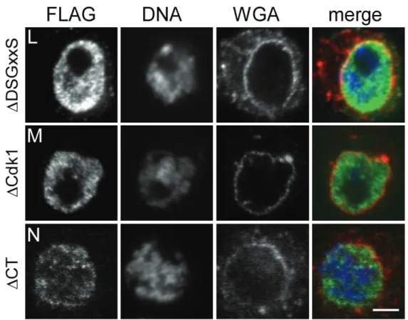
Rca1 is a nuclear protein, but the APC/C activiator Cdh1Fzr and the APC/C substrates, mitotic cyclins, accumulate in the cytoplasm during interphase (Grosskortenhaus and Sprenger, 2002). How Rca1 can act on Cdh1Fzr despite their spatial separation still remains unclear. Moreover, it has to be elucidated if nuclear accumulation is a prerequisite to inhibit APC/C-Cdh1Fzr activity during G2 or to promote the G1-S transition. A first step to address these questions is the identification of the protein domains that are responsible for targeting Rca1 to the nucleus. The Rca1 protein contains several motifs that are highly conserved throughout the Rca1/Emi1 family (Figure 3.1). A structure/function analysis of Rca1 revealed the relevance of each protein motif for its functions: First, as an APC/C- Cdh1Fzr inhibitor during G2 of Drososphila embryogenesis and second, as a G1-S regulator that can induce ectopic S phases resulting in a rough eye (results are summarised in Figure 3.1; Radermacher, 2007; Zielke et al., 2006; Zielke, 2007). The C-terminus harbours a zinc-binding region (ZBR) which is essential for both functions during G1 and G2.
Furthermore, a N-terminal truncations up to amino acid 255 result in a nonfunctional protein (Zielke et al., 2006; Zielke, 2007). The central part of Rca1 harbours a minimal D- box, a KEN-box and the DSGxxS degron which mediates Emi1, but not Rca1 degradation (Guardavaccaro et al., 2003; Margottin-Goguet et al., 2003; Zielke, 2007). Each of these protein motifs and all Cdk1 phosphorylation sites have been shown to be dispensable for Rca1 function (Zielke et al., 2006; Zielke, 2007). Deletion of amino acid 203-255 resulted in a protein which was not able to restrain APC/C-Cdh1Fzr activity during G2 of embryogenesis. However, it was still capable to induce S phases (Radermacher, 2007).
Deletion of the F-box alone or N-terminal truncations including the F-box cannot induce S