DISSERTATION
ZUR ERLANGUNG DES DOKTORGRADES DER NATURWISSENSCHAFTEN (DR. RER. NAT.)
DER FAKULTÄT FÜR BIOLOGIE UND VORKLINISCHE MEDIZIN
DER UNIVERSITÄT REGENSBURG
vorgelegt von
Matthias Kies
aus Eschenbach i. d. OPf.
im Jahr
2017
Das Promotionsgesuch wurde eingereicht am:
05.05.2017
Die Arbeit wurde angeleitet von:
Prof. Dr. Frank Sprenger
Unterschrift:
Für meine Eltern
Table of contents
1 Abstract ...9
2 Introduction ... 10
2.1 The eukaryotic cell cycle ... 10
2.2 Cell cycle regulation during Drosophila embryogenesis ... 11
2.3 Cyclin‐dependent kinases (Cdks) drive cell cycle progression ... 12
2.4 Checkpoints monitor cell cycle progression ... 14
2.5 Regulation of the G1‐S transition ... 14
2.6 Entry into mitosis ... 16
2.7 The ubiquitin proteasome pathway mediates degradation of cell cycle regulators ... 17
2.8 The Anaphase Promoting Complex/Cyclosome (APC/C) ... 20
2.9 The SCF complex ... 23
2.10 Cyclin‐dependent kinase inhibitors ... 25
2.11 The APC/C inhibitor Emi1 ... 27
2.12 Rca1, the Drosophila homologue of Emi1 ... 29
3 Results ... 33
3.1 Identification of Rca1 interaction partners ... 33
3.1.1 Aim ... 33
3.1.2 Rca1 is in complex with SCF components, Skp2, and Dap ... 33
3.1.3 Rca1 interacts F‐box dependent with polyubiquitinated substrates ... 37
3.2 Biochemical analysis of the interaction between Rca1 and Dap ... 38
3.2.1 Aim ... 38
3.2.2 Fusion of UBA domains to Rca1 does not stimulate binding of polyubiquitinated Dap ... 39
3.2.3 SCF‐Rca1 complex is not required for recruitment of Dap to Rca1 ... 40
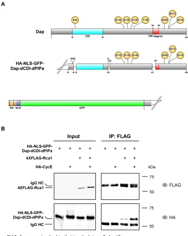
3.2.4 CycE/Cdk2 activity increases Rca1 binding to Dap ... 42
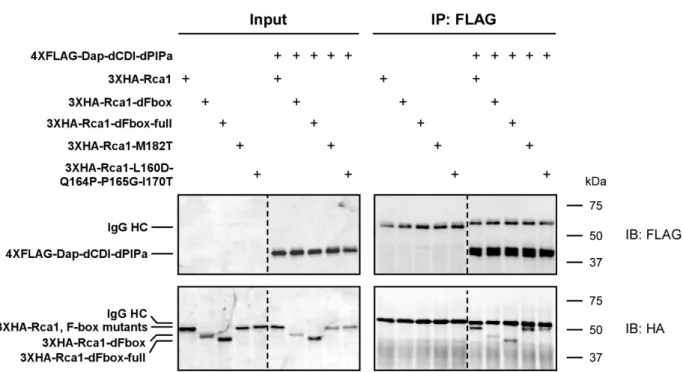
3.2.5 S/T‐P Cdk phosphorylation sites in Dap (S205, S214) are not required for Rca1 binding ... 44
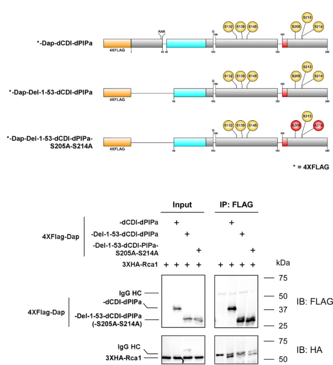
3.2.6 N‐terminal half of Dap is required but not sufficient to mediate interaction with Rca1 ... 47
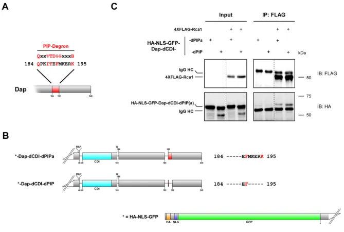
3.2.7 PIP‐degron in Dap is not required for interaction with Rca1 ... 49
3.2.8 The RXXL sequence in Dap is not a degron motif but mutations in this sequence reduces Rca1 binding... 50
3.2.9 Central region in Rca1 is required for Dap binding... 52
3.3 In vivo analysis of APC/C‐Fzr activity regulation by Rca1 and Dap ... 53
3.3.1 Aim ... 53
3.3.2 APC/C‐Fzr activity is inhibited by Rca1 and does not depend on Dap ... 54
3.3.3 Rca1 requires both ZBR and F‐box for efficient APC/C‐Fzr inhibition ... 56
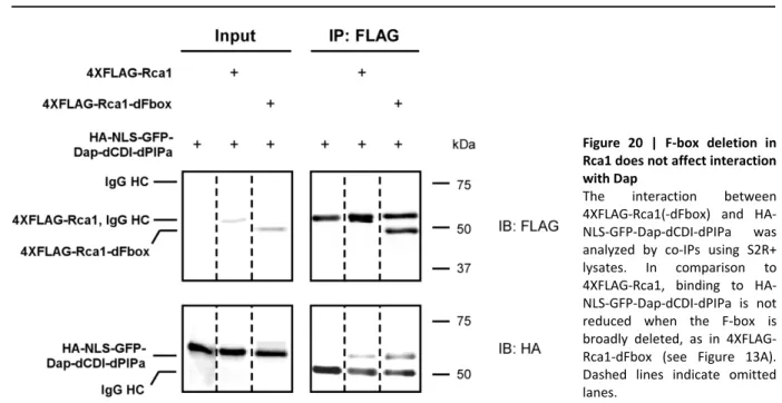
3.3.4 Loss in APC/C‐Fzr inhibition by F‐box deficient Rca1 is restored by downregulation of Dap activity ... 60
3.4 Protein stability analyses of potential SCF‐Rca1 target substrates by flow cytometry ... 62
3.4.1 Aim ... 62
3.4.2 Method for analysis of protein stability by flow cytometry ... 63
3.4.3 Analysis of Dap stability ... 67
3.4.3.1 PIP‐degron mutations result in Dap stabilization ... 67
3.4.3.2 N‐terminal PIP‐degron deletion (dPIPa) is sufficient for inactivation of the PIP‐degron .. 69
3.4.3.3 Mutation of the RXXL sequence stabilizes Dap ... 70
3.4.3.4 Dap is destabilized by Rca1 in an F‐box dependent manner ... 73
3.4.3.5 CycE/Cdk2 and Rca1 function synergistically to drive Dap degradation ... 74
3.4.3.6 A functional ZBR is required for stimulated Dap destabilization by CycE/Cdk2 ... 76
3.4.3.7 S/T‐P Cdk phosphorylation sites in Dap (S205, S214) are not required for its degradation ... 77
3.4.3.8 N‐terminal region in Dap is required for its degradation ... 78
3.4.4 Analysis of Skp2 stability ... 78
3.4.4.1 CycE/Cdk2 stimulates degradation of Skp2 ... 78
3.4.4.2 Skp2 stability does not depend on Rca1 ... 79
3.5 Establishment of an in vitro APC/C‐Fzr activity assay ... 80
3.5.1 Aim ... 80
3.5.2 Purification of 6XHIS‐TEV‐FLAG‐Fzr by baculovirus‐infected SF21 cells ... 81
3.5.3 Cdc16 precipitate from a stable S2R+ cell line provides APC/C‐Fzr activity when supplemented with in vitro translated 4XFLAG‐Fzr ... 82
4 Discussion ... 85
4.1 Establishment of a method for protein stability analyses during cell cycle progression ... 85
4.2 CRL4‐Cdt2 targets Dap for degradation ... 86
4.3 SCF‐Rca1 targets Dap for degradation ... 88
4.4 SCF‐Rca1 contributes to APC/C‐Fzr inhibition in G2‐phase ... 92
4.5 Dual regulation of APC/C‐Fzr inhibition by Rca1 ... 93
4.6 Establishment of an in vitro APC/C assay ... 96
4.7 Rca1 interacts with Skp2 and a broad range of further cell cycle proteins ... 97
5 Material ... 99
5.1 Chemicals ... 99
5.2 Kits 100 5.3 Proteins/Enzymes ... 101
5.4 Oligonucleotides ... 101
5.5 cDNA clones ... 101
5.6 Plasmids ... 102
5.7 Bacterial strains ... 106
5.8 Eukaryotic cell lines ... 106
5.9 Antibodies ... 107
5.9.1 Primary antibodies ... 107
5.9.2 Secondary antibodies ... 107
5.10 Solutions and buffers ... 107
5.11 Media and agar plates ... 113
5.12 Consumable material ... 114
5.13 Software ... 115
5.14 Equipment ... 115
6 Methods ... 118
6.1 DNA/RNA methods ... 118
6.1.1 Molecular cloning ... 118
6.1.2 DNA amplification by PCR ... 119
6.1.3 Agarose gel electrophoresis ... 119
6.1.4 Transformation of electrocompetent cells ... 120
6.1.5 Transformation of chemically competent cells ... 120
6.1.6 Preparation of E. coli cultures ... 120
6.1.7 Isolation of DNA ... 121
6.1.7.1 Mini scale isolation of plasmid DNA ... 121
6.1.7.2 Midi scale isolation of plasmid DNA ... 121
6.1.7.3 Preparative isolation of DNA fragments from agarose gels ... 122
6.1.7.4 Isolation of DNA fragments after PCR ... 122
6.1.8 Quantification of DNA ... 122
6.1.8.1 Photometric quantification of purified DNA ... 122
6.1.8.2 Quantification of DNA/RNA by gel analysis ... 122
6.1.9 Restriction digestion of DNA ... 123
6.1.10 Dephosphorylation of DNA‐ends ... 123
6.1.11 Blunting of DNA‐ends ... 123
6.1.12 Ligation of DNA fragments ... 124
6.1.13 Production of dsRNA for RNA interference ... 124
6.2 Protein methods ... 125
6.2.1 Analysis of interaction partners ... 125
6.2.1.1 Identification of protein interaction partners by mass spectrometry ... 125
6.2.1.2 Analysis of protein interaction partners by Co‐Immunoprecipitation ... 127
6.2.2 SDS‐PAGE ... 128
6.2.3 Coomassie staining of polyacrylamide gels ... 129
6.2.4 Western blotting ... 129
6.2.5 Antibody staining of Western blots ... 129
6.2.6 In vitro synthesis of proteins ... 129
6.2.7 Purification of heterologous expressed proteins by affinity chromatography ... 130
6.2.8 Dialysis of purified proteins ... 131
6.2.9 Concentration of protein samples ... 131
6.2.10 Storage of purified proteins ... 131
6.2.11 In vitro ubiquitination assay ... 131
6.2.12 Drosophila S2R+ cells ... 133
6.2.12.1 Production of stable S2R+ cell lines ... 133
6.2.12.2 Culturing of S2R+ Drosophila cells ... 133
6.2.12.3 Freezing of S2R+ cell stocks ... 133
6.2.12.4 Thawing of frozen S2R+ cell stocks ... 134
6.2.12.5 Splitting of cells ... 134
6.2.12.6 Seeding of cells ... 134
6.2.12.7 Determination of cell concentration ... 134
6.2.12.8 Transfection of cells ... 135
6.2.12.9 Harvesting of cells ... 135
6.2.12.10 Silencing of genes by RNA interference ... 135
6.2.12.11 In vivo APC/C‐Fzr activity assay ... 136
6.2.12.12 Preparation of cell extract ... 136
6.2.13 Spodoptera frugiperda SF21 cells ... 137
6.2.13.1 Maintenance of insect cells ... 137
6.2.13.2 Freezing of SF21 cell stocks ... 137
6.2.13.3 Thawing of frozen SF21 cell stocks ... 137
6.2.13.4 Counting of SF21 cells ... 137
6.2.13.5 Expression of eukaryotic proteins in baculovirus‐infected SF21 cells ... 137
6.3 Flow cytometry of S2R+ Drosophila cells ... 141
6.3.1 Preparation of living S2R+ Drosophila cells ... 141
6.3.2 Measurement procedure ... 141
6.3.3 Analysis of data ... 141
7 Abbreviations ... 143
8 Single and three letter code for amino acids ... 146
9 List of figures ... 147
10 List of tables ... 150
11 References ... 152
12 Supplement ... 164
12.1 Identification of Rca1 interaction partners by mass spectrometry ... 164
12.2 Identification of Dap interaction partners by mass spectrometry ... 173
13 Zusammenfassung ... 179
14 Danksagung ... 180
15 Eidesstattliche Erklärung ... 181
1 Abstract
The Anaphase‐Promoting Complex/Cyclosome (APC/C) drives degradation of a large variety of mitotic cell cycle regulators. Therefore, tight regulation of APC/C activity plays a central role in proper cell cycle progression. Suppression of APC/C activity during G2‐phase is crucial for entry into mitosis. This is achieved 1) by Cdk‐dependent phosphorylation of the APC/C activator subunit Fzr, which renders Fzr unable to interact with the APC/C, and 2) by direct inhibition of APC/C‐Fzr by Regulator of Cyclin A1 (Rca1). Previous studies suggest that not only the C‐terminal part, but also the central localized F‐box in Rca1 is fundamental for APC/C‐Fzr inhibition. F‐box proteins usually recruit substrates to SCF complexes, causing their polyubiquitination and subsequent degradation. To identify such substrates, an Rca1 precipitate from S2R+ cells was analyzed by mass spectrometry. Rca1 was found in complex with SCF components and other cell cycle regulators including Dacapo (Dap) and Skp2. Rca1 binds polyubiquitinated substrates in an F‐box dependent manner in vivo giving evidence for a functional SCF‐Rca1 complex. To analyze, whether SCF‐
Rca1 affects the protein stability of Dap and Skp2, a method for the stability analysis of GFP‐labeled proteins during cell cycle progression of S2R+ cells was established. Dap stability was analyzed using a cell cycle inactive GFP‐tagged version of Dap that lacks destabilization by CRL4‐Cdt2 during S‐phase (GFP‐Dap‐
dCDI‐dPIPa). Downregulation of Rca1 activity by RNAi resulted in upregulation of GFP‐Dap‐dCDI‐dPIPa protein levels. Conversely, overexpression of Rca1 stimulated degradation of GFP‐Dap‐dCDI‐dPIPa in an F‐box dependent manner. Overexpression of CycE enhanced the interaction between Rca1 and Dap‐dCDI‐
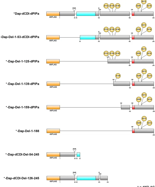
dPIPa, and further destabilized GFP‐Dap‐dCDI‐dPIPa. These studies indicate that SCF‐Rca1 functions synergistically with CycE/Cdk2 to target Dap for degradation. Two candidate Cdk phosphorylation sites in Dap, S205 and S214, were mutated but these mutations did neither affect the stability nor did they impair the interaction with Rca1. Interaction analyses with different truncated Rca1 and Dap constructs have revealed that the N‐terminal part of Dap and the central region in Rca1 are required for their interaction.
Loss of Rca1 binding by deletion of N‐terminal residues in GFP‐Dap‐dCDI‐dPIPa resulted in its complete stabilization. Flow cytometric based in vivo APC/C‐Fzr activity assays in S2R+ cells were used to analyze the functional interaction between SCF‐Rca1 and Dap. Loss of the F‐box reduced APC/C‐Fzr inhibition.
However, this defect in APC/C‐Fzr inhibition was suppressed by RNAi‐mediated downregulation of Dap activity. Taken together, these studies provide a model, in which SCF‐Rca1 targets Dap for degradation to release CycE/Cdk2 activity for APC/C‐Fzr inhibition. To analyze the biochemical inhibition of APC/C‐Fzr by Rca1 in more detail, an in vitro APC/C‐Fzr ubiquitination assay was further developed. Experiments in the past failed to purify active Drosophila APC/C‐Fzr from transgenic embryos. Therefore, a stable S2R+ cell line expressing a GFP‐tagged APC/C component, Cdc16‐MYC‐TEV‐GFP, was established. In addition, Fzr was in vitro translated in reticulocyte lysate and purified from baculovirus‐infected SF‐21 cells. APC/C‐Fzr‐
dependent ubiquitination activity was detected to some extend by using Cdc16 precipitate treated with in
vitro translated Fzr.
2 Introduction
2.1 The eukaryotic cell cycle
Each organism on earth either consists of a single cell or a body that is composed of a vast variety of highly specialized cell types. Reproduction of cells constitutes a fundamental aspect in each organism’s life. In unicellular organisms, cell division results in an entire new organism, whereas in multicellular organisms, cell divisions fulfill several functional aspects, such as sexual reproduction, growth, development and maintenance of life. During embryonic development, a high number of cells arises from a single founder cell through cell divisions, thereby forming differentiated cell communities used for the formation of tissues and organs. After completion of development, cell divisions are required for replacement of injured and aged cells. The process of cell division is highly regulated and evolutionarily conserved from unicellular organisms, such as yeast, to higher multicellular organisms, such as humans. A highly precisely regulated cell machinery, known as the cell cycle ensures that events during cell division occur in a timely ordered fashion. In eukaryotes, the typical cell cycle is divided into four distinct phases: G1‐, S‐, G2‐ and M‐phase (see Figure 1).
During S‐phase, the genetic material is replicated resulting in chromosomes that consist of two identical sister chromatids. Equal distribution of the replicated sister chromatids into two new daughter cells occurs during the following M‐phase. Two gap phases, G1‐ and G2‐phase, separate S‐ and M‐phases for growth and preparation. During G1‐phase, the first phase of the cell cycle, the cell grows and prepares for the following S‐phase by synthesizing substrates required for DNA synthesis. However, when growth factors and nutrients are low, the cell exits the cell cycle and enters the G0 phase, a quiescent stage, which keeps the cell in a non‐proliferative state. After completion of S‐phase, the G2‐phase follows, where cell growth continues and synthesis of proteins required for entry into M‐phase takes place. The first three phases of the cell cycle (G1‐, S‐ and G2‐phase) together are known as interphase and describe the period between
Figure 1 | The standard eukaryotic cell cycle The standard eukaryotic cell cycle consists of four distinct phases: G1‐, S‐, G2‐ and M‐phase. During S‐phase DNA replication occurs, whereas a series of distinct processes during M‐phase promote the formation of two genetically identical daughter cells. The M‐phase is comprised of mitosis (nuclear division) and cytokinesis (cell division). The two gap phases (G1‐ and G2‐phase) prepare the cell for the upcoming phase. Cell growth takes place during interphase, which includes G1‐, S‐ and G2‐phase. Cell cycle progression is governed by three major transitions that function in a switch‐like manner.
The G1/S transition initiates entry into S‐phase, the G2/M transition regulates entry into mitosis and the metaphase‐to‐anaphase transition controls segregation of the sister chromatids into the new daughter cells and stimulates exit from mitosis (1, 2).
the M‐phase of successive cell cycles. The M‐phase is composed of mitosis and cytokinesis. Mitosis leads to the division of the nucleus and is regulated by five consecutive stages (prophase, prometaphase, metaphase, anaphase and telophase). In prophase, the first stage of mitosis, the sister chromatids are prepared for their segregation by condensation of the chromatin. Furthermore, the two centrosomes separate and migrate to opposite sides of the nucleus. The separated centrosomes initiate the assembly of the mitotic spindle required for sister chromatid segregation. During the next phase, the prometaphase, the nuclear envelope breaks down, the sister chromatids become attached to the mitotic spindle and their migration to the central region of the mitotic spindle (metaphase plate) begins. In metaphase, all sister chromatids are attached to the mitotic spindle and aligned at the metaphase plate. During anaphase, the mitotic spindle promotes the segregation of the sister chromatids to the opposite poles of the cell. The final stage of mitosis occurs during telophase, when the mitotic spindle is disassembled, the single‐
chromatid chromosomes become decondensed and the nuclear envelope is reformed around the chromosomes. After mitosis, cell division is completed by cytokinesis, which physically divides the cytoplasm of the proliferating cell into two daughter cells sharing the same genetic information (1).
2.2 Cell cycle regulation during Drosophila embryogenesis
Different types of cell cycle regulation are established during development of the Drosophila embryo. After fertilization, embryogenesis begins with 13 rapid synchronous nuclear divisions that occur in a common cytoplasm called a syncytium. These divisions cycles lack gap phases and consist only of alternating S‐ and M‐phases (10, 11). The first eight divisions take place in the central portion of the egg. After that, most of the nuclei begin to migrate to the surface as they continue to divide. Some nuclei remain in the interior of the embryo to differentiate into yolk nuclei. After two further divisions (cycles 8 – 10) in synchrony with the migrating nuclei, yolk nuclei cease dividing and become polyploid (12). After nine divisions, a few nuclei move to the posterior pole of the embryo, where they form separate pole cells, the precursors of the germ cells, during division cycle 10. Meanwhile, the remaining migrating nuclei are evenly distributed in a monolayer under the cell membrane of the embryo, where they execute four further divisions (cycles 10 – 13) to form a syncytial blastoderm. During cell cycle 14, cellularization is initiated by the formation of a plasma membrane between all of the peripheral nuclei. The formed cells are progenitors of future somatic tissues. Following cellularization, gastrulation movement begins (10). From now on, maternal components are exhausted and cell cycle progression requires active transcription (13, 14). The following cell divisions (cycles 14 – 16) are no longer synchronous. Instead, for the first time, a G2‐phase is established during cell cycle 14 and entry into mitosis occurs in a spatiotemporal pattern of 25 mitotic domains (15). The duration of the G2 phase, in which cells of each domain reside, depends on the zygotic expression of String (Stg).
Stg belongs to the family of Cdc25 phosphatases, which trigger mitotic entry by immediate activation of
mitotic Cdk1 (16‐18). Following mitosis 16, the first G1 phase is established in cell cylce 17. Most of the
epidermal cells exit the cell cycle and arrest in G1 until end of embryogenesis (18). Only cells of the
developing nervous system enter S‐phase and continue to divide (19). Other cell types, including dorsal
cells as well as cells forming internal organs such as the gut and salivary glands switch their mitotic cycles
to endoreplication cycles (20). Endoreplication cycles are special cell cycle variants, in which cells undergo
continuing rounds of DNA replication consisting only of G‐ and S‐phases, thereby becoming polyploid (21).
2.3 Cyclin‐dependent kinases (Cdks) drive cell cycle progression
The cell cycle is a complex machinery that requires strict regulation to ensure that specific events during cell cycle progression are triggered in a timely ordered fashion. Three switch‐like transitions are distributed throughout the cell cycle that are precisely controlled by several cell cycle regulators (2). These transitions are primarily controlled by cyclin‐dependent kinases (Cdks) that function as key regulators during cell division (1, 2, 4‐6). Cdks are serine/threonine kinases that primarily phosphorylate proteins on the minimal consensus motif S/T‐P, with the optimal sequence being S/T‐P‐X‐R/K (6). An alternative phosphorylation motif is the so‐called non‐S/T‐P motif consisting of the minimal consensus sequence S/T‐X‐X‐R/K (22). Cdks are inactive alone and require binding of regulatory subunits, known as cyclins, to become active (1).
However, cyclin binding result only in partial activation of Cdks. For complete activity, Cdks must be in a particular phosphorylation state. This is achieved by Cdk‐activating kinases (CAKs) that stimulate Cdk activity by phosphorylation of a threonine residue adjacent to the kinase active site (T161). Furthermore, members of the Wee1 kinase family inhibit activated cyclin‐bound Cdk complexes through phosphorylation of specific tyrosine and threonine residues (Tyr15 in all Cdks, Tyr15 and Thr14 in Cdks of higher eukaryotes). Cdk activity is regained by phosphatases of the Cdc25 family that remove the inhibitory phosphate groups (1, 2).
Beside activation, cyclins also confer substrate specificity to the bound Cdks. This is well characterized for E‐ and A‐type cyclins, which contain a hydrophobic patch that mediates binding to specific substrates through interaction with the cy motif consisting of the short sequence R‐X‐L (6). In contrast, D‐type cyclins possess a L‐X‐C‐X‐E motif that is required for association with members of the pocket protein family (23, 24). Recruitment of target substrates with several Cdk phosphorylation sites can be stimulated by cyclin‐
dependent kinase subunits (Cks). Cks are highly conserved small adaptor proteins that interact with Cyc/Cdk complexes. Due to a phosphate anion‐binding site, Cks proteins are additionally able to associate with phosphorylated residues, thereby allowing multi‐phosphorylation of already phosphorylated target substrates (25). Mammals possess two Cks proteins, Cks1 and Cks2 (26), whereas in Drosophila Cks function is provided by the corresponding homologues Cks85A and Cks30A (27, 28). Another control of substrate recognition is achieved by subcellular localization of cyclin‐bound Cdk complexes (6). For instance, E‐type and A‐type cyclins activate Cdks in the nucleus (29‐31), while CycB1, a B‐type cyclin, traffics between the cytoplasm and the nucleus to stimulate phosphorylation of substrates in both compartments (30‐32) .
Cdk activity plays a central role in three major transitions that are distributed throughout the cell cycle (see Figure 1). These transitions behave in a switch‐like manner and are based on all‐or‐none responses that induce an irreversible series of events when a certain threshold of Cdk activity is reached. The decision to enter a new division cycle is made at the restriction point (mammals) or Start (yeast) in late G1‐phase.
Once a cell passes this “point of no return”, it is committed to a new round of the cell cycle and mitogenic stimuli are no longer required to drive progression through the cell cycle (33). Following the restriction point/Start, an increase in Cdk activity stimulates the G1/S transition (2, 34, 35). Upon DNA replication, a rapid release of high Cdk activity triggers the transition from G2‐ to M‐phase by which mitotic processes are initiated (1, 2). Conversely, initiation of the metaphase‐to‐anaphase transition, which promotes exit from mitosis is induced by Cdk inactivation (1, 2).
In contrast to Cdks that are present throughout the cell cycle, different types of cyclins are synthesized
and destroyed during specific cell cycle stages, resulting in periodic oscillations of active Cyclin/Cdk
complexes (1, 2, 4‐6). In yeast, cell division cycles are driven by a single Cdk, Cdc28 (Schizosaccharomyces pombe)/Cdc2 (Saccharomyces cerevisiae), and different cyclins that are expressed at specific cell cycle stages (1). During evolution, the number of Cdks and cyclins increased, resulting in a large variety of different Cyc/Cdk complexes (4). In mammals, three interphase Cdks (Cdk2, Cdk4 and Cdk6), a mitotic Cdk (Cdk1) and ten cyclins that belong to four different classes (D‐type: CycD1, D2 and D3; E‐type: CycE1, E2;
A‐type: CycA1, A2 and B‐type: CycB1, B2 and B3) are directly involved in cell cycle progression (1, 2, 4‐6).
The different cyclin classes that contribute to cell divisions are well conserved among eukaryotes, only the number of cyclin subtypes varies. For instance, Drosophila possesses only one version of the cyclin types D, E and A (CycD, CycE and CycA), and two versions of B‐type cyclins (CycB and CycB3) (1). According to the classical model of the mammalian cell cycle, specific Cyclin/Cdk complexes function during distinct stages of the cell cycle in order to trigger various events in their sequential order (see Figure 2).
Upon stimulation by growth factors, Cdk4 and Cdk6 are activated by D‐type cyclins and play an important role for progression through G1‐phase (36). Entry into S‐phase is stimulated by CycE/Cdk2 activity, while continuation and completion of S‐phase depends on CycA/Cdk2 activity. Both CycA and CycB complexed with Cdk1 are required for progression through mitosis (1, 2, 4‐6). Although this model of cell cycle regulation is widely distributed and accepted, studies with Cdk‐knockout mice demonstrated that cell cycle progression in most cell types does not require the interphase Cdks, Cdk2, Cdk4 and Cdk6. Only highly specialized cell types depend on these Cdk types (2, 4). Cdk2‐null mice are viable and does not reveal any cell cycle defects in proliferating cells (37, 38). Even triple knockout embryos for cdk2, cdk4 and cdk6 developed normally until mid‐gestation, but subsequently died due to heart and haematopoietic defects.
In contrast, embryos lacking Cdk1 activity were not able to perform any cell divisions (39). Thus, Cdk activity provided by the mitotic Cdk, Cdk1, is essential and sufficient to drive the mammalian cell cycle.
Furthermore, studies with cyclin‐knockout mice showed that D‐type and E‐type cyclins are dispensable for cell cycle progression (40, 41). Only loss of CycA2 or CycB1, respectively, resulted in lethality (42, 43). These studies suggest that only Cdk1, together with CycA2 and CycB1, is required for triggering events involved in cell division. Given that expression of both CycA and CycB steadily rise during interphase (1), together with the fact that a single cyclin is able to promote cell cycle progression of fission yeast (44), a minimal threshold level was established, in which the level of Cdk activity controls the entry into different cell cycle stages (see Figure 3).
Figure 2 | Classical model of mammalian cell cycle regulation by specific Cyc/Cdk complexes
Level of cyclin accumulation controls the activity of specific Cyc/Cdk complexes during cell cycle progression. Upon mitogenic stimulation, Cdk4 and Cdk6 complexed with D‐type cyclins regulate progression through G1‐phase (not shown) (3).
CycE/Cdk2 activity stimulates entry into S‐phase.
CycA/Cdk2 plays an important role in S‐phase progression. Finally, onset of mitosis requires both CycA/Cdk1 and CycB/Cdk1 activity (2, 4‐6).
Low Cdk1 activity is sufficient for initiation of S‐phase, whereby an increase in Cdk1 activity during S‐ and G2‐phase stimulates entry into mitosis. Threshold for initiation of S‐phase is achieved by low levels of CycA, while accumlation of CycA and CycB during interphase is necessary to trigger entry into mitosis. The requirement of only Cdk1 for proper cell cycle progression indicates that the basic principles in cell cycle regulation are highly conserved from yeast to higher eukaryotes.
2.4 Checkpoints monitor cell cycle progression
Proper cell cycle progression by Cyc/Cdk complexes is essential for the maintenance of life, as failure in cell cycle regulation may cause severe defects including unscheduled proliferation, genomic instability and chromosomal instability, leading in the worst case to death. Therefore, critical processes during progression through the cell cycle are monitored by checkpoints. These checkpoints control whether cell cycle events have been completed successfully without any complications before progression into the next cell cycle phase takes place. When this is not the case, and inconvenient conditions prevent appropriate cell divisions, the corresponding checkpoint is activated. Activation of checkpoints triggers arrest of the cell cycle through induction of several signal transduction pathways that modulate Cdk activity.
Progression through the cell cycle is halted until all failures compromising genomic integrity are completely eliminated (4).
The DNA damage checkpoint protects the cell from endogenous (reactive cellular metabolites) and environmental factors (ionizing or ultraviolet radiation, chemicals and drugs) that affect the integrity of the genomic DNA. Whenever unreplicated or damaged DNA is detected, signaling pathways pause cell cycle progression, thereby giving the cell time to repair the damage before cell division processes are resumed. Cell cycle arrests due to DNA damage occur during G1‐ or G2‐phase (4, 45). Upon successful DNA replication, the spindle assembly checkpoint that functions at the metaphase‐to‐anaphase transition ensures proper distribution of sister chromatids during mitosis. Only when all chromosomes are bipolarly attached to the mitotic spindle during metaphase, processes resulting in chromosome segregation are initiated. This includes the downregulation of Cdk activity, which contributes to the elimination of sister chromatid cohesion, thereby allowing the migration of sister chromatids to opposite poles of the cell. As long as the chromosomes are not correctly aligned to the mitotic spindle, Cdk activity is sustained by preventing CycB degradation (4).
2.5 Regulation of the G1‐S transition
The G1/S transition is characterized by a switch‐like transcriptional activation of G1/S genes that drive
Figure 3 | Minimal threshold model for general cell cycle regulation
Entry into S‐ or M‐phase, respectively, are triggered by different thresholds of Cdk1 activity.
Threshold required for S‐phase entry is accomplished by CycA or CycE complexed with Cdk1 or Cdk2. The M‐phase threshold is fulfilled by CycA and CycB in conjunction with Cdk1 (2).
family of transcription factors. E2F family members are divided into activator E2Fs (E2F1, E2F2 and E2F3a) stimulating expression of G1/S genes and repressor E2Fs (E2F3b, E2F4, E2F5, E2F6, E2F7, and E2F8) that inhibit the expression of the same G1/S genes (34). DNA binding to promoters of E2F target genes requires association with any member of the DP protein family (DP1 and DP2) (46), except E2F7 and E2F8, which bind DNA independently of the DP proteins (47, 48). During early G1‐phase, E2F transcription factors interact with members of the pocket protein family (RB, p107 and p130) to inhibit transcription (34).
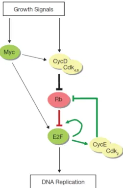
Activator E2F proteins are bound and inhibited by RB (49), while the E2F repressor proteins E2F4 and E2F5 associate with p107 and p130 to form transcriptional repressor complexes (50). In contrast, the repressor proteins E2F6, E2F7 and E2F8 do not require binding of pocket proteins to function as transcriptional repressors (47, 51, 52). An increase in Cdk activity during late G1‐phase triggers transcriptional activation of G1/S genes (34). Cdk activity is initially promoted by Cdk4 and Cdk6, which become activated by the accumulation of D‐type cyclins in response to growth factors (36). Phosphorylation of the pocket proteins by D‐type cyclin‐conjugated Cdk4/6 results in their release from the bound E2F transcription factors (9, 34). E2F repressors are replaced by E2F activator proteins, thereby inducing the transcription of G1/S genes, with CycE belonging to the first subset of G1/S proteins that is expressed (53). CycE activates Cdk2 and drives further phosphorylation of the pocket proteins (9, 34). Furthermore, E2F activator proteins stimulate their own transcription (54). By this, a positive feedback loop system is established that induces G1/S gene expression in a switch‐like manner (see Figure 4).
Upon transcriptional activation of G1/S genes, transcription is inactivated through several negative feedback loops. Expression of the E2F target gene cyclin A stimulates CycA/Cdk2 activity during S‐phase, resulting in inhibition of the activator E2F1 by phosphorylation (55). Both CycA/Cdk2 and CycE/Cdk2 mediate degradation of the cyclin‐dependent kinase inhibitor p27, thereby further enhancing Cdk2 activity (56, 57). Another negative feedback loop is established by the E2F target gene skp2 that stimulates formation of SCF‐Skp2, which targets E2F1 for degradation (58). Furthermore, accumulation of the E2F
Figure 4 | A bistable RB‐E2F switch regulates entry into S‐phase DNA replication is initiated by a feedback system that transforms growth signals into a bistable switch in response to growth signals. Here, a simplified version of the G1/S switch involving the core components RB and E2F is shown. During G1‐phase, E2F transcription factors are bound and inhibited by RB. Growth factors induce RB inactivation by CycD/Cdk4 and CycD/Cdk6, thereby releasing E2F activity that stimulates entry into S‐
phase through transcriptional activation of DNA replication genes. Positive feedback loops ensure rapid accumulation of G1/S gene products. E2F stimulates its own activity by directing its own transcription and the transcription of cycE, which results in further Cdk activity driving Rb inhibition (9).
targets E2F6, E2F7 and E2F8 during S‐phase contributes to inactivation of G1/S gene transcription (34).
The RB‐E2F pathway controlling the G1/S transition is functionally conserved among eukaryotes, but differs in its complexity. In Drosophila, G1/S gene transcription is regulated by two members of the E2F transcription family, E2F1 and E2F2, a single DP protein and two pocket proteins, RBF1 and RBF2 (59). E2F1 activates G1/S gene transcription, while E2F2 function as transcriptional repressor (60, 61).
As in the mammalian system, in Drosophila, entry into S‐phase requires Cdk activation. CycD/Cdk4 was identified as kinase able to provide Cdk activity during G1‐Phase. However, Cdk4 activity is not critical for triggering the switch into S‐phase, since loss of Cdk4 did not disturb proper cell cycle progression (62).
Entry into S‐phase rather depends on CycE/Cdk2 activity. Embryos mutant for cycE displayed a G1‐phase arrest, while ectopic overexpression of CycE induced entry into S‐phase (63, 64). Studies with mutant embryos have shown that in mitotically dividing cells, CycE stimulates E2F1 dependent transcription of DNA replication genes (65). CycE was also identified as an E2F1 target gene product in endocycling cells (66), suggesting that G1/S switch is also established in Drosophila. During S‐phase, CycE/Cdk2 stimulates its own inactivation by phosphorylation of CycE, which is recognized by SCF‐Ago for degradation (67, 68).
CycA plays an important role for S‐phase entry as well. Overexpression of CycA or loss of the CycA/Cdk1 inhibitor Roughex accelerated the G1/S transition (69, 70).
2.6 Entry into mitosis
Entry into mitosis is triggered by abrupt activation of Cdk1 at the end of G2‐phase (1). Studies in the mammalian system suggest that the release of high Cdk1 activity is achieved through a switch‐like feedback system that relies on the interplay of Cdk1 regulatory proteins (71). During G2‐phase, mitotic Cdk1 is kept in an inactive state by inhibitory phosphorylations on Tyr15 and Thr14. These phosphorylations are catalyzed by two kinases, Tyr15 by Wee1 (35) and both Tyr15 and Thr14 by Myt1 (72). Inhibition of both enzymes drives premature entry into mitosis (73). Before mitosis, members of the Cdc25 phosphatase family (Cdc25A, Cdc25B and Cdc25C) activate Cdk1 by removing the inhibitory phosphate groups (74). A switch‐like mechanism for Cdk1 activation is established by positive feedback loops that involve Cdk1‐mediated regulation of the Cdc25 proteins as well as the kinases Wee1 and Myt1 (see Figure 5).
Phosphorylation by Cdk1 activates Cdc25 phosphatases, but inhibits the kinase activity of Wee1 and Myt1.
Thus, once a small portion of Cdk1 becomes active, a sudden increase in Cdk1 activation follows, providing the Cdk1 activity required for robust progression through mitosis (1, 7, 71). Removal of inhibitory phosphate groups is regarded as initiator of the feedback system. Overexpression of a non‐
phosphorylatable version of Cdk1 (T14A and Y15F) caused rapid cell cycles with abortive mitoses (75).
Cdc25A is sufficient to trigger the switch into mitosis. Deletion of the other Cdc25 isoforms, Cdc25B and
Figure 5 | A molecular switch controls entry into mitosis
Inhibitory phosphorylations catalyzed by Wee1‐related kinases keep Cdk1 in an inactive state (Cyclin:Cdk1‐Y‐PO4) during G2‐phase. Before mitosis, dephosphorylation by Cdc25‐related phosphatases activate Cdk1 (Cyclin:Cdk1). Inhibition of Wee1 kinases and activation of Cdc25 phosphatases through phosphorylation by Cdk1 generates a positive feedback system that allows a switch‐like activation of Cdk1 (7).
activation of Cdc25 phosphatases to trigger the switch from a state of “no Cdk1 activity” to a state of “high Cdk1 activity” is not well understood. Studies in human culture cells point to CycA/Cdk2 as an essential component of such a trigger mechanism (1, 77). CycA2 raises during S‐phase (1, 2, 4‐6) and its downregulation resulted in a G2‐phase arrest due to inhibitory phosphorylation on CycB/Cdk1 (77).
Furthermore, CycA2/Cdk2 activity was not inhibited upon downregulation of CycB1 expression. Therefore, it is possible that CycA/Cdk2 initiates activation of Cdc25 phosphatases to trigger abrupt CycB/Cdk1 activation in late G2‐phase. In addition, CycB/Cdk1 promotes its activation by stimulating the activity of members of the Polo‐like kinase (Plk) family that support phosphorylation of Cdc25 phosphatases (1, 78, 79).
In Drosophila, CycA, B and B3 in conjunction with Cdk1 contribute to progression through mitosis (1, 80).
In vivo analyses of diverse mutant embryos have demonstrated the functional role of each cyclin. Embryos mutant for cycA were lethal due to a G2‐phase arrest before mitosis 16 during embryogenesis when maternal contributions are exhausted. Thus, CycA is essential for mitosis (81‐83). cycB mutant embryos were viable and displayed only minor defects in cell cycle progression, such as delay of mitosis or slightly abnormal mitotic spindles. Loss of CycB3 did not produce abnormalities during cell divisions. However, cycB cycB3 double mutants were lethal and showed defects in mitotic spindle assembly. Mutation of both cycA and cycB3 resulted in embryonic lethality with additional failures in chromosome condensation (80).
cycA cycB double mutant embryos even arrested before mitosis 15, a stage during which maternal supplied cyclin transcripts are still present (80, 81). Altogether, these studies indicate that all three cyclins cooperate to regulate progression through mitosis, with each cyclin possessing major functions during mitosis. CycA is mainly responsible for triggering mitosis, whereas CycB is particularly important for proper formation of the mitotic spindle. CycB3 plays a major role in chromosome condensation (80).
In Drosophila, two different Cdc25 homologues exists, String (Stg) and Twine. However, only Stg is responsible for Cdk1 activation in mitosis (17). Twine plays an important role in controlling Cdk1 activity in meiotic cells (84). As suggested for the mammalian system, activation of Stg plays also a critical role in triggering immediate entry into mitosis. During embryogenesis, when cells arrest in G2‐phase of cell cycle 14, Stg expression is required to drive cells into mitosis (16, 17).
2.7 The ubiquitin proteasome pathway mediates degradation of cell cycle regulators
Cell divisions are driven by the coordinated up‐ and downregulation of cell cycle regulatory proteins at
specific stages of the cell cycle. The presence of cell cycle proteins at non‐desirable time points during the
cell cycle might cause severe defects on the viability and functionality of the cell. In the worst case, the cell
cycle regulatory system becomes uncontrolled resulting in highly proliferating and genetically unstable
cells that finally give rise to cancer. Therefore, the existence of a system that regulates degradation of
regulatory proteins in a temporally and spatially specific manner constitutes an essential part in proper
cell cycle progression (85). The ubiquitin proteasome pathway mediates the degradation of specific
proteins and belongs to the major proteolytic mechanisms in eukaryotic cells. In this pathway, the
formation of specific polyubiquitin (polyUb) chains on target proteins label them for degradation by the
26S proteasome (see Figure 6). Ubiquitin (Ub) is a small protein of 76 residues and is highly conserved
among eukaryotes (86). The attachment of monoubiquitin on substrates, a process known as
ubiquitination, are catalyzed by a cascade of enzymatic reactions that involve the three enzymes E1
(ubiquitin‐activating enzyme), E2 (ubiquitin‐conjugating enzyme) and E3 (ubiquitin‐ligase). In these
reactions, an isopeptide bond between the carboxyl group of the C‐terminus of Ub (G76) and the ε‐amino group of a lysine residue within the substrate is formed. Repeated ubiquitination of the previously added Ub results in the formation of polyUb chains. Due to the presence of seven lysine residues in Ub, seven different types of lysine linkages (K6, K11, K27, K29, K33, K48, K63) can exist in polyUb chains. The lysine residue that is utilized for linkage determines the fate of the target substrate. PolyUb chains that consist of Ub linked by K6, K11, K29 and K48 target substrates for proteolytic destruction by the 26S proteasome.
However, K48‐conjugated polyUb chains are the primary signal for degradation (87). In contrast, K63 linkages are involved in non‐proteolytic processes, such as protein trafficking (88), DNA repair (89) and inflammation (90).
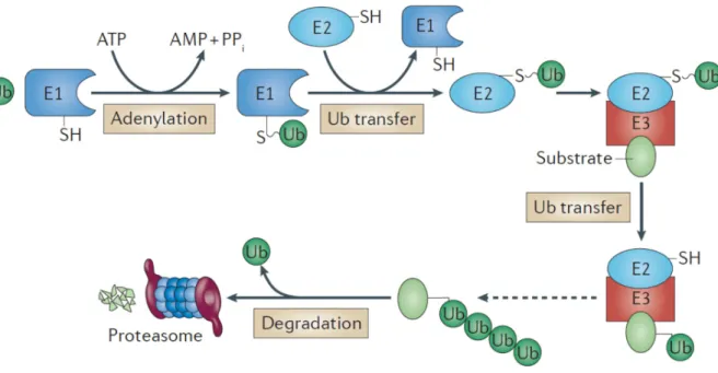
Figure 6 | The ubiquitin proteasome pathway
The ubiquitin proteasome pathway mediates degradation of proteins by the 26S proteasome. Polyubiquitin (polyUb) chains are assembled on target proteins by the subsequent action of three enzymes: E1 (ubiquitin‐activating enzyme), E2 (ubiquitin‐
conjugating enzyme) and E3 (ubiquitin‐ligase). First, the E1 catalyzes the formation of an Ubiquitin (Ub) adenylate intermediate (adenylation), which is then transferred to the catalytical cysteine in E1. This creates a reactive thioester bond between E1 and the C‐terminus of Ub, thereby releasing AMP and pyrophosphate (PPi). Next, the activated Ub is transferred to a cysteine residue in E2. Finally, E3 that is associated with a target substrate catalyzes the transfer of the E2‐bound Ub to a lysine residue in the substrate. Multiple cycles of Ub transfers to the previously attached Ub (preferentially through K48‐linkages) leads to the formation of polyUb chains (dashed arrow), which are recognized by the 26S proteasome for degradation. The tilde (~) represents a high‐energy bond (91).
The E1 enzyme catalyzes the initial step of the ubiquitination process, the ATP‐dependent activation of Ub.
This is achieved by two subsequent reactions. First, the E1 binds ATP and Ub and catalyzes the formation
of an Ub adenylate via adenylation of its carboxy‐terminal glycine residue, thereby releasing
pyrophosphate. Next, the catalytic cysteine in E1 attacks the Ub adenylate, resulting in a covalent E1∼Ub
thioester and release of AMP. The high‐energy thioester bond (∼ prepares Ub for further transfer
reactions (92, 93). The number of E1 enzymes that function in Ub activation depends on the organism. For
instance, in human, two E1 enzymes, Ube1 and Ube6, were identified. In Drosophila, Uba1 is the sole E1
enzyme (93).
The second step of the ubiquitination cascade is accomplished by the transfer of the activated Ub in E1∼Ub to an E2 enzyme. Upon E1∼Ub binding, a catalytic cysteine in E2 attacks the E1∼Ub thioester in a transthioesterification reaction, resulting in E2∼Ub, in which Ub is again covalently linked via a high‐energy thioester bond (94). In contrast to E1 enzymes, E2 enzymes comprise a large family that is defined by a conserved core ubiquitin conjugation (UBC) domain containing the catalytic cysteine (95). In human, 37 members of the E2 family are present, whereas Drosophila possesses 32 different E2 proteins (96).
The final step in the ubiquitination process requires the action of an E3 enzyme, which catalyzes the transfer of the active Ub from a specific E2∼Ub complex to a lysine residue within a specific target substrate. By this, an isopeptide bond is formed between the Ub C‐terminus and the ε‐amino lysine of the
substrate. Repeated ubiquitination of a lysine residue of the previously added Ub leads to the formation of a polyUb conjugated substrate. All E3 enzymes belong to three classes according to the domain structure they contain: (1) HECT (homologous to to the E6AP carboxyl terminus) domain containing E3s, (2) RING (really interesting new gene) and (3) RBR (RING‐between‐RING) domain containing E3s (97).The E3 families differ in the mechanism of Ub transfer. RING E3s transfer the Ub directly to the substrate by binding E2∼Ub and the target substrate simultaneously (98, 99). The RING domain associates with the E2‐Ub complex and stimulates the Ub transfer. Ring E3s are further subdivided into single‐subunit and
multi‐subunit E3 ligases (100). In contrast to RING E3s, HECT E3s ubiquitinate their substrates in a two‐step
reaction. After binding of E2∼Ub via the HECT domain, an E3∼Ub thioester intermediate on a catalytic cysteine residue within the HECT domain is first established, before the Ub is finally transferred to the target substrate (101). RBR E3s share features with both RING and HECT E3 ligases, using a RING‐HECT hybrid mechanism for ubiquitination. RBR domain consists of two RING domains, RING1 and RING2, which are separated by a conserved IBR (in‐between‐ring) domain. RING1 functions as interaction site for E2∼Ub, while RING2 contains the catalytic cysteine residue for the formation of an E3∼Ub thioester intermediate (102). Since all E3 enzymes contain individual domains for substrate recognition, E3s are primarily involvedin the selection of their target substrates. The mammalian genome encodes for more than 600 E3s to cover the ubiquitination of the vast variety of target substrates (103). In contrast, E2 enzymes determine the type of lysine linkage during ubiquitination, and thereby the fate of the substrate (104).
Polyubiquitinated proteins, especially those that are linked through K48, are degraded by the 26S proteasome, which consists of two sub‐complexes, the 20S core particle (CP) and two 19S regulatory particle (RP). The CP is capped by one or two RPs. The 20S CP is composed of four rings (two outer α‐ and
two inner β‐rings). Each ring consists of seven distinct subunits, α1‐α7 and β1‐ β7, respectively. The outer α‐rings function as entrance for the substrate and the removal of degraded peptides. The inner β‐
rings provide proteolytic activity by the β‐subunits β1, β2 and β5. The 19S RP is divided into two sub‐
structures, the base and the lid complex. The lid complex consists of Rpn subunits (Rpn3, Rpn5‐9, Rpn11,
Rpn12 and Rpn15). The base complex contains six AAA‐ATPase subunits (Rpt1‐Rpt6) and non‐ATPase
subunits (Rpn1, Rpn2, Rpn10 and Rpn13) (105, 106). ATPase activity is necessary for unfolding and translocation of the protein to be degraded into the 20S RP, where degradation into short peptides takes place (107). Recognition of polyubiquitinated substrates is accomplished by intrinsic receptors, 19S RP subunits (Rpn13, Rpn1, Rpn10, Rpt5 and Rpn15), and extra‐proteasomal shuttle proteins (Rad23, Dsk2 and Ddi1) that recruit polyubiquitinated substrates to the 26S proteasome. Shuttle proteins associate with polyubiquitin chains via their UBA domain, and interact with 19S RP subunits via their UBL domain (108).Poly‐Ub chains consisting of at least four Ub are required to provide an efficient signal for recognition
(109). During proteasomal degradation, polyubiquitin chains are disassembled by members of the DUB
(deubiquitinating enzymes) family (110). The 19S RP subunit Rpn11 functions as integral DUB of the 26S
proteasome (111), whereas Uch37 (112) and Ubp6 (113) associate with 19S RP subunits. Released polyubiquitin chains are broken down to single Ub monomers by the DUB protein IsoT, which thereby refills the pool of available free Ub for further protein modifications (114).
2.8 The Anaphase Promoting Complex/Cyclosome (APC/C)
The anaphase promoting complex or cyclosome (APC/C) is a multi‐subunit E3 ligase that belongs to the family of RING E3s. The vertebrate APC/C is a high molecular weight complex (1.22 MDa) that consists of 19 conserved subunits, of which 5 are present in two copies (see Figure 7). The subunits divide the complex into three structural domains: 1) the platform, which functions as scaffold for the different subunits, 2) the catalytic core, that binds an E2 enzyme to catalyze substrate ubiquitination, and 3) the tetratricopeptide repeat (TPR), also known as the arc lamp, which serves as further scaffold that associates with APC/C regulatory proteins (115).
Figure 7 | Schematic structure of the APC/C complex bound to its co‐activator subunit Cdc20/Cdh1
The APC/C is a multi‐subunit RING‐type E3 ligase that consists of several conserved subunits (19 subunits, of which 5 exist as duplicates), which divide the complex in three functional parts: 1) The platform (green subunits), 2) the catalytic core (yellow subunits), and 3) tetratricopeptide repeat (TPR) lobe (dark blue subunits), which is stabilized by TPR accessory and stability factors (light blue subunits). APC3, APC6, APC7, APC8 and APC12 are present as dimers. The platform and the TPR lobe have scaffolding functions. APC11 mediates binding to the E2 enzyme. Substrate recruitment occurs via APC10 and the associated co‐activator subunit Cdc20/Cdh1 (red subunit) (115).
In vertebrate, the APC/C interacts with three distinct E2 enzymes (UBCH5, UBCH10 and UBE2S) via the
RING domain of APC11 (116). Together, UBCH5/UBCH10 and UBE2S stimulate the formation of
polyubiquitin chains on bound target substrates. Both UBCH5 and UBCH10 bind to the APC/C to catalyze
the initial attachment of short polyubiquitin chains on specific lysine residues within the target substrate
(117). During cell cycle progression, UBCH10 plays a more prominent role than UBCH5. Loss of UBCH5 did
not show any mitotic defects in mammalian cells (118). Furthermore, UBCH10 is more efficient in
ubiquitination than UBCH5 (117). In Drosophila, UbcD1/Effete (119) and Vihar (120) represent homologues
of UBCH5 and UBCH10, respectively. After the assembly of short polyubiquitin chains, they are elongated
by UBE2S resulting in long polyubiquitin chains that target the substrate to the 26S proteasome for degradation (121, 122). UBE2S interacts with the APC/C scaffold subunit APC2, its co‐activator subunit Cdc20 or Cdh1 and binds on a specific surface in the RING domain of APC11 that does not compete with association of the initiator E2 enzymes (116, 123).
APC/C is an essential cell cycle regulator, since it mediates progression through the cell cycle by targeting other critical cell cycle regulators for degradation (see
Figure 8). APC/C substrates are degraded in a timely and ordered fashion to ensure controlled activation of cell cycle events. This is achieved by several regulatory mechanisms including binding of regulatory proteins to the APC/C, phosphorylation of APC/C subunits and subcellular localization of the APC/C (85).
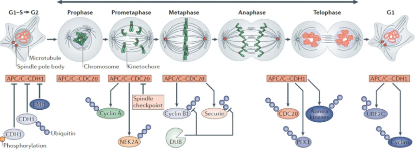
Figure 8 | Regulation of APC/C activity during cell cycle progression
Timely ordered degradation of APC/C substrates during cell cycle progression is primarily regulated by the APC/C co‐activator subunits Cdc20 and Cdh1, respectively. Cdc20 activates APC/C during early mitosis, while Cdh1 keeps the APC/C active during late mitosis and G1‐phase. Upon entry into mitosis, APC/C‐Cdc20 targets early mitotic substrates for degradation, such as Cyclin (Cyc) A and NEK2A. Destruction of the APC/C‐Cdc20 substrates CycB1 and Securin is inhibited by the Spindle checkpoint. When sister chromatids are bipolarly attached to the mitotic spindle during metaphase, the spindle checkpoint is inactivated and degradation of CycB1 and Securin is stimulated, leading to exit from mitosis and cytokinesis. In late mitosis, APC/C‐Cdh1 becomes activated and mediates degradation of additional late mitotic substrates including Cdc20, Plk1 and Aurora kinases. Degradation of cyclins establishes a stable G1‐phase. In late G1‐phase, Cdk activity is upregulated and APC/C‐Cdh1 activity is downregulated by APC/C‐
Cdh1 dependent destruction of UBE2C (UBCH10) and Cdh1, inactivation of Cdh1 by Cdk mediated phosphorylation and direct APC/C‐Cdh1 inhibition by Emi1 (115)