Functional analysis of the cell cycle regulator Rca1 in Drosophila melanogaster
Inaugural-Dissertation
zur
Erlangung des Doktorgrades
der Mathematisch-Naturwissenschaftlichen Fakultät der Universität zu Köln
vorgelegt von
Norman Zielke aus Bonn
Köln 2007
Berichterstatter: PD Dr. Frank Sprenger
Prof. Dr. Maria Leptin
Tag der mündlichen Prüfung: 10. Januar 2007
Abstract 1
1. Introduction ...3
1.1. Cell cycle regulation during Drosophila development...4
1.1.1. Adaptation of cell cycle regulation during Drosophila embryogenesis...5
1.1.2. Third instar eye imaginal disc display a linear arrangement of cell cycle stages ...7
1.1.3. Cell cycle regulation in wing imaginal discs ...8
1.1.4. Endoreplication cycles...9
1.2. The cell cycle control system...10
1.2.1. Initiation and execution of mitosis...11
1.2.2. Regulation of the G1-S transition ...14
1.2.3. Initiation and regulation of DNA replication ...15
1.3. The cell cycle regulator Rca1 ...18
1.3.1. The Rca1/Emi1 family...20
1.3.2. Regulation of Rca1/Emi1 activity...21
2. Aim ...24
3. Results ...25
3.1. Rca1 regulates mitotic entry in postblastoderm embryos in concert with Cyclin/Cdk complexes...25
3.1.1. Cyclin E dependent kinase activity contributes to downregulation APC/C-Fzr activity during G2...25
3.1.2. The Cyclin A phenotype is enhanced by downregulation of Cyclin E/Cdk2 activity...27
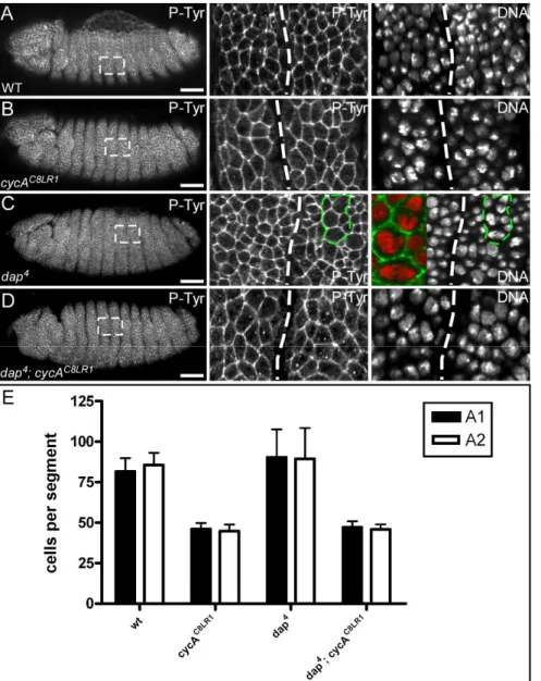
3.1.3. Downregulation of Dacapo activity is not sufficient to restore mitosis 16 in Cyclin A mutants ...30
3.2. Functional analysis of the Rca1 protein ...33
3.2.1. Rca1 inhibits APC/C-Fzr activity in G2 by an F-box independent mechanism ...33
3.2.2. Rca1 gets degraded by different mechanisms than Emi1...35
3.3. Rca1 is implicated in the G1-S transition during imaginal disc development ...37
3.3.1. Rca1 and Fzr are expressed in complementary domains during eye imaginal disc development ...37
3.3.2. Overexpression of Rca1 promotes S-phase entry in eye imaginal discs...38
3.3.3. The F-box is essential to drive cells prematurely into S-phase ...39
3.3.4. Excess Rca1 activity accelerates G1-S transition in wing imaginal discs ...42
3.3.5. Rca1 overexpression stabilizes mitotic cyclins in G1 ...43
3.3.6. Cyclin A accumulation accompanied with Rca1 overexpression relies on a functional F-box ...44
3.3.7. Rca1 gets degraded within the morphogenetic furrow...45
3.3.8. Rca1 lacking the F-box fails to restore the proliferation disadvantage of rca1
mutant clones...46
3.3.9. Rca1 promotes S-phase by a mechanism independent of Cyclin E/Cdk2 ...49
3.3.10. Fzr is not a target of the SCF/Rca1 complex ...50
3.4. Investigation of the S-phase promoting activity of Rca1 in endoreplicating cells....52
3.4.1. Rca1 is not required for endocycle progression ...52
3.4.2. Overexpression of Rca1 perturbs endocycle progression in a F-box dependent manner...53
3.4.3. Continuous expression of Rca1 increases Cyclin E levels in salivary glands ...55
3.4.4. Rca1 expression forces endoreplicating cells to re-enter a mitotic state ...57
3.4.5. Overexpression of Cyclin A cannot impair endoreplication in larval salivary glands ...60
3.4.6. The endocycle breakdown induced by Rca1 overexpression is due to impaired DNA licensing ...61
3.4.7. Continuous Cyclin E expression results in accumulation of Cyclin A and Cdk1...62
3.4.8. Oscillation of APC/C-Fzr activity is not required for endocycle progression ...63
4. Discussion ...65
4.1. Rca1 restrains APC/C-Fzr activity in postblastoderm embryos in concert with Cyclin/Cdk complexes...65
4.2. Structural requirements for APC/C-Fzr inhibition in G2 ...69
4.3. Rca1 degradation in G1 is achieved by a so far unknown mechanism...73
4.4. Rca1 promotes S-phase entry as part of an SCF-complex ...75
4.5. Rca1 might be required to maintain the mitotic state...79
4.6. Outlook...84
5. Material and Methods ...88
5.1. Material ...88
5.1.1. Chemicals ...88
5.1.2. Special chemicals and kits ...88
5.1.3. Electronic equipment, computer and software ...88
5.1.4. Media, solutions and buffers...89
5.1.5. Bacterial strains ...92
5.1.6. Oligonucleotides...93
5.1.7. Plasmids ...94
5.1.8. Fly Stocks...95
5.1.9. Antibodies ...97
5.2. Molecular cloning...99
5.2.1. Restriction digests of DNA...99
5.2.2. Dephosphorylation of DNA ends...99
5.2.3. Klenow fill in of DNA ends...99
5.2.4. Isolation of DNA fragments ...99
5.2.5. Agarose gel electrophoresis ...99
5.2.6. DNA ligation ...99
5.2.7. Preparation of electro-competent cells...100
5.2.8. Transformation of electro-competent E. coli ...100
5.2.9. Transformation of chemically-competent E. coli...100
5.2.10. Isolation of plasmid DNA...100
5.2.11. Amplification of DNA by PCR (Polymerase Chain Reaction) ...101
5.2.12. Site directed mutagenesis ...101
5.2.13. DNA sequencing ...101
5.2.14. Preparation of RNA probes for in situ hybridization ...101
5.3. Drosophila techniques...102
5.3.1. Maintenance of flies ...102
5.3.2. Generation of transgenic flies ...102
5.3.3. Collection and fixation of embryos...103
5.3.4. Antibody staining of embryos...103
5.3.5. Induction of clones ...104
5.3.6. Dissection of imaginal discs and salivary glands ...104
5.3.7. Antibody staining of imaginal discs and salivary glands ...105
5.3.8. BrdU-labelling...105
5.3.9. Flow cytometry...106
5.3.10. In situ hybridization of eye imaginal discs...106
5.3.11. Production of embryo extracts ...107
5.3.12. Production of salivary gland extracts...107
5.3.13. SDS-Page and western blot analysis...108
6. References ...109
Note added in proof 119
Abbreviations 120
Single and three letter code for amino acids 121
Zusammenfassung 122
Erklärung 124 Teilpublikationen 124
Lebenslauf 125 Danksagung 126
Abstract
Tight regulation of APC/C activity is essential for cell cycle progression. An important class of negative APC/C regulators are the Rca1/Emi1 family proteins. All members of the Rca1/Emi1 family share a conserved zinc binding region (ZBR) which is essential for their inhibitory activity. The Rca1/Emi1 proteins belong to the class of F-box proteins that are known to act as substrate recognition subunits in SCF-E3-ligase complexes. Emi1 and Rca1 interact in vitro with members of the Skp family via the F-box. However, no F-box dependent function has been ascribed to these proteins. In Drosophila, Rca1 is required in G2 to prevent premature activation of the APC/C by Fzr. Loss of Rca1 results in an arrest during G2 of the 16th embryonic cell cycle due to premature cyclin degradation. In order to map the essential domains for Rca1 function, a series of deletion constructs was tested for their ability to inhibit APC/C-Fzr activity in vivo. A C-terminal Rca1 fragment including the ZBR was sufficient to restore mitosis 16 in rca1 mutant embryos. This observation confirms that the ZBR is the only protein motif essential for APC/C-Fzr inhibition by Emi1/Rca1. Moreover, this result indicates that the F-box is dispensable for APC/C-Fzr inhibition during embryogenesis. However, analysis of Rca1 function during larval development revealed that Rca1 has a secondary role as an F-box protein. Using the MARCM technique, wing disc cells were generated in which endogenous Rca1 was replaced by an Rca1 construct lacking the F- box. These cells displayed a reduced proliferation rate and prolonged G1-phase. Conversely, overexpression of Rca1 accelerates the G1-S transition in imaginal discs in an F-box dependent manner. Hence, it is likely that Rca1 regulates S-phase entry as part of a yet uncharacterized SCF-complex. In addition, the effect of Rca1 on endoreplication was analyzed. Overexpression of Rca1 during salivary gland development leads to a reduction of polyploidization. This phenotype also depends on a functional F-box. Endoreplication cycles are driven by oscillating waves of Cyclin E/Cdk2 activity, whereas Cdk1 and the mitotic cyclins are transcriptionally downregulated. Furthermore, APC/C-Fzr activity seems not to be required once the endoreplication program has been initiated. Cells overexpressing Rca1 displayed elevated levels of Cyclin E, although Cyclin E is not a target of the APC/C-Fzr complex. It has been shown that continuous expression of Cyclin E interferes with DNA- licensing. Thus, the reduced DNA content in Rca1 overexpressing cells might be due to elevated Cyclin E levels. Additionally, Rca1 overexpressing cells displayed markers for mitotic cells such as Cdk1 and nuclear Cyclin A. The accumulation of Cdk1, Cyclin A and Cyclin E cannot simply be explained by APC/C inhibition. It rather appears that Rca1
activates the transcription of these genes by an unknown mechanism. Nevertheless it cannot be excluded that the APC/C-Fzr complex indirectly contributes to this process. Altogether, Rca1 might act as an F-box protein in an SCF complex that is involved in maintaining diploidy.
1. Introduction
All living organisms are comprised of cells that reproduce by the interplay of cell growth and cell division. Division of already existing cells is the only possibility to generate novel cells and consequently the only way to inherit the genome of the progenitor cell. Thus, the capability of cell division is a fundamental prerequisite for the continuance of life. In unicellular organisms each cell division results in the production of an entire new organism.
In multicellular organisms, however, it requires numerous divisions to create a novel organism from a fertilized oocyte. Since cell division and cell differentiation cannot occur simultaneously, cell division has to be tightly coordinated with the developmental program.
In multicellular organisms, cell division occurs not only during development but is rather required throughout the whole lifespan. Continuous replacement of dead and degenerated cells is a fundamental process to maintain the health of an organism. Hence, impaired cell division can result in severe defects during development as well as in adult organisms.
In principle, cell division requires two different steps that have to be orchestrated. In the first step, cells duplicate their DNA, which is equally distributed between both daughter cells during the actual division step. Since both events occur in an ordered fashion and cannot be separated from each other, the reproduction process of a eukaryotic cell is generally referred as cell cycle (for a general review see Morgan, 2006). The eukaryotic standard cell cycle is divided into four distinct phases, which are named G1, S, G2 and M-phase. The actual division process takes places during M-phase, which is subdivided into mitosis and cytokinisis. The period between two subsequent divisions is termed interphase and is comprised of the three remaining phases. Interphase begins in G1, where the cells are highly metabolic active and increase their cell mass. After completion of this gap phase, cells undergo S-phase to duplicate their DNA. DNA-replication results in the generation of two sister chromatids that will be evenly distributed during mitosis. Before initiation of mitosis, cells enter the G2-phase, in which they undergo further growth. Mitosis is also referred as nuclear division since it only results in the formation of two new nuclei. In the following cytokinisis, these daughter nuclei are then distributed into separate cells.
Based on morphological criteria, mitosis is subdivided into five sections: The first part of mitosis, where the DNA begins to condensate, is named prophase. It is followed by prometaphase and correlates with the initiation of nuclear envelope breakdown. During
metaphase, sister chromatids are attached to the mitotic spindle and form the metaphase plate in the middle of the cell. In the following anaphase, sisterchromatids separate and are subsequently pulled towards the spindle poles. The formation of new nuclei marks the terminal phase of mitosis and is termed telophase. At this time the DNA starts to decondensate, a process that persists till onset of interphase.
Figure 1 The eukaryotic standard cell cycle (adapted from Morgan, 2006). The standard cell cycle is comprised of four phases. In S-phase the DNA of the cell becomes duplicated. During M-phase, the replicated DNA is equally distributed between both daughter cells. S and M-phase are separated by two Gap-phases (G1 and G2) in which the cell increases their mass by growth. The M-phase is dived into two sections, mitosis and cytokinisis. During mitosis sisterchromatids are distributed into two daughter nuclei which become, in the following cytokinisis separated, by a new cell wand. Mitosis is subdivided into five phases, whereby metaphase and anaphase are of particular interest. During metaphase, sister chromatids are held in the middle of the cell by the mitotic spindle. In the following anaphase, sister chromatids fall apart and are drawn to opposite poles of the cell.
1.1. Cell cycle regulation during Drosophila development
The eukaryotic standard cell cycle is not the only of cell cycle mode that is applied. The cells of multicellular organisms rather display different types of cell cycle regulation. This plasticity is necessary to adapt the cell cycle to the different demands of a certain tissue or a particular developmental process. The fruit fly, Drosophila melanogaster is an outstanding model organism to study the mechanisms coordinating cell proliferation with the developmental program (for review see Edgar and Lehner, 1996; Lee and Orr-Weaver, 2003;
Swanhart et al., 2005). During the course of Drosophila development, cells exhibit a variety of different cell cycle types. Even during the short period of embryogenesis, the cells have to undergo three different modes of cell cycle regulation (Figure 2). After completion of
embryogenesis a feeding larvae arises from the embryo. The larvae grows dramatically, but instead of increasing cell numbers, most cells of the larval tissues undergo a specialized cell cycle called endocycle. Most of the adult structures derive from imaginal discs which divide mitotically during the larval period. Of particular interest are the imaginal discs that comprise of the prospective eye (Figure 3) and wing (Figure 4), since they display a cell cycle mode that resembles the standard cell cycle. Finally, nurse and follicle cells of the Drosophila ovary have been proofed as very useful for the investigation of DNA replication and endoreplication, respectively.
1.1.1. Adaptation of cell cycle regulation during Drosophila embryogenesis
The first ten cell cycles during Drosophila embryogenesis are nuclear divisions that occur in a common cytoplasm and give rise to a syncytium. These syncytial cell cycles are very rapid, since they consist only of S and M-phases without intervening Gap-phases. At the onset of embryogenesis, all nuclei are located in the centre of the syncytium. However, at the end of the seventh division cycle three-quarters of the nuclei start to migrate to the surface. The remaining nuclei, by contrast, develop into yolk nuclei that exit the cell cycle after completion of the tenth cycle and then initiate endoreplication. During endoreplication, mitosis is bypassed resulting in an increased DNA content (for review see Edgar and Orr- Weaver, 2001; Lilly and Duronio, 2005). In the course of the ninth division cycle, the first migrating nuclei arrive at the posterior pole of the zygote. During the following cell cycle, cellularization becomes initiated and thereby these nuclei lose their synchrony with the remainder of the nuclei. These early forming posterior cells are termed as pole cells and give rise to the germ cells. The remaining nuclei reach the surface at the beginning of the tenth cell cycle. These nuclei undergo four additional syncytial divisions until they initiate cellularization. These divisions originate at the poles and spread then wavelike to the middle of the embryo. Similar to the first ten divisions, these cell cycles (10-13) lack any intervening Gap-phases, but are a bit slower.
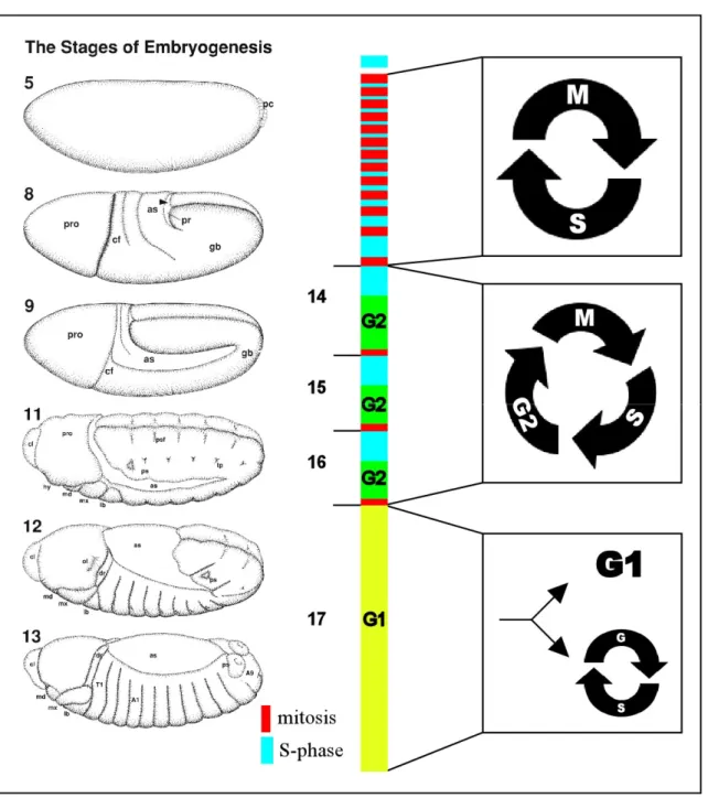
Figure 2 Cell cycle regulation during Drosophila embryogenesis. The first thirteen cell cycles are very rapid and only consist of S- and M-phases. At the stage of cellularization, the midblastula transition occurs and thereby cells change from maternal to zygotic transcription. The following three divisions are termed postblastoderm cell cycles. During these stages, cells undergo a distinct G2-phase, but enter S-phase without an intervening G1-phase. Most of the epidermal cells persist in this terminal G1-phase until the end of embryonic development, while the cells of certain internal tissues such as the gut or the salivary glands enter endocycles. An exception are the cells of the developing nervous system which remain mitotic. The drawings of embryonic stages on the right side are adapted from Hartenstein (1993).
During the 14th embryonic cell cycle the remaining nuclei initiate cellularization, a stage that is called cellular blastula. With the onset of cell cycle 14, the nuclei lose their synchrony and the divisions occur in an invariant spatiotemporal pattern of 25 mitotic domains (Foe, 1989).
The first 13 divisions are driven by maternal stockpiles. At the cellular blastula stage these maternal transcripts are exhausted and zygotic gene expression becomes essential (Merrill et
al., 1988; Wieschaus and Sweeton, 1988). The initiation of zygotic transition is called midblastula transcription and results in the introduction of the first G2-phase (Edgar and O'Farrell, 1989; Edgar and O'Farrell, 1990). The first G1-phase is established after completion of mitosis16 (Edgar and O'Farrell, 1990). Most of the epidermal cells reside in this terminal G1 phase until the end of embryogenesis. Groups of 10-50 imaginal cells that develop into adult structures are separated from the remainder of cells and re-initiate proliferation only upon onset of larval development. By contrast, cells that give rise to the larva proper (e.g. gut, fat body and salivary glands) initiate endoreplication cycles and become polyploid (Smith and Orr-Weaver, 1991). The cells of the developing nervous system are an exception, because they continue to proliferate mitotically during late embryogenesis.
1.1.2. Third instar eye imaginal disc display a linear arrangement of cell cycle stages
The imaginal discs of Drosophila are monolayered epithelial sacs that undergo extensive proliferation during the larval stages. The adult eyes as well as some structures of the head originate from the posterior part of the eye-antenna disc. Eye imaginal discs are an excellent system to study cell cycle control during organ development. A major advantage of the eye imaginal disc is that alterations of the cell cycle often results in aberrant eye morphology (de Nooij and Hariharan, 1995). Eye phenotypes are easy recognizable and therefore facilitate the identification of defects in the cell cycle program. During the first two larval stages, eye imaginal disc cells proliferate in a unpatterned manner that resembles the standard cell cycle.
However, during the third instar stage the differentiation of eye disc cells is initiated. The differentiation into photoreceptor cells is coordinated by the movement of the morphogenetic furrow. The morphogenetic furrow sweeps from anterior to posterior and thereby creates a linear arrangement of cell cycle stages (Figure 3). Undifferentiated cells anterior to the morphogenetic furrow divide asynchronously, whereas cells in the posterior part initiate differentiation. The cells within the morphogenetic furrow are synchronized in G1. A subset of these cells, termed as preclusters, exits the cell cycle and differentiates into the photoreceptor cells R8, R2, R5, R3 and R4 (Ready et al., 1976; Wolff and Ready, 1991a).
The remainder of cells enter a terminal cell cycle called second mitotic wave. The cells of this second mitotic wave give rise to the photoreceptor cells R1, R6, R7, the cone cells, the pigment cells as well as the precursors of the mechanosensory bristles (Ready et al., 1976;
Wolff and Ready, 1991a). The remaining undifferentiated cells undergo apoptosis (Wolff and Ready, 1991b).
Figure 3 Eye imaginal disc cells exhibit morphologically distinguishable G1-S transition. The eye imaginal disc represents the posterior part of the eye-antenna disc. During the third instar stage the morphogenetic furrow (MF) moves from posterior to anterior and thereby initiates the differentiation into photoreceptor cells (PRC).
Cells in the posterior part of the disc divide asynchronously until they become synchronized in G1 by the anterior sweeping furrow. The cells within the morphogenetic furrow subsequently separate into two subpopulations. One fraction terminates proliferation and initiates differentiation immediately, while the remaining cells enter a terminal cell cycle called second mitotic wave (SMW).
1.1.3. Cell cycle regulation in wing imaginal discs
Wing imaginal discs gives rise to two different adult structures. The ventral part of the disc develops into the wing, whereas the dorsal part differentiates into the notum. Wing imaginal disc cells exhibit a mode of cell cycle regulation that resembles the proliferation behaviour of vertebrate cells. Cells in wing imaginal disc undergo a standard cell cycle with four distinct phases. Moreover, unlike embryonic divisions, these cell cycles are accompanied with cell growth ensuring that these cells maintain a constant size during proliferation. Wing imaginal disc cells undergo apoptosis only occasionally, therefore it is thought that most of the disc growth results from proliferation (James and Bryant, 1981). In contrast to the cells of the developing eye, cell division in wing imaginal discs occurs in a largely unpatterned fashion (for review see Milan, 1998). Wing disc cells rather divide in clusters of synchronized cells that are randomly distributed through out the disc. However at the end of larval development, a stripe of cells at the dorso-ventral boundary enter a developmentally programmed cell cycle arrest (O'Brochta and Bryant, 1985) and therefore this region of the disc was named “zone of none proliferating cells” ( ZNC; Figure 4). Cells of the ZNC cease proliferation around 30
hours earlier than remaining cells, but re-initiate the cell cycle for a couple of division during the pupal stage (Hartenstein and Posakony, 1989; O'Brochta and Bryant, 1985). The zone of none proliferating cells is subdivided into four domains (Figure 4; Johnston and Edgar, 1998).
Cells in the centre of the anterior part as well as the whole posterior part of the ZNC undergo a G1 arrest, whereas the two outer cell rows of the anterior part stay in the G2-phase.
Figure 4 Cell division during wing development. Cells in wing imaginal discs proliferate extensively during the larval period. Thereby, these cells undergo a standard cell cycle with four distinct phases. Cell division occurs in an asynchronous and unpatterned man- ner. Only at the end of larval develop- ment cells of the ZNC undergo a de- velopmentally programmed cycle arrest. The cells in the middle of the anterior part of the ZNC arrest in G1, while the adjacent cell rows undergo a G2 arrest. Cells in the posterior domain of the ZNC uniformly arrest in G1.
1.1.4. Endoreplication cycles
The endoreplication cycle or endocycle is a cell cycle variant that is employed by various tissues in Drosophila (for review see Edgar and Orr-Weaver, 2001; Lilly and Duronio, 2005).
During endoreplication, cells undergo repeated rounds of DNA replication without intervening mitosis which result in increased DNA contents. Endocycling cells exhibit a distinct Gap-phase, in which no DNA-replication occurs. Endoreplication is an effective strategy of cell growth and is therefore frequently found in cells that give rise to tissues with high metabolic activity (for review see Edgar and Nijhout, 2004). Cells of several internal organs such as the gut, fat body, malipighian tubules and salivary glands, initiate endoreplication cycles during late embryogenesis and maintain endoreplication cycles during the larval period (Smith and Orr-Weaver, 1991). In addition, several adult tissues like the gut and the ovary harbor endoreplicating cells (Micchelli and Perrimon, 2006; Ohlstein and Spradling, 2006). Endoreplication results in multiple copies of the genome that can be organized in different chromosomal arrangements. Generally, it is distinguished between polyploidy and polyteny. Polyploid cells contain multiple copies of their chromosomes that
are clearly distinguishable from each other. In polytene cells, by contrast, sisterchromatids remain closely associated. A well known example of polyteny, are the giant chromosomes of the larval salivary gland that exhibit a DNA content of 1024-2048C. The border between polyploidy and polyteny is uncertain and numerous intermediate configurations can be observed. An interesting example for the plasticity of the chromosome arrangement is found in the nurse cells of the Drosophila ovary, which switch from polyteny to polyploidy. In the first five endoreplication cycles during nurse cell development the chromosomes remain aligned to each other (Dej and Spradling, 1999). After S-phase of the fifth cycle, however, the chromosomes condense and separate from each other. From this point on the chromosomes of the nurse cells maintain the polyploid configuration and continue endoreplication until they reach a DNA content of 1024 C. Besides the nurse cells, the Drosophila ovary harbors another cell type that becomes polyploid. During oogenesis, the somatic follicle cells first undergo five mitotic divisions and then initiate endoreplication. Follicle cells execute five endocycles that give rise to DNA content of 16C. However, DNA replication stops completely at this stage. Several loci maintain DNA replication and become amplified.
Among these amplified loci are genes required for the formation the chorion of the eggshell.
Therefore, this process was termed chorion gene amplification (Calvi et al., 1998).
1.2. The cell cycle control system
During cell division the cell is confronted with numerous problems challenging the correct inheritance of the genetic information of the progenitor cell. Cells have to ensure that the genome is only duplicated once per cell cycle. Then, the chromosomes must be distributed evenly between both daughter cells and thereby each cell must receive a full copy of the genome. I most cases, the cell cycle must be coordinated with cell growth to maintain a constant cell size. In order to prevent the inheritance of severe chromosomal defects, safeguard mechanisms have to interrupt cell cycle progression after genomic damage. In multicellular organisms proliferation must be coordinated with the demands of the developmental program and the housekeeping mechanisms, respectively. Finally, it must be ensured that these events occur in a ordered manner and that the cell cycle proceeds only in one direction. To achieve all these tasks eukaryotic cells have evolved a tightly regulated cell cycle control system that is basically conserved throughout the animal kingdom (for review see Morgan, 2006; Murray, 2004). The heart of the cell cycle control system are the Cyclin dependent kinases (Cdk) and their regulators, the cyclins. At certain stages of the cell cycle,
Cdk’s become activated by interaction with a particular Cyclin. Cdk levels remain constant throughout the cell cycle, whereas cyclin levels oscillate. Thus, it is achieved that Cyclin/Cdk activity peaks at specific points of the cell cycle and thereby initiates the next series of cell cycle event. Generally, it is distinguished between two types of cyclins. Cyclins involved in mitosis are referred as mitotic cyclins, whereas cyclins implicated in the initiation of DNA replication are termed G1 cyclins. In Drosophila, mitotic cyclins A, B and B3 as well as the G1 cyclins D and E have been identified. Moreover, orthologues of Cdk1, Cdk2 and Cdk2/4 are know in Drosophila (for review see Edgar and Lehner, 1996; Lee and Orr-Weaver, 2003;
Swanhart et al., 2005).
1.2.1. Initiation and execution of mitosis
Mitosis is initiated in late G2 by the activation of Cyclin/Cdk1 complexes (Minshull et al., 1989; Murray and Kirschner, 1989). Cdk1 only forms complexes with a subset of cyclins, which were generally referred as mitotic cyclins. In Drosophila, entry into mitosis is regulated by Cdk1 and the mitotic cyclins A, B and B3 (Jacobs et al., 1998; Knoblich and Lehner, 1993; Lehner and O'Farrell, 1989; Lehner and O'Farrell, 1990a; Lehner and O'Farrell, 1990b). It is thought that the mitotic cyclins partly overlap in their functions, since only loss of cyclin A results in embryonic lethality (Jacobs et al., 1998; Knoblich and Lehner, 1993;
Lehner and O'Farrell, 1989; Lehner and O'Farrell, 1990b). Transcription of mitotic cyclins is initiated during late S-phase and results in the accumulation of cyclin/Cdk1 complexes during G2. To prevent premature entry into mitosis, Cdk1 activity is restrained by inhibitory phosphorylation at threonine residue 14 and tyrosine residue 15. These phosphorylations are mediated by kinases of the conserved Wee1/Myt1 family (Morgan, 1995). To initiate mitotic entry, these inhibitory phosphorylations are removed by a Cdc25 phosphatase (Russell and Nurse, 1986). The Drosophila genome bears two different isoforms of Cdc25, named string and twine. String activity becomes essential for the first time after midblastula transition and is then required for all mitotic divisions throughout development (Edgar et al., 1994a; Edgar and O'Farrell, 1990). By contrast, the activity of Twine is restricted to meiosis (Edgar and Datar, 1996)
After mitotic entry, the Cyclin/Cdk1 complex must be inactivated to allow proper progression through mitosis (Murray et al., 1989). The downregulation of Cdk1 activity is achieved by degradation of the Cyclin subunit (Glotzer et al., 1991). In Drosophila, mitotic cyclins are
sequentially degraded (Figure 5; Sigrist et al., 1995). Cyclin A degradation is initiated in metaphase just before chromosome separation. Cyclin B degradation occurs in early anaphase when the chromosomes are separated. Cyclin B3 gets degraded in late anaphase after chromosome segregation. The degradation of B-type cyclins depends on a conserved protein motif named destruction box (Glotzer et al., 1991; King et al., 1996). Deletion of this destruction box (D-box) results in a stable Cyclin B protein. Overexpression of this stable Cyclin B in Drosophila embryos leads to an arrest in early anaphase (Sigrist et al., 1995).
Moreover, overexpression of a stable version of Cyclin B3 specifically arrest the cell cycle in late anaphase (Sigrist et al., 1995). So far no particular motif that mediates Cyclin A destruction has been identified. However, a N-terminal truncated version of Cyclin A is refractory to degradation and overexpression of this fragment results in an metaphase arrest (Sigrist et al., 1995). Based on these observations, it has been proposed that the successive steps required for the completion of mitosis are ordered by the sequential degradation of mitotic cyclins (Sigrist et al., 1995).
Figure 5 Regulation of the anaphase promoting complex/cyclosome (APC/C). To initiate mitosis, Cyclin/Cdk1 is activated by the phosphatase String/Cdc25. The APC/C gets subsequently phosphorylated and thereby activated through Cdc20/Fzy binding. The activated APC/C-Cdc20 complex initiates in turn the sequential degradation of mitotic cyclins and thus the downregulation of Cdk1 activity. After depletion of Cdk1 activity the APC/C forms a complex with another activator protein, Cdh1/Fzr. The activity of APC/C-Cdh1complex is crucial for the initiation of the following G1-phase.
The degradation of mitotic cyclins and numerous other proteins is mediated by the 26S proteasome, a multi-subunit protease specific for multi-ubiquitinated substrates (for review see Baumeister et al., 1998; Coux et al., 1996; Hochstrasser, 1996). In order to mark proteins for proteasomal degradation, multi-ubiquitin chains are transferred in a three-step reaction to the substrate. In the first step, ubiquitin is activated by forming a high-energy thioester between a cysteine of its active site and the C-terminus of ubiquitin. The activated ubiquitin is subsequently transferred to one of several ubiquitin-conjugating enzymes that are also named E2-enzymes. Finally, the ubiquitin is covalently attached to the substrate protein by an ubiquitin-protein ligase or E3-enzyme, respectively. The ubiquitin ligase mediating the proteasomal degradation of mitotic cyclins is called anaphase promoting complex or cyclosome (for review see Peters, 2006; Pines, 2006; Zachariae and Nasmyth, 1999). The anaphase promoting complex (APC/C) is a high molecular weight complex that consists of at least eleven subunits (Gieffers et al., 2001; Passmore et al., 2005). The activity of the APC/C depends of its phosphorylation state and the abundance of two WD40 activator proteins, Cdc20 and Cdh1 (Schwab et al., 1997; Visintin et al., 1997). The APC/C-Cdc20 complex gets only activated during mitosis to mediate the proteasomal degradation of mitotic cyclins and other cell cycle regulators. The activation of the APC/C by Cdc20 depends on the phosphorylation state of the APC/C (Kraft et al., 2003; Peters et al., 1996). Cdc20 can only bind to the APC/C once several APC/C subunits have been phosphorylated (Kramer et al., 2000) (Kramer et al., 1998). In vertebrates, APC/C phosphorylation is achieved by mitotic kinases such as Cyclin/Cdk1 and polo like kinase 1 (Descombes and Nigg, 1998; Patra and Dunphy, 1998). Hence, APC/C-Cdc20 activity is restricted to early mitosis. By contrast, Cdh1 can only activate the APC/C at stages with low Cdk activity (Kramer et al., 2000;
Zachariae et al., 1998). It is thought that Cdk1 and Cdk2 phosphorylate Cdh1 thereby preventing APC/C activation (Kramer et al., 2000; Sorensen et al., 2000; Zachariae et al., 1998). Thus, the APC/C-Cdh1 complex is only active during late mitosis and G1, when Cyclin/Cdk activity is dampened. In addition to a destruction box, substrates of the APC/C- Fzr complex frequently contain a KEN-box (Burton and Solomon, 2001; Hilioti et al., 2001;
Pfleger et al., 2001). The Drosophila orthologues of Cdc20 and Cdh1 are encoded by the genes fizzy (fzy) and fizzy-related (fzr), respectively. Cells in fizzy mutant embryos fail to downregulate mitotic cyclins and subsequently arrest in metaphase (Dawson et al., 1993;
Dawson et al., 1995; Sigrist et al., 1995). Epidermal cells of embryos lacking fizzy-related cannot establish the terminal G1-phase and undergo an additional seventeenth mitosis (Sigrist and Lehner, 1997). Closer inspection of fizzy-related mutants revealed that completion of this
additional mitosis (including cyclin degradation) does not require fizzy-related (Jacobs et al., 2002). Moreover, cells in eye discs derived from hypomorphic fizzy-related mutants fail to become synchronized in G1 and enter ectopic S-phases (Pimentel and Venkatesh, 2005).
Therefore APC/C-Fzr activity is thought to be only required for establishment and maintenance of the G1 state (Figure 5).
1.2.2. Regulation of the G1-S transition
In vertebrates, the transition from G1 to S-phase is regulated by three different kinases, Cdk2, Cdk4 and Cdk6. In response to external growth signals Cyclin D expression is stimulated (Matsushime et al., 1994; Sherr, 1993). Cyclin D activates Cdk4/6 and members of retinoblastoma (Rb) tumour suppressor family are subsequently inhibited. The inhibition of Rb leads to the release of a transcription factor of the E2F family (Attwooll et al., 2004; Blais and Dynlacht, 2004; Kato et al., 1993). E2F stimulates the transcription of Cyclin E and Cyclin A (DeGregori et al., 1995; Pagano et al., 1992). Besides these cyclins, E2F activates the transcription of numerous other genes required for DNA replication such as ribonucleotide reductase (RNR) and the DNA polymerase δ accessory subunit, PCNA (DeGregori et al., 1995). Cyclin E and Cyclin A activate Cdk2 and initiate in turn DNA replication (Dutta and Stillman, 1992; Pagano et al., 1992). Moreover, Cdk2 also phosphorylates Rb and enhances thereby its own activation by increased Cyclin E and A transcription. In Drosophila, single genes for Cdk4 and Cyclin D have been identified (Datar et al., 2000; Meyer et al., 2000; Sauer et al., 1996). Drosophila Cyclin D/Cdk4 is not directly implicated in the transition from G1 to S-phase, although it can phosphorylate Rb (Datar et al., 2000; Meyer et al., 2000; Xin et al., 2002). It rather appears that the Cyclin D/Cdk4 complex is involved in growth regulation, but it remains to be clarified how this function is achieved (Datar et al., 2000; Meyer et al., 2000). Recently it has been demonstrated that this pathway requires mitochondrial activity and Hph, a hydroxylase implicated in the cellular response to low oxygen (Frei and Edgar, 2004; Frei et al., 2005). In Drosophila, S-phase induction mainly relies on Cyclin E/Cdk2 activity (Knoblich et al., 1994; Richardson et al., 1995). Cyclin E/Cdk2 phosphorylates the sole Rb ortholog in Drosophila (Rbf) and stimulates thereby transcription of S-phase genes via E2F1 (Du et al., 1996; Duronio and O'Farrell, 1994; Duronio and O'Farrell, 1995; Duronio et al., 1995). In addition, S-phase can be induced by Cyclin A overexpression or by loss of the Cyclin A/Cdk1 inhibitor Roughex, respectively (Foley et al., 1999; Sprenger et al., 1997; Thomas et al., 1997). Exit from S-
phase is facilitated by downregulation of Cyclin E dependent kinase activity. After autophosphorylation, Cyclin E is therefore targeted for degradation by an SCF ubiquitin ligase complex (Koepp et al., 2001; Moberg et al., 2001; Schwab and Tyers, 2001). In Drosophila, Cyclin E degradation is mediated by the F-box protein Archipelago that was initially identified in a screen for mutants causing overproliferation (Moberg et al., 2001).
The SCF ubiquitin ligases are named by their core subunits Skp, Cullin, and F-box protein (for review see Ang and Wade Harper, 2005; Jackson et al., 2000; Vodermaier, 2004). Apart from the three core subunits, SCF complexes contain a RING finger protein as well as an E2- enzyme (Jackson et al., 2000). SCF complexes are implicated in a plethora of processes such as cell cycle regulation, signalling pathways, circadian rhythms and apoptosis (Grima et al., 2002; Koepp et al., 1999; Maniatis, 1999; Nateri et al., 2004). SCF complexes are only distinguishable by their F-box proteins that confer substrate specificity (Skowyra et al., 1997).
F-box proteins are characterized by a conserved motif that was first identified in Cyclin F and thus named F-box (Bai et al., 1996). In addition F-box proteins frequently contain protein motifs involved in protein-protein interaction (Jin et al., 2004; Winston et al., 1999). It is thought that these domains are required for substrate binding. Substrate recognition by many F-box proteins depends on phosphorylation of the substrate, thereby allowing temporal control of degradation (Orlicky et al., 2003; Skowyra et al., 1997). The F-box protein is attached to the cullin scaffold by an Skp protein that recognizes the F-box (Schulman et al., 2000; Zheng et al., 2002). Mammals and yeast have only a single Skp gene (Skp1), while six Skp proteins (SkpA-F) have been identified in Drosophila (Nayak et al., 2002; Yamanaka et al., 2002). So far only SkpA has been characterized in greater detail (Murphy, 2003) and it remains to be elucidated whether the other homologues are implicated in SCF complexes.
1.2.3. Initiation and regulation of DNA replication
At the transition from G1 to S-phase, DNA replication is initiated by increasing Cdk activity.
DNA synthesis occurs at specific sites of the chromosomes which are named origins of replication. During late mitosis and early G1 the pre-replicative complex (pre-RC) gets recruited to the replication origins. This process is also known as DNA licensing. Upon S- phase entry the pre-RC gets activated and subsequently triggers DNA replication. The pre-RC contains the helicase that unwinds the DNA and promotes assembly of the actual replication machinery. The formation of the pre-RC relies on the origin recognition complex (ORC). The
ORC complex consists of six subunits and is constantly bound to the replication origins. The ORC can only promote pre-RC formation during late G1 (Figure 6; Bell and Dutta, 2002;
Chesnokov et al., 1999; Gossen et al., 1995). In late mitosis and early G1, the actual licensing process begins with binding of Cdt1 and Cdc6 to the ORC complex (Figure 6). After recruitment of Cdt1 and Cdc6 to the ORC, minichromosome maintenance (MCM) proteins are loaded onto the DNA and remain associated with the DNA until S-phase (Figure 6). The MCM2-7 proteins are arranged in a bilobed hexameric structure that surrounds the DNA (Fletcher et al., 2003; Pape et al., 2003). Once the MCM2-7 complex is associated with the DNA the licensing process is completed and the pre-RC can be activated. The components of the pre-RC, namely ORC1-6, Cdt1, Cdc6 and MCM2-7, are conserved among all eukaryotes, including Drosophila (Bell and Dutta, 2002; Chesnokov et al., 1999; Gossen et al., 1995; Su et al., 1996). The Drosophila ortholog of Cdt1 is also known as double-parked (dup) (Thomer et al., 2004; Whittaker et al., 2000).
Figure 6 Assembly of the pre-replicative complex (adapted from Morgan, 2006). DNA replication begins at specific sites on the chromosomes named origins. The ORC1-6 complex binds constantly to the replication origins and serves as scaffold for the formation of the pre-replicative complex (pre-RC). In late mitosis and early G1, Cdt1 and Cdc6 are recruited to the ORC complex. Upon binding of Cdt1 and Cdc6, the MCM2-7 helicases are loaded to the DNA.
The precise duplication of the genome is crucial for the survival of an organism. In multicellular organisms any genome instability potentially gives rise to cancer and thus compromises the life of the whole organism. Therefore, several safeguard mechanisms have been evolved ensuring that the DNA is only replicated once per cell cycle (for review see Bell and Dutta, 2002; Blow and Dutta, 2005). After initiation of DNA replication, the MCM2-7
complexes are release from the replication origin and move with the replication fork along the DNA. The release of the MCM2-7 complexes results in inactivation of the pre-RC. Thus, new MCM2-7 complexes cannot be recruited until formation of a novel pre-RC. The assembly of pre-RCs is restricted to late mitosis and early G1, thereby ensuring that mitosis proceeds DNA-replication. In multicellular organisms this is largely achieved by two mechanisms. In late G1, Geminin binds to Cdt1 and prevents the formation of pre-RCs (McGarry and Kirschner, 1998). Geminin is a target of the APC/C which mediates its proteasomal degradation during late mitosis and G1 (McGarry and Kirschner, 1998). At the end of the G1 phase, the APC/C is inactivated, resulting in accumulation of Geminin and subsequent inhibition of Cdt1. A Drosophila orthologue of Geminin has been identified, but it remains to be clarified whether Drosophila Geminin levels oscillate throughout the cell cycle (Quinn et al., 2001). Since APC/C activity depends on Cdk phosphorylation, the assembly of pre-RCs is indirectly coupled to Cyclin/Cdk activity. However, Cyclin dependent kinase activity contributes also directly to the formation pre-RC. It is assumed that APC/C activity is dispensable for the endoreplication cycles in Drosophila (Edgar and Orr-Weaver, 2001; Lilly and Duronio, 2005). However, overexpression of Cyclin E promotes MCM2-7 loading and prevents thereby endoreplication (Follette et al., 1998; Su and O'Farrell, 1998; Weiss et al., 1998). Although phosphorylation of pre-RC subunits appears to be important to inhibit pre- RC assembly, the mechanisms are only poorly understood. In higher eukaryotes (including Drosophila), Cdt1 protein levels fluctuate throughout the cell cycle (Nishitani et al., 2001;
Thomer et al., 2004). Evidence from several organisms suggest that two different ubiquitin- ligase complexes contribute to the proteasomal degradation of Cdt1. In human cells, a Skp2 containing SCF-complex interacts with Cdt1, whereby Cdt1 binding requires phosphorylation by Cdk2 or Cdk4 (Li et al., 2003; Liu et al., 2004; Nishitani et al., 2001; Sugimoto et al., 2004). In C. elegans, an SCF-like complex based on Cullin 4 has been identified that is required to downregulate Cdt1 at the end of G1 (Zhong et al., 2003). In Drosophila Dup/Cdt1 degradation seems to be Cyclin E/Cdk1 dependend, but it remains to be clarified whether SCF-E3 ligases are implicated in this process (Thomer et al., 2004).
1.3. The cell cycle regulator Rca1
Tight regulation of APC/C activity is crucial to ensure normal cell cycle progression (for review see Peters, 2006; Pines, 2006; Zachariae and Nasmyth, 1999). The activity of the APC/C depends on its phosphorylation state and the presence of the WD40 activator proteins Fzy/Cdc20 and Fzr/Cdh1 (Schwab et al., 1997; Visintin et al., 1997). During mitosis, phosphorylation of APC/C subunits is prerequisite for its interaction with Fzy, whereas activation of APC/C by Fzr is prevented via phosphorylation (Kramer et al., 2000; Sorensen et al., 2000; Zachariae et al., 1998). Therefore, APC/C-Fzr activity is restricted to later mitotic stages and G1 when Cdk activity is low. Several additional molecules have been identified that regulate APC/C activity by other mechanisms (for review see Peters, 2006;
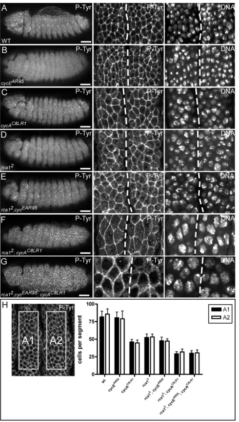
Pines, 2006). Prominent members among these regulators are the vertebrate Emi proteins that restrict APC/C activity at different cell cycle stages (for review see Schmidt et al., 2006). The Drosophila rca1 gene encodes an APC/C-Fzr inhibitor that is related to the Emi1 proteins (Grosskortenhaus and Sprenger, 2002). Embryos homozygous mutant for rca1, fail to execute the 16th mitosis of Drosophila embryogenesis. Due to this G2 arrest, rca1 mutants display a reduced number of epidermal cells compared to wild-type (Dong et al., 1997). Since this phenotype resembles mutants for Cyclin A (Lehner and O'Farrell, 1989), the gene was named regulator of Cyclin A 1 (rca1). The G2 arrest in rca1 mutants is caused by premature degradation of the mitotic cyclins A and B (Grosskortenhaus and Sprenger, 2002). In Drosophila, degradation of mitotic cyclins is mediated by the APC/C and the two activator proteins, Fizzy (Fzy) and Fizzy-related (Fzr) (Dawson et al., 1995; Sigrist et al., 1995; Sigrist and Lehner, 1997). Mutants for fzr fail to establish the terminal G1-phase and execute an extra cell cycle (Sigrist and Lehner, 1997). Since overexpression of Cyclin A abolishes the terminal G1 arrest (Sprenger et al., 1997), this additional mitosis 17 is probably due to accumulation of Cyclin A. By contrast, overexpression of Fzr prevents accumulation of mitotic cyclin and entry into mitosis 16 (Sigrist and Lehner, 1997). Double mutants for rca1 and fzr display epidermal cell numbers similar to wild-type, indicating that mitosis 16 occurs normally in these embryos (Grosskortenhaus and Sprenger, 2002). Moreover, simultaneous overexpression of Fzr and Rca1 allows normal cyclin accumulation and execution of mitosis 16 (Grosskortenhaus and Sprenger, 2002). Hence, these experiments demonstrated that Rca1 has a negative effect on Fzr. In addition, co-immunoprecipitation experiments revealed that Rca1 and Fzr also interact physically (Grosskortenhaus and Sprenger, 2002). Altogether, these data give rise to the model that Rca1 restrains APC/C-Fzr activity during G2 of cell cycle 16 to allow cyclin accumulation and subsequent entry into terminal mitosis (Figure 7).
Moreover, overexpression of Rca1 can overcome the G2 arrest in Cyclin A mutant embryos (Dienemann and Sprenger, 2004). This suggests that Cyclin A/Cdk1 activity also contributes to APC/C-Fzr inhibition in G2 of cell cycle 16 (Figure 7).
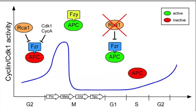
Figure 7 Rca1 prevents untimely activation of APC/C-Fzr complex. APC/C activity is crucial for execution of mitosis and the establishment of the G1-phase. At the metaphase-anaphase transition the APC/C is activated by Fzy and mediates the degradation of mitotic regulators such as cyclins. Since APC/C activation by Fzy requires high kinase activity, APC/C-Fzy activity is restricted to mitosis. By contrast, Fzr can only bind to the APC/C at stages with low kinase activity as found in late mitosis, G1 and G2. During G2, APC/C-Fzr activity is dampened by Rca1 to allow the accumulation of Cyclin/Cdk1 activity and entry into mitosis. In addition, recent evidences suggest that Cyclin/Cdk1 activity also contributes to APC/C-Fzr inhibition in G2. In G1, Rca1 activity has to be restricted to allow APC/C-Fzr activity.
Rca1 was initially identified in a screen for suppressors of the roughex eye phenotype (Dong et al., 1997). Roughex is an inhibitor of Cyclin A depend kinase activity (Foley et al., 1999).
Flies carrying weak alleles of roughex display a rough eye phenotype (Thomas et al., 1994).
In roughex mutants, cells of eye imaginal discs enter S-phase prematurely because they fail to downregulate Cyclin A/Cdk1 activity in G1 (Thomas et al., 1997). Moreover, eye imaginal disc cells in hypomorphic fzr mutants fail to undergo the G1 arrest in the morphogenetic furrow and display elevated levels of mitotic cyclins (Pimentel and Venkatesh, 2005). Hence, demonstrating that two different mechanisms contribute to the inhibition of Cyclin A/Cdk1 activity in G1. On the one hand, Cdk1 activity is restricted by the action of the Cdk1 inhibitor Roughex (Sprenger et al., 1997; Thomas et al., 1997). On the other hand, S-phase entry is prevented by APC/C-Fzr complex which mediates the destruction of Cyclin A. Mitotic cyclins accumulate upon entry into S-phase, suggesting that APC/C-Fzr activity is
downregulated after progression through G1. It has been demonstrated that Cyclin A/Cdk1 acts negatively on APC/C-Fzr activity (Dienemann and Sprenger, 2004). However, if APC/C- Fzr inactivation is required for Cyclin A accumulation, this rises the question how Cyclin A/Cdk1 activity can accumulate and inactivate Fzr. In human cell culture, it has been shown that Emi1, the vertebrate ortholog of Rca1, promotes S-phase entry by inhibiting the APC/C- Cdh1 complex (Hsu et al., 2002). In late G1-phase, E2F stimulates the transcription of Emi1 to allow Cyclin A accumulation and subsequent entry into S-phase (Hsu et al., 2002).
Depletion of Emi1 levels by RNAi prevents S-phase entry while cells overexpressing Emi1 progress faster through G1 (Hsu et al., 2002). Overexpression of Rca1 also drives cells into ectopic S-phases (Dong et al., 1997). Furthermore, the premature entry into S-phase in roughex mutants can be suppressed by reduced activity of Rca1 (Dong et al., 1997).
Therefore, it seems conceivable that Rca1 might have a similar function at the G1-S transition as Emi1. Moreover, a recent study proposed that Emi1 might have a secondary function at the G1-S transition beyond APC/C inhibition and this could be possible for Rca1 (Rape and Kirschner, 2004).
1.3.1. The Rca1/Emi1 family
Rca1 shares limited homology (18% identity) to the vertebrate Emi1 proteins (Reimann et al., 2001a). Emi1 was initially identified as an APC/C inhibitor specific for early mitotic stages, but further work revealed that Emi1 is also implicated in the transition from G1 to S-phase (Hsu et al., 2002; Reimann et al., 2001a). In contrast to Rca1, the Emi1 proteins are able to inhibit Cdc20 and Cdh1 dependent APC/C activity (Hsu et al., 2002; Reimann et al., 2001a;
Reimann et al., 2001b). Despite their low identity, Rca1 and Emi1 display an intriguingly similar arrangement of their functional domains (Figure 8). All Rca1/Emi1 proteins contain an F-box in their central region followed by a zinc binding region (ZBR) in the C-terminal part. The ZBR is assumed to be involved in protein-protein interaction. Additionally, they harbor different putative nuclear localization signals as well as several sequence motifs that might be involved in degradation. Finally, they contain several potential Cdk1 phosphorylation sites distributed throughout the protein. Two recent reports described the identification of a novel meiosis specific homologue of Emi1 called Emi2/XErp1 (Schmidt et al., 2005; Tung et al., 2005). Emi2/XErp1 is an inhibitor of the APC/C-Fzy complex that seems to be required for maintenance of the cyctostatic factor (CSF) arrest during Xenopus oocyte maturation (Rauh et al., 2005; Schmidt et al., 2005; Tung et al., 2005). The C-terminal
part of Emi2/XErp1 shows significant homology to Emi1 and display a ZBR as well as an F- box domain (Schmidt et al., 2005; Tung et al., 2005).
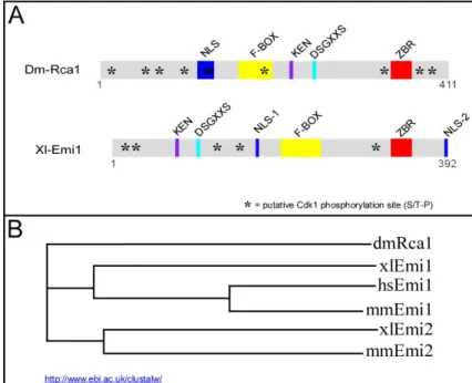
Figure 8 Rca1 is related to APC/C inhibitors of the Emi1/Emi2 family.
(A) Overview of the conserved motifs in Rca1 and Xenopus Emi1. Both pro- teins contain an F-Box and a C- terminal zinc binding region (ZBR).
The N-terminal part of the xlEmi1 harbours a conserved DSGxxS that is crucial for Emi1 degradation. Rca1 displays a similar motif and a KEN- box in its central region. Finally, both proteins contain various numbers of putative Cdk phosphorylation sites marked by an asterisk. xlEmi1 was used as showcase and the features described for xlEmi1 apply basically to all Emi1/Emi2 proteins. (B) Rca1 is a distant relative of the Emi1/Emi2 family. The phylogenetic tree was generated by the ClustalW multiple sequence alignment tool.
All Rca1/Emi family members belong to the class of F-box proteins. F-box proteins are part of SCF (Skp-Cullin-F-box) ubiquitin ligases that are involved in targeting of numerous substrates for degradation (Ang and Wade Harper, 2005; Kipreos and Pagano, 2000;
Maniatis, 1999). Emi1 was identified in a screen for Skp1 interaction partners and deletion of the F-box prevents Skp1 binding in vitro (Reimann et al., 2001a). In addition, yeast two- hybrid data indicated that Emi2/XErp1 interacts also with Skp1 in a F-box dependent manner (Schmidt et al., 2005). A genome wide yeast two-hybrid analysis demonstrated that Rca1 interacts with Drosophila SkpA and B (Giot et al., 2003). These observations indicate that the Rca1/Emi proteins contain functional F-box domains. However, several studies demonstrated that the F-box of the Emi1 proteins is dispensable for its inhibitory effect on the APC/C (Reimann et al., 2001a; Schmidt et al., 2005). Thus, the in vivo function of the F-box remains unclear. It has to be elucidated whether Rca1/Emi proteins act as classical F-box proteins in an SCF complex that targets proteins for proteasomal degradation.
1.3.2. Regulation of Rca1/Emi1 activity
Since APC/C-Fzr activity is crucial for the establishment of the G1-phase (Jacobs et al., 2002;
Pimentel and Venkatesh, 2005), Rca1 activity has to be eliminated at this stage. During embryogenesis, Rca1 is degraded specifically at the stage when the epidermal cell enter the
terminal G1-phase (Grosskortenhaus and Sprenger, 2002). At the moment it is unclear which pathways mediates Rca1 degradation in G1 and how this is regulated. However, much more is known about the regulation of Emi1 and these mechanisms could also apply to Rca1 (Figure 9).
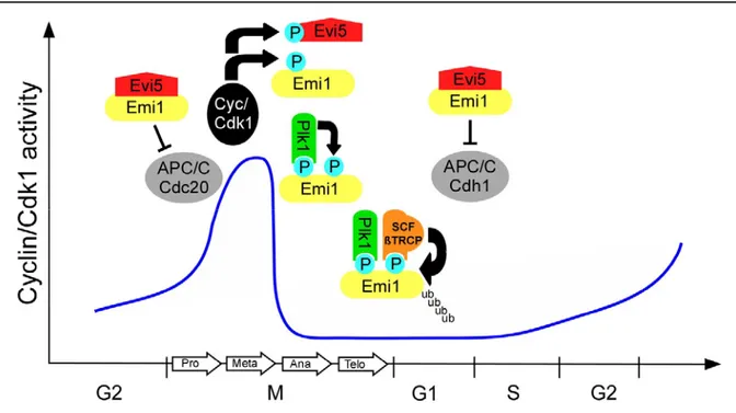
Figure 9 Emi1 degradation is regulated at multiple levels. During early mitosis, Emi1 inhibits APC/C-Cdc20 activity and enables thereby the accumulation of mitotic cyclins. In addition, Emi1 promotes S-phase entry by APC/C-Cdh1 inhibition. The stabilizing factor Evi5 accumulates in late G1 and maintains Emi1 levels during S/G2. During prophase Evi5 is phosphorylated by polo like kinase 1 (plk1) and becomes subsequently degraded.
After Cdk1 phosphorylation, Emi1 is then also phosphorylated by Plk1 1. Phosphorylated Emi1 is recognized by the SCF/ßTRPC complex, which subsequently targets Emi1 for proteasomal degradation.
Emi1 activity is necessary for normal progression through mitosis and promotes the transition from G1 to S-phase (Hsu et al., 2002; Reimann et al., 2001a). In early mitosis Emi1 prevents premature APC/C-Cdc20 activation to facilitate the increase of Cyclin/ Cdk1 activity (Reimann et al., 2001a). To allow normal cyclin destruction in mitosis, Emi1 is degraded during prophase (Reimann et al., 2001a). The proteasomal degradation of Emi1 is mediated by the SCF/ßTRCP complex and persists until G1 (Guardavaccaro et al., 2003; Margottin- Goguet et al., 2003). The F-box protein ßTRCP specifically recognizes a DSGxxS consensus site in Emi1. ßTRCP can only bind to the DSGxxS degron when both serines have been phosphorylated by polo like kinase 1 (Plk1) (Hansen et al., 2004; Moshe et al., 2004). Plk1 activity originates in G2 and persists until early G1. Therefore, premature activation of Emi1 degradation must be prevented in early mitosis. The initiation of Emi1 degradation by Plk1 requires the previous phosphorylation by Cyclin/Cdk1 and is thereby directed to later mitotic
stages (Margottin-Goguet et al., 2003; Reimann et al., 2001a). In addition, the Evi5 oncogene has been identified as a stabilizing factor for Emi1 (Eldridge et al., 2006). Evi5 accumulates in early G1 and shields Emi1 from Plk1 phosphorylation by binding to a site adjacent to the DSGxxS degron. After progression through early mitosis, Evi5 degradation is triggered by Plk1 and Emi1 is then accessible for Plk1. The basic components of this pathway such as ßTRCP and Plk1 are conserved in Drosophila (Barr et al., 2004; Jiang and Struhl, 1998).
Moreover, Rca1 also contains a putative DSGxxS degron in its central region (Figure 8).
Therefore it is conceivable that Rca1 degradation is achieved in a similar manner.
2. Aim
APC/C activity is crucial for normal progression through mitosis and establishment of the G1- phase. During mitosis, APC/C activity depends on the WD40 proteins Cdc20/Fzy, while in G1 the APC/C is activated by Fzr. Previous work has shown that Rca1 is an inhibitor of the APC/C-Fzr complex. During G2 of the terminal cell cycle of Drosophila embryogenesis, Rca1 prevents the untimely activation of the APC/C-Fzr complex . At this stage APC/C-Fzr activity is also antagonized by Cyclin A/Cdk activity. Using different genetic approaches, it should be addressed whether this also applies to earlier cell cycles. Moreover, it should be elucidated whether other Cyclin dependent kinases contribute to APC/C-Fzr inhibition.
Overexpression of Rca1 during eye development promotes S-phase entry, suggesting that Rca1 might be implicated in the transition from G1 to S-phase. A major goal of this study is to determine whether Rca1 has a second function at the G1-S transition and whether this function relies on its inhibitory effect on the APC/C-Fzr complex. Furthermore, Rca1 contains several conserved protein motifs. The role of these domains for Rca1 function and regulation should be elucidated in a structure/function analysis. Since Rca1 contains a conserved F-Box with so far unknown function, this analysis should be particularly focused on this F-box motif.