Cell cycle
Cell cycle model of regenerating hepatocytes in mammals D I S S E R T A T I O N
zur Erlangung des akademischen Grades Dr. rer. nat.
im Fach Theoretical Biology eingereicht an der
Institut für Theoretische Biologie Humboldt-Universität zu Berlin
von
M.Tech. Anuradha Chauhan
geboren am 27.05.1979 in Bhopal, Indien
Präsident der Humboldt-Universität zu Berlin:
Prof. Dr. Dr. h.c. Christoph Markschies Dekan der Institut für Theoretische Biologie:
Prof. Dr. Andreas Herrmann Gutachter:
1. Prof. Dr. Hanspeter Herzel 2. Prof. Dr. Thomas Höfer 3. Dr. Nils Blüthgen
Tag der mündlichen Prüfung: 09.12.2010
Dedicated to my parents Ms. Suman Chauhan and Prof. K.P.S. Chauhan
Abstract
Mathematical models have been used to study the cell cycle for the last 50 years. Now it is well established that cell replication is a controlled process with sequential and timely activation and degradation of cyclins leading to swift transitions between the phases of the cell cycle. The essential achievement in identifying the key components and in dissecting the mechanisms of the cell cycle circuitry has been attributed to the simultaneous use of model systems like yeast, frogs, sea urchin, starfish, and flies. Present understanding of the cell cycle needs to be extended to investigate whether those findings also apply to mam- malian in-vivo models like mice. Some mammalian cell cycle models exist focusing on specific check points or transitions, but there is no integrated model yet where the cell cycle is induced after injury in the mammalian cells.
Liver regeneration is one of the most synchronised cell proliferation phenomenon, where 95% of the cells simultaneously enter cell cycle after being induced by injury. Therefore cell cycle in regenerating livers was chosen as the model system. Focusing on how injury induced pro-inflammatory signalsprimethe cells in G1 phase and consequently both cy- tokine and growth factor induced pathways lead to further cell cycle progression, the G1-S phase transition was modeled. The model was further extended to mitotic events leading to the all-or-none G2-M transition and mitotic exit. I focussed on the emerging role of Cdh1 in the mammalian cell cycle. Cdh1 is known to play a key role in maintaining quiescence during G1. The role of Cdh1 in the G2 delay was further investigated. Cdh1 was suggested to be at the core of the cell cycle machinery controlling cyclin dynamics.
This model is an attempt in understanding core machinery of the mammalian cell cycle.
Better understanding of the cell cycle control system in mammalian cells would enable studying cell physiology in a larger context of response to the environmental changes and heterogeneous cell proliferation at the tissue level. This leads to the major goal of cell cycle modeling in understanding perturbations of the human cell cycle machinery which lead to diseases like cancers.
Zusammenfassung
Während der letzten 50 Jahre wurden mathematische Modelle zum Studium des Zell- zyklus verwendet. Nach dem heutigen Verständnis ist die Zellreplikation ein kontrollierter Prozess aus sequentieller und zeitlich koordinierter Aktivierung und Abbau von Zyklinen, die einen schnellen Übergang zwischen den Zyklusphasen ermöglichen. Dabei ist der Er- folg bei der Ermittlung der wichtigsten Komponenten und Aufgliederung der Schaltmecha- nismen im Wesentlichen auf die gleichzeitige Anwendung von Modellsystemen wie Hefe, Frosch, Seeigel, Seestern und Fliege zurückzuführen. Das heutige Verständnis des Zellzy- klus muss erweitert werden, um zu überprüfen ob die Erkenntnisse auch auf in-vivo Mo- delle von Säugetieren wie der Maus zutreffen. Es existieren solche Modelle, die sich auf spezifische Kontrollpunkte oder Übergänge konzentrieren, allerdings noch kein integriertes Modell, in dem der Zellzyklus durch eine Verletzung im Säugetier induziert wird.
Das Modellsystem der Leberregeneration bei Nagern wurde gewählt, da es sich durch das am höchsten verbreitete Phänomen der Synchronisation der Zellproliferation auszeich- net. Hierbei treten 95% der Zellen nach einer Verletzung gleichzeitig in den Zellzyklus ein.
Mit dem Fokus auf die Frage, wie die Zellen durch pro-inflammatorische Signale nach Ver- letzungen insPrimingin der G1 Phase eintreten, gingen wir in einen durch Zytokine und Wachstumsfaktoren induzierten Säugetier-Zellzyklus über. Dank der gut untersuchten G1- S-Phase der Leberregeneration konnten wir die nachgelagerten, durch pro-inflammatorische Zytokine und Wachstumsfaktoren induzierte Signalwege modellieren, die nach einer Ver- letzung zum G1/S- Übergang in der Leber führen. Weiterhin wurden mitotische Ereignisse modelliert, die zum Alles-oder-Nichts G2/M Übergang und dem mitotischen Ausgang füh- ren. Wir konzentrieren uns auf die vielversprechende Funktion von Cdh1 in der Zellzyklus- kontrolle, welches bekanntlich eine Schlüsselrolle in der Aufrechterhaltung der Ruhephase während der G1 Phase spielt. Weiterhin haben wir dessen Rolle bei der Verzögerung der G2 Phase untersucht. Wir vermuten eine zentrale Rolle von Cdh1 im Zellzyklus durch die Kontrolle der Dynamik der Zykline.
Das Modell ist ein Versuch, die Kernmechanismen der Zellzykluskontrolle bei Säugetie- ren zu verstehen. Besseres Verständnis der Mechanismen in der Säugetierzelle würde das Studium der Zellphysiologie im größeren Zusammenhang der Antwort auf Umweltverände- rungen und der heterogenen Zellproliferation auf Gewebeebene ermöglichen. Diese Schritte führen zum großen Ziel der künftigen Zellzyklusmodellierung im Hinblick auf Störungen der humanen Zellzyklusmaschinerie, welche zu Krankheiten wie Krebs führen.
Contents
1 Liver regeneration 1
1.1 The liver . . . 1
1.2 Liver regeneration . . . 3
1.3 Experimental animal models of liver regeneration . . . 4
1.4 Patterns of DNA synthesis and gene expression during regeneration . . . 5
1.5 Autonomy vs circadian control of liver regeneration . . . 6
1.6 Metabolic pathways and liver regeneration . . . 7
2 G1 and S phase of the cell cycle during liver regeneration 9 2.1 Introduction . . . 9
2.2 Cell cycle during liver regeneration . . . 10
2.2.1 Priming phase . . . 11
2.2.2 Progression Phase . . . 14
2.2.3 CKIs during liver regeneration . . . 16
2.2.4 Cyclins during liver regeneration . . . 17
2.3 Proteolytic degradation . . . 18
3 S phase and mitosis 21 3.1 Introduction . . . 21
3.2 E2Fs regulating transcription of cyclins . . . 24
3.3 Cyclin dependent kinase inhibitors (CKIs) . . . 26
3.4 Cyclins . . . 27
3.5 Proteolytic degradation . . . 28
3.6 Additional feedback controls . . . 31
4 Cell cycle modeling: state of the art 37 4.1 Introduction . . . 37
4.2 Cell cycle differences between different model organisms . . . 37
4.3 Cell cycle models . . . 38
4.4 Modeling methodologies . . . 40
4.5 Advances and challenges in cell cycle modeling . . . 41
4.6 Perspective . . . 43
5 A mesoscale model of G1-S phase transition in liver regeneration 45 5.1 Introduction . . . 45
5.2 The model . . . 47
5.3 Results . . . 50
5.3.1 Kinetics and extent of DNA synthesis . . . 51
5.3.2 Simulations of knockouts . . . 52
5.3.3 Sensitivity analysis of the model and robustness of liver regeneration . 54 5.3.4 Filtering properties of the model . . . 55
5.4 Discussion . . . 57
6 The integrated mammalian cell cycle model of regenerating liver 59 6.1 Introduction . . . 59
6.2 The model mechanisms and temporal organization . . . 61
6.2.1 Mitosis . . . 61
6.2.2 E2Fs regulating sequential activation of cyclins . . . 63
6.2.3 CKIs at the interphase of S phase and mitosis . . . 63
6.2.4 Cyclins . . . 64
6.2.5 Cdc25 and Wee1 . . . 65
6.2.6 APC and SCF: the degradation oscillators controlling the cell cycle . . 66
6.3 Results . . . 67
6.3.1 Simulations of mitosis model . . . 67
6.3.2 Simulations of knockouts . . . 72
6.3.3 Irreversibility of mitotic exit . . . 79
6.4 Discussion . . . 81
7 Conclusions and outlook 83 Appendix 89 1 Model Equations . . . 89
2 G1-S model . . . 90
3 G1/S model extension . . . 92
4 Mitosis model . . . 95
5 Mutant parameters . . . 99
6 Bistability . . . 100
1 Liver regeneration
Synopsis
Liver regeneration after surgical resection is one of the most studied models of cell, organ, and tissue regeneration. The complexity of the signaling pathways initiating and terminating this process have provided paradigms for regenerative medicine. Many aspects of the signaling mechanisms involved in hepatic regeneration are under active investigation. The purpose of this chapter is to introduce regeneration phenomena observed in rodents giving insights into the signals controlling the proliferation, function and structure of the liver during liver regeneration.
1.1 The liver
Figure 1.1:
Liver anatomy. Liver consists of two main lobes: left lobe and right lobe. Blood from heart and intestines is supplied to liver through the portal vein and purified blood leaves through the hepatic vein. Upon injury the cell proliferation first begins in the cells that surround the portal vein of the liver lobule and then proceed towards the hepatic artery.
Figure 1.2:
Liver lobule. Lobule is the functional unit of liver. Hepatocytes are the main cell types in the lobules which arrange themselves into hepatic plates. Sinusoids surround these hepatic plates and maintain the flow of blood between branches of portal and hepatic veins supplying essential nutrients to the cells.
The liver is an important organ of the body that has a central role in metabolic homeostasis, as it is responsible for the metabolism, synthesis, storage and redistribution of nutrients, carbohy- drates, fats and vitamins. The liver produces large numbers of serum proteins including albumin, acute-phase proteins, enzymes and cofactors. Importantly, it is the main detoxifying organ of the body removing wastes and xenobiotics by metabolic conversion and biliary excretion. There are two distinct sources that supply blood to the liver: 1) oxygenated blood flows in from the hepatic artery; 2) nutrient-rich blood flows in from the hepatic portal vein (Fig. 1.1).
At the central area the common bile duct, portal vein, and hepatic artery enter the liver. Branches of the hepatic artery and portal vein guide blood to the periportal regions of the lobules. From there, it flows through microvessels, the sinusoids, along hepatocyte columns that are lined with endothelial cells (generally known as sinusoidal cells), and drains into the central vein. The liver consists of two main lobes. These lobes are organized in repetitive functional units called liver lobules, which besides its main constituents, hepatocytes, consists of sinusoidal endothelial cells, Kupffer, stellate, and bile duct cells. Upon injury the cell proliferation first begins in the cells that surround the portal vein of the liver lobule and then proceed towards the central
1.2 Liver regeneration vein. The complex lobule architecture ensures a maximal exchange area between blood and hepatocytes in healthy liver (Fig. 1.2). In liver disease, such as hepatocellular cancer, the contact surface between hepatocytes and sinusoidal cells decreases and contributes to compromised liver function (Hoehme et al., 2010) .
1.2 Liver regeneration
Figure 1.3:
Liver regeneration legend: Prometheus revisited. Liver is the main detoxifying organ of the body. Thus nature provides it with a remarkable regeneration capacity against injury from ingested toxins. First depiction of liver regeneration capacity can be traced back in the Greek myth. Prometheus was punished by Gods of Olympus for stealing the secret of fire from them. A portion of his liver was eaten up by an eagle daily, which regenerated overnight and provided eagle with eternal food and Prometheus with eternal pain (Taub, 2004).
Being the main detoxifying organ of the body, it is quite susceptible to get damaged by ingested toxins. In order to maintain its architecture and function, nature provides it with a remarkable capacity to regenerate after injury by counterbalancing the cell death with compensatory cell division. Earliest recognition of its extraordinary regenerative capacity can be found in Greek myth of Prometheus. Gods of Olympus punished prometheus for stealing the secret of fire from them. A portion of his liver was eaten up by an eagle daily, which regenerated overnight, thus providing eagle with eternal food and Prometheus with eternal pain (Fig. 1.3). Although adult hepatocytes are long lived and normally do not undergo cell division, they maintain the ability to proliferate in response to toxic injury and infection.
Remarkable capacity of liver to regenerate after injury and to adjust its size to match its host has intrigued scientists since many years. The regenerative process is compensatory because the
size of the resultant liver is determined by the demands of the organism, and, once the original mass of the liver has been re-established, proliferation stops. Adding to its adaptive capacity, in some cases, transplanting liver from a baboon to a human, caused liver to grow to the size of the human liver and transplanting liver from a large dog to a small dog led to loss of liver mass until it reached the size appropriate to the small dog (Michalopoulos and DeFrances, 1997). There are various central questions regarding the process of liver regeneration. What triggers the process of liver regeneration? How the size and function of liver is maintained during regeneration?
What turns off the phenomenon once the liver mass is reconstituted? Understanding the process might assist in treatment of serious liver diseases and may also find implications for certain types of gene therapies.
1.3 Experimental animal models of liver regeneration
Most studied experimental models of liver regeneration are those of rodents (mouse and rat).
Studies with hepatic resections in larger animals (dogs and primates) and humans have estab- lished that the regenerative response is proportional to the amount of liver removed. Studies on transplantation of liver from other bigger animals to humans (Francavilla et al., 1992; Starzl et al., 1993) demonstrate that liver mass is precisely regulated and that signals from the body can have negative as well as positive effects on liver mass until the correct size is reached. Type of liver injury inflicted to the animal can be classified into two types: 1) Partial hepatectomy and 2) Cell necrosis .
Partial hepatectomy Partial hepatectomy is the surgical removal of a part of liver. Bucher and Swaffield (1964) show that the extent of hepatocyte replication in the regenerating liver of adult rats is proportional to the amount of tissue resected for resections involving 40-70% of the liver. Removal of 30% of the liver lies below this threshold and does not elicit a clear wave of DNA replication. Liver regeneration phenomena is most clearly shown by the 70% partial hepatectomy model in rodents, which was pioneered by Higgins and Anderson in 1931 (Higgins and Anderson, 1931) . It is a simple operation (partial hepatectomy, PH) in which two-thirds of the liver of a rat is removed. Specific liver lobes are removed intact, without damage to the lobes left behind. The residual lobes enlarge to make up for the mass of the removed lobes, though the resected lobes never grow back. The whole process lasts 5 to 7 days.
Cell necrosis In this model hepatocytes are directly damaged and thereby induced to undergo necrosis, similar growth-factor- and cytokine mediated pathways are activated as occurs after partial hepatectomy (Dabeva and Shafritz, 1993). Proliferation of hepatocytes is also involved in the liver regeneration that occurs after massive hepatocyte necrosis, or apoptosis that is induced by hepatic toxins such as CCl4or systemically introduced Fas ligand, but the cell-cycle response
1.4 Patterns of DNA synthesis and gene expression during regeneration is not as synchronized (Fausto, 1999). As expected, there are also significant changes in liver architecture during liver regeneration, both after partial hepatectomy and liver necrosis.
1.4 Patterns of DNA synthesis and gene expression during regeneration
Figure 1.4:
Patterns of DNA synthesis and gene expression during rat liver regeration. (a) After partial hepatectomy, DNA synthesis in hepatocytes (H; green) peaks around 24 hours, whereas DNA synthesis in the non-parenchymal cells (NP; yellow) peaks around 36–48 hours. Re-accumulation of liver mass (red) is complete within a week. (b) The induction pattern of gene expression for growth-regulated genes, such as -actin. (c) The induction pattern of gene expression for cell-cycle-regulated genes, such as insulin-like-growth-factor-binding protein-1 (IGFBP1). (d) Some genes, such as that encoding the isoform of CCAAT-enhancer-binding protein (C/EBP), are downregulated during the period of maximal growth and are re-expressed after the growth phase has occurred (Taub, 2004). Note that in mice DNA synthesis peaks 12–16 hours later compared to rat (Weglarz and Sandgren, 2000)
Cell division is rarely seen in hepatocytes in the normal adult liver, as these cells are in the G0 phase of the cell cycle (Michalopoulos and DeFrances, 1997; Webber et al., 1994). However,
after partial hepatectomy approximately 95% of hepatic cells, which are normally quiescent, rapidly re-enter the cell cycle. In the rat liver, the rate of DNA synthesis in hepatocytes begins to increase after about 12 hours as they enter the S phase of the cell cycle and peaks around 24 hours (Fig. 1.4). However, the induction of DNA synthesis occurs later in the non-parenchymal cells (at 48 hours for Kupffer and biliary epithelial cells, and at 96 hours for endothelial cells).
Subsequent levels of DNA synthesis in hepatocytes are lower, as complete restoration of liver mass requires an average of 1.6 cycles of replication in all cells. By comparison, the peak in DNA synthesis in mice occurs later (36–40 hours after partial hepatectomy) and varies between strains. The onset of DNA synthesis is well-synchronized in hepatocytes, beginning in cells that surround the portal vein of the liver lobule and proceeding towards the central vein. The inci- dence of mitosis (M phase) is lower than is predicted on the basis of the number of hepatocytes that undergo DNA synthesis, and the ploidy of hepatocytes and percentage of binucleate cells increases with successive rounds of DNA synthesis, which ultimately limits further regenera- tion. Most of the increase in liver mass has occurred by 3 days after partial hepatectomy and mass restoration is complete in 5–7 days (Taub, 2004).
Changes in gene expression associated with regeneration are observed within minutes of hepatic resection (Fig. 1.4). Growth regulated genes demonstrate elevated expression throughout the entire growth phase and expression returns to normal after about three days. Cell cycle regulated genes show a sharp peak of expression that coincides with the G1 phase of hepatocyte cell cycle, including the first round of replication and a second smaller round of replication that occurs 48 hours after 2/3 PH. In this way a cell cycle regulated cascade of gene and protein expression allows cells to progress through the G1 phase of cell cycle. Ultimately, changes in the levels of cyclins and their regulatory kinases allow for transition through the late phases of G1 (D-type cyclins) into S phase (Cyclin E) (Taub, 2004).
1.5 Autonomy vs circadian control of liver regeneration
The extent and timing of liver regeneration are known to vary according to circadian rhythms (Barbason et al., 1995); a recent study has identified a mechanism by which these rhythms control hepatocyte proliferation after PH. In these experiments, the peak of DNA replication after PH in mice always occurred 36 hours after the operation, regardless of the time of the day at which the procedure was performed. The entry of cells that had replicated their DNA (G2 cells) into mitosis, however, always occurred at the same time of day. This finding suggests that a circadian clock controls the G2/M transition (Matsuo et al., 2003b).
On the other hand, the timing of DNA replication, which is not under the control of circadian rhythms, appears to be an intrinsic property of hepatocytes. Rats and mice differ in the timing of DNA replication after PH, which is 12 to 16 hours earlier in rats. Weglarz and Sandgren (2000) transplanted rat hepatocytes into the livers of mice after PH and found that rat hepatocytes replicated earlier than mouse hepatocytes in the resultant chimeric liver. These results indicate
1.6 Metabolic pathways and liver regeneration that the timing of hepatocyte DNA replication after PH is an autonomous process, primarily guided by intrinsic signals .
1.6 Metabolic pathways and liver regeneration
Liver regeneration after PH is a perfectly calibrated response whose apparent sensor is the body’s requirement for liver function. The increased metabolic demands imposed on the liver remnant after PH are likely connected with activation of the machinery directly involved in DNA replica- tion. mTOR (mammalian target of rapamycin) is part of a complex that senses nutrient or energy status, and also integrates growth factor signals. In the regenerating liver, rapamycin – a phar- maceutical agent that is known to block hepatic regeneration – inhibits the activation of Cyclin D through the inhibition of mTOR, thereby preventing progression through G1 and entry into the DNA-synthesis phase of the cell cycle (Taub, 2004). The mTOR complex may regulate liver regeneration by modulating cell growth and proliferation in response to the energy demands of the remaining liver, given that rapamycin, an inhibitor of mTOR, inhibits DNA replication after PH.
Several of the liver-restricted immediate-early genes encode enzymes and proteins that are in- volved in regulating the gluconeogenic response of the liver. Gluconeogenesis results in the net production of glucose by the liver, which increases the serum glucose level and can also be used to produce glycogen, glycoproteins and other sugars. The induction of gluconeogenic genes by partial hepatectomy represents an adaptive response of the liver whereby the remaining third of the liver compensates to produce sufficient glucose for the whole organism. Liver-specific transcription factors have an important role in determining liver-specific functions, including the level of glucose production, by regulating the expression of genes that encode liver-specific enzymes (such as metabolic enzymes) and liver-specific secreted proteins (such as albumin).
The adaptive response of the liver during regeneration, which allows for the maintenance of metabolic homeostasis, is accomplished by the interplay between different sets of transcription factors. Specifically, this involves those transcription factors that are induced by the regenera- tive response, and those that are normally expressed in the liver, to regulate the differentiated functions of the hepatocyte (Taub, 2004).
Expression of many liver-specific genes – such as those which encode IGFBP1, glucose 6- phosphatase andα-fibrinogen is regulated in the basal state by hepatic nuclear factor-1 (HNF1), which is a liver-specific transcription factor. Transcriptional activity of HNF1 is upregulated during liver regeneration, which is accomplished by binding of HNF1 to the growth-induced transcription factors STAT3 and AP1. So, together, these two types of transcription factors – growth-induced (STAT3 and AP1) and tissue-specific (HNF-1) – provide an adaptive response to liver injury and amplify the expression of hepatic genes that are important for the homeo- static response during organ repair (Taub, 2004). Such mechanisms enable the liver to maintain metabolic function, despite the loss of two thirds of its functional mass.
2 G1 and S phase of the cell cycle during liver regeneration
Synopsis
During liver regeneration, normally quiescent hepatocytes enter cell cycle. A large number of genes are involved in the cell cycle control during liver regeneration. The essential circuitry required for the process comprises cytokine and growth factor induced pathways. The innate immune system plays an important role in the initiation of liver regeneration after an induced external damage. Injured sites release cytokines whichprimethe hepatocytes to readily respond to growth factors and enter the cell cycle. This chapter summarizes the known molecular and cellular mechanisms of liver regeneration which lead to initiation of the cell cycle machinery and DNA synthesis.
2.1 Introduction
Figure 2.1:
Hepatocyte cell cycle. Hepatocyte cell cycle can be divided into two parts: primingand progression. Pro- inflammatory cytokines like TNF, IL-6 prime the quiescent cell to G1 phase from where cells can return to quiescence.
At late G1, upon being induced by growth factors such as HB-EGF, HGF, cells commit themselves irreversibly to further progression. Growth factor HB-EGF mainly lies at the interface ofprimingand progression (Fausto et al., 1995).
In a normal adult liver cells rarely divide and are considered to be in a quiescent state, i.e., in the G0 phase of cell cycle. However, after 2/3 PH 95 % of the hepatic cells rapidly enter cell cycle (Fig. 2.1). The eukaryotic cell cycle is traditionally divided into four phases: S-G1-M-G2. S is the synthesis phase during which the DNA replicates and M is the mitosis phase during which chromosomal separation occurs and cells finally divide. S and M phase are separated in time by two gaps (G1 and G2 phases). During G1 and G2 phases cells prepare for the next phase, synthesizing needed protein and increasing their mass. The transition of quiescent cells from G0 phase to G1 phase, which is often calledpriming(Fausto, 2000), is reversible and from here cells can return to quiescence upon growth factor withdrawal. Once it crosses G1 check point cell is committed to DNA synthesis and completion of a cell division cycle (Fig. 2.1).
2.2 Cell cycle during liver regeneration
Liver regeneration is a complex process, involving activation of multiple pathways, including those induced by cytokines (e.g. interleukin-6 (IL-6), tumor necrosis factor (TNF-α)), growth factors (e.g. hepatocyte growth factor (HGF), epidermal growth factor (EGF), heparin binding growth factor (HB-EGF), transforming growth factor (TGF-α)), thyroid hormones, insulin and norepinephrine (Fig. 2.2). The entry of quiescent hepatocytes into the cell cycle can be divided into two parts:Primingand progression (Fig. 2.1).
1. Priming:DuringPrimingcells exit the G0 quiescent state (G0-G1 transition) and become sensitive towards growth factors. Cells do not respond to growth factors before being primed. Primingis triggered by TNF-α and IL-6 released from Kupffer cells. IL-6/TNF- α elicit immediate early gene expression in hepatocytes (Fausto, 2000).
2. Progression: Growth factors trigger primed cells to enter DNA synthesis. Growth fac- tors including HGF, HB-EGF help the cell to pass through G1 restriction point. Primed cells with growth factors progress through G1 phase with appearance of Cyclin D in the mid-G1 phase. Cyclin D is important for progression beyond a late G1 restriction point.
Appearance of Cyclin E in the late G1 phase and early S phase and its tightly regulated expression makes it the most important marker for G1-S transition. Eventually, Cyclin D and Cyclin E levels are brought down by a family of cyclin dependent kinase inhibitors (CKIs) (Fausto, 2000).
Besides cytokines and growth factors there are many other signals from various cell types am- plifying hepatocyte activity. Insulin and norepinephrine amplify the mitogenic response of EGF and HGF. Norepinephrine rises rapidly in the plasma within 1 hour after PH (Cruise et al., 1987).
Norepinephrine induces secretion of EGF from the Brunner’s glands of the duodenum (Olsen et al., 1985). Pancreatic islets supply insulin to the liver continually through portal vein. Insulin infusion corrects liver atrophy by a process involving hepatocyte replication (Gupta et al., 1988).
2.2 Cell cycle during liver regeneration
Figure 2.2:
Liver regeneration as interplay of several pathways. After liver injury, several signals are initiated simultaneously in the liver. Gut-derived factors, such as LPS, are upregulated after liver injury or hepatectomy and reach the liver through the portal blood supply. They activate hepatic non-parenchymal cells (including Kupffer cells and stel- late cells) and increase the production of TNFα and IL-6. Other factors are released from the pancreas (insulin), duodenum or salivary gland (EGF), adrenal gland (norepinepherine), thyroid gland (T3) and stellate cells (HGF).
Cooperative signals from these factors allow the hepatocytes to overcome cell-cycle checkpoint controls and move from G0, through G1, to the S phase of the cell cycle. This leads to DNA synthesis and hepatocyte proliferation.
TGFβsignaling, which inhibits hepatocyte DNA synthesis, is blocked during the proliferative phase but is restored at the end of the process of regeneration by helping to return hepatocytes to the quiescent state (Taub, 2004).
2.2.1 Priming phase
Pro-inflammatory cytokine pathway (PICs)
The PICs induced initiation of liver regeneration: Cytokines which are small secreted pro- teins which mediate and regulate immunity, inflammation, and hematopoiesis. They are pro- duced in response to an immune stimulus. Pro-inflammatory cytokine are a general term for those immuno-regulatory cytokines that favor inflammation. Tumor necrosis factor (TNF) and interleukin-6 (IL-6) are two major pro-inflammatory cytokines that are responsible for early re- sponses. They are mainly produced by Kupffer cells. Pro-inflammatory network is initiated through the binding of TNF to its receptor TNFR1, leading to activation of NF-κB in Kupffer cells NF-κB activation results in upregulation of IL-6 transcription in Kupffer cells. IL-6 is re- leased into the serum and activates the neighboring hepatocytes by binding to its receptor, IL-6R.
Activation of IL-6R which is a complex of gp80 and gp130 subunits leads to phosphorylation of STAT3 (signal transducer and activator of transcription 3) monomers by JAKs (janus-associated
kinases). STAT3 then homodimerizes and translocates to the nucleus, where it induces transcrip- tion of a number of target genes. lL-6 is also able to signal via the Map kinase pathway (Fig.
2.3) (Taub, 2004).
Figure 2.3:
Cytokine pathways activated during liver regeneration. The figure illustrates interactions in cytokine pathways be- tween Kupffer cells and hepatocytes in the regenerating liver (other non-parenchymal cells also may be involved).
TNF binds its receptor TNFR1 on Kupffer cells, leading to the activation of NF-κB. Components of immune system such as C3a, C5a, and MyD88 also can activate NF-κB after PH. IL-6 and TNF are both NF-κB target genes; IL-6 is subsequently released into the serum, and binds to its receptor on hepatocytes. Activation of gp130, which is one of the subunits of the receptor complex , leads to phosphorylation of STAT3 monomers by JAKs. STAT3 then homod- imerizes and translocates to the nucleus, where it induces transcription of a number of target genes. In parallel with STAT3 phosphorylation, gp130 activation also leads to activation of ERK and MAPK signaling. All these signaling events lead to transcription of several genes involved in various liver regeneration processes (adapted and extended from (Fausto et al., 2006) and (Taub, 2004)).
Why PICs are important for initiation of liver regeneration Evidence for the importance of pro-inflammatory cytokines during this phase of regeneration includes (1) increase in liver mRNA and serum levels of TNF and IL-6 after PH (Akerman et al., 1992; Trautwein et al., 1996; Iwai et al., 2001); (2) activation of the transcription factors NF-κB and STAT3 (Fitzgerald
2.2 Cell cycle during liver regeneration and Kreutzer, 1995; Cressman et al., 1995); (3) inhibition of DNA replication by anti-TNF antibodies[20]; (4) blockage of liver regeneration in IL-6 and TNF receptor type I (Tnfr1) KO mice (Cressman et al., 1996; Yamada et al., 1997); and (5) correction of the defect in TNFR1 KO mice by IL-6 injection (Yamada et al., 1997).
Further evidence that cytokines are important for regeneration arises from the fact that certain cytokines have the ability to prime resting hepatocytes for cell division without PH. Hepatocytes in the normal liver are quiescent (G0 phase) and exhibit only a minimal response to potent in vitro mitogens, such as transforming growth factor alpha (TGF), epidermal growth factor (EGF), and hepatocyte growth factor (HGF). However, growth factor infusion into rats preceded by a single TNF injection induces replication in up to 40% of hepatocytes in the normal liver (Webber et al., 1998).
The precise role of IL-6 in liver regeneration has been particularly difficult to define. It has been calculated that almost 40% of the immediate early genes expressed in the regenerating liver (Su et al., 2002; White et al., 2005) may be IL-6 dependent, (Li et al., 2001) suggesting that the role of IL-6 in this process is complex. The primary function of IL-6 in regeneration was originally shown to be proliferative, as IL-6 KO mice had a striking deficit in DNA replication after PH (Blindenbacher et al., 2003; Klein et al., 2005; Wallenius et al., 2001; Sakamoto et al., 1999;
Wuestefeld et al., 2003).
IL-6 production is needed only for a short period to induce hepatocyte growth, as uncontrolled synthesis would lead to continuous acute phase response and catabolism detrimental to the health of the host. The main production machinery of IL-6, Kupffer cells, are adapted to control their own IL-6 production through PGE2 (Goss et al., 1992). PGE2 is induced by IL-6 activated by Kuppfer cells(Fennekohl et al., 2000). When PGE2 is significantly elevated it starts inhibiting IL-6 in a negative feedback loop fashion (Goss et al., 1992, 1993). PGE2 negatively regulates the production of both TNF-α (Tanaka et al., 1996) and IL-6 (Callery et al., 1990; Goss et al., 1992).
Triggering the PICs: Role of components of the innate immune system Because cytokine activation participates in the initiation of liver regeneration, identifying the mechanisms that trigger the activation of this network is important. A logical candidate for a master upstream molecule is lipopolysaccharide (LPS), which is released from enteric bacteria into the portal circulation (Cornell, 1985). Indeed, Cornell (1990) found that rats with restricted production of LPS and mice that are naturally hypo-responsive to LPS (C3H/HeJ mice) have a delay in regeneration after PH. The LPS resistance of C3H/HeJ mice (Poltorak et al., 1998) was later found to be the consequence of a point mutation in the gene for Toll-like receptor 4 (TLR4), a member of a class of receptors that bind various microbial products. LPS binding to TLR4 activates multiple intracellular signaling pathways, some of which are dependent on myeloid differentiation factor 88 (MyD88), an adapter protein that mediates intracellular signals from several TLRs (Akira et al., 2001). Myd88 KO mice failed to activate TNF and IL-6 (Seki et al.,
2005; Campbell et al., 2006). STAT3 activation and expression of important STAT3 target genes, such as Socs3 and acute phase response genes, were also blocked in Myd88 KO mice after PH.
Identifying the ligand and receptor that signal through MyD88 early after PH is an exciting challenge, and perhaps in doing so the mechanisms that initiate the liver regeneration cytokine cascade will be identified.
Other components of the innate immune system appear to be critical for normal regeneration as well; mice deficient in the C3 and C5 components of complement display significant deficits after PH (Strey et al., 2003). In these animals, diminished activation of the cytokine pathway is manifested by lack of increases in TNF and IL-6 levels, and in impaired NF-B and STAT3 activity. Whether and how these two aspects of innate immunity, TLR-MyD88 signaling and the complement cascade, converge to initiate cytokine signaling in liver regeneration is not clear.
2.2.2 Progression Phase
Cell cycle progression induced by growth factors The cytokine network acts at thepriming phase of liver regeneration, which corresponds to the passage of quiescent hepatocytes into the cell cycle (G0 to G1). Cell cycle progression is then driven by growth factors, which override a restriction point in late G1. Passage from G1 to S phase is associated with retinoblastoma (Rb) phosphorylation, increased expression of the Rb family members and of Cyclin D, -E, and -A, and formation of Cyclin D and Cyclin E complexes with their cyclin dependent kinase partners Cdk4 and Cdk2 respectively (Menjo et al., 1998; Albrecht et al., 1993, 1998). Rb dependent regulation of cyclins is covered in more details in section 3.2.
HGF and the EGF receptor (EGFR) ligand familyare important growth factors that drive cell cycle progression during liver regeneration (Matsumoto and Nakamura, 1992; Michalopoulos and Khan, 2005). HGF is produced by mesenchymal cells and acts on hepatocytes in a paracrine or endocrine fashion. Its effects are multiple and have been grouped into morphogenic, mito- genic, and mitogenic categories. HGF is known to be the most potent mitogenic growth factor, with meticulously maintained expression by activation and inhibition via urokinse-type plas- minogen activator (uPA) and plaminogen activator inhibitor (PAI), respectively (Shimizu et al., 2001). PAI is induced by mediators of acute phase response such as IL-6 and plays an important role in early stages of liver regeneration. PAI suppresses the active form of HGF thus negatively controlling its activity. HGF expression is stimulated by several factors, which are seen to be elevated during immediate early phase including interleukines (IL-6, TNF-α) (Liu et al., 1994;
Ohira et al., 1996), C/EBP-β (Jiang and Zarnegar, 1997; Liu et al., 1994) and a range of growth factors (EGF, TGFα) (Gohda et al., 1994; Matsumoto et al., 1992). Via its receptorMet, HGF activates many signaling molecules (Ras/Erk/MAPK, PI3K/Akt) and immediate early genes (c- jun, c-fos) (Borowiak et al., 2004). Few signal transduction molecules (for example, ERK, JNK, MAPK), transcription factors (for example AP1,C/EBP-β) and many other downstream targets seem to be shared and synergistically activated by cytokines and growth factors (Taub, 2004).
2.2 Cell cycle during liver regeneration Studies of liver regeneration in mice with hepatocyte-specific deletion of c-met, the gene for the HGF receptor, were conducted (Huh et al., 2004; Borowiak et al., 2004). Borowiak et al.
(2004) demonstrated that HGF/c-met signaling is essential for cell cycle entry after PH, and that it is responsible for the activation of extracellular signal-regulated kinase 1/2 (ERK1/2). In contrast, Huh et al. (2004) reported that hepatocyte c-met-deficient mice had massive mortality after PH, and thus examined the role of this pathway in other liver injury models. They conclude that HGF/c-met signaling is important in hepato-protection from apoptosis, and in facilitating healing after CCl4 administration. The discrepancy in post-operative survival between the two reports is most likely related to the different surgical techniques used by the two groups, as noted by Borowiak et al. (2004). Until additional data become available, deciding whether HGF/c-met signaling functions primarily in mitogenesis, or whether it maintains hepatocyte homeostasis and thus facilitates cell replication, is not possible.
The family of ligands that bind the EGFR, in addition to EGF, includes TGF, heparin-binding EGF-like growth factor (HB-EGF), and amphiregulin (AR). TGF is an autocrine growth factor, both produced by and active on hepatocytes (Mead and Fausto, 1989). Although TGF has ef- fects on cell motility and vascularization, its main effect is the stimulation of cell proliferation.
Transgenic mice that overexpress TGF display constitutive hepatocyte proliferation and even- tually develop cancer (Webber et al., 1994). TGF expression increases after PH in wild-type mice, but TGF KO mice have no defects in liver regeneration (Russell et al., 1996). The normal regeneration seen in these animals is likely a consequence of compensation by other EGFR lig- ands, although the roles of these growth factors after PH are not entirely redundant, as discussed below.
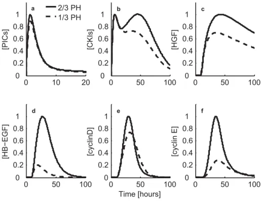
HB-EGFis an important growth factor during hepatic regeneration. HB-EGF induction is sen- sitive to the degree of damage (1/3 PH vs 2/3 PH) (Mitchell et al., 2005a) and other kind of damages (Temizer et al., 1992; Kiso et al., 1995, 1996), whilepriminggenes like IL-6, TNF-α, c-jun,c-fos,c-myc are similarly expressed in 1/3 PH and 2/3 PH. HB-EGF expression is induced by Raf/MAPK pathway (Ellis et al., 2001; McCarthy et al., 1997, 1995), which is activated by HGF viamet signaling, as mentioned earlier.
HB-EGF is expressed earlier than HGF and TGF after PH and appears to have a unique role in liver regeneration (Kiso et al., 1995, 2003). A 30% PH does not result in coordinated DNA replication, despite activation of the cytokine cascade (Bucher and Swaffield, 1964; Mitchell et al., 2005a). A single injection of HB-EGF 24 hours after 30% PH can override this blockage betweenPrimingand cell cycle progression, eliciting a wave of DNA replication. Interestingly, this effect cannot be accomplished by similarly injecting HGF or TGF (Mitchell et al., 2005b).
In addition, HB-EGF KO mice have a delay in DNA replication after 70% PH, although this deficiency is partially compensated by an earlier increase in TGF expression in these animals.
Both c-met and the EGFR are receptor tyrosine kinases, which recruit enzymes and scaffolding proteins to phosphorylated intracellular domains of each receptor. Multiple intracellular sig- naling pathways are thus activated, which regulate a multitude of transcription factors, initiate
translation, and regulate metabolic pathways. One mitogenic signal transduction pathway that is of particular interest, because it may integrate cytokine signals as well as growth factor signals, is the Ras-Raf-MEK cascade, which results in the activation of ERK1/2. ERK1/2 activation is correlated with hepatocyte DNA replication in vivo and hepatocyte proliferation in vitro (Ta- larmin et al., 1999; Thoresen et al., 2003; Li et al., 2002; Coutant et al., 2002). Moreover, growth factors such as HGF and TGF and cytokines such as TNF and IL-6 stimulate ERK1/2 activity in primary hepatocytes and hepatocyte cell lines (Argast et al., 2004; Francavilla et al., 1986;
Scheving et al., 2002).
2.2.3 CKIs during liver regeneration
Assembly of cyclins with their activating partners, cyclin dependent kinases (cdks) ,and their en- zymatic activity are regulated by a number of small proteins termed CKIs. To date, two different families of CKIs have been described in mammalian cells that differ in structure, mechanism of inhibition, and cdk target specificity. The Ink family of CKIs includes the tumor suppres- sor protein p16Ink4a, as well as p15Ink4b, p18Ink4c, and p19Ink4d, that appear to specifically target the G1 phase Cyclin D-Cdk4/Cdk6 complexes (Sherr and Roberts, 1995, 1999). The structurally and functionally distinct Cip/Kip family comprises three proteins: p21 (also known as Cip1, Waf1, Sdi1) (el Deiry et al., 1993; Harper et al., 1995; Noda et al., 1994; Xiong et al., 1993), p27Kip1 (Polyak et al., 1994; Toyoshima and Hunter, 1994), and p57Kip2 (Lee et al., 1995; Matsuoka et al., 1995). These proteins are able to bind and inhibit with different efficacy the activity of most cyclin-cdk complexes including Cdk2, Cdk3, Cdk4, and Cdk6 (Harper et al., 1995; Matsuoka et al., 1995).
A wide variety of environmental signals can regulate expression and activity of CKIs. Interest- ingly, either growth arrest resulting from DNA damage, cell senescence, and terminal differenti- ation or cell cycle entry and progression after stimulation with growth factors were accompanied by p21 gene activation (Macleod et al., 1995; Noda et al., 1994; Nourse et al., 1994; Sherr, 1994) through various transcription factors including p53 (el Deiry et al., 1993; Macleod et al., 1995).
Beyond its role as a CKIs, at low stoichiometric concentrations, p21 may act as an assembly factor for active Cyclin D-Cdk4/6 complexes and could potentially function as an activator of these kinases (Cheng et al., 1999; LaBaer et al., 1997; Sherr and Roberts, 1999). In contrast to p21, expression of p27 protein generally declines in several cell types in response to mito- genic stimulation (Agrawal et al., 1995; Nourse et al., 1994). Furthermore, inhibitory activity of this protein increases by different anti-mitogenic signals, such as TGF- or by contact inhibition (Polyak et al., 1994; Poon et al., 1995).
Several reports (Dotto, 2000; Glaise et al., 1998; Macleod et al., 1995; Matsuoka et al., 1995;
Sherr and Roberts, 1995) highlight the important difference that exists between cell types con- cerning regulation of these CKIs, expression, and role during development and differentiation.
Particularly, these proteins are involved in coordinate regulation of cell proliferation and dif- ferentiation and maintenance of terminally differentiated cells in quiescent state (Zhu and Sk-
2.2 Cell cycle during liver regeneration oultchi, 2001). Because of the high proliferative potential of mature hepatocytes at the adult stage, a specific regulation of CKIs is intended. Several prior studies (Albrecht et al., 1997, 1998, 1999; Ehrenfried et al., 1997; Jaumot et al., 1999; Kato et al., 1998; McIntyre et al., 1999;
Pujol et al., 2000; Timchenko et al., 1997) have documented the pattern of CKIs expression in proliferating hepatocytes including p21, p27, and p57. However, although some investigators demonstrated that p21 decreased in regenerating rat liver (Timchenko et al., 1997), others re- ported its upregulation during cell cycle progression of rat hepatocytes in vivo (Albrecht et al., 1999; Jaumot et al., 1999; Kato et al., 1998). In addition, most reports described expression of CKIs in vivo during regeneration of rat or mouse liver. A lack of immunohistochemical analysis could not fully support the conclusion that observed patterns of CKIs expression in regenerating liver are fully derived from hepatocytes. Only a very limited number of reports documented patterns of p21 and p27 expression in hepatocytes during rat liver regeneration by immuno- cytochemistry (Jaumot et al., 1999; Pujol et al., 2000). Although a few reports described some aspects of CKIs expression in primary mouse and rat hepatocytes in pure culture (Albrecht et al., 1999; McIntyre et al., 1999), the functional role of these proteins in proliferating cells and their regulation by different signal transduction pathways have not yet been investigated .
2.2.4 Cyclins during liver regeneration
Cell cycle progression is regulated by sets of cyclin and cdk complexes. Cyclin D, -E, and -A are cyclically synthesized during G1, G1-S, and S phases, respectively. Cyclin D, with its catalytic partner Cdk4/6, leads to phosphorylation of Rb, releasing transcriptional factors such as E2F that activate genes required for entry into S-phase. (Fausto, 2000; Sherr, 1993). Concomitant binding of Cyclin E and A with their catalytic partner Cdk2 contributes to the initiation and progression of S-phase (Kitamura et al., 1998).
Quiescent cells contain low levels of D-type cyclins. Upon mitogenic stimulation, the expression of D-type cyclins increases and then Cyclin D-Cdk4 complexes can assemble and translocate to the nucleus (Michalopoulos and DeFrances, 1997; Fausto, 2000). The activation of Cyclin D- Cdk4 complexes at mid G1 is responsible for the phosphorylation of Rb and the other members of the pocket family (p107 and p130). As a result of this phosphorylation transcription factors of the E2F family are released and the expression of genes necessary for cell cycle progres- sion is induced (Thompson et al., 1986; Alcorn et al., 1990; Morello et al., 1990; Feldenberg et al., 1999; Cressman et al., 1995). Cyclin D-Cdk4 activity is negatively regulated by CKIs, which consist of two major families. The Ink4 inhibitors (p15, p16, p18, and p19) specifically inhibit Cdk4 and Cdk6 by preventing complex formation with the D-type cyclins (Sherr and Roberts, 1995). The Cip/Kip family of proteins (p21, p27, and p57) bind and inhibit numerous cyclin-cdk complexes. The interplay between Cyclin D-Cdk4 and other G1-regulatory proteins is complex and incompletely understood . Cyclin D proteolysis requires phosphorylation by GSK3beta at Thr-286; additional work recently established that p286-D1 is a substrate for the SCF(Fbx4/alphaB-crystallin) E3 ligase (Barbash and Diehl, 2008).
Figure 2.4:
Proteolytic ligase activity during G1 and early S phase. G1 stability is sustained by APCCdh1dependent degradation of G1 cyclins and SCFSk p2 dependent degradation of G1-S cyclins. The transition into S phase is mediated by a change in activity of the SCFSk p2 complex upon inhibition of APCCdh1. Thus, proteolytic targets are altered in a phase-dependent manner according to the selectivity of the active ubiquitin ligase at that time. Maintenance of subsequent phases and their transitions also involves similar mechanisms of phase specific selectivity of ligases (Ang and Harper, 2004).
Cyclin E-Cdk2 complexes formed at mid-late G1 also phosphorylate the pocket proteins. How- ever, the major role of these complexes is accomplished in the G1-S transition, possibly by phosphorylating key proteins involved in the firing of DNA replication (Lundberg and Wein- berg, 1998; Krude et al., 1997). Cyclin A-Cdk2 complexes are necessary for S phase progression although the putative substrates for these complexes are still unknown (Sherr, 1994).
2.3 Proteolytic degradation
Ubiquitin mediated proteolysis begins through an enzymatic cascade involving E1 ubiquitin activating enzymes, E2 ubiquitin conjugating enzymes, and E3 biquitin ligase enzymes. These enzymes ultimately serve to mediate the covalent attachment of Ub molecules by the E3 ligase to a lysine residue in the target substrate, or on the growing multi-ubiquitin chain extending from the "tagged" protein. This Ub chain serves as a signal for the 26S proteasome to unfold and digest the target. Most of the target selectivity of this system is conferred by the E3 ligase because of its direct interaction with the ubiquitination substrate. The two most prominent E3 ligases in cell cycle control are the anaphase-promoting complex (APC) and the SCF complexes [reviewed in (Hershko and Ciechanover, 1998)].
APC The APC core consists of 12 subunits and is regulated by two activating subunits: Cdc20 (also known as Fizzy) and Cdh1 (also known as Hct1 or Fizzy-related) (reviewed in (Vodermaier and Peters, 2004; Harper et al., 2002; Zachariae and Nasmyth, 1999)). These activating subunits
2.3 Proteolytic degradation each confer differential substrate selectivity to APC. Moreover, each associates with the APC core under different circumstances: Cdc20 is more likely to associate with the APC core during mitosis under conditions of high Cdk activity, and Cdh1 is more likely to activate the APC core during mitotic exit and G1 phase under conditions of low Cdk activity. Substrate specificity of APC and its stage specific activation is discussed in more details in Chapter 3. Here we limit our discussion to Cdh1 associated APC and its role during G1 phase.
Cdh1 recognizes various proteins in late M and G1 phases, such as mitotic cyclins, Cdc20, Cdh1, Aurora A, Aurora B, Plk1, Skp2 (reviewed in (Castro et al., 2005)). Cdh1 levels are relatively constant throughout the cell cycle (Zachariae et al., 1998; Jaspersen et al., 1999).
Cdh1 activity is regulated by cell cycle dependent phosphorylation and dephosphorylation, being unphosphorylated in late M and G1 phases, and then phosphorylated during S, G2 and early M phases (Zachariae et al., 1998; Jaspersen et al., 1999). The phosphorylation of Cdh1 by cyclin-cdk complexes inhibits APC activation by preventing Cdh1 from binding to the core APC subunits, whereas dephosphorylation of Cdh1 due to degradation of Cyclin B at late M phase and inactivation of Cdk1, induces APC activation by allowing Cdh1 to access the core APC subunits (Kramer et al., 2000). After Cdc20 is completely degraded at the end of mitosis, APCcdh1 remains as the active form of the APC during G1 and also during the first part of S- phase (Jaspersen et al., 1999; Kramer et al., 2000).
SCF The SCF E3 ligase comprises three subunits at its core; Skp1, Cullin, and Rbx1. This core interacts with modular F-box proteins, which all share an F-box sequence motif. F-box proteins directly bind to the target substrate and bridge the interaction between the E3 ligase and target substrate, and so the identity of the F box determines the target of SCF. There are various F-box proteins, including subfamilies with WD40 domains (Fbws) and those with leucine-rich-repeats (Fbls). F-box proteins frequently recognize their substrates through phosphorylation-dependent mechanisms. In the case of Fbws, the WD40 motif recognizes a phosphodegron domain on the substrate that forms after appropriate phosphorylation events [reviewed in (Deshaies, 1999;
Koepp et al., 1999)].
The F-box-protein Skp2 bound to SCF complexes is most well studied among over 70 F-box proteins identified in human. SCF-Skp2 mainly ubiquitinates and degrades CKIs as well as G1- S cyclin (Frescas and Pagano, 2008). Controlling Skp2 activity is clearly important for proper cell cycle control. Skp2 can function as an oncogene in model systems and is overexpressed in a number of tumor types (reviewed in (Pagano and Benmaamar, 2003)). Bashir et al. (2004) and Wei et al. (2004) have indicated a Cdh1 dependent mechanism by which G1 cells maintain low levels of SCFSk p2 , thereby putting a break on S-phase entry until the criteria for S-phase entry have been met. Thus, G1 stability is maintained by APCCdh1 dependent degradation of G1 cyclins and SCFSk p2dependent degradation of G1-S cyclins. The transition into S phase is mediated by a change in activity of the SCFSk p2complex upon inhibition of APCCdh1.
Therefore, proteolytic targets are altered in a phase-dependent manner according to the selec-
tivity of the active ligase at that time. Maintenance of subsequent phases of cell cycle and their transitions also involves similar mechanisms of phase specific selectivity of ligases(Fig. 2.4).
3 S phase and mitosis
Synopsis
Mitotic activation in mammalian cells is promoted by multiple redundant controls at the tran- scriptional, posttranslational and degradation level, but the relative contribution of these sepa- rate pathways to the decision to enter mitosis is currently not well resolved. There is a lot of emerging FoxM1 mediated transcriptional and Cdh1 mediated proteolytic control of Cyclin B in mammalian cells which is developing a new level of understanding for the mammalian cell cycle. This chapter summarizes the known molecular and cellular pathways observed in the con- trol of mitotic cyclins and the mechanisms leading to activation, inactivation and degradation of cyclins.
3.1 Introduction
Early in the cell cycle, the DNA is replicated and chromosomes duplicated in the S phase. The second major phase of the cell cycle is the M phase, which typically consists of two events:
nuclear division (mitosis) and cell division (cytokinesis). The period between the end of one M phase and the beginning of the next is called interphase.
Mitosis is a complex and beautiful process that distributes the duplicated chromosomes equally into a pair of daughter nuclei. The pairs of sister-chromatids are attached in early mitosis to the mitotic spindle, a bipolar array of protein polymers called microtubules. By the midpoint of mitosis (metaphase), sister-chromatids in each pair are attached to microtubules coming from op- posite poles of the spindle. At the next stage (anaphase), sister-chromatid cohesion is destroyed resulting in sister-chromatid separation. The microtubules of the spindle pull the separated sis- ters to opposite ends of the cell (sister-chromatid segregation) and the two sets of chromosome are each packed into new daughter nuclei. Following mitosis cell itself divides by cytokinesis.
Mitosis is preceded by gap phase called G2 when cells prepare themselves for mitosis (Fig. 3.1).
Cell cycle events are regulated at three regulatory checkpoints. First is the G1-S checkpoint which we have discussed earlier in chapter 2. There are two checkpoints during the G2/M transition where the progression of cell cycle can be blocked. Failure to complete DNA repli-
Figure 3.1:
Events of cell division cycle. The central events of cell reproduction are chromosome duplication, which takes place in S phase, followed by chromosome segregation and nuclear division (cytokinesis), which are collectively called M phase. G1 is the gap phase between S and M phases; G2 is the gap phase between S and M phases. At metaphase sister chromatids are aligned on the mitotic spindle and during sister chromatids get separated and pulled to opposite spindle poles (Morgan, 2007).
cation blocks the cell in G2 phase from entry into mitosis. Delay in spindle assembly blocks the metaphase-to-anaphase transition, thereby preventing sister-chromatid segregation until the spindle is ready. Cell cycle can thus be viewed as a linked series of tightly regulated molecular switches, each of which triggers the initiation of cell cycle progression at a specific regulatory checkpoint.
Cyclin oscillations regulating the cell cycle control system The whole process of cell divi- sion is mainly orchestrated by cdks. As the cells progress through the cell cycle, abrupt changes in the enzymatic activities of these kinases lead to changes in phosphorylation state and thus the state of activation of proteins that govern the cell cycle processes. Concentrations of cdks are constant throughout the cell cycle. Oscillations in their activity depend on the corresponding oscillations in the levels of their respective cyclin subunits. Different cyclins are produced at different cell cycle stages with additional controls imposed on them by various other cell cycle regulators (Fig. 3.3), resulting in a series of cyclin-cdk complexes which govern distinct cell cycle events (Fig. 3.2).
Cyclin D and Cyclin E form active complexes with cdks during G1-S transition which trigger DNA synthesis as discussed in chapter 1. The rise of G1-S cyclins is accompanied by the appear- ance of Cyclin A which also forms complexes with cdks. Cyclin A-Cdk2 is thought to initiate chromosome condensation during prophase. Towards the end of S phase, Cyclin B expression
3.2 E2Fs regulating transcription of cyclins
Cyclin D, Cyclin E Cyclin A Cyclin B
SCF
G1/S APC APC
Figure 3.2:
Overview of the cell cycle control system. Levels of three major cyclin types oscillate during the cell cycle (top), providing the basis for oscillations in the cyclin-cdk complexes that drive cell cycle events (bottom). In general, cdk levels are constant and in large excess over cyclin levels. Thus, cyclin-cdk complexes form in parallel with cyclin levels. The enzymatic activities of cyclin-cdk complexes also tend to rise and fall in parallel with cyclin levels, although in some cases cdk inhibitor proteins or phosphorylation introduce a delay between the formation and activation of cyclin-cdk complexes. Formation of active G1-S cyclin-cdk complexes commits the cell to a new division cycle at G1-S checkpoint. G1-S cyclin-cdks then activate the S cyclin-cdk complexes that initiate DNA replication at the beginning of S phase. M-cdk cyclin activation occurs after the completion of S phase, resulting in progression through the G2/M checkpoint and assembly of the mitotic spindle. Proteolytic ligases (APC and SCF) impose another level of control on cell cycle oscillations. At the metaphase-to-anaphase transition APC activation triggers sister-chromatid separation. APC activation also causes the destruction of S and M cyclins and thus the inactivation of cdks, which promotes the completion of mitosis and cytokinesis. APC activity is maintained in G1 until G1-S cyclin-cdk activity rises again and commits the cell to the next cell cycle. G1-S cyclins are further degraded by SCF (adapted from Morgan (2007)).
is switched on, leading to accumulation of Cyclin B-Cdk1 complexes during G2. Switch like activation of Cyclin B-Cdk1 complexes trigger the G2-M transition (Fig. 3.4). Spindle assembly and other early mitotic events lead to the alignment of duplicated sister chromatids on the mitotic spindle in metaphase. In addition to that Cyclin B-Cdk1 eventually stimulates activation of APC, which triggers the metaphase-to-anaphase transition by stimulating the destruction of proteins that hold sister-chromatids together. The APC also causes destruction of S and M phase cyclins, resulting in the inactivation of all major cdk activities in late mitosis. Increased production of CKIs also occurs in late mitosis. The resulting inactivation of cdks allows dephosphorylation of their mitotic targets, which is required for spindle disassembly and the completion of M phase.
Low levels of cdks are maintained until late in the following G1, when rising G1-S cyclins again commit the cell to a new cycle (Morgan, 2007).
CKIs
Cdc25
Wee1 APC-Cdc20
APC-Cdh1 SCF
Figure 3.3:
Regulators of cell cycle control system. Cyclins are periodically expressed during cell cycle. Cyclin-cdk complexes are also formed in parallel with cyclins. Most crucial for the cell cycle transitions are the activity levels of cyclin- cdks. Properties like switch-like activation and delay in activation are introduced by various cell cycle regulators.
CKIs prevent premature activation of Cyclin D, Cyclin E and Cyclin A by stoichiometrically inhibiting them. Cdc25 activates and Wee1 inactivates Cyclin B-Cdk1 in a positive feedback manner by phosphorylation-dephosphorylation events enabling switch-like activation of Cyclin B-Cdk1 at G2/M transition. Proteolytic degradators APC and SCF control the timely phase specific degradation of cyclins. APC with its activator subunit Cdc20 degrades Cyclin B at mitosis. With its another activator subunit Cdh1, APC degrades Cyclin B and Cyclin A till G1. SCF degrades Cyclin D and Cyclin E from Late G1 to S phase (adapted from Vermeulen et al. (2003)).
3.2 E2Fs regulating transcription of cyclins
E2F family of transcription factors are known to control the expression of various genes respon- sible for entry into and progression through S phase (e.g. Cyclin D, Cyclin E, Cyclin A). The applications of new technologies such as DNA microarray analysis, chromatin immunoprecipi- tation (ChIP) techniques and bioinformatics has enlarged the view of the number and nature of genes potentially regulated by E2F, including various G2 (e.g. Cyclin A) and M phase genes (e.g. Cdc2, Cyclin B) (Ren et al., 2002; Zhu et al., 2004; Osterloh et al., 2007).
E2F proteins can be (1) activators of transcription (E2F1, E2F2 and E2F3a) or (2) repressors of transcription (E2F3b, E2F4, E2F5 and E2F6) (Calzone et al., 2008a). For simplicity we only talk about the activator E2Fs and lump all three of them into one entity named E2F. E2F activity is tightly controlled by binding to retinoblastoma protein (Rb). Rb belongs to a family of pocket
3.2 E2Fs regulating transcription of cyclins
Figure 3.4:
Switch-like activation of mitotic players at G2/M transition. At G2/M transition the cell enters mitosis in an all-or- none fashion which is enabled by switch-like activation of Cdk1 associated with Cyclin B. Increased Cdk1 activity triggers the activation of APCCdc20, causing rapid Cyclin destruction and Cdk1 inactivation. Cdk1 inactivation, leads to APCCdh1activation, which further degrades Cyclin B till S phase (adapted from Morgan (2007)).
proteins which have the ability to bind proteins to their pockets. The protein binding function of Rb is regulated by phosphorylation. Rb sequesters E2F and inhibits its transcriptional activity.
This hold of Rb on E2F depends on its phosphorylation level; higher the Rb phosphorylation level more the E2F is released from its hold and is available for transcriptional activation of further downstream genes.
Phosphorylation of Rb is regulated by various cdks. Complete phosphorylation and inactivation of Rb via cyclins occurs in a sequential and cooperative manner (Knudsen and Wang, 1996;
Zarkowska and Mittnacht, 1997). At mid G1 Cyclin D-Cdk4/6 complexes initiate the phospho- rylation of Rb. Cyclin D can only achieve partial phosphorylation of Rb. Complete phosphory- lation of Rb and subsequent release of E2F in excess requires further phosphorylation of Cyclin D hypophosphorylated Rb by Cyclin E. Cyclin E hyperphosphorylates Rb during G1-S phase (Lundberg and Weinberg, 1998) (Fig. 3.5).
E2F together with B-myb is required for the activation of Cyclin B gene in G2. B-myb is an E2F target gene expressed at G1-S, but is not fully active until phosphorylated by Cyclin A-Cdk2 in S phase. This provides a possible explanation for delayed transcriptional activation of Cdc2 and Cyclin B in G2 (Zhu et al., 2004; Osterloh et al., 2007). Thus E2F also provides a link between G1-S and G2-M specific transcription and also provides a mechanism by which the temporal distinction is achieved. However, for simplicity reasons we do not explicitly consider B-myb mediated regulation of Cyclin B via Cyclin A.
Figure 3.5:
Sequential activation of Rb-E2F. Phosphorylation of Rb is regulated by various cyclin-associated-kinases. Complete phosphorylation and inactivation of Rb via cyclins occurs in a sequential and cooperative manner. At mid G1, Cyclin D-Cdk4/6 complexes initiate the phosphorylation of Rb. Cyclin D can only achieve partial phosphorylation of Rb.
Complete phosphorylation of Rb and release of E2F in excess requires phosphorylation of Cyclin D hypophosphory- lated Rb by Cyclin E. Cyclin E hyperphosphorylates Rb during G1-S phase.
3.3 Cyclin dependent kinase inhibitors (CKIs)
Cyclin dependent kinase inhibitors (CKIs) provide additional regulation to the timely cell cycle dependent expression of cyclins. CKIs are grouped into two categories: Ink4 and Cip/Kip. Ink4 proteins inhibit Cyclin D associated kinase activity. Cip/Kip family consists of three members:
p21, p27 and p57. These proteins share a homologous inhibitory domain, which is both neces- sary and sufficient for binding and inhibition of Cdk4- and Cdk2 containing complexes. These proteins act as stoichiometric inhibitors of Cdk2 and Cdk1; and they preferentially act on Cdk2 complexes (Vidal and Koff, 2000). p21 binds to all four cyclin-cdk complexes with a preference for those containing Cdk2 and inhibits their activation by generally blocking their catalytic sites.
However, the mechanism behind CKIs dependent negative regulation of Cyclin B levels is still not clear (Gillis et al., 2009; Tyner, 2009).
p21 is predominantly transcriptionally regulated via IL-6 dependent STAT3, Myc and E2F (Gar- tel and Tyner, 1999; Coller et al., 2000), but recently its post-transcriptional control is reported to be equally important for its stability and degradation(Jascur et al., 2005; Sheaff et al., 2000). p27 mRNA levels remains majorly constant. Regulation of p27 protein is more complex involving