The role of liver regeneration-associated protein ALR, Augmenter of Liver Regeneration,
in cholestatic liver diseases
Dissertation to obtain the Degree of Doctor of Natural Sciences (Dr. rer. nat.)
from the Faculty of Chemistry and Pharmacy University of Regensburg
Presented by
Sara Ibrahim from Damascus (Syria)
December 2018
The work presented in this thesis was carried out at the University Children Hospital in Regensburg from January 2015 to December 2018 under the supervision of Prof. Dr. Thomas S. Weiß.
This work was printed and published thanks to financial support from the German Academic Exchange Service (DAAD)
Gedruckt bzw. Veröffentlicht mit Unterstützung des Deutschen Akademischen Austauschdienst (DAAD)
Date of colloquium: 10.12.2018
Board of examiners: Chairman: Prof. Dr. Jens Schlossmann
First Examiner: Prof. Dr. Achim Göpferich
Second Examiner: Prof. Dr. Thomas S. Weiß
Dedicated to the memory of my Mother, Emtithal Saleh, and my
Father, Mohammad Ibrahim.
"Do the best you can until you know better. Then when you know better, do better."
Maya Angelou (1928-2014)
Table of contents
Chapter 1 Introduction 1
Chapter 2 Aims of the thesis 53
Chapter 3 Regulation of ALR by bile acids 57
Chapter 4 Regulation of ALR by cholestasis-related cytokines 79 Chapter 5 Role of cytosolic sfALR in altering cholestatic injury 95 Chapter 6 Role of recombinant ALR in reducing bile acids toxicity
during liver regeneration 119
Chapter 7 Conclusion and summary 135
Appendix Abbreviations 143
Expression plasmids: Backbone maps 149
Acknowledgements 155
Publications and Presentations 159
Eidesstattliche Erklärung 163
Chapter 1
Introduction
Chapter 1
1. Introduction - Augmenter of Liver Regeneration (ALR)
Liver regeneration is a well-orchestrated process that is triggered by hepatocellular death and can be induced by toxins, viral infections or by tissue loss due to trauma or surgical resection [1]. The phenomenon of liver regeneration has been known since the myth of Prometheus who stole fire from Zeus and gave it to mankind. As a punishment, Zeus’s eagle would feast on Prometheus’s liver each day, and his liver would regrow at night until the eagle’s return [2].
In the early twentieth century, scientists became interested in identifying the factors that regulate hepatic growth and regeneration. The first scientific evidence of the liver’s ability to regenerate was confirmed by performing partial hepatectomy in rats and showing the restoration of the liver mass [3]. In 1975 LaBrecque and Pesch [4]
prepared an extract from weanling rat livers and demonstrated that injecting this substance into partially hepatectomized rats stimulates liver regeneration and therefore named it Hepatic Stimulatory Substance (HSS). Several attempts were applied to further identify and characterize the molecule responsible for the stimulation of liver regeneration [5-7]. After progressive purification of HSS, a fraction with augmenting effect following a 40% hepatectomy in rats was obtained containing three protein bands (14, 30 and 35 kDa) [8]. The 30 kDa band was chosen for further sequencing and was published in 1994 by by Hagiya et al., who first coined the term
“Augmenter of liver Regeneration, ALR” [9]. The sequenced rat protein consisted of 125 amino acids with a molecular weight of 15 kDa under reducing conditions and 30 kDa under non-reducing conditions [9]. The published rat sequence was then corrected in 1995 [10] and proposed the presence of three ATGs and therefore opened the possibility of multiple isoforms of ALR. Afterwards, the sequence of human and mouse ALR was published in 1996 [11]. Moreover, Hepatopoietin (HPO) was cloned and sequenced from human fetal liver tissue in 1997 and was proved to be identical with ALR, therefore HPO is also referred to as ALR [12].
ALR protein is encoded by GFER (growth factor erv1 homolog of s. cerevisiae) gene
that consists of 3 exons and 2 introns [10, 13] and can be regulated by different
stimuli. In this review, we aimed to systemically review the literature discussing ALR
structure, regulation, function and clinical importance. We included original articles,
published in English, investigating ALR, HPO or GFER and presenting a clear
indication of ALR isoform and molecular size. This is of note and critical, since there
studies, a purified protein fraction (HSS) instead of a single sequenced protein (ALR) (e.g. [14-16]). Furthermore, a few reports indicated that HSS has a molecular size of 15 kDa but then claimed that ALR is different from HSS [17], without presenting a sequence for HSS. Due to the ambiguity about HSS protein/fraction, articles on HSS were only taken into consideration, if a clear indication of gene name and molecular size are given.
Although a considerable size of work has been devoted to summarizing the knowledge on ALR (see previous reviews [1, 18, 19]), an investigation of ALR expression in different organs as well as delineation between the ALR isoforms and the function of endogenous and exogenous ALR is still missing. Therefore, we aspired to revise the knowledge about ALR and gather evidence of the expression of different ALR isoforms and the functional role of the endogenous or exogenously applied ALR as well as to highlight the clinical importance of this protein.
2. ALR isoforms and structure
The first gene sequence of ALR was reported in 1994 [9] and was later corrected in 1995, where an additional G was added at position 266. This generated two additional in-frame ATG initiation sites and raised the possibility of different ALR isoforms [10]. Indeed, the expression of different ALR isoforms in mouse, rat and human organs has been thoroughly investigated and the existence of three isoforms of ALR has been shown (Table I). Furthermore, the sequence of full length ALR shows 74% homology to mouse ALR and 73% homology to rat ALR (Fig. 1A). The longest transcript of ALR encodes a 205 amino acid protein corresponding to a molecular weight of 23 kDa [20]. Moreover, the smallest transcript of ALR encodes a 125 amino acid protein corresponding to a molecular weight of 15 kDa (short form ALR, sfALR) [13]. The sfALR is 80 amino acids shorter than lfALR at the N terminus [20] (Fig. 1B). Those 80 amino acids are also referred to as “N80”. N80 includes (i) N- terminal (N-N80) domain which is described as an IDD (intrinsically disordered domain) and acts as a crucial targeting signal from the cytosol into the mitochondria [21]. (ii) C-terminal of N80 (C-N80) contains the CRAC motif (71-74 aa) (Fig. 1B).
CRAC motif acts as a recognition site in the disulfide relay system of mitochondrial
inter-membrane space (IMS). CRAC mediates the “recycling” of Mia40, an
oxido-reductase, in charge of oxidative protein folding in the IMS [21-23]. Moreover, it
was previously shown that mutating the cysteine residues (C71 or C74) in ALR’s
Chapter 1 CRAC motif leads to preventing the reaction with Mia40 [23]. Nevertheless, Mia40 can still exchange electrons with ALR through the CEEC motif (142-145 aa) (corresponds to CXXC motif in Erv1, yeast homologue of ALR) (Fig. 1B) but this exchange is less efficient due to the “buried” structure of the CEEC motif. This argues for a specific hydrophobicity near the CRAC motif of long form ALR that drives the reaction between Mia40 and the N terminus of ALR [23]. In addition, Lu et al. have demonstrated that interaction between ALR and JAB1 occurs via amino acids 81-143 (corresponds to 1 to 63 aa in sfALR) [24] (Fig. 1B). Furthermore, the CEEC motif (Fig. 1B) neighbors the FAD binding domain and is affected by the reaction between Mia40 and FAD. Moreover, the “CEEC-FAD” region of ALR is referred to as “redox active center” that donates electrons to Mia40 [23].
Additionally, disulfide bonds formation in ALR (Fig. 1B) is of importance for ALR
functionality. C95 and C204 are responsible of forming head-to-tail dimers of all
isoforms of ALR and therefore stabilizing the structure [25]. The dimerization also
facilitates the binding to the FAD molecule by non-covalent bonds [26, 27]. Other
cysteins, C171 and C188 form “intramolecular” disulfide bonds that flank the FAD
molecule. It is worth to mention that C154 and C165 (non-conserved cysteins) in
human ALR (Fig. 1A) may account for oligomers formation ofter seen during the
preparation of recombinant human ALR and mutating those cysteins prevented the
formation of oligomers in recombinant human ALR [23].
Fig. 1. (A) An illustration of ALR sequence homology in human, mouse and rat is shown. Sequences of rat, mouse and human ALR were obtained from Pubmed search tool. Dashes indicate absence of the amino acid, black frames the non-conserved cysteins and * the initial methionines. (B) The structure of human ALR isoforms. Long forms (23 and 21 kDa) of ALR posess additional 80 amino acids (N80) at the N terminus of the short form ALR (15 kDa). N80 harbors the IDD and CRAC motif, which are responsible for mitochondrial localization and Mia40 recognition. The CEEC motif (corresponds to CXXC in yeast homologue Erv1) and the neighboring FAD binding domain, which “recycle” Mia40 by catalyzing the electron transfer from ALR to Mia40. Disulfide bonds formed by cysteins are indicated by arrows; D: functional dimerization; O: Oligomerization (non-conserved cysteins) and IM: intramoleclular bonds. IDD: intrinsically disordered domain.
Chapter 1
3. Regulation of ALR
The human ALR gene (GFER) (growth factor erv1-like) has been mapped on chromosome 16 next to the polycystic kidney disease gene (PKD1) [28]. Analysis of ALR gene (Fig. 2) has shown that it consists of 3 exons and 2 introns [10, 13]. ALR gene promoter has been shown to be TATA-less and to have some characteristics of an oncogene and a growth factor [29]. It further showed an initiator-like element with three tandem repeats (-66, -1) [29]. It was shown that regulatory elements of ALR promoter might exist in the region between -447 and -49bp. interestingly, it was found that a putative AP1/AP4 binding site (-375, -369) is responsible for negative transcriptional activity of ALR by AP1 (Activator Protein 1) in HepG2 cells and AP4 (Activator Protein 4) in COS-7 cells [30]. Additionally, it was reported that ALR expression could be attenuated by EGF (epidermal growth Factor) via C/EBP
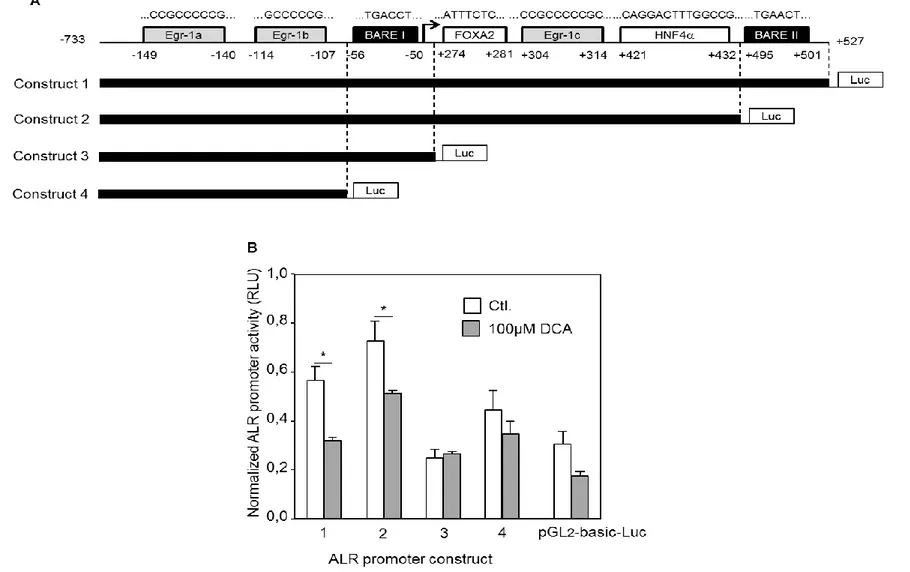
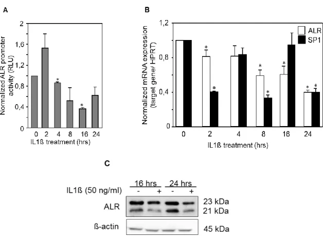
(CCAAT/enhancer binding proteins) binding to its binding site (-292, -279) within ALR promoter [31]. Analyzing a selected promoter region (-252, -49) from the trans- criptional start site has shown that HNF4 (hepatocyte nuclear factor 4 alpha) binds to a binding site (-209, -204) and reduces ALR expression [32]. In addition, SP1 (specific protein 1) positively regulates ALR expression by binding to its binding site at (-152, -145) [32]. Interestingly, a recent study including the first intron of GFER gene (-733 to +527 bp) [33] investigated the potential regulatory elements in the introns that may affect the expression of different genes [34, 35]. It was demonstrated that HNF4 binds at (+421, +432) to ALR promoter and induces ALR expression [33] despite the presence of an upstream HNF4 binding site with repressing activity. However, the inducing effect of HNF4 is diminished upon SHP (Small Heterodimer Partner) activation e.g. by bile acids [33]. Exposure of hepatocytes to oxidative stress induces Nrf2 (nuclear factor erythroid 2-related factor 2), which in turn, by binding to an anti-oxidant response element (ARE) located between (-27, -19), induces ALR expression [36]. Interestingly, involvement of Nrf2 in ALR regulation was further confirmed by studies showing ALR induction upon treatment with known Nrf2 inducers such as tBHQ (tertiary butylhydroquinone), HBV (hepatitis B virus) infection [36], phenethyl isothiocyanate and sulforaphane [37, 38].
In addition, FOXA2 (Forkhead Box A2, also known as HNF3) binds to a binding site
(+276, +282) within ALR promoter inducing ALR expression and this binding is
response protein-1) that binds to its binding site (+304, +314) and induces ALR expression [33].
Furthermore, phenylbutyrate, a HDAC1 (histone deacetylase 1) inhibitor and demethylation reagent, was shown to induce the expression of ALR in the brain of a Huntington's disease mouse model [40]. This proposes the possibility that ALR could be regulated by histone acetylation and/or methylation [40] and therefore by epigenetic mechanisms [41, 42]. Additionally, MIF (macrophage Migration Inhibitory Factor), a pluripotent cytokine involved in cell cycle, cell proliferation and liver regeneration, has been demonstrated to increase the promoter activity of ALR [43].
Interestingly, a recent report has suggested that ALR may be regulated by miRNAs and showed that Toluene exposure up-regulated. miR7109-5p, which may regulate ALR [44].
Fig. 2. Schematic illustration of ALR promoter region, transcription factors and response elements involved in ALR regulation. SP1, FOXA2, Egr-1 and HNF4 (+421, +432) are inducers of ALR expression, however, the induction by HNF4 (+421, +432) is reversed by SHP which is activated by bile acids. Furthermore, AP1/AP4, HNF4 (-209, -204) and C/EBPβ (induced by EGF) are repressing factors of ALR expression. IL6-RE-BP increases the activating effect of FOXA2 whereas ARE (-27, -19) induces ALR expression by binding of Nrf2 to this response element. The latter could be activated by HBV infection, tBHQ, sulforaphane and phenethyl isothiocyanate. Moreover, HDAC1-inhibitor phenylbutyrate and MIF activate the expression of ALR whereas Toluene exposure represses ALR expression via miR7109-5p. MIF: macrophage migration Inhibitory Factor, HDAC1: histone deacetylase 1, C/EBPβ: CCAAT/enhancer binding proteins.
4. Location of ALR
Expression of ALR mRNA was detected in several tissues and was found to be
highly expressed in the liver, testis, brain and kidneys. Moreover, low levels of ALR
mRNA were detected in the heart, muscles, spleen and lungs [10, 11]. Interestingly, it
Chapter 1 was shown that mRNA expression of ALR in muscle tissues differs between males and females between 18 and 35 years of age [45]. A variety of studies were published analyzing the expression of ALR protein expression, including its isoforms and their respective cellular localization. Table (I) summarizes reports with detailed information about ALR isoform (molecular weight), organ and organelle affiliation. In the rat brain, ALR was found to expressed in its long forms (23 and 21 kDa) [46, 47]
in neurons and glial cells located in the nucleus and mitochondria [47]. Furthermore, this was partly confirmed in a study demonstrating cytosolic expression of these isoforms in a human glioma cell line [48]. In addition, cytosolic expression of 15 kDa ALR (short form ALR, sfALR) was detected in human neuroblastoma cell lines [49].
All ALR isoforms (23, 21 and 15 kDa) have been identified in rat muscle tissue [46]
and other studies performing electron microscopy demonstrated ALR localization in the mitochondrial inter-membrane space or associated to mitochondrial cristae in human muscle fibers [45, 50]. Moreover, all three ALR isoforms were detected in rat kidneys [46] and the short form ALR (15 kDa) expression was reported in the renal cortex as well as medulla of rat kidneys after injury [51, 52]. In addition, testis of mouse [11, 46] and rat [46] also showed positive expression of long form isoforms (23 and 21 kDa). However, Since ALR was first detected in the livers of weanling rats, the hepatic expression of ALR isoforms has been thoroughly investigated. The first report demonstrating ALR protein expression by Giorda et al. [11] showed western blots with ALR bands of 40, and 23 kDa under non-reduced conditions in rat and mouse liver tissue, and a 28 kDa band was also detected in rat liver. In the meantime, the expression of ALR under reducing conditions showed 2 bands (~23 and ~21 kDa) in mouse [36, 53, 54] and rat [46, 54-56]. Using non-reducing conditions revealed expression of 3 bands (40, 38 and 36 kDa) of ALR in rats [57]
and in normal as well as disease-mouse models (NASH and ALD) [53, 57].
Interestingly, in a study that used liver-specific knock-out of ALR (by an
albumin-Cre/LoxP system) it was shown that ALR protein expression, after being
repressed at birth, reappeared in the livers of the KO-mice after 1 year. The same
study indicated the presence of oval cells (hepatic progenitor cells, HPC) in the
KO-mice [57] which might explain the recurred expression of ALR by activation of
HPCs and consequently giving rise to cholangiocytes or hepatocytes [58]. Gandhi
and his colleagues did not describe ALR expression in cholangiocytes [53, 57, 59],
both in hepatocytes and cholangiocytes [36, 60, 61], a potential expression of ALR in HPC might be proposed. So far, expression of ALR in non-parenchymal liver cells could not be detected [36, 61] except noted in one study [59] with low expression of ALR in stellate, Kupffer and hepatic endothelial cells, which was not detectable after 2 days of cell culture. It is worth to mention that in human liver tissue, hepatocytes and hepatoma cell lines under reducing conditions ALR is expressed up to all three isoforms (23, 21, 15 kDa) and in several bands (40, 38, 36 kDa) under non-reducing conditions [57]. Furthermore, it was demonstrated that hepatoma cell lines (HepG2, Huh7 and Hep3B) express the 23 and 21 kDa ALR [33, 36, 54, 62] in the mitochondria as well as cytosol [54]. Moreover, primary human hepatocytes (PHH) were analyzed and showed expression of 3 isoforms of ALR in the cytosol, whereas 23 and 21 kDa were additionally found in the mitochondria [54]. Another study by Li et al. using human fetal, adult normal and tumorous liver tissue reported 23 kDa ALR in the cytosol and its 15 kDa isoform in the nucleus [63]. In addition, expression of ALR was investigated in normal [54, 61, 62], HCC (hepatocellular carcinoma) [61-64], CCC (cholangiocellular carcinoma) [61], and HBV positive [36] liver tissue and demonstrated reduced ALR expression in normal liver compared to diseased tissues.
The expression of three isoforms of ALR (23, 21, 15 kDa) is attributed most likely to the presence of 3 initial in-frame ATG initiation sites in ALR gene [10]. Therefore it is of interest to find out why more or less isoforms of ALR are expressed in different tissues and under conditions of disease. It was hypothesized that in rats the 22 kDa isoform is post-translationally modified by dimerization to form the observed 40, 38 and 36 kDa bands under non-reducing conditions [59]. Recently published data by the same group showed two isoforms of ALR (22 and 20 kDa) under reducing conditions [53]. Considering the estimated molecular sizes on SDS-PAGE gells and the detection of 22 and 20 kDa bands of ALR under reducing conditions, the three bands (40, 38, 36 kDa), detected under non-reducing conditions, may be due to the formation of homo- and hetero-dimers of the 22 and 20 kDa ALR.
Finally, contradictory evidence about the expression of ALR isoforms may be due to
technical limitations like resolution of gels used in SDS-PAGE or the specificity and
sensitivity of anti ALR antibodies for immune detection. For example, our group
demonstrated the expression of a 23 kDa ALR in normal, cirrhosis, HCC and CCC
samples under reduced conditions, but also showed multiple unresolved bands under
non-reducing conditions [61]. Nevertheless, more recent studies of our group using
Chapter 1
various new developed anti-ALR antibodies have shown that ALR is expressed in
human liver in 3 different isoforms: 23, 21 and 15 kDa [54, 62]. Therefore, the
variable findings in detecting ALR isoforms might in part be attributed to reduction of
protein lysates, the quality of gel resolution and the quality of antibodies.
Table (I): Summary of studies investigating expression of ALR protein isoforms in different organs
Organ Species Remarks Cell type Sub-cellular localization Isoform (kDa) References
Brain
Rat
diencephalon, cerebellum, brainstem
neurons, glial cells nucleus, external envelope of mitochondria
23, 21
[47]
brain, cerebellum - - 23, 21 [46](1)
Human
- neuroblastoma cell line
SH-5YSY
Cytosol 15
[49](2)
- glioma cell line (T98G) Cytosol 23, 21 [48]
Skeletal Muscle
Rat - - - 23, 21, 15 [46]
Human mitochondrial myopathy patients muscle fibers
mitochondrial
innermembrane and cristae, cytosol
- [45, 50]
Kidney Rat
- - - 23, 21, 15 [46]
induced expression of ALR in renal cortex and outer strip of outer medulla upon acute kidney injury (AKI) (Gentamicin- or I/R- induced injury)
renal tubular epithelial cells
after AKI ALR was detected in the cytosol and apical plasma membrane
15 [51, 52]
induced expression of ALR after
obstructive nephropathy 15 [65]
Testis Mouse - - soluble,
insoluble fractions
40, 38, 23
23 (n.r.)(3) [11](4)
Chapter 1
seminiferous tubules spermatogonia , elon- gated sperm cells
mitochondrial inner mem- brane
23, 21
[46]
Rat - - - 23, 21 [46]
Blood
Mouse blood plasma after hemorrhagic shock
- - ELISA(5)
[66]
Rat
blood from liver (negative in venous and arterial blood)
- - 23, 21
[46]
serum after PCS (portacaval shunt), endotoxemia, gram- negative sepsis
- - ELISA(5)
[66]
serum after partial hepatectomy - - 21 [56]
serum after partial hepatectomy - - ELISA(5) [59]
culture medium of hepatocytes upon TNFα, LPS, Actinomycin D or MMS treatment
- - ELISA(5)
[66]
Human
serum of patients - - ELISA(5) [67]
serum of patients - - ELISA(5) [60]
- leukemic T cells - 23 [68]
serum of patients with various liver diseases
- - ELISA(5)
[69]
- multiple Myloma cell 23, 21
[70]
Liver
Mouse
- - - 40, 23 (n.r.)(3) [11]
- - - 23, 21 [36]
Liver
normal, ALD, NASH liver tissue hepatocytes - 40, 38, 36 (n.r.)(3) [57]
normal and after EtOH feeding - 40, 38, 36 (n.r.)
and 22, 20 (r.)(3) [53]
- - - 22, 20 [54]
Rat
- - - 40, 28, 23 (n.r.)(3) [11]
- - - 23, 21 [46]
- hepatocytes, sparely in
non-paranchymal cells
cytosol 40, 38, 36 (n.r.)(3)
[59]
- - cytosol 23, 21 [56]
- - - 23, 21 [55]
- - - 22, 20 [54]
Human
hepG2 cells cytosol, perinucleus region - [24]
human liver (fetal, adult and tumor)
- cytosol, nucleus 23 (cytosol),
15 (nucleus) [63]
- hepatocytes,
cholangiocytes
cytosolic, perinuclear immunostaining
23 [61]
- cell line SMMC-7721 mitochondria 23 (mitochondria),
15 [71](6)
Chapter 1
- hepatocytes, rarely in
cholangiocytes and endothelial cells
cytosol, negative in nucleus -
[60]
HepG2 mitochondria, cytosol 23, 21 [33, 36, 54, 62]
Liver
human liver samples (normal and HCC)
- - 23, 21, 15
[62]
human liver sections (normal, ALD, HBV postive)
hepatocytes and cholangiocytes
- -
[36]
human liver sections hepatocytes cytosol, barely in nucleus - [64]
human liver samples (normal, ALD, NASH, HCV)
- - 40, 38, 36 (n.r.)(3)
[57]
PHH lfALR in mitochondria,
cytosol, sfALR in cytosol
23, 21, 15
[54]
Hep3B - 23, 21 [54]
normal liver tissue - - 23, 21, 15 [54]
- Huh7 - 23, 21 [54]
Cervix Human - Hela cells mitochondria 23 [72]
(1) Klissenbauer et al. have investigated the expression of ALR protein in different rat tissues, 23 kDa ALR was detected in heart. Moreover, 23 and 21 kDa ALR were detected in fat tissue, spleen and lungs.
(2) Plomino et al. (2009) have pointed out the possibility of ALR up-take upon treatment with recombinant ALR (15 kDa).
(3) (n.r.) stands for non-reduced conditions and demonstrated analysis of ALR dimers, whereas (r.) stands for analysis of ALR under reduced conditions.
(5) ALR detection using ELISA includes all isoforms of ALR.
(6) Cao et al. (2009) investigated the expression of ALR in SMMC-7721 cell line and detected 15 and 23 kDa ALR, the latter was localized in the mitochondria. Nevertheless, a recent report has suggested not considering the studies including BEL7402, SMMC7721 or SKHEP1 cell lines due to the highly possible contamination with Hela cells which was reported in different laboratories in Asia and Europe. [73].
Chapter 1
5. Clinical importance of ALR 5.1. Mutations in GFER gene
Over the last years, the clinical importance of ALR has been underlined by studies that identified several mutations in ALR gene (GFER) (Fig. 3) leading to severe mitochondrial disorders. The first described mutation in ALR gene (GFER) (R194H) was reported to cause an infantile mitochondrial disorder [74]. Di Fonzo et al.
reported three children who suffered from, among others, progressive myopathy, partial combined respiratory chain deficiency and development delays. Later on, the mutation (R194H) was characterized structurally and enzymologically in both lfALR (23 kDa) and sfALR (15 kDa) and was demonstrated to adversely affect the stability of both isoforms of ALR (characterized by weaker FAD binding and lower thermal stability) with minimal effects on its enzyme activity (characterized by minor changes to interaction with Mia40 and cytochrome C) [75]. However, in a more recent study it was emphasized that the mutated site (R194H in human ALR that corresponds to R182H in yeast Erv1) causes lower thermal stability and the quick release of the FAD cofactor but also pointed out the loss of enzyme activity (interaction with Mia40) [76].
Furthermore, North et al. [77] described a patient at the age of 18 months with
neonatal onset of lactic acidosis, poor feeding, irritability, hepatomegaly, bilateral
cataracts and adrenal insufficiency. This patient was diagnosed with a mitochondrial
disorder, but the genetic etiology was unknown. At the age of 19 years old, the
patient was presented with significant muscle weakness in the face, neck, trunk and
proximal portions of all extremities [78]. Using “Next generation sequencing” (NGS)
the R194H mutation as well as another mutation in GFER gene (Q125X; C.373C>T)
(Fig. 3) were reported. This mutation (Q125X) was predicted to cause loss of normal
function of ALR protein either by premature protein truncation or nonsense mediated
mRNA decay. In addition, three further GFER mutations (C.259-25_259-24delCA,
C.219delC, C.217delG) (Fig. 3) were identified in a study and were suggested to
cause frame shift variations or truncation of ALR [79]. Interestingly, in a recent report,
the authors suggested using rapid targeted genomics to analyze genetic defects in
GFER gene in critically ill infants, since they identified R194H mutation in a 5 day old
infant suffering from lactic acidosis and dysmorphic features [80]. This might open the
possibility that investigating ALR mutation may contribute to adjustment of treatment
plans and improvement in pre-symptomatic and prenatal testing.
Fig. 3. Schematic illustration demonstrating the localization of reported mutations in patients.
C.217delG and C.219delC (p.(Ala73Profs*77) and p.(Cys74Alafs*76) respectively) are of truncating nature, C.373C>T (p.(Gln125X)) causes either premature protein truncation or non-sense mediated mRNA decay. C.259-25_259-24delCA causes frame shift variations and C581G>A (p.(Arg194His)) causes loss of stability or enzyme inactivation. Adapted from Nambots et al. 2017 [79].
5.2. ALR and liver diseases
The mRNA and protein expression of ALR have been investigated in specific organs linked to various diseases and using different injury models. For example, performing an ischemia/reperfusion injury (IRI) in mice and a hypoxia/re-oxygenation (H/R) model induced expression of 15 kDa of ALR in the kidneys [51, 52, 65, 81]. Acute kidney injury (AKI) also induced the expression of ALR [51, 52]. Moreover, the expression of ALR was reported to be induced in muscle biopsies from patients with mitochondrial myopathies [50] and was shown to be inversely correlated with the tumor grading in samples from patients with colorectal carcinoma and colon cancer cell lines [82].
ALR expression was thoroughly analyzed in samples from patients with various liver diseases (see summary in Table II) and was found to be strongly increased in diseases associated with normal or abnormal regeneration like cirrhosis or HCC.
Several reports have shown that the expression of ALR is increased in liver tissue
from patients with HCC [60-62, 83], CCC [61] and cirrhosis [61, 69, 84]. Interestingly,
ALR protein expression in HCC correlates inversely with tumor grading and its
angio-invasion [62, 83] and this was found presumably to be attributed to expression
of the cytosolic 15 kDa ALR isoform (sfALR) [62]. Furthermore, ALR mRNA [54] and
protein [57] expression were shown to be reduced in the liver of patients with
Chapter 1 liver diseases (ALD) ALR expression was either reduced [57] or not altered [36]
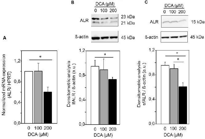
compared to normal liver tissue. Moreover, ALR mRNA expression has been investigated in liver tissues from patients with cholestatic liver diseases and was found to be significantly reduced (chapter 5).
Infection with hepatitis B virus leads to induction of several genes among them target genes of Nrf2 such as ALR [36]. Hence increased ALR mRNA and protein expression was found in HBV positive liver tissues [36]. Furthermore, ALR serum levels were found to be induced in patients with HBV-related acute, chronic and severe hepatitis or cirrhosis [69]. Serum levels of ALR might be considered a candidate for prognosis of HBV related-liver failure since it was found to be induced in patients with HBV-related ACLF (acute-on chronic liver failure) compared to normal patients and to be also induced in the surviving ACLF patients compared to dead patients [85]. In addition, patients with improved hepatic failure (HF) show increased serum levels of ALR protein compared to patients with deteriorating HF [60]. On the other hand, HCV infection impairs induction of Nrf2 [86] and therefore as expected, ALR expression is not altered in HCV positive liver tissue [36, 57]. Interestingly, hepatic ALR expression is low or even unchanged after insulting the liver by fat, alcohol or HCV infection, respectively. But as a consequence of these insults progression to cirrhosis or even tumorigenesis occurs with induction of ALR expression as part of the regenerative response that occurs regardless of the underlying etiology [61].
5.3. ALR and in vivo models
Multiple studies have reported the use of mouse and rat models that mimic diseases conditions in order to explore the effect of ALR in those diseases. It was reported that ALR serum levels are elevated in rats after PCS (portacaval shunt) surgery, endotoxemia or sepsis, and in a mouse model of hemorrhagic shock [66].
Furthermore, ALR serum levels are increased in rats after tissue loss by partial hepatectomy (PH) [56, 59] and return to normal levels after 12 hours [59] or 36 hours [56]. On the contrary, hepatic level of ALR after PH remains unchanged (after 40%
PH) or decreases (after 70% PH) [59]. ALR liver-specific knock-out mice have been shown to endure accelerated steatosis and to develop liver tumors after 1 year [57].
The same mouse model was used to investigate the alcohol-induced injury and was
shown to accelerate injury [53]. Interestingly, using hepatocytes transfected with ALR
of anti-tumor agents [87] and reduces IRI injury in steatotic liver [88]. Further, the subcutaneous-injected hepatoma cells expressing sfALR (15 kDa, [62]) or lfALR (23 kDa, [83]) showed reduced tumor growth in mice due to their reduced metastatic ability. Injecting ALR (23 kDa) expression plasmid into mice with CCl
4-induced injury suppresses apoptosis and attenuates the acute injury [84] which is underlined with a study that reported increased ALR serum levels after CCl
4-injury in rats [85].
Interestingly, ALR-knock-in and knock-out models in Zebrafish have been established
and demonstrated the functional role of ALR in vertebrate hepatogenesis [89]. It is
worth to mention that the physiological effects of ALR in spermatogenesis were
investigated by an ALR transgenic mouse model driven by the human TSPY (testis-
specific protein, Y-encoded) promoter that allows the transgene to be specifically
activated in the testes [90]. This study highlighted that temporal expression of ALR is
required for normal testicular development and spermatogenesis.
Chapter 1
Table (II): Summary of studies investigating hepatic expression of ALR in various liver diseases with the methods used in the investigation.
Liver disease Study subject Target Methods Reference
Hepatocellular carcinoma (HCC)
HCC vs. corresponding surrounding non-tumorous tissue (n=4), HCC vs. surrounding
cirrhotic tissue (n=5) mRNA PCR
[61]
normal vs. HCC (n=5) protein IHC, wb
normal (n=10) vs. HCC (n=20) serum ELISA
[60]
HCC vs. HF and normal mRNA PCR
HCC vs. HF protein IHC
normal (n=1) vs. HCC (w/o angioinvasion n=2 and with angioinvasion n=2) protein Wb
[62]
HCC liver sections (n=53) (high vs. Low expression of ALR) protein IHC
non-tumorous (w/o HBV nor alcohol, n=5) vs. tumorous (w/o HBV nor alcohol, n=12) protein IHC [36]
HCC (n=22) vs. para-cancerous liver tissue (n=22) mRNA PCR
[64]
HCC vs. para-cancerous liver tissue protein IHC
HCC with low ALR (n=19) vs. HCC with high ALR (n=25) mRNA PCR
[83]
HCC w/o angioinvasion (n=29) vs. HCC w angioinvasion (n=14) mRNA PCR
HCC w/o angioinvasion (n=4) vs. HCC w angioinvasion (n=4) protein IHC, wb
Cholangiocyte carcinoma (CCC)
CCC vs. corresponding non-tumorous tissue (n=5) mRNA PCR
[61]
normal vs. CCC (n=3) protein IHC, wb
Hepatitis B virus (HBV) infection
normal (n=10) vs. Chronic HBV infection (n=20) serum ELISA [85]
HCV (n=10) vs. HBV (n=10) mRNA PCR
[36]
non-tumorous with HBV (n=8) vs. tumorous (with HBV, n=7) protein IHC
non-tumorous w/o HBV (n=5) vs. non-tumorous with HBV (n=8) protein IHC
Hepatitis C virus
(HCV) infection normal (n=5) vs. HCV infection (n=7) protein Wb [57]
Non-alcoholic fatty liver diseases (NAFLD)
normal (n=5) vs. NASH (n=5) protein Wb [57]
normal (n=17) vs. Steatosis (n=27) and NASH (n=29) mRNA PCR [54]
Alcoholic liver diseases (ALD), alcoholic steatohepatisis (ASH)
non-tumorous with alcohol (n=4) vs. HCC with alcohol (n=14) protein IHC(1) [36]
normal (n=5) vs. ALD (n=5) protein Wb [57]
Cholestatic liver
diseases (CLD) normal (n=13) vs. Cholestasis (n=45) mRNA PCR chapter 5
Hepatic failure (HF) and Acute liver diseases (ALD)
improved HF (n=6) vs. deteriorating HF (n=12) serum ELISA [60]
normal (n=10) vs. HBV related ACLF (n=40) serum ELISA
[85]
survival ACLF patients (n=10) vs. Dead ACLF patients (n=10) serum ELISA
normal (n=2) vs. Acute liver failure (n=2) protien Wb [84]
Cirrhosis
normal (n=18) vs. Cirrhosis (n=25, with n=14 alcohol-induced cirrhosis, n=7 HCV-induced
cirrhosis and n=4 autoimmune diseases induced cirrhosis) mRNA PCR
[61]
normal vs. Alcohol-induced cirrhosis (n=5) protein IHC, wb
normal (n=10) vs. Cirrhosis (n=30) serum ELISA [69]
normal (n=2) vs. Liver cirrhosis (n=2) protein Wb [84]
Wb = western blot; PCR = qRT-PCR; IHC = immunohistochemistry
(1) Dayoub et al. (2013) investigated ALR protein expression in liver sections from patients with HCC and non-tumorous tissue (with or without HBV or alcohol) by IHC and quantitatively evaluated the intensity of anti-ALR staining (from 0 to 3).
Chapter 1
6. Function of ALR
Different isoforms of ALR have been associated with different subcellular localizations and therefore specific functions. Several studies have investigated the role of the 23 kDa ALR (long form, lfALR) and the 15 kDa ALR (short form, sfALR) related to different diseases and pointed out the beneficial effects of the individual ALR isoforms. In the next paragraph we summarize the studies that investigated the over-expression of lfALR and sfALR as well as the studies that explored the impact of ALR silencing (effects all isoforms) on cellular functions.
6.1. The functional role of mitochondrial lfALR (23 kDa)
As implied before, the 23 kDa of ALR is located in the mitochondria (see the location of ALR) and the mitochondrial targeting of ALR is attributed to the IDD domain in the initial 80 amino acids (N80) at the N terminal of lfALR (see the structure of ALR). In mammalian cells, lfALR is a part of the disulfide relay system that recycles Mia40 (mitochondrial import and assembly 40) in the inter-mitochondrial space (IMS) while transporting proteins into the IMS. Mia40 interacts with its substrates by its CPC motif [91, 92] and oxidizes those substrates which leads to substrate folding and trapping in the IMS. Furthermore, reduced Mia40 is re-oxidized -“recycled”- by the CRAC and CXXC motif in ALR, which itself obtains electrons from cytochrome C [22, 23, 93]. For more detailed information about the role of ALR in mitochondrial biogenesis we would like to refer to the recent review article of Kallegri et al. 2014 [94] and Erdogan et al. 2017 [95].
Besides its role in mitochondrial protein import, there has been contradictory evidence about the role of ALR in the maturation and export of cytosolic Fe-S cluster proteins. Fe-S proteins were reported to be involved in enzymatic catalysis, DNA synthesis and repair, iron homeostasis and heme synthesis [96]. Lange et al. [97]
have suggested that ALR, may be involved in the maturation of cytosolic Fe-S proteins. The authors claimed that ALR interacts with Atm1 (an ABC transporter in the inner membrane of the mitochondria) and facilitates the export of Fe-S proteins to the cytosol [97]. However, later it was suggested that, in yeast cells, ALR plays neither a direct nor an indirect role in the maturation of Fe-S cluster [98].
Nevertheless, a recent study in mammalian cells has pointed out the role of ALR in
exporting MitoNEEt to the outer mitochondrial membrane (OMM). MitoNEEt is a
Fe-S protein that is synthesized in the mitochondrial matrix. Upon synthesis, MitoNEEt translocates through the inner membrane (IM) of the mitochondria by ABCb7 and then through the IMS by ALR to the OMM where it contributes to cell proliferation [72]. However, whether ALR is involved in the maturation and export of other Fe-S protein still requires further investigations.
Furthermore, endogenous lfALR (23 kDa) has been investigated in various disease models and Table (III) summarizes the studies about over-expression of lfALR and its role in different diseases. Expression of lfALR promotes liver growth during hepatogenesis [89], reduces fibrotic injury [99, 100], protects against radiation-induced oxidative stress [71] and attenuates acute liver injury by acetaminophen or CCl
4[84, 101]. The latter has been attributed to autophagy activation (by promoting p62 degradation and LC3II conversion) and apoptosis repression [84, 101]. In addition, lfALR has been reported to reduce ER stress [102]
and promote the anti-tumor effects of doxorubicin by increasing its cellular retention [87]. Furthermore, lfALR has been shown to increase the pluripotency of embryonic stem cells (ESC) by preserving the mitochondrial integrity and function in ESCs [103]
On the other hand, it was shown that lfALR does not contribute to reducing the bile acid-induced apoptosis [104].
It is worth to mention that over-expression of lfALR in the testis of the mice
(transgenic mouse) was reported to cause abnormal spermatogenesis and reduced
fertility in those mice [90]. Interestingly, it was suggested that continuous
over-expression of lfALR in mammalian cells leads to the accumulation of lfALR not
only in the mitochondria but also in the cytosol [105]. Since no exclusive
mitochondrial localization of lfALR is demonstrated in previously mentioned studies,
the question rises whether the previously reported effects e.g. altering gene
expression by endogenous lfALR may be due to its cytosolic accumulation rather
than its mitochondrial localization.
Chapter 1
Table (III): Summary of the studies investigating the over-expression of lfALR (23 kDa)
Vehicle Host Disease / Model Function Molecular explanation Reference
pcDNA3 injection into rat caudal vein
liver fibrosis (induced by colchicine)
protects against fibrosis, en- hances regeneration
reduces the expression of TIMP1, collagen I
and collagen III [99]
pcDNA3.1/ Myc- HisB vector in Lentivirus
cell lines (HepG2, SMMC-7721, BEL- 7402, 293FT, L02)
radiation (oxidative stress)
protects against radiation- induced oxidative stress, improves cell viability
reduces ROS production, mitochondrial dysfunction, apoptosis; reduction of
cytochrome C release and caspase 3/7 activity
[71]
pcDNA3.1/ Myc- HisB
transfection into Huh7-NTCP cells
cholestasis (GCDCA) no reduction of caspase 3/7 activity
- [104]
MSCV-IHRES-GFP retrovirus
transfection into mouse ESC (Embryonic stem cells)
- promotes ESC pluripotency,
preserves mitochondrial structural integrity and function
reduces expression of Drp1 (Dynamine- related protein 1)
[103]
mRNA of ALR injection into 1-2 cell stage Zebrafish embryos
hepatogenesis promotes liver growth -
[89]
Minicircle vector or pcDNA3.1
transfection into SMMC-7721, HSC- T6 (rat hepatic stel- late cells), injection into rat tail
liver fibrosis reduces the fibrotic injury, increases the survival rate via deactivation of HSC
-
[100]
Lentivirus injection into mouse tail
acute liver injury by acetamiophen (APAP) over-dose (oxidative stress)
protects liver by activation of autophagy and reduction of apoptosis
increases p62 degradation and LC3II conversion, reduces release of AIF,
cytochrome C and caspase 3 activation [101]
pcDNA3.0 injection in mouse tail or transfection into AML12 cells
CCl4-induced acute liver injury
protects against acute liver injury by autophagy induction, suppression of apoptosis, pro- moting proliferation
induces p62 degradation, LC3II conversion and levels of ATG5, ATG7 and Beclin-1.
[84]
p-Adxsi-Flag tagged vector
transfection into HepG2 cells (xeno- grafts in mice)
HCC and anti-tumor agents (doxorubicin)
sensitizes hepatocytes to anti- tumor effects of doxorubicin
reduces expression of ABCB1 and ABCG2 (MDR proteins) partially by blocking Snail/Akt pathway
[87]
pcDNA 3.0 transfection into HepG2 cells
lipotoxicity (palmitic acid treatment)
reduces ER stress by reducing Ca+2 release to the cytosol
reduces IP3R expression, induces IP3R and Bcl2 interaction, reduces mitochondrial Bax expression
[102]
flag tagged pAdxsi into Adenovirus
transfection into mice or HepG2 cells
IRI liver injury and steatosis
reduces inflammation and necrosis caused by IRI
enhances oxygen consumption and ATP
production [88]
- transfection into
MHCC97H and xenografts in mice
HCC and metastasis reduces HCC metastasis blocks ERK pathway
[83]
Chapter 1 6.2. The functional role of cytosolic sfALR (15 kDa)
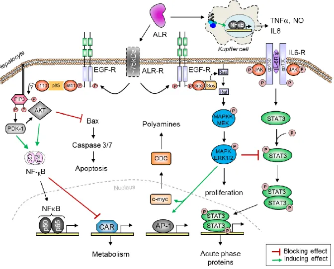
Short form ALR (sfALR, 15 kDa) is located in the cytosol [54] and presumably in the nucleus [63], due to the lack of the IDD (responsible for mitochondrial targeting) in the initial 80 amino acids (N80) found only in lfALR (see ALR structure). However, the potential effects (summarized in Table IV) and molecular functions (Fig. 4) of sfALR have been investigated in different models. sfALR was shown to induce the response of hepatocytes to IL6 signaling by induced STAT3 (signal transducer and activator of transcription 3) phosphorylation, which increases the expression of e.g.
acute phase proteins (APP) like fibrinogen (FGB) and haptoglobin (HP) [106]. In addition, the activation of STAT3 might account for the anti-apoptotic effect of sfALR by reduced DR5 (death receptor 5) expression and caspase 3/7 activity during cholestasis (chapter 5) and free fatty acid (FFA)-induced toxicity [54]. Interestingly, the reduced DR5 expression by sfALR may also be due to reduced CHOP (C/EBP- homologous protein, an inducer of DR5 expression [107]) which is repressed upon attenuation of FFA-induced ER-stress by sfALR [54].
In addition, sfALR was attributed to have anti-metastatic effects by reducing the migratory and invasion activity of hepatoma cells and as well as maintaining an epithelial like state with less signs of aggressive proliferation. sfALR mediates this by reducing Snail expression which induces the expression of epithelial cell markers like CDH1 (E-Cadherin) and ZO-1 (Zona Occludens-1) [62]. Moreover, the hepatic metabolic capacity is affected by endogenous sfALR by reducing cholesterol catabolism into bile acids due to reduced CYP7A1 (rate-limiting enzyme in bile acid synthesis) expression and reduced HNF4α expression (chapter 5). Additionally, lipid metabolism genes (CPT1, FABP1, ELOVL6) were reported to have altered expression in sfALR over-expressing cells under steatotic conditions [54], which could be due to repressed activation of JNK [54, 108] or the activation of miRNA expression e.g. miR122 [108].
The interactions of sfALR protein with other molecules have been also reported in multiple studies (Fig. 4). Endogenous sfALR interacts with JAB1 (c-Jun-activating domain binding protein 1) [24, 109-111] in the nucleus and might include the whole CSN (COP9 signalosome, consisting of COP9 and JAB1) [111]. This interaction results in AP1 activation [109, 111] in a MAPK- and JNK-independent manner [24].
Furthermore, it was shown that ALR activation of AP1 is dependent on the CXXC
strated that binding of ALR to JAB1 blocks JAB1 interaction with p27
kip1, which promotes cell cycle arrest and restricts the abnormal proliferation of HSC and the subsequent exhaustion [110]. This was confirmed by ALR knock-down that caused reduced p27
kip1nuclear retention and therefore a hyper-proliferative response of the HSC [110]. Moreover, cytosolic sfALR interacts with MIF (macrophages migration inhibitory factor) [43], which also results in AP1 activation due to MIF’s ability to block AP1 [43], this interaction might as well block MIF effects on p53 and results in tumor suppression [112] which may contribute to the described anti-metastatic effects of ALR [62]. Furthermore, ALR interacts with thioredoxin (TRX) [113] by its CXXC motif (C142-C145) and contributes to the maturation of viral proteins [113]. This interaction results in oxidized TRX which therefore induces AP1, c-Jun and NFB activity [26].
Additionally, ALR interacts with the pro-apoptotic protein BNIPL (Bcl-2/adenovirus
E1B) and leads to growth inhibition in hepatoma cells (BEL-7402) [114].
Chapter 1
Table (IV): Summary of the studies investigating over-expression of sfALR (15 kDa)
Host, traget cell Vehicle Disease/ model Function of sfALR Molecular explanation Reference infection of BS-C-1
cells (Kidney)
pTargeT vector controlled by T7 promoter in vaccinia virus
- interacts with thioredoxin interacts with thioredoxin (TRX) by the CXXC motif
[113]
transfection into COS-7 and HepG2 cells
pAS vector - increases c-Jun
phosphorylation and AP-1 activation
interacts with JAB1 (c-Jun activation domain- binding protein 1) in the nucleus, activates AP1 independent of MAPK, activates c-Jun independent of ERK1/2 and JNK pathways
[24]
transfection into COS-7 cells
pCMV-Myc vector activates AP1 interacts with JAB1 and activates AP1 via the
CXXC motif [109]
transfection into BEL-7402 cells
pGEX-5x-1 vector - interacts with BNIPL
(apoptotic protein)
- [114]
transfection into yeast cells and HepG2 cells
pAS2-1, pACT2 or pFlag-CMV-2 vector
- activates AP1 interacts with MIF in the cytosol
[43]
transfection into COS-7 and HepG2 cells
pCMV-Myc vector - activates AP1 interacts with JAB1 which activates AP1 in a
MAPK independent manner [111]
transfection into COS-7 cells and Yeast Y190 cells
pcDNA3.1A, pEGFP, pGEX-4T, pFlag-CMV or pAS vectors
- activates AP1, NFB and c-
Jun phosphorylation
interacts with thioredoxin
[26]
Intravenous injection into rats after CCl-induced
pcDNA3.1 vector acute hepatic injury, hepatic failure
increases cell proliferation and survival rates
-
[115]
transfection into HSC
(hematopoietic stem cells)
murine stem cell virus (MSCV)
- restricts abnormal proliferation
of HSC
interacts with JAB1 and inhibits the JAB1- p27kip1 interaction
[110]
transfection into HepG2 cells
pcDNA3.1 vector HCC and metastasis reduces cell motility and EMT (epithelial mesenchymal transition)
reduces Snail expression, induces
E-Cadherin and ZO-1 expression [62]
mice on MCD diet pcDNA3.1 vector NAFLD reduces the severity of fatty acid injury
suppresses JNK phosphorylation and fatty
acid synthesis genes, induces miR122 levels [108]
transfection into HepG2 cells
pcDNA3.1 vector acute phase response (APR)
induces expression of APP:
fibrinogen β and haptoglobin induces STAT3 phosphorylation
[106]
transfection into HepG2 and Huh7 cells
pcDNA3.1 vector NAFLD attenuates ER stress and lipoapoptsis, alters lipid metabolism genes, reduces TAG levels
reduces DR5, Bax and CHOP expression and JNK phosphrylation, increases ATP synthesis, alters expression of CPT1α, FABP1 and ELOVL6
[54]
transfection into HepG2 cells
pcDNA3.1 vector cholestasis reduces bile acid synthesis and bile acid-induced apoptosis
preserves STAT3 activation, reduces HNF4α
and DR5 expression chapter 5
Chapter 1
Fig. 4. An illustration of how endogenously expressed sfALR affects hepatocytes. ALR promotes IL6 signaling by increasing STAT3 phosphorylation and thereby regulating STAT3 target genes. Furthermore, STAT3 activation by sfALR blocks HNF4 and reduces its trans-activating effects. sfALR also reduces the expression of SNAIL and CHOP (by reducing ER stress) and thereby their target genes. ALR interacts with MIF and blocks its inhibitory effects on p53 and AP1, which results their activation and tumor suppression by p53. ALR also interacts with thioredoxin and promotes AP1 and NFB activation. Moreover, ALR interacts with JAB1 in the nucleus and thereby activates AP1. MIF and thioredoxin bind to JAB1 to prevent JAB1 interaction with p27kip1 which increases the nuclear retention of p23kip1 and therefore induces cell cycle arrest at G1 phase. In addition, the pro-apoptotic protein BNIPL interacts with ALR and MIF and regulates cell survival. BNIPL: Bcl-2/adenovirus E1B, MIF: macrophage migration inhibitory factor, CHOP: C/EBP-homologous protein, STAT3: signal transducer and activator of transcription 3, TRX: thioredoxin, JAB1: c-Jun-activating domain binding protein 1, CSN5: COP9 signalosome subunit 5, HNF4: hepatocyte nuclear factor 4
6.3. ALR silencing
In order to further clarify the role of ALR for cellular functions, many studies have explored the effect of ALR silencing (all isoforms) and monitored the outcome of its absence on the development of various diseases. Table (V) summarizes the main finding of the studies that inspected the effect of silencing ALR.
In the kidney, it was shown that ALR knock-down had no effect on HK-2 cells viability [81, 116] but aggravated the H/R injury by increased ROS production and apoptosis due to the activation on AMPK/mTOR pathway [116]. Morevoer, ALR silencing inhibited cytokine production and the inflammatory response after H/R injury by blocking MAPK pathway [81]. Furthermore, it was suggested that ALR silencing decreases NKB nuclear translocation in kidney cells [81]. On the contrary, it was reported that over-expression of sfALR (15 kDa isoform) does not affect NFB’s nuclear translocation in hepatocytes during cholestasis (chapter 5), which might argue for organ specific effects of ALR or more likely for a specific role of different ALR isoforms due to their location and function.
The anti-oxidative role of ALR was further evidenced in human derived glioma cells
(T98G) which showed that silencing of ALR reduces the expression of anti-oxidative
protein clusterin and induces the activity of caspase 3 and 9 [48]. Additionally, ALR
silencing sensitizes HepG2 cells to radiation-induced oxidative stress [71]. ALR
silencing suppresses liver growth [89], reduces cell viability and induces apoptosis in
rat hepatocytes [117] as well as HepG2 cells [118]. In the liver, after partial
hepatectomy ALR silencing resulted in reduced compensatory hepatocellular
proliferation, increased pro-apoptotic proteins and caspase activity [55]. Furthermore,
liver specific ALR knock-out (KO) mice developed steatosis accompanied by altered
expression of lipid metabolism genes, enhanced ROS production, mitochondrial
dysfunction, increased Bax expression and NK as well as CD8
+cell recruitment to
the liver [57]. Interestingly, it was shown that ALR-KO mice develop liver tumors
within 6 months of age and therefore ALR silencing links non-alcoholic liver disease
to hepato-carcinogenesis [57]. The same group showed later that ALR-KO mice
suffer accelerated alcohol injury, which they attributed to changes in lipid metabolism
genes, altered alcohol metabolism by ADH1 (alcohol dehydrogenase 1), ALDH1
(aldehyde dehydrogenase 1
)and CYP2E1 (cytochrome P450 2E1) and augmented
oxidative stress upon injury [53]. In addition, it was shown that diminishing ALR
expression enhances free fatty acid-induced ER stress and lipoapoptosis [102] and
Chapter 1
reduces the anti-tumor effects of doxorubicin due to enhanced expression of export
transporters and therefore reduced cellular retention of doxorubicin [87]. In contrast
to Tang et al. [118], who showed reduced tumor growth of HepG2 in nude mice after
ALR silencing, others reported promoted cell growth and EMT (epithelial
mesenchymal transition) [83]. It is worth to mention that silencing ALR in HSC
(Hematopoietic stem cells) caused a hyper-proliferative response due to reduced
JAB1 binding and therefore reduced nuclear retention of p27
kip1[110]. In addition,
ALR silencing in mouse embryonic stem cells (ESC) increases Drp1 (Dynamine-
related protein 1) expression and activates mitochondrial autophagy, which results in
enhanced apoptosis of ESC [119]. Interestingly, a chemical screen identified a
molecule that is capable of inhibiting ALR activity, MitoBloCK-6 [120]. MitoBloCK-6
was shown to induce apoptosis by cytochrome C release in ESCs but not in
differentiated cells which suggests a vital role of ALR in the ESC homeostasis [120].
Table (V): Summary of studies investigating ALR silencing (all ALR isoforms)
Organ Host Vehicle disease/
model
Effect of ALR silencing molecular explaination Reference
Liver
rat heptocytes antisense oligo- nucleotide
hepatocytes survival
reduced cell viability and induced apoptosis
induced rounding, detachment of cells, increased LDH and cytochrome C re- lease, Caspase 3 activity, reduced ATP
[117]
HepG2, L02 cells lentivirus with shRNA
radiation (oxidative stress)
inhibited cell viability, induced sensibility to radiation (HepG2), minimal effects on normal cell line
- [71]
HepG2 cells, xenografts in nude mice
siRNA in pSIALR-A plasmid
HCC growth inhibited growth in HepG2 and xeno- grafted HCC tumors in nude mice
- [118]
Zebrafish embryos
Morpholino antisense oligonucleotides
hepatogenesi s
suppressed liver growth reduced hepatocyte proliferation without affecting apoptosis
[89]
HepG2 cells (and xenografts in mice)
shRNA doxorubicin
treatment
reduced caspase 3 activity due to reduced cellular retention of doxorubicin
induced snail and therefore ABCB1 and ABCG2 (export pump) expression
[87]
HepG2, Huh7, Huh7-sfALR cells
siRNA - reduced expression of the three
isoforms of ALR
- [54]
HepG2 cells PLVX- shRNA- plasmid
lipotoxicity (palmitic acid treatment)
induced ER stress by increasing Ca+2 release into the cytosol
induced expression of IP3R and Bax, reduced expression of Bcl2
[102]
HepG2, MHCC- 97H cells xeno- grafts in mice
shRNA HCC and
metastasis
promoted cell growth and migration in vivo and in vitro
induced EMT in hepatoma cells due to activated ERK pathway
[83]
rat adenovirus with partial reduced hepatocyte proliferation and - [55]
Chapter 1
shRNA hepatectomy polyamine synthesis, increased pro- apoptotic proteins and caspase activity
Liver
mice liver-specific ALR knock out
(Albumin Cre- lox)
Steatosis ALR-KO in mice results in accelerated hepatic steatosis after 2 weeks, development of liver tumors after 6 months of age
increased ratio of Bax /Bcl2, induced IL1, TNFα and IL6, increased recruit- ment of NK cells and CD8+ cells, in- creased ROS, mtDNA damage, re- duced ATP levels and expression of ACACA, SREBP1c, CPT1α and PPARα
[57]
mice liver-specific ALR knock out
(Albumin Cre- lox)
alcohol induced injury
accelerated alcohol induced liver injury in ALR-KO mice
reduced expression of FASN, ACACA, SREBP1c, CPT1α (lipid metabolism genes), and activity of ADH1, ALDH1 and CYP2E1 (alcohol metabolism genes), augmented oxidative stress, fibrosis, inflammation by EtOH feeding
[53]
Stem cells
murine ESC (Embryonic stem cells)
FG12-Lentivirus–
GFP–shRNA - loss of mitochondrial function in ESC, reduced proliferation and enhanced apoptosis
triggered mitochondrial autophagy or mitophagy by increased Drp1 (Dynamine-related protein 1)
[119]
hematopoietic stem cells, bone marrow trans- plantation in mice
FG12-Lentivirus–
GFP–shRNA
- hyper-proliferative response and exhaustion in hematopoietic stem cells (HSC)
reduced p27kip1 expression through its binding to JAB1
[110]
ESC, Hela cells, and Zebrafish embryos
MitoBloCK-6 („ALR inhibitor“)
- induced apoptosis by cytochrome C release and impaired cardiac development in Zebrafish embryos
Inhibition of ALR activity
[120]
HK-2 cells lentivirus with shRNA/ALR
H/R injury no effect on viability, inhibited
inflammatory response and cytokines production after H/R injury
blocked MAPK pathway and decreased nuclear translocation of NFB [81]
HK-2 cells lentivirus with shRNA/ALR
I/R kidney injury
no effect on cell viability, increased autophgy and ROS production in vitro
activated AMPK/mTOR pathway [116]
Glioma cells
T98G cells (human)
siRNA H2O2
(oxidative stress)
reduced expression of clusterin (anti- oxidative protein) and Bcl2, induced Caspase 3 and 9 activity
- [48]
Myeloma cells
U266 cells shRNA - promoted apoptosis induced Bax expression, reduced Bcl2
expression and IL6 synthesis
[70]