in Dictyostelium discoideum mutants
INAUGURAL-DISSERTATION zur
Erlangung des Doktorgrades
der Mathematisch-Naturwissenschaftlichen Fakultät der Universität zu Köln
vorgelegt von
Nandkumar Krishna Khaire aus Solapur, Indien
2003
Referees/Berichterstatter Prof. Dr. Angelika A. Noegel
Prof. Dr. Siegfried Roth
Date of oral examination/ 04.12.2003 Tag der mündlichen Prüfung
The present research work was carried out under the supervision and the direction of Prof. Dr.
Angelika A. Noegel in the Institute of Biochemistry I, Medical Faculty, University of Cologne, Cologne, Germany, from October 2000 to December 2003.
Diese Arbeit wurde von Oktober 2000 bis Dezember 2003 am Biochemischen Institut I der Medizinischen Fakultät der Universität zu Köln unter der Leitung und der Betreuung von Prof. Dr.
Angelika A. Noegel durchgeführt.
cytoskeleton plays a key role in almost all cellular processes, including motility, cytokinesis, cell-to-cell and cell-substrate adhesions and intracellular transport. Several actin binding proteins are also involved in these processes, among them are actin crosslinking proteins (for example, filamin and α-actinin). Filamin (also known as ddfilamin or gelation factor or ABP 120) consists of an actin binding domain and six rod repeats of 100 amino acids, its last repeat being responsible for the formation of the homodimer. Both the domains are necessary for the actin crosslinking activity of filamin. Dictyostelium mutants lacking filamin have severe defects in multicellular slug migration towards light, phototaxis, and preferable temperature, thermotaxis. To study the phototaxis defect in filamin− mutants at the molecular level we expressed various domains in the mutant and tested their rescue potential. Expression of C terminally truncated and point mutated (at a putative phosphorylation site) filamin rescued the phototaxis defect partially. Full-length filamin when expressed under the control of the ecmA promoter in the anterior tip of the slug rescues the phototaxis, but not when expressed under the control of the cotB promoter which allows expression in the posterior ¾th of the slug.
Phototaxis is a complex phenomenon, which includes more than 55 genes. To identify genes involved in this process we carried out a microarray analysis. Amoung 65 genes we selected in microarray analysis, 40 genes were up regulated and 25 genes were down regulated. From the functions of most of theses genes, we conclude that the phototactic behaviour of slugs is controlled by extracellular cAMP, Ca2+ ions and cell adhesion. To further focus on filamin's function in phototaxis we searched for proteins interacting with filamin by a yeast two hybrid screen and by immunoprecipitation. TipA, GAPA and SapA proteins were pulled down in the immunoprecipitation approach while the FIP, filamin interacting protein, was found earlier in a yeast two hybrid screen. Biochemical studies suggest that FIP is associated with F-actin and may function in vesicle trafficking. Detailed analysis of the mutants of LIM proteins, villidin and filamin for chemotactic migration towards cAMP, led us to conclude that alteration in chemotactic motility of individual cells may not affect the phototactic migration of the slug.
To My Family
My first, and most earnest, acknowledgment must go to my esteemed advisor, Prof.
Dr. Angelika A. Noegel, Institute of Biochemistry I, Medical Faculty, University of Cologne, Cologne, Germany. Her valuable guidance, creative suggestions, constructive criticism and constant encouragement during the course of present investigation are not only praiseworthy but also unforgettable. Her analytical perusal of the manuscript is highly acknowledged. It was indeed a pleasure for me to work under her superb guidance.
I pay my sincere thanks to Dr. Francisco Rivero, who helped me in learning the microscopic work and provided Rac GTPases for checking the interactions with ddfilamin.
He has always been the first person I usually run into when I am faced with complicated assays, he always left whatever he was doing to listen to me.
I thank Dr. Ludwig Eichinger, for providing me the lab facilities to carry out microarray analysis and his helpful discussion regarding protein kinases and protein phosphorylation. I also would like to thank Patrick Farbrother, for his help through out the microarray analysis.
I pay my sincere thanks to Dr. Andreas Hasse, Dr. Akis Karakesisoglou and Dr.
Budi Tunggal for their suggestions and fruitful discussion during lab meetings and journal clubs.
I am thankful to Rolf for providing technical assistance when required. My thanks are also due to Rosi, for her help during yeast two hybrid screening and monitoring cleanliness in laboratory as well as sitting room. I am also thankful to Monika for introducing the yeast two hybrid system.
The constant cooperation, motivation and nice company provided by Dr. Sonia Ramos, Dr. Michael Leichter, Dr. Somesh Baggavalli and Maria Marko is acknowledged.
My colleagues Henning, Dhamodharan, Hameeda, Sabu, Kumar, Sunil, Deen and Yogikala were very friendly and helpful. Thanks for their company especially during tea and lunchtime.
I also owe a huge debt of gratitude to Bettina Lauss, who made all the necessary official things easy and fast. Far too many people to mention individually have assisted in so many ways during my work at Institute of Biochemistry I can be acknowledged, in particular Maria, Berthold, Kathrin, Bärbel and Roberto.
A penultimate thank-you goes to my wonderful parents for their understanding, endless patience and encouragement when it was most required.
My final, and most heartfelt, acknowledgment must go to my wife Pratibha, for her support, and companionship. For all that, and for being everything I am not, she has my everlasting love. Thanks to little Gouri, my loving daughter for her affection.
Finally, the financial assistance received by me from the DFG is highly acknowledged.
Cologne Nandkumar K. Khaire
18/09/2003
Table of Contents
Chapter Description Page(s)
I. INTRODUCTION 1-8
1.1 Dictyostelium, model organism 1
1.2 Phototaxis in Dictyostelium 3
1.3 Chemotaxis in Dictyostelium 7
II. MATERIALS AND METHODS 9-41
1 Materials 9
1.1 Laboratory materials 9
1.2 Instruments and equipments 10
1.3 Kits 11
1.4 Enzymes, antibodies, substrates, inhibitors and antibiotics 11
1.5 Chemicals and reagents 13
1.6 Media and buffers 13
1.6.1 Media and buffers for Dictyostelium culture 14
1.6.2 Media for E. coli culture 14
1.6.3 Media and buffers for Yeast culture 15
1.6.4 Buffers and other solutions 16
1.7 Biological materials 18
2 Cell biological methods 19
2.1 Growth of Dictyostelium 19
2.1.1 Growth in liquid nutrient medium 19
2.1.2 Growth on SM agar plates 19
2.2 Development of Dictyostelium 19
2.3 Preservation of Dictyostelium 20
2.4 Transformation of Dictyostelium cells by electroporation 20
2.5 Quantitative Phagocytosis assays 21
3 Molecular biological methods 21
3.1 Purification of plasmid DNA 21
3.2 Digestion with restriction enzymes 22
3.3 Generation of blunt ends in linearised plasmid DNA 22
3.4 Dephosphorylation of DNA fragments 23
3.5 Setting up of ligation reaction 23
3.5.1 Generation of the point mutation 23
3.6 DNA agarose gel electrophoresis 24
3.7 Recovery of DNA fragments from agarose gel 25
Chapter Description Page(s)
3.8 RNA formaldehyde-agarose gel electrophoresis 25
3.9 Transformation of E. coli 26
3.9.1 Transformation of E. coli cells by the CaCl2 method 26 3.9.2 Transformation of E. coli cell by electroporation 27
3.10 Glycerol stock of bacterial culture 27
3.11 Construction of vectors 28
3.11.1 Vector for the expression of ddfilamin GFP fusion protein 28
3.12 DNA sequencing 28
3.13 Computer ananlysis 28
4 Methods for Yeast Two Hybrid System 29
4.1 Transformation of Yeast by Lithium Acetate Method 29
4.2 DNA isolation from Yeast 29
4.3 β-galactosidase colony lift assay 30
4.4 Yeast strain maintenance 30
5 Biochemical Methods 31
5.1 Preparation of total protein from Dictyostelium 31
5.2 SDS-polyacryamide gel elctrophoresis 31
5.3 Native or Nondenaturating PAGE 32
5.3.1 Coomassie blue staining of SDS-polyacrylamide gels 33
5.3.2 Silver staining polyacrylamide gels 33
5.4 Immunoprecipitation from Dictyostelium cell lysate 33
5.5 Detection of phosphorylation 34
5.6 Western blotting using the semi-dry method 34 5.7 Immunodetation of membrane-bound proteins 34
5.8 Video imaging and chemotaxis assay 35
5.9 Qualitative phototaxis assay 35
6 Immunological methods 36
6.1 Direct immunofluorescen of Dictyostelium cells 36
6.1.1 Preparation of Dictyostelium cells 36
6.1.2 Methanol fixation 36
6.1.3 Picric acid-paraformaldehyde fixation 36
6.1.4 Immunolabeling of fixed cells 37
6.1.5 Mounting of coverslips 37
6.2 Preparation of heat-killed yeast cells 38
6.3 TRITC-labeling of heat-killed yeast cells 38
Chapter Description Page(s)
7 Microscopy 38
7.1 Live cell imaging of Dictyostelium cells expressing RN/GFP 39 7.2 Live cell imaging of RN-GFP during phagocytosis 39
7.3 Microscopy of the fixed preparations 39
8 Microarray analysis 39
8.1 RNA preparation 40
8.2 Spiking of internal mRNA controls 40
8.3 Quantitation, normalization and data analysis 41
8.4 Signal Quantification 41
III. RESULTS 42-72
1 Biochemical studies 42
1.1 Overexpression of domains of filamin 42
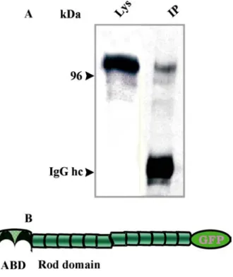
1.2 Localisation and distribution of ddfialmin domains during growth 44 1.3 Expression of full-length ddfilamin GFP fusion protein in GA1
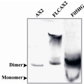
compensates the loss of α-actinin 44 1.3.1 The ddfilamin rod domain GFP fusion protein (RN-GFP)
does not localise properly in HG1264 cells 46 1.3.2 Ddfilamin rod domain GFP fusion protein (RN-GFP)
forms heterodimers with the endogenous protein 49 1.3.3 A GFP tag located at the C-terminus does not interfere
with the dimerisation property of filamin 49 1.4 Detection of phosphorylation and generation of a point mutation 50
1.4.1 Generation of the point mutation 51
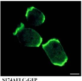
1.4.2 Expression and localisation of the mutated protein 51
2 Role of filamin in phagocytosis 52
2.1 Overexpression of the filamin rod domain reduces relative
phagocytosis in AX2 as well as in mutant cells 52
3 Phototaxis in mutant strain and rescue experiments 54 3.1 Expression of full-length ddfilamin and full-length ddfilamin
with a mutation at the predicted phosphorylation site rescues the
phototaxis defect. 55
3.2 Rescue of phototactic defect with filamin domains 56 3.3 The requirement of the dimerisation domain for the rescue
behavior of ddfilamin 57
3.4 Expression of full length ddfilamin at the anterior tip of
the slug is necessary to rescue the phototactic defect 58
Chapter Description Page(s)
4 Identification and characterisation of binding partners for
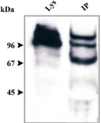
filamin 59 4.1 FIP, a novel protein interacts with filamin 60
4.1.1 Construction of a full-length FIP cDNA 60 4.1.2 Localisation of FIP does not change in HG1264 62 4.1.3 FIP may have a role in vesicle transport 62 4.1.4 FIP is associated with the actin cytoskeleton 63 4.4 Immunoprecipitation revealed the identification of TipA, SapA,
and GAPA as ddfilamin associated proteins, suggesting a role for
filamin in cell morphogenesis and actin remodeling. 64 4.3 Checking the interaction of filamin with Rho GTPases by yeast
two hybrid screening. 65
5 Microarray analysis suggests that mutant slugs have elevated
extracellular cAMP levels and lowered cell adhesion. 67
6 Chemotactic motility of mutants in actin binding proteins 69
6.1 Filamin minus cells chemotax normally 69
6.2 LimD- cells have a defect in chemotaxis but have normal
phototaxis 69 6.3 The villidin− mutant has a defect in chemotaxis as well as in
phototaxis 71
IV. DISCUSSION 73-83
1. Subcellular localisation of ddfilamin and its domains. 73 2. F-actin crosslinking by ddfilamin is essential for the rescueof the
phototaxis defect in ddfilamin− mutants. 75 3. Filamin in the tip region is essential for slug movement and
phototaxis. 78 4. Interaction partners of filamin suggest a diverse function for the
protein. 78
5. FIP may function in membrane traffic. 80
6. Microarray analysis suggests that the phototaxis defect in ddfilamin− mutants may be due to reduced cell adhesion and
defective cAMP wave propogation. 80
Chapter Description Page(s)
V. SUMMARY/ZUSAMMENFASSUNG 84-88
VI. BIBLIOGRAPHY 89-102
VII. ABBREVIATIONS 103
Erklärung -
Curriculum Vitae/Lebenslauf -
I. Introduction
1. Dictyostelium as a model organism for cytoskeletal studies
Dictyostelium discoideum, a soil living social amoeba feeds on yeast and bacteria in its natural habitat and has become an attractive tool for the study of the actin cytoskeleton and related proteins because of the following reasons: (i) The organism can be grown in the laboratory easily on bacterial lawns or in shaking culture (axenically). (ii) It undergoes a developmental cycle, which allows researchers to study a variety of changes that occur during development of a multicellular organism. (iii) Manipulation of genes can be done by homologous recombination or REMI (Restriction Enzyme Mediated Integration) (Kuspa and Loomis, 1992) and is easy because the organism is haploid. Overexpression of gene/s using nonintegrating vectors can be easily achieved, which allows studying the live dynamics of proteins or domains of protein by fusing with Green Fluorescence Protein (GFP). (iv) Dictyostelium carries all classes of actin binding proteins, which can be found in all eukaryotes and thus can be compared with mammalian cells. (v) The ongoing Dictyostelium genome project, which is likely to be finished by the end of this year and the Japanese cDNA project add great advantage to the study of the organism (Schleicher and Noegel, 1992 and Noegel and Luna, 1995).
Figure 1: The asexual life cycle of Dictyostelium discoideum. a, Dictyostelium amoebae live off bacteria in the soil. b, When the bacteria are consumed and starvation is imminent, the amoebae stop dividing and activate several genes that allow them to aggregate by chemotaxis towards cyclic AMP diffusing from centrally located cells. These aggregates, which can contain up to 100,000 cells, transform into motile slugs (c, d) and finally into fruiting bodies (e–g). The fruiting bodies contain 80,000 viable spores supported by 20,000 dead stalk cells.
(Graphic from Kessin 2000.) Blue and yellow colours for the cells indicate two different natural isolates. For a dynamic view of Dictyostelium development see http://dicty.cmb.nwu.edu/dicty/pnd/
The actin cytoskeleton has a dynamic role in the life cycle of Dictyostelium, it gives shape to the cells and controls cell movement, cell division and intracellular vesicle transport. A variety of actin-binding proteins are present in the cells controlling assembly and disassembly of actin. Among these actin-binding proteins are F-actin cross-linking proteins, capping protein, severing proteins, monomer-binding proteins, membrane anchors and motor proteins.
The localization and crosslinking of filamentous actin (F-actin) into bundles and networks is mediated by multiple families of cytoskeletal proteins, of which several share an α-actinin- like conserved F-actin binding domain (ABD) (Matsudaira, 1991, Otto 1994 and Troys et al.
1999). To date, eleven different actin crosslinking proteins have been identified in Dictyostelium, including a filamin-like protein (Hock and Condeelis, 1987), spectrin-like protein (Bennett and Condeelis 1988), ddfilamin (also termed as ddFLN gelation factor or ABP120) (Condeelis et al. 1981 and Noegel et al. 1989), α-actinin (Brier et al. 1983 and Noegel et al. 1987), elongation factor 1a (Demma, 1990), comitin (Weiner et al. 1993), a 30
Fechheimer, 1987, Fechheimer et al. 1992), cortexellin I and II (Faix et al. 1992) and fimbrin (Prassleret al. 1997).
** *
CAP*
* *
**
*
*
*
*
* *
*
*
34 kD*
Figure 2: Actin-binding proteins and their function (Schleicher et al. 1995). Actin-binding proteins influence the equilibrium between monomeric G-actin and filamentous F-actin as well as the structural organization of the network of actin filaments by either binding to monomeric actin and inhibiting polymerisation, or crosslinking, capping, severing, anchoring or moving the actin filaments by binding to actin filaments. The asterisk indicates actin- binding proteins that have been reported in Dictyostelium.
1.2 Phototaxis in Dictyostelium
Dictyostelium discoideum is widely used as a simple model organism for multicellular development (Maeda et al. 1997 and Gross, 1994). Dictyostelium live as individual amoebae in soil, preying on bacteria. But when food runs out and starvation is imminent, the previously independent amoebae form dendriticaggregation streams, which break up into groups of up to 105 cells (Shaffer, 1957 and Kessin, 2001) before they form cylindrical migrating slugs. Slugs are sensitive to light, pH, and even slight differencesin temperature, which allows them to migrate toward an optimal location for fruiting. Slugs are polar with a tip at the anterior consisting of prestalk cells whereas the posterior consists predominantely of prespore cells.
Phototactic turning is initiated in the tipand slugs sense the light only at the anterior prestalk zone (Francis, 1964, Poff and Loomis, 1973 and Fisher et al. 1984). Using the tools that are available in Dictyostelium, phototaxis and thermotaxis can be addressed at a molecular level.
Several groups isolated membrane bound photoreceptors and analysed the absorption spectrum (Poff and Butler, 1974, Poff et al. 1974 and Hader and Lebert, 1994). Schlenkrich et al.1995a isolated a 45.5 kDa putative photoreceptor from Dictyostelium, the absorption spectrum of which closely resembled the action spectrum for Dictyostelium phototaxis Schlenkrich et al.1995b, leading to the conclusion that this protein might play an important role in the photoreception of Dictyostelium amoebae.
Two models have been proposed to explain phototactic turning (Poff and Loomis, 1973, Fisher et al. 1984 and Fisher 1997). The optical model assumes thatthe pseudoplasmodium acts as a cylindrical lens, causing stronger stimulation of light locally and speeding up cell movement in the tip thus leadingto bending of the anterior zone toward the light source. The signal transduction or sign reversal model assumes that light acts directly on physiological processes of the cell and cell-cellsignalling by shifting the position of the organising centre in the tip. In agreement with both of these hypotheses Miura and Siegert (2001) found that light acts directly on the cAMP-signalling system and cell movement. Upon light irradiation, aggregating cells change their periodicity of cAMP signalling and cells in the slug tip release cAMP. They also found that concomitant changes in cell movement occurred in slug cells.
But these results do not explain the thermotaxis (Smith et al. 1982), where slugs migrate to a preferred temperature in the dark, neither do they explain data obtained with ddfilamin mutant cells, which have a clear phototaxis deficiency and migrate over shorter distances in the darkness as well as in horizontally directed light (Wallraff and Wallraff, 1997).
The anterior tip of the slug mostly consists of prestalk cells that control the migration of the slug. The tip shows average high concentration of cAMP, while the posterior prespore cells contain lower average concentration (Dormann and Weijer, 2001). Elevated level of cAMP is necessary for the maintenance of the tip, generation of a scroll wave and for the migration of anterior like cells (ALCs) from posterior region cells of the slug. Consistent with these observations, the multicellular aggregates overexpressing extracellular phosphodiesterase do not form a tip. Dormann and Weijer (2001) injected cAMP into the tip of slugs and found that higher concentrations (10-2 M) of cAMP are needed to disturb the wave generated at the tip.
Darcy et al. (1994) suggested an intermediary role for cGMP in photosensory and thermosensory processing in slugs and amoebae. They found all phototaxis mutant strains
cGMP responses to light and heat were unaltered in their cGMP response to a cAMP pulse, concluding that cAMP and light/heat regulate cGMP via independent pathways.
Several experiments have suggested a role for RasD in the correct proportioning of the prespore and prestalk cells in differentiation (Reymondet al. 1986 and Louis et al. 1997).
Furthermore, RasD− null cells exhibit a phototaxis and thermotaxis defect and were not able to orient correctly, suggesting a role for RasD in the modulation of morphogenetic signaling by the photo- and thermo-receptors (Willkins et al. 2000).
Villidin is a novel multidomain protein (190 kDa) containing a N-terminal WD repeat, three PH domains in the middle of the molecule, and five gelsolin-like segments, followed by a villin-like headpiece at the C-terminal end. The protein is prominently expressed in the slug stage (Gloss et al. 2003). The villidin-minus slugs exhibited a phototaxis defect. The mutant slugs migrate over a shorter distance and not as directed towards light as wild type slugs. The expression of the WD domain in the mutant background caused a more direct movement of the slugs towards the light source, whereas the gelsolin/villin region did not exhibit any rescuing potential.
Recently, another Dictyostelium protein has been described that has some similarity to villidin, GRP125, a new gelsolin-related protein. In the absence of GRP125, slugs fail to readjust their orientation correctly. Analysis of the GRP125-deficient mutant showed that GRP125 is required for coupling photodetection to the locomotory machinery of slugs (Stocker et al. 1999).
Li et al. (2001) explained the role of Sphingosine-1-phosphate (S-1-P) lyase, an enzyme that functions in fatty acid metabolism and controls the slug migration. S-1-P lyase null mutant show extremely limited directional migration, the slugs developed directly at the site of aggregation. The authors suggest that the phototaxis defect might be due to aberrant actin distribution and abnormal cell morphogenesis in mutant slugs.
Wallraff and Wallraff (1997) tested behavioural deficits in the slugs of three mutant strains of Dictyostelium lacking different F-actin binding proteins, among them two were F-actin crosslinking proteins. Two strains, defective in the production of either α-actinin or severin (an actin capping and severing protein), did not show changes in slug behavior. Slugs of the
mutant lacking ddfilamin, however, migrated shorter distances in the darkness as well as in horizontally directed light. More remarkably, they migrated at an angle of approximately 45°
to the left or right of the incident light, whereas wild-type slugs migrated on fairly straight paths towards the light. The author concluded that, as these mutants are known to lack a constituent of the cytoskeleton, alterations in the cytoskeletal architecture might cause alterations in the optical properties of cell structures, which are of importance in the specific mechanism underlying slug phototaxis.
Genetic analysis of slug behaviour suggests that as many as 55 genes are involved in phototaxis and that several of the encoded proteins regulate signal transduction pathways involving the intracellular messengers cAMP, cGMP, IP3 and Ca2+ (Darcy 1994, Fisher et al.
1997 and Fisher, 1997). But ddfilamin emerged as the only protein directly associated with the actin cytoskeleton, which had a significant role in Dictyostelium phototaxis.
Structurally filamins are homodimers with large polypeptide chains that associate at their carboxyl termini (Gorlin et al. 1990 and Fucini et al. 1999). Their conserved amino-terminal actin binding domains consist of two calponin homology domains common to the members of the α-actinin/spectrin superfamily of actin-binding proteins (Hartwig, 1994). The rest of the polypeptide forms 24 repeated domains of ~96 amino acids, each made up of seven antiparallel β-strands that produce an immunoglobulin fold (Fucini et al. 1997). The Dictyostelium filamin has a shorter elongated domain, consisting of only six rod like repeats of 100 amino acids (Noegel et al. 1989). Its dimerisation is mediated via the formation of intramolecular β-sheets between the rod domains 6, leading to an antiparallel arrangement of the two monomer chains (McCoy et al. 1999 and Fuciniet al. 1999). Dictyostelium mutants lacking filamin have defects in the structure of the actin cytoskeleton and exhibit reduced cross-linking of actin filaments, leading to reduced size and frequency of pseudopods. This results in a decreased motility, chemotaxis and phagocytosis of the mutant cells (Cox et al.
1992 and 1995).
Recent studies suggested that the function of the filamins is not only in maintaining the cortical actin network but also the organisation and stabilisation of these networks by interwebbing them with membrane proteins and receptors. Thus filamins interact with several membrane receptors: the cytoplasmic part of the glycoprotein Ib IX complex, the receptor for von Willebrand factor (Andrews and Fox, 1991 and Takafuta et al. 1998), β1- and β2-integrins
were all identified as ligands for filamin. Filamins also affect intracellular trafficking of proteins and signal transduction. Filamin influences the activity of furin, a protease that is involved in the proteolytic processing of many proproteins, by promoting its internalisation (Zent et al. 2000). It binds to presenilin-1, a protein involved in early onset familial Alzheimer's disease and in the notch signalling pathway (Zhang et al. 1998, Schwarzmanet al. 1999) and interacts with caveolin-1, a multifunctional protein with roles in caveolae biogenesis, endocytic events, cholesterol transport and various signal transduction processes (Stahlhutand Deurs, 2000). Furthermore, the involvement of filamin in signal transduction is confirmed by its interaction with several components of the NFκB pathway (Edwards et al.
1997, Marti, 1997 and Leonardi et al. 2000) and the small GTPases RhoA, Rac1, Cdc42 and RalA (Ueda et al. 1992, Ohta et al. 1999, Bellangeret al. 2000 and Pi et al. 2002).
Human filamin is also strongly phosphorylated in vivo, which affects its interaction with several proteins such as GTPases and might also affect the F-actin binding capacity and crosslinking activity. Phosphorylation is mainly on serine and threonine residues and is achieved by a variety of kinases (cAMP-kinase, PKC and CaM-kinase II) (Ohta and Hartwig, 1995 and Tiggs et al. 2003).
To understand the role of ddfilamin in phototaxis we performed rescue studies by expressing the actin binding domain, rod domain, C terminally truncated ddfilamin (which can no longer crosslink actin filamients) and point mutated ddfilamin (at suspected phosphorylation site). As the anterior tip of the slug controls its migration, we expressed ddfilamin under the control of cell type specific promoters to test whether it plays a functional role there. We also studied global gene expression pattern in the mutant by microarray analysis in order to identify genes that involved in this process. Furthermore, we checked the interaction of this protein with small GTP binding proteins by using the yeast two hybrid system. With an immunoprecipitation assay we found three proteins interacting with ddfilamin, which might help ddfilamin to perform its role in the phototactic migration.
1.3 Chemotaxis in Dictyostelium
Cells sense and respond to signals in the extracellular environment. Many cells have the capacity to detect the direction and intensity of an extracellular chemical gradient and respond by directed migration toward the source of the chemical. This process is called chemotaxis.
Chemotaxis results from a localised polymerisation of F-actin leading to the formation of a
new lamellipod or pseudopod, cell polarisation, and the forward protrusion of the leading edge. Phosphoinositide 3-Kinase plays a central role in establishing and maintaining cell polarity by regulating the subcellular localisation and activation of down stream effectors that are essential for regulating cell polarity and proper chemotaxis. In addition, other proteins, among them actin-binding proteins, are crucial for the establishment of polarity and for chemotaxis.
Here we have analysed chemotactic migration of LimC−, LimD−, LimC−/D− and villidin mutants. LimC and LimD are LIM domain containing proteins that directly associate with F- actin. LimD− and LimC−/D− were found to have a defect in chemotaxis. Reexpression of LimD-GFP in both cells rescued the defect stating that the defect found is due to the loss of function of LimD (Khurana et al. 2002). Villidin is a novel protein with WD and PH domains, which are associated with signal transdution and a C-terminal gelsolin like domain followed by a villin headpiece, which normally bundles F-actin very strongly. The headpiece in villidin however, does not bind to F-actin (Vardar et al., 2002). We also studied the migration behaviour of villidin mutants and found that was also altered (Gloss et al. 2003).
II. Materials and Methods
1. Materials
1.1 Laboratory materials
Cellophane sheet, Dry ease Novex
Centrifuge tubes, 15 ml, Greiner
Coverslips (glass), Ø12 mm, Ø18 mm, Ø55 mm Assistent
Corex tube, 15 ml, 50 ml Corex
Cryo tube, 1 ml Nunc
Electroporation cuvette, 2 mm electrode gap Bio-Rad
Gel-drying frames Novex
Microcentrifuge tube, 1.5 ml, 2.2 ml Sarstedt
Micropipette, 1-20 µl, 10-200 µl, 100-1,000 µl Gilson
Micropipette tips Greiner
Multi-channel pipette Finnigan
Needles (sterile), 18G–27G Terumo, Microlance
Nitrocellulose membrane, BA85 Schleicher and Schuell
Nitrocellulose-round filter, BA85, Ø82 mm Schleicher and Schuell
Nuclepore membrane filter Nuclepore
Nylon membrane Biodyne B Pall
Parafilm American Nat Can
Pasteur pipette, 145 mm, 230 mm Volac
PCR soft tubes, 0.2 ml Biozym
Petri dish (35 mm, 60 mm, 100 mm) Falcon
Petri dish (90 mm) Greiner
Plastic cuvette, semi-micro Greiner
Plastic pipettes (sterile), 1 ml, 2 ml, 5 ml, 10 ml, 25 ml Greiner
Quartz cuvette Infrasil Hellma
Quartz cuvette, semi-micro Perkin Elmer
Saran wrap Dow
Scalpels (disposable), Nr. 10, 11, 15, 21 Feather
Slides, 76 x 26 mm Menzel
Syringes (sterile), 1 ml, 5 ml, 10 ml, 20 ml Amefa, Omnifix Syringe filters (Acrodisc), 0.2 µm, 0.45 µm Gelman Sciences
Whatman 3MM filter paper Whatman
X-ray film, X-omat AR-5, 18 x 24 mm, 535 x 43 mm Kodak
1.2. Instruments and equipments Centrifuges (microcentrifuges):
Centrifuge 5417 C Eppendorf
Centrifuge Sigma B Braun
Cold centrifuge Biofuge fresco Heraeus Instruments Centrifuges (table-top, cooling, low speed):
Centrifuge CS-6R Beckman
Centrifuge RT7 Sorvall
Centrifuge Allegra 21R Beckman
Centrifuges (cooling, high speed):
Beckman Avanti J25 Beckman
Sorvall RC 5C plus Sorvall
Centrifuge-rotors:
JA-10 Beckman JA-25.50 Beckman SLA-1500 Sorvall SLA-3000 Sorvall SS-34 Sorvall
Dounce homogeniser, 10 ml B. Braun
Electrophoresis power supply, Power-pac-200, -300 Bio-Rad
Electroporation unit Gene-Pulser Bio-Rad
Fluorimeter PTI
Freezer (-80 °C) Nunc
Freezer (-20 °C) Siemens, Liebherr
Gel-documentation unit MWG-Biotech
Heating block DIGI-Block JR NeoLab
Heating block, Dry-Block DB x 20 Techne
Hybridization oven Hybaid
Ice machine Ziegra
Incubators:
Incubator, microbiological Heraeus
Incubator with shaker Lab-Therm Kuehner
Laminar flow, Hera Safe (HS 12) Heraeus
Magnetic stirrer, MR 3001 K Heidolph
Microscopes:
Light microscope, CH30 Olympus
Light microscope, DMIL Leica
Light microscope, CK2 Olympus
Fluorescence microscope, DMR Leica
Fluorescence microscope, 1X70 Olympus
Confocal laser scanning microscope, DM/IRBE Leica
Stereomicroscope, SZ4045TR Olympus
Oven, conventional Heraeus
PCR machine, PCR-DNA Engine PTC-200 MJ Research
pH-Meter Knick Refrigerator Liebherr Semi-dry blot apparatus, Trans-Blot SD Bio-Rad
Shakers GFL Kuehner
Sonicator, Ultra turrax T25 basic IKA Labortechnik
Speed-vac concentrator, DNA 110 Savant
Spectrophotometer, Ultraspec 2000, UV/visible Pharmacia Biotech
Ultracentrifuges:
Optima TLX Beckman
Optima L-70K Beckman
Ultracentrifuge-rotors:
TLA 45 Beckman
TLA 100.3 Beckman
SW 41 Beckman
UV-crosslinker UVC 500 Hoefer
UV- transilluminator TFS-35 M Faust
Vortex, REAX top Heidolph
Video cameras
JAI CV-M10 CCD Camera Stemmer Imaging
SensiCam PCO Imaging
Waterbath GFL X-ray-film developing machine, FPM-100A Fujifilm
1.3 Kits
FairPlayTM Microarray labeling kit Stratagene
Nucleobond AX Macherey-Nagel
NucleoSpin Extract 2 in 1 Macherey-Nagel
Nucleotrap Macherey-Nagel
Original TA Cloning Invitrogen
pGEM-T Easy Promega
Qiagen Midi- and Maxi-prep Qiagen
Qiagen RNeasy Midi/Mini Kit Qiagen
Stratagene Prime It II Stratagene
1.4 Enzymes, antibodies, substrates, inhibitors and antibiotics Enzymes used in the molecular biology experiments:
Calf Intestinal Alkaline Phosphatase (CIAP) Roche
Klenow fragment (DNA polymerase) Roche
Lysozyme Sigma
Protein A Sepharose CL-4B Amersham
Proteinase K Sigma
Restriction endonucleases Amersham,
New England Biolabs
Ribonuclease A (RNase A) Sigma
S1-nuclease Amersham
T4 DNA ligase Roche
Taq-polymerase Roche Primary antibodies:
Mouse anti-actin monoclonal antibody Act 1-7 (Simpson et al. 1984) Mouse anti-GFP monoclonal antibody K3-184-2 (Gloss et al. 2003) Mouse anti-filamin-monoclonal Antibody 82-421-5 (Brink et al. 1989) Mouse anti-filamin- monoclonal antibody 82-454-12(Brink et al. 1989) Mouse anti-csA monoclonal antibody 33-294-17 (Bertholdt et al. 1985) Mouse anti-α-actinin monoclonal antibody 47-60-8 (Schleicher et al. 1984) Mouse anti-FIP230 monoclonal antibody K12-349-7 (Knuth, 2002 Ph. D Thesis) Mouse anti-FIP230 monoclonal antibody K12-362-6 (Knuth, 2002 Ph. D Thesis) Mouse anti-FIP230 monoclonal antibody K12-454-2 (Knuth, 2002 Ph. D Thesis) Goat anti-GST antibody (Amersham).
Rabbit Phospho-Serine/Threonine polyclonal Antibody (Cell Signaling Technology) Rabbit Phospho-Threonine polyclonal Antibody (Cell Signaling Technology)
Secondary antibodies:
Goat anti-mouse IgG, peroxidase conjugated Sigma Goat anti-rabbit IgG, peroxidase conjugated Sigma Mouse anti-goat IgG, peroxidase conjugated Sigma Sheep anti-mouse IgG, I125 conjugatedAmersham
Inhibitors:
Benzamidin Sigma β-glycerophosphate Sigma
Complete Mini®, Protease inhibitor cocktail tablets Roche
Diethylpyrocarbonate (DEPC) Sigma
Leupeptin Sigma Pepstatin Sigma
Phenylmethylsulphonyl fluoride (PMSF) Sigma
Sodium Fluoride Sigma
Sodium orthovanadate Sigma
Sodium pyrophosphate Sigma
Antibiotics:
Ampicillin Gruenenthal
Blasticidin S ICN Biomedicals
Chloramphenicol Sigma Dihydrostreptomycinsulfate Sigma
Geneticin (G418) Life Technologies
Kanamycin Sigma, Biochrom
Tetracyclin Sigma
Most of the chemicals and reagents were obtained either from Sigma, Fluka, Difco, Merck, Roche, Roth or Serva. Those chemicals or reagents that were obtained from companies other than those mentioned here are listed below:
Acetic acid (98-100%) Riedel-de-Haen
Acrylamide (Protogel: 30:0,8 AA/Bis-AA) National Diagnostics
Agar-Agar (BRC-RG) Biomatic
Agarose (Electrophoresis Grade) Life Technologies
3-Amino-1, 2, 4-triazol Sigma
Amino acids Sigma
Bacto-Agar Difco
Bacto-Pepton Difco
Bacto-Trypton Difco 5-Brom-4-chlor-3-indolyl-β-D-galactopyranosid (X-Gal) Roth
Bromphenolblue (Na-Salt) Serva
BSA (Bovine serum albumin) Roth
Calciumchlorid-Dihydrat Merck
Chloroform Riedel-de-Haen
Coomassie-Brilliant-Blue G 250 Roche
Coomassie-Brilliant-Blue R 250 Serva
p-Cumaric acid Fluka
Cyclohexamide Sigma
1,4-Dithiothreitol (DTT) Gerbu
Dimethylformamide Riedel-de-Haen
Ethanol Riedel-de-Haen
Ethylen diamine tetraaceticacid (EDTA) Merck
Ethylenglycolbis [2-aminoethylether]-
Glycerine Riedel-de-Haen Isopropyl-D-thiogalactopyranoside (IPTG) Loewe Biochemica
Methanol Riedel-de-Haen Morpholino propane sulphonic acid (MOPS) Gerbu
N- [2-Hydroxyethyl] piperazine-N’-2-
-ethanesulfonic acid (HEPES) Biomol
Sodium dodecyl sulphate (SDS) Serva
Sodium hydroxide Riedel-de-Haen
Triton X-100 Merck
Tween 20 Roth
Yeast Nitrogen Base Difco
Radiolabelled nucleotide:
α-32P-deoxyadenosine triphosphate, (10 mCi/ml) Amersham
1.6 Media and buffers
All media and buffers were prepared with deionised water filtered through an ion-exchange unit (Membra Pure). The media and buffers were sterilized by autoclaving at 120 ºC and
antibiotics were added to the media after cooling to approx. 50 ºC. For making agar plates, a semi-automatic plate-pouring machine (Technomat) was used.
1.6.1 Media and buffers for Dictyostelium culture AX2-medium (Claviez et al. 1982), pH 6.7:
7.15 g yeast extract 14.3 g peptone (proteose) 18.0 g maltose
0.486 g KH2PO4
0.616 g Na2HPO2.2H2O
add H2O to make 1 litre
Soerensen phosphate buffer (Malchow, 1972), pH 6.0:
2 mM Na2HPO4
14.6 mM KH2PO4
Phosphate agar plates, pH 6.0: 9 g agar
add Soerensen phosphate buffer, pH 6.0 to make 1 litre
Salt solution (Bonner, 1947): 10 mM NaCl 10 mM KCl 2.7 mM CaCl2
SM agar plates(Sussman, 1951), pH 6.5: 9 g agar 10 g peptone 10 g glucose 1 g yeast extract 1 g MgSO4.7 H2O 2.2 g KH2PO4
1 g K2HPO4
add H2O to make 1 litre
1.6.2 Media for E. coli culture
LB medium (Sambrook, 1989), pH 7.4: 10 g bacto-tryptone 5 g yeast extract 10 g NaCl
adjust to pH 7.4 with 1 N NaOH
add H2O to make 1 litre
For LB agar plates, 0.9% (w/v) agar was added to the LB medium and the medium was then autoclaved. For antibiotic selection of E. coli transformants, 50 mg/l ampicillin, kanamycin or chloramphenicol was added to the autoclaved medium after cooling it to approximately 50ºC.
For blue/white selection of E. coli transformants, 10 µl 0.1 M IPTG and 30 µl X-gal solution (2% in dimethylformamide) was spread per 90 mm plate and the plate was incubated at 37ºC for at least 30 min before using.
SOC medium (Sambrook, 1989), pH 7.0: 20 g bacto-tryptone 5 g yeast extract 10 mM NaCl 2.5 mM KCl
dissolve in 900 ml deionised H2O adjust to pH 7.0 with 1 N NaOH
The medium was autoclaved, cooled to approx. 50ºC and then the following solutions, which were separately sterilized by filtration (glucose) or autoclaving, were added:
10 mM MgCl2.6 H2O 10 mM MgSO4.7 H2O 20 mM glucose
add H2O to make 1 litre
1.6.3 Media and buffers for Yeast culture
YEPD-Medium: YEPD-Agar plates:
20 g/l Difco Pepton 20 g/l Difco Pepton 10 g/l Yeast extract 10 g/l Yeast extract
18 g/l Agar agar
100 x Adenine solution: 100 x Tyrosine solution:
200 mg (1,1 mmol) Adenine in 100 ml 300 mg (1,7 mmol) Tyrosin in 100 ml
water dissolve with addition of little amounts dissolve with addition of NaOH solution and of HCl and filter sterilize. filter sterilize.
100 x Histidine solution: 100 x Leucine solution:
200 mg (1 mmol) Histidine in 100 ml 1000 mg (7,6 mmol) Leucin in 100 ml Water and filter sterilize. Water and filter sterilize.
100 x Tryptophan solution: 100 x Uracil solution:
200 mg (1 mmol) Tryptophan in 100 ml 200 mg (1,8 mmol) Uracil in 100 ml
Water filter sterilize. Water dissolve by warming in water bath and
filter sterilize.
1 M 3-Amino-1,2,4-triazol solution: 100 x Cycloheximide solution:
8,4 g 3-Amino-1,2,4-triazol in 100 ml 1 mg/ml Cycloheximid in Water solution Water, filter once and filter sterilize. filter sterilize.
10 x Amino acid solutions:
300 mg (2,3 mmol) Isoleucine 1500 mg (1,1 mmol) Valine 200 mg (0,9 mmol) Arginine 300 mg (1,6 mmol) Lysine 200 mg (1,34 mmol) Methionine 500 mg (3 mmol) Phenylalanine 2000 mg (16,8 mmol) Threonine, make volume to 1l and filter sterilize.
The composition of the selection media and agar plates is indicated in following table. Agar agar and Yeast extract without nitrogen base dissolved in water was autoclaved. The glucose solution was made in water and filter sterilised. The addition of the remaining stock solutions took place after cooling to 55 °C.
Selection plates
Reagents SD/-Leu SD/-Leu+Cyh SD/-Leu/-Trp SD/-Trp SD/-Leu/-His/-Trp/+3-AT
Yeast Nitrogen Base [g] 6,7 6,7 6,7 6,7 6,7
Agar agar [g] 20 20 20 20 20
Water [ml] 750 750 770 750 745
20% Glucose [ml] 100 100 100 100 100
10 x Amino acid solution [ml] 100 100 100 100 100
100 x Adenine [ml] 10 10 10 10 10
100 x Tyrosine [ml] 10 10 10 10 10
100 x Uracile [ml] 10 10 10 10 10
100 x Histidine [ml] 10 10 10 10 #
100 x Leucine [ml] # # # 10 #
100 x Tryptophane [ml] 10 10 # # #
Cycloheximide solution [ml] # 10 # # #
3-AT-solution [ml] # # # # 25
1.6.4 Buffers and other solutions
The buffers and solutions that were commonly used during the course of this study are mentioned below.
10x NCP-Buffer, pH 8.0: 12.1 g Tris/HCl 87.0 g NaCl 5.0 ml Tween 20 2.0 g sodium azide
add H2O to make 1 litre
PBG, pH 7.4: 0.5 % bovine serum albumin 0.1 % gelatin (cold-water fish skin) in 1x PBS, pH 7.4
1x PBS, pH 7.4: 8.0 g NaCl 0.2 g KH2PO4
1.15 g Na2HPO4
0.2 g KCl
dissolve in 900 ml deionised H2O adjust to pH 7.4
add H2O to make 1 litre, autoclave
1.2 M Phosphate buffer, pH 6.8: 1.2 M Na2HPO4, pH 9.1 was mixed with 1.2 M
NaH2PO4, pH 4.02 at the ratio of 2:1.
20x SSC, pH 7.0 3 M NaCl
0.3 M sodium citrate
TE buffer, pH 8.0: 10 mM Tris/HCl, pH 8.0 1 mM EDTA, pH 8.0
10x TAE buffer, pH 8.3: 27.22 g Tris
13.6 g sodium acetate 3.72 g EDTA
add H2O to make 1 litre
MES buffer, pH 6.5 20 mM 2-[N-morpholino]ethane sulphonic acid, pH 6.5 1 mM EDTA
250 mM sucrose
Homogenisation buffer, pH 7.4: 30 mM Tris/HCl, pH 7,4 2 mM DTT
2 mM EDTA 4 mM EGTA 5 mM Benzamidin 0,5 mM PMSF
1 Tablet complete® mini Protease Inhibitor Mix (Roche) per 10 ml buffer
add 30 % sucrose, prepare fresh before use.
Lysis Buffer (phosphorylation): 50 mM Tri-HCl, pH 7.6 150 mM NaCl
1 % Nonidet P-40 1 mM sodium orthovanadate 10 mM sodium fluoride 5 mM sodium pyrpphosphate
10 mM β-glycrephosphate
5 x Immunoprecipitation Buffer: 0.5 m Potassium phosphate buffer 0.375 M NaCl
25 mM EDTA 5 mM Benzamidine 2.5 mM PMSF
Adjust the pH to 7.9, prepare fresh.
Hybridisation solution (50 µl): Hybridisation buffer 48 µl Fish sperm DNA [10 mg/ml] 1µl Oligo dA (18 mer, 100 µM) 1µl mix well
Hybridisation buffer: 1.2M Phosphate buffer, pH 6.8 2mM EDTA
50 % Formamide
1% Na-Laurylsarcosinate 0.2 % SDS
4 x Denhadt’s Reagent
100 x Denhardt’s reagent: 2 % Ficoll 400
2 % Polyvinylpyrolidone 2 % bovine serum albumin
1.2 M Phosphate buffer, pH 6.8: 2 volumes 1.2 M Na2HPO4 1 volume 1.2 M NaH2PO4
Blocking Solution (282 ml): 270 ml 1-Methy-2-pyrrolidinone (solution should be colourless)
0.4 g Succinic anhydride (store desiccated) 12.1 ml 1M Sodium borate, pH 8.0 (42mM)
1.7 Biological materials
Bacterial strains: E. coli JM 38 (Vieira and Messing, 1982) E. coli DH5α (Hanahan, 1983)
E. coli XL1 blue (Bullock,1987)
Klebsiella aerogenes (Williams KL and Newell, 1976)
Dictyostelium discoideum strain:
AX2-214 An axenically growing derivative of wild strain, NC-4 (Raper, 1935). Commonly referred to as AX2.
GHR, Filamin-Mutant fromAX2: Eichinger et al. 1996
HG1264, Filamin¯mutant from AX2: Brink et al. 1990
GA1 Filamin¯ and α-actinin¯ double
Mutant: Rivero et al. 1996
Yeast Strains : Y190 (Flick et al. 1990, Harper et al. 1993)
Y187 (Harper et al. 1993)
2 Cell biological methods
2.1 Growth of Dictyostelium
2.1.1 Growth in liquid nutrient medium(Claviez et al. 1982)
Dictyostelium discoideum AX2 and the derived transformants were grown in liquid AX2 medium containing dihydrostreptomycin (40 µg/ml) and other appropriate selective antibiotics (depending upon mutant) at 21°C either in a shaking-suspension in Erlenmeyer flasks with shaking at 160 rpm or on petri dishes. For all the cell biological works, cultures were harvested at a density of 3-5 x 10⁶ cells/ml.
2.1.2 Growth on SM agar plates
In general, Dictyostelium cells were plated onto SM agar plates overlaid with Klebsiella aerogenes and incubated at 21 ºC for 3-4 days until Dictyostelium plaques appeared on the bacterial lawns. To obtain single clones of Dictyostelium, 50-200 cells were suspended in 100 µl Soerensen phosphate buffer and plated onto Klebsiella-overlaid SM agar plates. Single plaques obtained after incubation at 21ºC for 3-4 days were picked up with sterile tooth-picks, transferred either to new Klebsiella-overlaid SM agar plates or to separate petri dishes with AX2 medium supplemented with dihydrostreptomycin (40 µg/ml) and ampicillin (50µg/ml) (to eliminate the bacteria) and any other appropriate selective antibiotic (depending upon mutant).
2.2 Developoment of Dictyostelium
Development in Dictyostelium is induced by starvation. Cells grown to a density of 2-3 x 10⁶ cells/ml were pelleted by centrifugation at 2,000 rpm (Sorvall RT7 centrifuge) for 2 min at 4ºC and were washed two times in an equal volume of cold Soerensen phosphate buffer in order to remove all the nutrients present in the culture medium. For development in suspension culture, the cells were resuspended in Soerensen phosphate buffer at a density of 1 x 10⁷ cells/ml and were shaken at 160 rpm and 21 ºC for desired time periods. For development on nitrocellulose filters, 0.5 x 10⁸ cells were deposited on nitrocellulose filters (Millipore type HA, Millipore) and allowed to develop at 21 °C as described Newell (Newell et al. 1969).
2.3 Preservation of Dictyostelium
Dictyostelium cells were allowed to grow in AX2 medium to a density of 4-5 x 10⁶ cells/ml. 9 ml of the densely grown culture were collected in a 15 ml Falcon tube on ice and supplemented with 1 ml horse serum and 1 ml DMSO. The contents were mixed by gentle pipetting, and aliquoted in cryotubes (1 ml). The aliquots were incubated on ice for 60 min, followed by incubation at –20 ºC for at least 2 hr. Finally the aliquots were transferred to –80 ºC for long term storage.
For reviving the frozen Dictyostelium cells, an aliquot was taken out from –80 ºC and thawed immediately at 37 ºC in a waterbath. In order to remove DMSO, the cells were transferred to a Falcon tube containing 30 ml AX2 medium and centrifuged at 2,000 rpm (Sorvall RT7 centrifuge) for 2 min at 4 ºC. The cell pellet was resuspended in 250 µl of AX2 medium and the cell suspension was plated onto SM agar plates overlaid with Klebsiella. The plates were incubated at 21 ºC until plaques of Dictyostelium cells started to appear.
2.4 Transformation of Dictyostelium cells by electroporation
The electroporation method for transformation of Dictyostelium cells described by de Hostos143 was followed with little modifications. Dictyostelium discoideum cells were grown axenically in suspension culture to a density of 2-3 x 10⁶ cells/ml. The cell suspension was incubated on ice for 20 min and centrifuged at 2,000 rpm (Sorvall RT7 centrifuge) for 2 min at 4 ºC to collect the cells. The cells were then washed with an equal volume of ice-cold Soerensen phosphate buffer followed by an equal volume of ice-cold electroporation-buffer.
After washings, the cells were resuspended in electroporation-buffer at a density of 1 x 10⁸ cells/ml. For electroporation, 20-25 µg of the plasmid DNA was added to 500 µl of the cell suspension and the cell-DNA mixture was transferred to a pre-chilled electroporation cuvette (2 mm electrode gap, Bio-Rad). Electroporation was performed with an electroporation unit (Gene Pulser, Bio-Rad) set at 0.9 kV and 3 µF without the pulse controller. After electroporation, the cells were immediately spread onto a 100-mm petri dish and were allowed to sit for 10 min at 21 ºC. Thereafter, 1 ml of healing-solution was added dropwise onto the cells and the petri dish was incubated at 21 ºC on a shaking platform at 50 rpm for 15 min. 10 ml of AX2 medium was added into the petri dish and the cells were allowed to recover overnight. The next day, the medium was replaced by the selection medium containing appropriate antibiotic. To select for stable transformants, selection medium was replaced every 24-48 hr until the control plate (containing cells electroporated without any DNA) was clear of live cells.
Electroporation-buffer: 0.1 M Potassium phosphate buffer:
100 ml 0.1 M potassium phosphate buffer 170 ml 0.1 M KH2PO4
17.12 g sucrose 30 ml 0.1 M K2HPO4
add distilled H2O to make 1 litre adjust to pH 6.1 autoclave
Healing-solution:
150 µl 0.1 MgCl2
150 µl 0.1 CaCl2
10 ml electroporation-buffer
2.5 Quantitative phagocytosis assays
Phagocytosis was performed according to Maniak et al. (1995). Briefly Dictyostelium cells were grown to <5 x10⁶/ml over 5 generations in axenic medium. Cells were centrifuged and resuspended at 2 x 10⁶ cells/ml in fresh axenic medium at 21 °C. TRITC-labelled yeast cells prepared according to Materials and Method 6.3 were added in a 5 fold excess (109 yeast cells/ml stock). Cells were incubated on a rotary shaker at 160 rpm. Samples were taken at different intervals and the fluorescence of non internalized yeasts were quenched by incubating for 3 min with 100 µl trypan blue (2 mg/ml). Cells were centrifuged again, resuspended in phosphate buffer and the fluorescence was measured using a fluorimeter (544 nm excitation / 574 nm emission).
3 Molecular biological methods
3.1 Purification of plasmid DNA
In general, for small cultures (1 ml) of E. coli transformants, the alkaline lysis method of Holmes and Quigley (1981) was used to extract plasmid DNA. This method is good for screening a large number of clones simultaneously for the desired recombinant plasmid.
Briefly, single transformants were picked up from the culture plate and were grown overnight in 1 ml of LB media containing suitable antibiotic. The E. coli cells grown overnight were pelleted by centrifuging at 6,000 rpm in a microcentrifuge for 3-5 min. The pellet was then resuspended completely in 250 µl STET/lysozyme buffer and the suspension was incubated at room temperature for 10 min to lyse the bacterial cells. The bacterial lysate was boiled at 100ºC for 1 min and was then centrifuged in an Eppendorf centrifuge at maximum speed for 15 min at room temperature. The plasmid DNA present in the supernatant was precipitated by
adding an equal volume of isopropanol and incubating at room temperature for 10 min. The precipitated DNA was pelleted in the Eppendorf centrifuge at 12,000 rpm for 15 min and the DNA pellet was washed with 70 % ethanol, dried in a speed-vac concentrator and finally resuspended in 40 µl TE, pH 8.0, containing RNase A at 1 µg/ml.
STET/lysozyme buffer, pH 8.0:
50 mM Tris/HCl, pH 8.0 50 mM EDTA
0.5% Triton-X-100 8.0% Sucrose
Add lysozyme at 1 mg/ml at the time of use.
Alternatively, for pure plasmid preparations in small and large scales (for sequencing, PCR or transformations), kits provided either by Macherey-Nagel (Nucleobond AX kit for small scale plasmid preparations) or by Qiagen (Qiagen Midi- and Maxi-Prep kit for large scale plasmid preparations) were used. These kits follow basically the same approach: first overnight culture of bacteria containing the plasmid is pelleted and the cells are lysed by alkaline lysis. The freed plasmid DNA is then adsorbed on a silica matrix, washed with ethanol, and then eluted with TE, pH 8.0. This method avoids the requirement of caesium chloride or phenol- chloroform steps during purification.
3.2 Digestion with restriction enzymes
All restriction enzymes were obtained from NEB, Amersham or Life Technologies and the digestions were performed in the buffer systems and temperature conditions as recommended by the manufacturers. The plasmid DNA was digested for 1-2 hr.
3.3 Generation of blunt ends in linearised plasmid DNA
For many cloning experiments, it was necessary to convert the 5’ or 3’ extensions generated by restriction endonucleases into blunt ends. Repair of 5’ extensions was carried out by the polymerase activity of the Klenow fragment, whereas repair of 3’ extensions was carried out by the 3’ to 5’ exonuclease activity of the Klenow fragment.
Reaction-mix for 5’ extensions: Reaction-mix for 3’ extensions:
1-4 µg linearised DNA 1-4 µg linearised DNA 5 µl 10x High salt buffer 5 µl 10x High salt buffer 1 µl 50x dNTP-mix (each 4 mM) 2 U Klenow fragment 2 U Klenow fragment add H2O to make 50 µl add H2O to make 50 µl
500 mM Tris/HCl, pH 7.5
1 M NaCl 100 mM MgCl2
10 mM DTT
The reaction was carried out at 37ºC for 25-30 min. After incubation, the reaction was immediately stopped by heat-inactivating the enzyme at 75 ºC for 10 min or by adding 1µl 0.5 M EDTA. This was followed by phenol/chloroform extraction and precipitation of DNA with 2.5 volume ethanol.
3.4 Dephosphorylation of DNA fragments
To avoid self-ligation of the vector having blunt ends or that has been digested with a single restriction enzyme, 5’ ends of the linearised plasmids were dephosphorylated by calf- intestinal akaline phosphatase (CIAP). Briefly, in a 50 µl reaction volume, 1-5 µg of the linearised vector-DNA was incubated with 1 U calf-intestinal alkaline phosphatase in CIAP- buffer (provided by the manufacturer) at 37 ºC for 30 min. The reaction was stopped by heat- inactivating the enzyme at 65 ºC for 10 min. The dephosphorylated DNA was extracted once with phenol-chloroform and precipitated with 2 vol. ethanol and 1/10 vol. 2 M sodium acetate, pH 5.2.
3.5 Setting up of ligation reactions
A DNA fragment and the appropriate linearised plasmid were mixed in approximately equimolar amounts. T4 DNA ligase and ATP were added as indicated below and the ligation reaction was left overnight at 10-12 ºC.
Ligation reaction: 5x Ligation buffer:
Linearised vector DNA (200-400ng) Supplied with the enzyme
DNA fragment by manufacturers
4 µl 5x ligation buffer 1µ 0.1 M ATP
1.5 U T4 Ligase
and water to make up to 20 µl.
3.5.1 Generation of the point mutation
The point mutation was generated to replace serine 174 in ddfilamin with alanine by using an overlapping PCR approach, involving the following steps.
i) Two fragments between nucleotides 270-487 and 461-574 were generated, which has 26 nucleotides overlapping end.
ii) The point mutation was generated by introducing a mismatch nucleotide in the overlapping ends.
iii) The mutation at the EcoRV site in position 548 bp was made to destroy the restriction enzyme site without changing the amino acid.
iv) Overlaping PCR was done to join the two fragments using primers at the extreme ends.
v) The PCR product was cloned into a pGEM-T Easy vector and transformed into E. coli.
vi) DNA obtained from the bacterial transformants was sequenced to confirm the mutation.
vii) The 290 bp fragment obtained by a EcoRV digestion from the pGEM-T Easy vector was ligated to full-length ddfilamin cDNA with C-terminal GFP tag, digested with the EcoRV.
viii) The orientation of the cloned fragment was confirmed by PCR using a forward primer at the 5’ end of the full-length cDNA and one of the reverse primer used for generation of the 290 bp fragment.
ix) Clones were further confirmed by sequence analysis.
3.6 DNA agarose gel electrophoresis
Agarose gel electrophoresis was performed according to the method described by Sambrook to resolve and purify the DNA fragments. Electrophoresis was typically performed with 0.7 % (w/v) agarose gels in 1x TAE buffer submerged in a horizontal electrophoresis tank containing 1x TAE buffer at 1-5 V/cm. Only for resolving fragments less than 1,000 bp, 1 % (w/v) agarose gels in 1x TAE buffer were used. A DNA-size marker was always loaded along with the DNA samples in order to estimate the size of the resolved DNA fragments in the samples. The gel was run until the bromophenol blue dye present in the DNA-loading buffer had migrated the appropriate distance through the gel. The gel was examined under UV light at 302 nm and was photographed using a gel-documentation system (MWG-Biotech)
DNA-size marker:
1 kb DNA Ladder (Life Technologies): 12,216; 11,198; 10,180; 9,162; 8,144; 7,126;
6,108; 5,090; 4,072; 3,054; 2,036; 1,636; 1,018;
506; 396; 344; 298; 220; 201; 154; 134; 75 bp
DNA fragments from restriction enzyme digests or from PCR reactions were separated by agarose gel electrophoresis and the gel piece containing the desired DNA fragment was carefully and quickly excised while observing the ethidium bromide stained gel under a UV transilluminator. The DNA fragment was then purified from the excised gel piece using the Macherey-Nagel gel elution kit (NucleoSpin Extract 2 in 1), following the method described by the manufacturers.
3.8 RNA formaldehyde-agarose gel electrophoresis
The formaldehyde-agarose denaturing electrophoresis (Lehrach et al. 1977) is used for separation and resolution of single stranded RNA.
Sample preparation for electrophoresis:
In general, 30 µg of purified total RNA was mixed with an equal volume of RNA-sample buffer and denatured by heating at 65 ºC for 10 min. After denaturation, the sample was immediately transferred to ice and 1/10 vol. of RNA-loading buffer was added. Thereafter, the RNA samples were loaded onto a denaturing formaldehyde-agarose gel.
Formaldehyde-agarose gel preparation:
For a total gel volume of 150 ml, 1.8 g agarose (final concentration 1.2%) was initially boiled with 111 ml DEPC-H2O in an Erlenmeyer flask, cooled to 60 ºC and then 15 ml of RNA-gel- casting buffer, pH 8.0 and 24 ml of 36 % formaldehyde solution were added. The agarose solution was mixed by swirling and poured into a sealed gel-casting chamber of the desired size. After the gel was completely set, denatured RNA samples were loaded and the gel was run in 1x RNA-gel-running buffer, pH 7.0, at 100 V until the bromophenol blue dye had migrated the appropriate distance through the gel.
A test gel was sometimes run with 5 µg of total RNA to check the quality of the RNA samples. In such a case, 10 µg/ml ethidium bromide was added to the RNA-sample buffer during sample preparation and after electrophoresis the gel was examined under UV light at 302 nm and was photographed using the gel-documentation system.
10x RNA-gel-casting buffer, (pH 8.0): 10x RNA-gel-running buffer, (pH 7.0):
200 mM MOPS 200 mM MOPS
50 mM sodium acetate 50 mM sodium acetate
10 mM EDTA 10 mM EDTA
adjust pH 8.0 with NaOH adjust pH 7.0 with NaOH
autoclaved autoclaved
RNA-sample buffer: RNA-loading buffer:
50% formamide 50% sucrose, RNase free
6% formaldehyde 0.25% bromophenol blue
in 1x RNA-gel-casting buffer, pH 8.0 in DEPC-H2O
Internal RNA-size standard:
26S rRNA (4.1 kb) 17S rRNA (1.9 kb)
3.9 Transformation of E. coli
3.9.1 Transformation of E. coli cells by the CaCl2 method Preparation of CaCl2-competent E. coli cells:
Chemical competent cell were prepared according to Hiroaki et al. 1990. An overnight grown culture of E.coli (0.5 ml) was inoculated into 50 ml LB medium and incubated at 21 ºC, with shaking 250 rpm until an OD600 of 0.4-0.6 was obtained. The bacteria were then pelleted at 4ºC for 10 min at 4,000 rpm (Beckman Avanti J25, rotor JA-25.50) and the bacterial pellet was resuspended in 20 ml of ice-cold TB buffer and incubated on ice for 15 min. The bacterial cells were again pelleted and gently resuspended in 2 ml of ice-cold TB, DMSO was added with gentle swirling to a final concentration of 7 % and aliquoted in 200 µl/tube. The aliquots were then quickly frozen in a dry ice/ethanol bath and immediately stored at –80 ºC.
Transformation Buffer (TB): 10 mM Pipes, 55 mM MnCl2, 15 mM CaCl2, 250 mM KCl, all the components except MnCl2 were mixed and pH was adjusted to 6.7 with KOH. Then, MnCl2 was dissolved, the solution was sterilised by filtration through a prerinsed 0.45 µm filter unit and stored at 4 0C. All salts were added as solids.
Transformation of CaCl2-competent E. coli cells:
Plasmid DNA (~50-100 ng of a ligase reaction or ~10 ng of a supercoiled plasmid) was mixed with 100-200 µl of CaCl2 -competent E.coli cells and incubated on ice for 30 min. The cells were then heat-shocked at 42 ºC for 45 s and immediately transferred to ice for 2 minutes.
The cells were then mixed with 1 ml of pre-warmed (at 37 ºC) SOC medium and incubated at 37 ºC with shaking at ~150 rpm for 45 min. Finally, 100-200 µl of the transformation mix, or an appropriate dilution, was plated onto selection plates and the transformants were allowed to grow overnight at 37 ºC.
Preparation of electroporation-competent E. coli cells:
An overnight grown culture of E. coli (5 ml) was inoculated into 1,000 ml of LB medium and incubated at 37 ºC with shaking at 250 rpm until an OD600 of 0.4-0.6 was obtained. The culture was then incubated on ice for 15-20 minutes. Thereafter, the culture was transferred to pre-chilled 500-ml centrifuge bottles (Beckman) and the cells were pelleted by centrifugation at 4,200 rpm (Beckman Avanti J25, rotor JA-10) for 20 min at 4 ºC. The bacterial pellet was washed twice with an equal volume of ice-cold water and the cells were resuspended in 40 ml of ice-cold 10% glycerol, transferred to a pre-chilled 50-ml centrifuge tube and centrifuged at 4,200 rpm (Beckman Avanti J25, rotor JA-25.50) for 10 min at 4 ºC. Finally, the cells were resuspended in an equal volume of 10 % chilled glycerol and aliquoted (50-100 µl) in 1.5 ml Eppendorf tubes that have been placed in a dry ice/ethanol bath. The frozen aliquots were immediately transferred to –80 ºC for long-term storage.
Transformation of electroporation-competent E. coli cells:
Plasmid DNA (~20 ng dissolved in 5-10 µl ddH2O) was mixed with 50-100 µl electroporation-competent E. coli cells. The transformation mix was transferred to a 2 mm BioRad electroporation cuvette (pre-chilled) and the cuvette was incubated on ice for 10 min.
The DNA was then electroporated into competent E. coli cells using an electroporation unit (Gene Pulser, Bio-rad) set at 2.5 KV, 25 µF, 200Ω. Immediatly after electroporation, 1 ml of pre-warmed (37 ºC) SOC medium was added onto the transformed cells and the cells were incubated at 37 ºC with shaking at ~150 rpm for 45 min. Finally, 100-200 µl of the transformation mix, or an appropriate dilution, was plated onto selection plates and the transformants were allowed to grow overnight at 37 ºC.
3.10 Glycerol stock of bacterial cultures
Glycerol stocks of all the bacterial strains/transformants were prepared for long-term storage.
The culture was grown overnight in LB medium with or without the selective antibiotic (depending upon the bacterial transformation). 850 µl of overnight culture was added to 150 µl of sterile glycerol in a 1.5 ml microcentrifuge tube, mixed well by vortexing and the tube was frozen on dry ice and stored at –80 °C.