The role of Dictyostelium discoideum annexin C1 in cellular responces
INAUGURAL-DISSERTATION zur
Erlangung des Doktorgrades
der Mathematisch-Naturwissenschaftlichen Fakultät der Universität zu Köln
vorgelegt von
Marko Marija
aus Belgrad, Serbien und Montenegro 2003
Referees/Berichterstatter Prof. Dr. Angelika A. Noegel Prof. Dr. M. Melkonian Date of oral examination/ 13.02.2004.
Tag der mündlichen Prüfung
The present research work was carried out under the supervision and the direction of Prof.
Dr. Angelika A. Noegel in the Institute of Biochemistry I, Medical Faculty, University of Cologne, Cologne, Germany, from October 2000 to December 2003.
Diese Arbeit wurde von Oktober 2000 bis Dezember 2003 am Institut für Biochemie I der Medizinischen Fakultät der Universität zu Köln unter der Leitung und der Betreuung von Prof. Dr. Angelika A. Noegel durchgeführt.
__________________________________________________
ABBREVIATIONS 1-2
I. INTRODUCTION 3-8
1.1 The protein family of annexins 3
1.2 The molecular structure of annexins 4
1.3 Biochemical properties and functions of annexins 6
1.4 Dictyostelium discoideum annexin 7
1.5 The aim of the work 7
II. MATERIAL AND METHODS 9-38 1.0 Material 9
1.1 Laboratory material 9
1.2 Instruments and equipments 9
1.3 Kits 11
1.4 Enzymes, antibodies, substrates, inhibitors and antibiotics 11
1.5 Chemicals and reagents 12
1.6 Media and buffers 12
1.6.1 Media and buffers for Dictyostelium culture 12
1.6.2 Media for E. coli culture 13
1.6.3 Buffers and other solutions 14
1.7 Biological materials 15
1.8 Plasmids 15
1.9 Oligonucleotide primers 15
2.0 Cell biological methods 16
2.1 Growth of Dictyostelium 16
2.1.1 Growth in liquid nutrient medium 16
2.1.2 Growth on SM agar plates 16
2.2 Development of Dictyostelium 17
2.3 Preservation of Dictyostelium 17
2.4 Transformation of Dictyostelium cells by electroporation 18
2.5 Endocytosis and exocytosis assays 19
3.0 Molecular biological methods 19
3.1 Purification of plasmid DNA 19
__________________________________________________
Chapter Description Page(s)
__________________________________________________
3.2 Digestion with restriction enzymes 20
3.3 Dephosphorylation of DNA fragments 21
3.4 Setting up of ligation reaction 21
3.5 PCR and generation of mutations by PCR-based site directed mutagenesis 21
3.6 DNA agarose gel electrophoresis 22
3.7 Recovery of DNA fragments from agarose gel 23
3.8 Transformation of E. coli 23
3.8.1 Standard protocol for the preparation and transformation of competent cells by SEM 23
3.8.2 Transformation of E. coli cells by electroporation 24
3.9 Glycerol stock of bacterial culture 25
3.10 Construction of vectors 25
3.10.1Vectors for expression of annexin as GFP-fusion proteins 25
3.11 DNA sequencing 26
3.12 Computer analysis 26
4.0 Biochemical methods 26
4.1 Preparation of total protein from Dictyostelium 26
4.2 Subcellular fractionation 27
4.3 Triton X-100 extraction of Dictyostelium cells 27
4.4 Gelfiltration using the Smart System 29
4.5 Isolation of phagosomes 29
4.6 Immunoprecipitation experiments 29
4.7 2D gel electrophoresis 30
4.8 SDS-polyacrylamide gel electrophoresis 31
4.9 Western blotting using the semi-dry method 32
4.10 Immunodetection of membrane-bound proteins 33
5.0 Immunological methods 34
5.1 Indirect immunofluorescence of Dictyostelium cells 34
5.1.1 Preparation of Dictyostelium cells 34
5.1.2 Methanol fixation 34
5.1.3 Immunolabelling of fixed cells 34
5.1.4 Mounting of coverslips 35
5.2 Immunolabelling of GFP-annexin expressing Dictyostelium cells fixed during phagocytosis 35
6.0 Microscopy 36
6.1 Live cell imaging of Dictyostelium cells expressing
GFP-annexin 37
6.1.2 Live cell imaging of GFP-annexin during pinocytosis 37 6.1.3 Manipulation of the cytoplasmatic pH of GFP-annexin
expressing living cells 37
6.2 Microscopy of agar plates 38
III. RESULTS 39-64
1 Biochemical studies 39
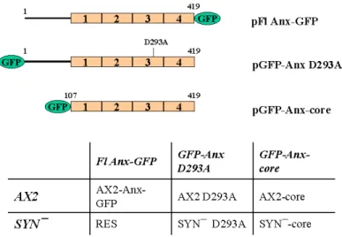
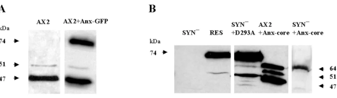
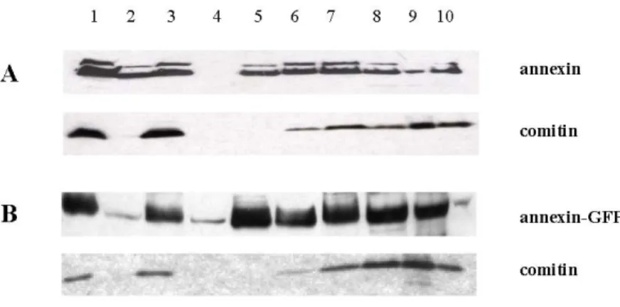
1.1 Expression of the full lenght annexin, a mutated annexin and
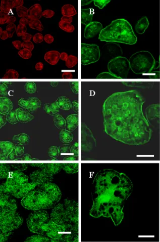
the annexin core as GFP-fusion proteins in AX2 and SYN¯ cells 39 1.2 Subcellular localization of the constructs 41 1.3 Association of annexin with intracellular membranes 43
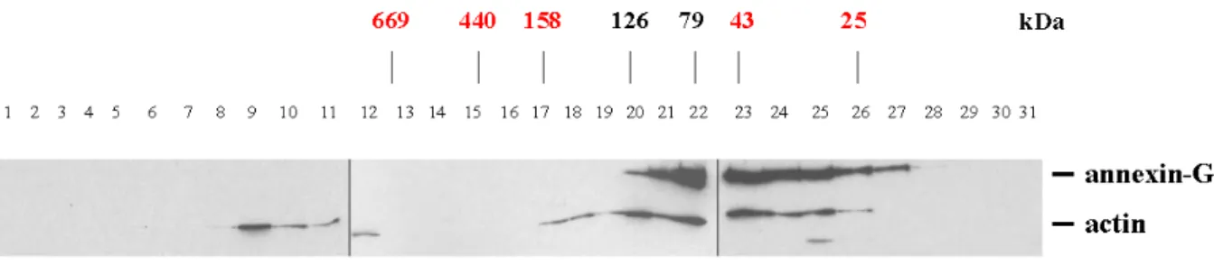
1.4 Gelfiltration experiments 45
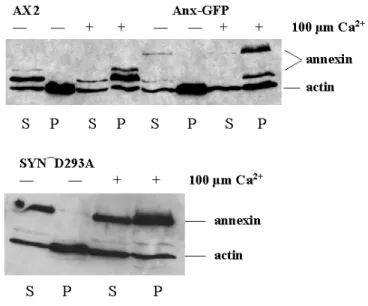
1.5 Annexin associates with Triton X-100 insoluble fraction in the
presence of calcium ions 46
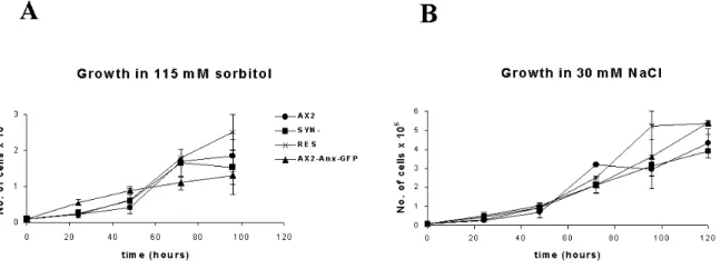
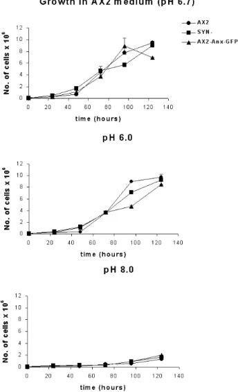
1.6 pH-dependent shuttling of annexin between the cytoplasm and plasma membrane and effect of the pH value of the medium on growth
of Dictyostelium cells 48
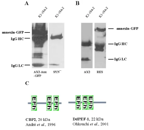
1.7 Detection of annexin binding partners 51 2 Localization of annexin during pinocytosis and phagocytosis 52
3 Phosphorylation of annexin 55
4 Development of SYN¯ cells 57
4.1 Development on phosphate agar plates 57
4.2 Localization of annexin in slugs and fruiting bodies 62
IV. DISCUSION 65-74
4.1 Subcellular localization of Dictyostelium annexin C1 66 4.2 Annexin localizes on pinosomes and phagosomes 67 4.3 Ca2+-independent membrane binding of annexin C1 68
4.4 Nonlipid annexin ligands 70
4.5 The role of annexin during development 72
__________________________________________________
Chapter Description Page(s)
__________________________________________________
4.6 Phosphorylation of annexin C1 73
V. SUMMARY/ZUSAMMENFASSUNG 75-78
BIBLIOGRAPHY 79-89
Erklärung 90
Curriculum Vitae/Lebenslauf 91-92
AP alkaline phosphatase APS ammonium persulphate ATP adenosine 5’-triphosphate bp base pair(s)
BSA bovine serum albumine Bsr blasticidin resistance cassette cDNA complementary DNA
dNTP deoxyribonucleotide triphosphate DABCO diazobicyclooctane
DMSO dimethylsulphoxide DNA deoxyribonucleic acid DTT 1,4-dithiothreitol
ECL enzymatic chemiluminescence EDTA ethylenediaminetetraacetic acid
EGTA ethyleneglycol-bis (2-amino-ethylene) N,N,N,N-tetraacetic acid G418 geneticin
GFP green fluorescent protein
HEPES N-[2-Hydroxyethyl] piperazine-N’-2-ethanesulphonic acid HRP horse radish peroxidase
IgG immunoglobulin G
IPTG iso-propylthio-galactopyranoside kb kilo base pairs
kDa kilodalton
MES morpholinoethansulphonic acid β-ME beta-mercaptoethanol
Mw molecular weight OD optical density
PAGE polyacrylamide gel electrophoresis PCR polymerase chain reaction
PIPES piperazine-N,N’-bis [2-ethanesulphonic acid]
PMSF phenylmethylsulphonylfluoride RNA ribonucleic acid
RNase ribonuclease rpm rotations per minute SDS sodium dodecyl sulphate
TEMED N,N,N’,N’-tetramethyl-ethylendiamine TRITC tetramethylrhodamine isothiocyanate UV ultra violet
vol. volume
v/v volume by volume w/v weight by volume
X-gal 5-bromo-4-chloro-3-indolyl-D-galactopyranoside
Abbreviations 2
Units of Measure and Prefixes Unit Name
0C degree Celsius D Dalton
g gram h hour L litre m meter min minute s sec V volt
Symbol Prefix (Factor)
k kilo (103) c centi (10-2) m milli (10-3) µ micro (10-6) n nano (10-9) p pico (10-12)
1.1 The protein family of annexins
Annexins are intracellular Ca2+- and phospholipid-binding proteins composed of four (or eight in annexin A6) so called “annexin repeats” following an unique N-terminal domain. The members of this big protein family are described to be present in a great variety of organisms ranging from fungi and protists to plants and vertebrates including humans. So far, more than 160 annexins present in more than 65 different species are described. In 1999, a new annexin nomenclature was proposed and accepted at the 50th Harden Conference on Annexins held in the United Kingdom (Table 1.).
1. Introduction 4
Table 1. (Previous page) The new annexin nomenclature. The five major annexin groups are shown (A-E). The nomenclature is proposed by Morgan R. and Fernandez P. The picture is taken from the review published by Gerke and Moss (2002). Although vertebrate annexins are unlikely to be widely represented in invertebrate species, the oldest of this group like, annexins A7, A11 and A13 are possible exceptions. The annexin A11 ortholog has been described in Aplysia.
1.2 The molecular structure of annexins
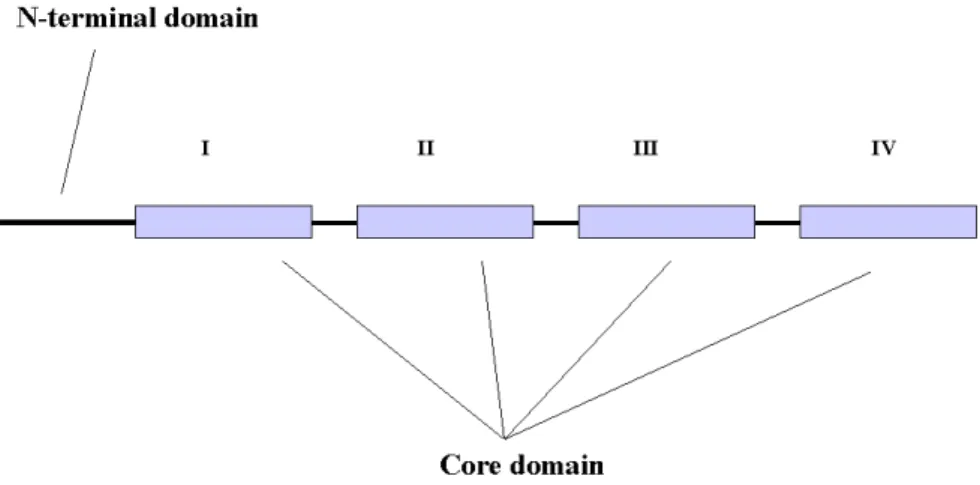
The property of annexins to bind phospholipids and Ca2+ is based on their structure. Annexins are composed of two typical domains, a divergent amino terminal domain and a conserved carboxy terminal core domain. This later domain is formed by a fourfold repeat (eightfold in annexin A6), with each repeat extending over approximately 70 amino acids and having a Ca2+-binding site.
The core domain forms a highly α-helical disc with its type II and type III Ca2+-binding sites (Weng et al., 1993) facing the membrane when the protein associates with phospholipids. The other (concave) side is facing the cell cytosol thus being open for interactions with possible binding partners. The unique amino terminal domains of different annexins vary in their length and amino acid composition. The C-terminal part of annexins is responsible for Ca2+- and phospholipid-binding and the N-terminal part is thought to confer functional diversity (Raynal and Pollard, 1994).
Fig.1. Primary structure of annexins. The core domain consists of four conserved repeats, eight in annexin A6.
The core domain is responsible for Ca2+- and phospholipid-binding. The core domains from different annexins show 45-55 % of homology while the N-terminal domains are variable.
Numerous crystal structures of annexin core domains which revealed the conservation of annexin's three-dimensional fold are described (Huber et al., 1990; Lewit-Bentley et al.,
1992; Weng et al., 1993; Kawasaki et al., 1996). Furthermore, site directed mutagenesis gave valuable contribution to the characterization of certain amino acid residues and their importance in the maintenance of the overall fold of annexin cores. For example, conserved arginine residues present in each of the four annexin repeats were shown to be of crucial importance for stabilizing the tertiary structure of annexin A5. Substitution of different serine and threonine residues by alanine and substitution of the unique tryptophane in the same annexin, resulted in an altered membrane binding underlining the role of these amino acids in intermolecular contacts (Campos et al., 1998; Campos et al., 1999). There are two types of Ca2+-binding sites present in annexin core domains. They differ not only in their architecture but also in their affinity for the bivalent cation. Three so-called type II sites are found in annexin repeats 2, 3 and 4, respectively. Two so- called type III sites are located in the first repeat (Jost et al., 1994, Huber et al., 1990, Weng et al., 1993). Site directed mutational analysis revealed that type II sites show higher affinity for Ca2+ compared to type III sites. Annexin A2 mutants, for example, with defects in the type II and/or type III sites also show different subcellular distributions. The annexin A2 type III mutant protein acquires the typical location in the cortical cytoskeleton observed for the wild-type molecule. In contrast, mutation in the type II Ca2+-binding site of annexin A2 changes localization of the protein which then remains essentially cytosolic, as does a mutant protein containing defects in both type II and type III Ca2+-binding sites (Jost et al., 1994). Furthermore, thermodynamic analysis revealed cooperativity in the binding of Ca2+ to annexin A1 (Rosengarth et al., 2001).
Fig.2. Crystal structure of human annexin A5. Highly α-helical folding of the protein core forms a slightly curved disk. The four annexin repeats are presented in different colors (repeat I: green; repeat II: blue; repeat III: red; repeat IV: violet/cyan). The N-terminal domain is unstructured, presented in green and it extends along the concave side of the molecule. Bound Ca2+ are depicted as yellow balls. The high and low Ca2+ forms are shown in violet and green
1. Introduction 6
respectively, revealing the conformational change in repeat III, which leads to an exposure of Trp-187 (Gerke and Moss, 2002).
A similar approach in characterizing the N-terminal domain of annexin showed that replacement of Trp-5 by alanine in the unique N-terminal domain of annexin A3 led to a much stronger phospholipid binding (Hofmann et al., 2000). In accordance with this finding it is possible that N-terminal domains of the smaller annexins affect the Ca2+-dependent phospholipid binding through stabilizing or destabilizing slightly different conformations of the molecule. Also, unique N-terminal domains can carry the binding sites for protein ligands (Mailliard et al, 1996;
Seemann et al., 1996) or posses sites for posttranslational modifications in these regions (Becker et al., 1990).
1.3 Biochemical properties and functions of annexins
Ca2+-dependent phospholipid binding is shared by all annexins. However, individual members differ in their Ca2+ sensitivity and phospholipid headgroup (phosphatidylserine, phosphatidylinositol) specificity (Raynal and Pollard, 1994). Additional features of annexins were revealed in in vitro studies of annexin A5 and annexin B12. These annexins exhibit a Ca2+- independent binding to negatively charged phospholipids under low pH conditions. In these studies it has been proposed that protonation of a certain residue could induce a reversible conformational change in the protein and lead to insertion into phospholipid bilayers (Köhler et al., 1997; Isas et al., 2000).
Although annexins have been known for more than 20 years their function remains elusive. A role in regulation of membrane-cytoskeleton dynamics has been suggested (Alvarez-Martinez et al., 1997; Gerke and Moss, 1997; Babiychuk et al., 1999). Since some annexins bind actin, an involvement in exocytosis (Chasserot-Golaz et al., 1996; Rosales and Ernst, 1997; Iino et al., 2000; Caohuy and Pollard, 2001; Salzer et al., 2002), in endocytosis (Futter et al., 1993; Grewal et al., 2000) and phagocytosis (Diakonova et al., 1997; Majeed et al., 1998; Pittis and Garcia, 1999) has been proposed. Furthermore, annexins could participate in signaling events since, for example, annexin A6 is found in association with activated PKC-α (Schmitz- Pfeiffer et al.,
1998) and annexin A7 interacts with GTP and catalyzes its hydrolysis mediating thus the Ca2+/ GTP signal during exocytotic membrane fusion (Caohuy et al., 1996). Recently, mutant mice have been generated that either overexpress or lack a specific annexin. These studies showed that overexpression of annexin A6 in the heart causes cardiac hypertrophy in mice (Gunteski-Hamblin et al., 1996) whereas a mouse strain lacking this protein is rather mildly affected and suffers from an increase in myocyte contractility and faster diastolic Ca2+ removal from the cytoplasm (Song et al., 2002). Mouse mutants lacking annexin A7 were reported by two groups. Whereas in one case the knockout mice were inviable and even in the heterozygous condition animals suffered from a defect in Ca2+ signal transduction and in insulin secretion (Srivastava et al., 1999), in the other case annexin A7 (-/-) mice were viable and exhibited an altered cell shortening-frequency relationship when cardiomyocytes were stimulated with high frequencies (Herr et al., 2001).
1.4 Dictyostelium discoideum annexin
Dictyostelium cells harbor an annexin gene, which gives rise to two isoforms of approximately 47 and 51 kDa. At the time of its discovery this protein exhibited highest homology to annexin A7 (synexin) and hence it was called annexin 7 / synexin (Döring et al., 1991). This homology extended not only along the annexin core domain but also throughout the amino terminal region, which is unusually long in these two proteins. Moreover, the amino terminal sequences of both proteins are subject to differential splicing yielding two isoforms (Bonfils et al., 1994; Magendzo et al., 1991a; Magendzo et al., 1991b; Selbert et al., 1996). With the description of further annexins phylogenetic studies became feasible and allowed a new classification of the annexin proteins. This placed Dictyostelium annexin together with the Neurospora protein into a separate group and is now called annexin C1 (proposed by Reginald Morgan and Maria-Pilar Fernandez, 1999).
1.5 The aim of the work
Dictyostelium discoideum annexin was previously described and characterized by Döring et al.
(1995). A Dictyostelium mutant in which the annexin gene was inactivated has been generated
1. Introduction 8
(SYN¯). The mutant cells were nearly indistinguishable from wild type cells under normal laboratory conditions. Only development was noticeably affected and retarded by more than four hours. The cells were able to undergo development and to form fruiting bodies containing viable spores. However, only a limited number of the cells participated in fruiting body formation.
Furthermore the fruiting bodies were significantly smaller. When the processes that involved membrane trafficking such as phagocytosis, exocytosis, pinocytosis and secretion were tested, a significant impairment was observed when the Ca2+ concentrations in the surrounding medium were strongly reduced, suggesting that annexin is involved in maintaining Ca2+ homeostasis (Döring et al., 1995).
This study was initiated to answer further questions regarding the cellular and subcellular localization of Dictyostelium annexin, its dynamic behaviour and possible interactions with other proteins.
To get further insight into the in vivo role of Dictyostelium annexin we have made use of a GFP- tagged annexin fusion protein and studied its involvement in cellular processes in Dictyostelium in vivo and in vitro, with emphasis on processes like pinocytosis, phagocytosis, osmoregulation and development. Also, we studied the role of full length annexin and its core domain as well as a mutated annexin variant in rescuing the previously described impairment in SYN¯ development.
To accomplish this, we have generated strains that express annexin as a full length annexin-GFP fusion, the annexin core domain fused to GFP, or a mutated variant of full length annexin fused to GFP as well. Moreover, the GFP-tag enabled easy visualization of fusion proteins in living and fixed cells.
The ability of some annexins (A5 and B12) to bind to phospholipid vesicles independently of Ca2+ but under low pH conditions have been reported (Köhler et al., 1997; Isas et al., 2000). We analyzed whether relocalization of annexin from the cytoplasm to the plasma membrane upon a decrease of the cytosolic pH could be observed as well in vivo.
Finally, in order to understand better the function of annexin, we focused also on the identification of its possible binding partners.
1. Material 1.1 Laboratory materials
Cellophane sheet, Dry ease Novex
Centrifuge tubes, 15 ml, 50 ml Greiner
Coverslips (glass), Ø12 mm, Ø18 mm, Ø55 mm Assistent
Corex tube, 15 ml, 50 ml Corex
Cryo tube, 1 ml Nunc
Electroporation cuvette, 2 mm electrode gap Bio-Rad
Gel-drying frames Novex
Hybridisation bag Life technologies
Microcentrifuge tube, 1.5 ml, 2.2 ml Sarstedt Micropipette, 1-20 µl, 10-200 µl, 100-1,000 µl Gilson
Micropipette tips Greiner
Needles (sterile), 18G–27G Terumo, Microlance
Nitrocellulose membrane, BA85 Schleicher and Schuell
Parafilm American National Can
Pasteur pipette, 145 mm, 230 mm Volac
PCR softtubes, 0.2 ml Biozym
Petri dish (35 mm, 60 mm, 100 mm) Falcon
Petri dish (90 mm) Greiner
Plastic cuvette, semi-micro Greiner
Plastic pipettes (sterile), 1 ml, 2 ml, 5 ml, 10 ml, 25 ml Greiner
Quartz cuvette, Infrasil Hellma
Quartz cuvette, semi-micro Perkin Elmer
Saran wrap Dow
Scalpels (disposable), Nr. 10, 11, 15, 21 Feather
Slides, 76 x 26 mm Menzel
Syringes (sterile), 1 ml, 5 ml, 10 ml, 20 ml Amefa, Omnifix Syringe filters (Acrodisc), 0.2 µm, 0.45 µm Gelman Sciences Tissue culture flasks, 25 cm2, 75 cm2, 175 cm2 Nunc Tissue culture dishes, 6 wells, 24 wells, 96 wells Nunc
Whatman 3MM filter paper Whatman
X-ray film, X-omat AR-5, 18 x 24 mm, 535 x 43 mm Kodak 1.2 Instruments and equipments
Centrifuges (microcentrifuges):
Centrifuge 5417 C Eppendorf
Centrifuge Sigma B. Braun Biotech Instruments
Cold centrifuge Biofuge fresco Heraeus Instruments Centrifuges (table-top, cooling, low speed):
2. Material and Methods 10
Centrifuge CS-6R Beckman
Centrifuge RT7 Sorvall
Centrifuge Allegra 21R Beckman
Centrifuges (cooling, high speed):
Beckman Avanti J25 Beckman
Sorvall RC 5C plus Sorvall
Centrifuge-rotors:
JA-10 Beckman
JA-25.50 Beckman
SLA-1500 Sorvall
SLA-3000 Sorvall
SS-34 Sorvall
Electrophoresis power supply, Power-pac-200, -300 Bio-Rad
Electroporation unit, Gene-Pulser Bio-Rad
Freezer (-80 0C) Nunc
Freezer (-20 0C) Siemens, Liebherr
Gel-documentation unit MWG-Biotech
Heating block, DIGI-Block JR NeoLab
Heating block, Dry-Block DB x 20 Techne
Ice machine Ziegra
Incubators:
Incubator, microbiological Heraeus
Incubator with shaker, Lab-Therm Kuehner
Laminar flow, Hera Safe (HS 12) Heraeus
Magnetic stirrer, MR 3001 K Heidolph
Microscopes:
Light microscope, CH30 Olympus
Light microscope, DMIL Leica
Light microscope, CK2 Olympus
Fluorescence microscope, DMR Leica
Fluorescence microscope, 1X70 Olympus
Confocal laser scan microscope, DM/IRBE Leica
Stereo microscope, MZFLIII Leica
Stereomicroscope, SZ4045TR Olympus
Oven, conventional Heraeus
PCR machine, PCR-DNA Engine PTC-2000 MJ Research
pH-Meter Knick
Refrigerator Liebherr
Semi-dry blot apparatus, Trans-Blot SD Bio-Rad
Shakers GFL Kuehner
Sonicator, Ultra turrax T25 basic IKA Labortechnik Sonicator (water bath), Sonorex RK 52 Bandelin
Speed-vac concentrator, DNA 110 Savant
Ultracentrifuges:
Optima TLX Beckman
Optima L-70K Beckman
Ultracentrifuge-rotors:
TLA 45 Beckman
TLA 100.3 Beckman
SW 41 Beckman
UV- transilluminator, TFS-35 M Faust
Vortex, REAX top Heidolph
Waterbath GFL
X-ray-film developing machine, FPM-100A Fujifilm 1.3 Kits
Nucleobond AX Macherey-Nagel
NucleoSpin Extract 2 in 1 Macherey-Nagel
Nucleotrap Macherey-Nagel
Original TA Cloning Invitrogen
pGEM-T Easy Promega
Qiagen Midi- and Maxi-prep Qiagen
Stratagene Prime It II Stratagene 1.4 Enzymes, antibodies, substrates, inhibitors and antibiotics
Enzymes used in the molecular biology experiments:
Calf Intestinal Alkaline Phosphatase (CIAP) Roche
Klenow fragment (DNA polymerase) Roche
Lysozyme Sigma
Restriction endonucleases Amersham
Ribonuclease A (RNase A) Sigma
T4 DNA ligase Roche
Taq-polymerase Roche
Primary antibodies:
Mouse anti-actin monoclonal antibody, Act 1-7 (Simpson et al., 1984) Mouse anti-GFP monoclonal antibody, K3-184-2 unpublished
Mouse anti-comitin monoclonal antibody 190-340-2 (Weiner et al., 1993) Mouse anti-CsA monoclonal antibody 33-294-17 (Bertholdt et al., 1985) Mouse anti-vacuolin monoclonal antibody 221-1-1 (Rauchenberger et al., 1997) Mouse anti-V/H+ ATPase monoclonal antibody (Jenne et al., 1998)
Secondary antibodies:
Goat anti-mouse IgG, peroxidase conjugated Sigma
2. Material and Methods 12
Sheep anti-mouse IgG, Cy3 conjugated Sigma Substrates:
Hydrogen peroxide (H2O2) Sigma
Inhibitors:
Phenylmethylsulphonylfluoride (PMSF) Sigma
Sodium fluoride Sigma
Sodium orthovanadate Sigma
Antibiotics:
Ampicillin Gruenenthal
Blasticidin S ICN Biomedicals
Dihydrostreptomycinsulphate Sigma
Geneticin (G418) Life technologies
Tetracyclin Sigma
1.5 Chemicals and reagents
Most of the chemicals and reagents were obtained either from Sigma, Fluka, Difco, Merck, Roche, Roth or Serva. Those chemicals or reagents that were obtained from companies other than those mentioned here are listed below.
Acetic acid (98-100%) Riedel-de-Haen
Acrylamide (Protogel: 30:0,8 AA/Bis-AA) National Diagnostics
Agar-Agar (BRC-RG) Biomatic
Agarose (Electrophoresis Grade) Life technologies
Chloroform Riedel-de-Haen
Ethanol Riedel-de-Haen
Glycerine Riedel-de-Haen
Glycine Riedel-de-Haen
Isopropypl-β-D-thiogalactopyranoside (IPTG) Loewe Biochemica
Methanol Riedel-de-Haen
N- [2-Hydroxyethyl] piperazine-N’-2-
-ethanesulfonic acid (HEPES) Biomol
Nonidet P40 Fluka
Peptone Oxoid
Sodium hydroxide Riedel-de-Haen
TRIzol Gibco BRL
Yeast extract Oxoid
1.6 Media and buffers
All media and buffers were prepared with deionised water filtered through an ion-exchange unit
were added to the media after cooling to approx. 50 0C. For making agar plates, a semi-automatic plate-pouring machine (Technomat) was used.
1.6.1 Media and buffers for Dictyostelium culture
AX2-medium, pH 6.7: 7.15 g yeast extract (Claviez et al., 1982) 14.3 g peptone (proteose)
18.0 g maltose 0.486 g KH2 PO4
0.616 g Na2HPO4.2H2O add H2O to make 1 liter Phosphate agar plates, pH 6.0: 9 g agar
add Soerensen phosphate buffer, pH 6.0 to make 1 liter
Salt solution: 10 mM NaCl
(Bonner, 1947) 10 mM KCl
2.7 mM CaCl2
SM agar plates, pH 6.5: 9 g agar
(Sussman, 1951) 10 g peptone
10 g glucose 1 g yeast extract 1 g MgSO4.7H2O 2.2 g KH2PO4
1 g K2HPO4
add H2O to make 1 liter Soerensen phosphate buffer, pH 6.0: 2 mM Na2HPO4
(Malchow et al., 1972) 14.6 mM KH2PO4
1.6.2 Media for E. coli culture
LB medium, pH 7.4: 10 g bacto-tryptone (Sambrook et al., 1989) 5 g yeast extract
10 g NaCl
adjust to pH 7.4 with 1 N NaOH add H2O to make 1 liter
2. Material and Methods 14
For LB agar plates, 0.9 % (w/v) agar was added to the LB medium and the medium was then autoclaved. For antibiotic selection of E. coli transformants, 50 mg/l ampicillin, kanamycin or chloramphenicol was added to the autoclaved medium after cooling it to approx. 50 0C. For blue/white selection of E. coli transformants, 10 µl 0.1 M IPTG and 30 µl X-gal solution (2 % in dimethylformamide) was plated per 90 mm plate and the plate was incubated at 37 0C for at least 30 min before using.
SOC medium, pH 7.0: 20 g bacto-tryptone (Sambrook et al., 1989) 5 g yeast extract
10 mM NaCl 2.5 mM KCl
dissolve in 900 ml deionised H2O adjust to pH 7.0 with 1 N NaOH
The medium was autoclaved, cooled to approx. 50 0C and then the following solutions, which were
separately sterilized by filtration (glucose) or autoclaving, were added:
10 mM MgCl2.6H2O 10 mM MgSO4.7H2O 20 mM Glucose
add H2O to make 1 liter 1.6.3. Buffers and other solutions
The buffers and solutions that were commonly used during the course of this study are mentioned below.
10 x NCP-Puffer (pH 8.0): 12.1 g Tris/HCl 87.0 g NaCl
add H2O to make 995 ml
add 5.0 ml Tween 20 after autoclaving
PBG (pH 7.4): 0.5 % bovine serum albumin
0.1 % gelatin (cold-water fish skin) in 1 x PBS, pH 7.4
1 x PBS (pH 7.4): 8.0 g NaCl
0.2 g KH2PO4
1.15 g Na2HPO4 0.2 g KCl
adjust to pH 7.4
add H2O to make 1 liter, autoclave TE buffer (pH 8.0): 10 mM Tris/HCl, pH 8.0
1 mM EDTA, pH 8.0 10 x TAE buffer (pH 8.3): 27.22 g Tris
13.6 g sodium acetate 3.72 g EDTA
add H2O to make 1 liter 1.7 Biological materials
Bacterial strains: E. coli DH5α Hanahan, 1983
E. coli XL1 blue Bullock et al., 1987 Klebsiella aeorgenes Williams and Newell,
1976 Dictyostelium discoideum strain:
AX2-214 An axenically growing derivative of wild strain, NC- 4 (Raper, 1935). Commonly referred to as AX2.
1.8 Plasmids
pDdA15gfp Neujahr et al., 1997
pDEX-RH Westphal et al., 1997
pGEM-T Easy Promega
pUC Pharmacia
pT7-7 Tabor, S. & Richardson, C. C. (1985) 1.9 Oligonucleotide primers
The oligonucleotide primers were designed on the basis of sequence information available and ordered for synthesis to Sigma. Following is a list of the primers used for PCR, sequence analysis or site directed mutagenesis during the course of the present investigation. The position and orientation of the primers are indicated in the text when discussed.
2. Material and Methods 16
Name Sequence Analysis
AnnA1-AccI 5’ 5’-CTGTCGACATGTCCTATCCACCAAACC-3’ Site dir. mut. & PCR AnnA2-PstI 3’ 5’-TCTGCAGTACCAATTTTACCTTCACCAGCTTTGTAGAGATC-3’ Site dir. mut. &
Seq.
AnnB1-AccI 3’ 5’-TGTCGACTTATGAGATAATATCTAATAATAATTTTTTG-3’ Site dir. mut. & PCR AnnB2-PstI 5’ 5’-GTAAAATTGGTACTGCAGAAAAGGAATTCATCAAAATTCTC-3’Site dir. mut. &
Seq.
AnnCore-AccI 5’ 5’-CTGTCGACGGATATCATCAA-3’ PCR SP6 universal 5’-ATTTAGGTGACACTATAG-3’ Seq.
Syngr 3’ 5’-CGCGGATCCTGAGATAATATCTAATAATAATTTT-3’ PCR Syngu 5’ 5’-CGCGGATCCATGTCCTATCCACCAAAC-3’ PCR T7 universal 5’-TAATACGACTCACTATAGGG -3’ Seq.
2. Cell biological methods
2.1 Growth of Dictyostelium
2.1.1 Growth in liquid nutrient medium (Claviez et al., 1982)
Dictyostelium discoideum AX2 and the derived transformants were grown in liquid AX2 medium containing dihydrostreptomycin (40 µg/ml) and other appropriate selective antibiotics at 21 0C either in a shaking-suspension in Erlenmeyer flasks with shaking at 160 rpm or on petri dishes. For all the cell biological works, cultures were harvested at a density of 3-5 x 10⁶ cells/ml.
2.1.2. Growth on SM agar plates
Dictyostelium cells were plated onto SM agar plates overlaid with Klebsiella aerogenes and incubated at 21 0C for 3-4 days until Dictyostelium plaques appeared on the bacterial lawns.
To obtain single clones of Dictyostelium, 50-200 cells were suspended in 100 µl Soerensen phosphate buffer and plated onto Klebsiella-overlaid SM agar plates. Single plaques obtained after incubation at 21 0C for 3-4 days were picked up with sterile tooth-picks, transferred either to new Klebsiella-overlaid SM agar plates or to separate petri dishes with AX2 medium supplemented with dihydrostreptomycin (40 µg/ml) and ampicillin (50 µg/ml) (to eliminate the bacteria) and any other appropriate selective antibiotic (depending upon mutant).
2.2 Development of Dictyostelium
Development in Dictyostelium is induced by starvation. Cells grown to a density of 2-3 x 10⁶ cells/ml were pelleted by centrifugation at 2,000 rpm (Sorvall RT7 centrifuge) for 2 min at 4 0C and were washed two times in an equal volume of cold Soerensen phosphate buffer in order to remove all the nutrients present in the culture medium. 5 x 10⁷ cells/ml were then resuspended in 3 ml Soerensen phosphate buffer and evenly distributed onto phosphate-buffered agar plates (90 mm) or water agar plates (90 mm). The plates were air dried and any excess liquid was carefully aspirated without disturbing the cell layer. The plates were then incubated at 21 0C.
Different stages of development were observed and the images were captured at indicated time points. For development in suspension culture, the cells were resuspended in Soerensen phosphate buffer at a density of 1 x 10⁷ cells/ml and were shaken at 160 rpm
and 21 0C for desired time periods.
2.3 Preservation of Dictyostelium
Dictyostelium cells were allowed to grow in AX2 medium to a density of 4-5 x 10⁶ cells/ml. 9 ml of the dense grown culture were collected in a 15 ml Falcon tube on ice and supplemented with 1 ml horse serum and 1 ml DMSO. The contents were mixed by gentle pipetting, and aliquoted in cryotubes (1 ml). The aliquots were incubated on ice for 60 min, followed by incubation at –20
0C for at least 2 hours. Finally, the aliquots were transferred to –80 0C for long term storage.
For reviving the frozen Dictyostelium cells, an aliquot was taken out from –80 0C and thawed immediately at 37 0C in a waterbath. In order to remove DMSO, the cells were transferred to a Falcon tube containing 30 ml AX2 medium and centrifuged at 2,000 rpm (Sorvall RT7 centrifuge) for 2 min at 4 0C. The cell pellet was resuspended in 10 ml of AX2 medium and 200 µl of the cell suspension was plated onto SM agar plates overlaid with Klebsiella while the remaining cell suspension was transferred into a 100-mm petri dish (Falcon) and antibiotics were added when appropriate. Cells in the petri dish were allowed to recover overnight at 21 0C and the medium was changed the next day to remove the dead cells and the traces of DMSO, whereas, the SM agar plates coated with cell suspension and bacteria were incubated at 21 0C until plaques of Dictyostelium cells started to appear.
2. Material and Methods 18
2.4 Transformation of Dictyostelium cells by electroporation
The electroporation method for transformation of Dictyostelium cells described by de Hostos et al. (1993) was followed with little modifications. Dictyostelium discoideum cells were grown axenically in suspension culture to a density of 2-3 x 10⁶ cells/ml. The cell suspension was incubated on ice for 20 min and centrifuged at 2,000 rpm (Sorvall RT7 centrifuge) for 2 min at 4
0C to collect the cells. The cells were then washed with an equal volume of ice-cold Soerensen phosphate buffer and afterwards with an equal volume of ice-cold electroporation- buffer. After washings, the cells were resuspended in electroporation- buffer at a density of 1 x 10⁸ cells/ml. For electroporation, 20-25 µg of the plasmid DNA was added to 500 µl of the cell suspension and the cell-DNA mixture was transferred to a pre-chilled electroporation cuvette (2 mm electrode gap, Bio-Rad). Electroporation was
performed with an electroporation unit (Gene Pulser, Bio-Rad) set at 0.9 kV and 3 µF without the pulse controller. After electroporation, the cells were immediately spread onto a 100-mm petri dish and were allowed to sit for 10 min at 21 0C. Thereafter, 1 ml of healing-solution was added dropwise onto the cells and the petri dish was incubated at 21 0C on a shaking platform at 50 rpm for 15 min. 10 ml of AX2 medium was added into the petri dish and the cells were allowed to recover overnight. The next day, the medium was replaced by the selection medium containing appropriate antibiotic. To select for stable transformants, selection medium was replaced every 24-48 hr until the control plate (containing cells electroporated without any DNA) was clear of live cells.
Electroporation-buffer: 0.1 M Potassium phosphate buffer:
100 ml 0.1 M potassium phosphate buffer 170 ml 0.1 M KH2PO4 17.12 g sucrose 30 ml 0.1 M K2HPO4 add distilled H2O to make 1 litre adjust to pH 6.1 autoclave
Healing-solution:
150 µl 0.1 MgCl2 150 µl 0.1 CaCl2
10 ml electroporation-buffer
2.5 Endocytosis and exocytosis assays
Fluid-phase endocytosis assays were performed according to the methods of Aubry et al. (1994).
Fluid-phase efflux assays were performed as described using TRITC-dextran (Buczynski, 1997).
Briefly, Dictyostelium cells were grown to < 5 x 10⁶ cell/ml. The cells were centrifuged and resuspended at 5 x 10⁶/ml in fresh axenic medium at 21°C and incubated for 15 min on a shaker to allow cells to recuperate. Then TRITC-dextran was added to a final concentration of 2 mg/ml. Samples were withdrawn at different time intervals and the cells were pelleted after incubating for 3 min with 100 µl of trypan blue (2 mg/ml) to remove non-specifically bound marker. The pellet was resuspended in phosphate buffer and the fluorescence was measured using a fluorimeter (544 nm excitation/574 nm emission). For fluid-phase exocytosis assays, cells were pulsed with TRITC-dextran (2 mg/ml) for 2 hours, washed and resuspended in fresh axenic medium. Fluorescence from the marker remaining in the cells was measured at different time intervals as explained above.
Phagocytosis was performed according to Maniak et al. (1995). Briefly, Dictyostelium cells were grown to < 5 x10⁶/ml over 5 generations in axenic medium. Cells were centrifuged and resuspended at 2 x 10⁶/ml in fresh axenic medium at 21 0C. TRITC-labeled yeast cells prepared according to Material and Methods 5.3 were added in a 5 fold excess (109 yeast cells/ml stock). Cells were incubated on a rotary shaker at 160 rpm. Samples were taken at different intervals and the fluorescence of non internalized yeasts was quenched by incubating for 3 min with 100 µl trypan blue (2 mg/ml). Cells were centrifuged again, resuspended in phosphate buffer and the fluorescence was measured using a fluorimeter (544 nm excitation/574 nm emission).
3. Molecular biological methods
3.1 Purification of plasmid DNA
In general, for small cultures (3 ml) of E. coli transformants, the boiling method described by Holmes and Quigley (1981) was used to extract plasmid DNA. This method is good for screening
2. Material and Methods 20
a large number of clones simultaneously for the desired recombinant plasmid. Briefly, single transformants were picked up from the culture plate and were grown overnight
in 3 ml of LB media containing suitable antibiotic. Next day, the overnight grown E. coli cells were pelleted by centrifugation at 6,000 rpm in a microcentrifuge for 3-5 min. The pellet was then resuspended completely in 250 µl STET/lysozyme buffer and the suspension was incubated at room temperature for 10 min to lyse the bacterial cells. The bacterial lysate was boiled at 100
0C for 1 min and was then centrifuged in an eppendorf centrifuge at maximum speed for 15 min at room temperature. The plasmid DNA present in the supernatant was precipitated by adding an equal volume of isopropanol and incubating at room temperature for 10 min. The precipitated DNA was pelleted in the eppendorf centrifuge at 12,000 rpm for 15 min and the DNA pellet was washed with 70 % ethanol, dried in a speed-vac concentrator and finally resuspended in 40 µl TE, pH 8.0 containing RNase A at 1 µg/ml.
STET/lysozyme buffer, pH 8.0:
50 mM Tris/HCl, pH 8.0 50 mM EDTA
0.5 % Triton X-100
8.0 % SucroseAdd lysozyme at 1 mg/ml at the time of use.
Alternatively, for pure plasmid preparations in small and large scales (for sequencing, PCR or transformations), kits provided either by Macherey-Nagel (Nucleobond AX kit for small scale plasmid preparations) or by Qiagen (Qiagen Midi- and Maxi-Prep kit for large scale plasmid preparations) were used. These kits have the following approach: first overnight culture of bacteria containing the plasmid is pelleted and the cells are lysed by alkaline lysis. The freed plasmid DNA is then adsorbed on a silica matrix, washed with ethanol, and then eluted with TE, pH 8.0 or water. This method avoids the requirement of caesium chloride or phenol-chloroform steps during purification.
3.2 Digestion with restriction enzymes
All restriction enzymes were obtained from NEB, Amersham or Life Technologies and the
digestions were performed in the buffer systems and temperature conditions as recommended by the manufacturers. The plasmid DNA was digested for 1-2 hr.
3.3 Dephosphorylation of DNA fragments
To avoid self-ligation of the vector having blunt ends or that has been digested with a single restriction enzyme, 5’ ends of the linearised plasmids were dephosphorylated by calf-intestinal akaline phosphatase (CIAP). Briefly, in a 50 µl reaction volume, 1-5 µg of the linearised vector-DNA was incubated with 1 U calf-intestinal alkaline phosphatase in CIAP-buffer (provided by the manufacturer) at 37 0C for 30 min. The reaction was stopped by heat- inactivating the enzyme at 65 0C for 10 min. The dephosphorylated DNA was extracted once with phenol-chloroform and precipitated with 2 vol. ethanol and 1/10 vol. 2 M sodium acetate, pH 5.2.
3.4 Setting up of ligation reactions
A DNA fragment and the appropriate linearised plasmid were mixed in approximately equimolar amounts. T4 DNA ligase and ATP were added as indicated below and the ligation reaction was left overnight at 10-12 0C.
Ligation reaction 5 x Ligation buffer Linearised vector DNA (200-400 ng) Supplied with the enzyme DNA fragment by manufacturers
4 µl 5x ligation buffer 1µl 0.1 M ATP
1.5 U T4 Ligase
and water to make up to 20 µl.
3.5 PCR and generation of mutations by PCR-based site directed mutagenesis
For polymerase chain reaction (PCR) plasmid carrying annexin C1 cDNA (pT7-7) was used.
Dictyostelium annexin C1 sequence carrying a mutation at the position 293 where aspartic acid (D) was replaced by alanine (A) was generated from wild type cDNA by PCR-based site directed mutagenesis. In two separate PCR reactions using the primers AnnA1-AccI and AnnA2-PstI in the first and AnnB2-PstI and AnnB1-AccI in the second, two halves of the annexin cDNA carrying wanted mutation are generated. The products of both steps were used as a template for a
2. Material and Methods 22
new round of PCR in which AnnA1-AccI and AnnB1-AccI were used as the forward and reverse primers, respectively. PCR products were cloned into the pGEM-T easy vector and verified by sequencing.
PCR conditions:
Reaction-mix (50 µl final volume): Reaction programme:
1 µl template (50-200 ng DNA) 1-step 92 0C for 2 min 1 µl 5’ primer (5 pmol/µl) 2-step 30 cycles of- 1 µl 3’ primer (5 pmol/µl) 92 0C for 1 min
1 µl dNTP-mix (10 mM ) 51 0C for 1 min
5 µl 10x PCR buffer 72 0C for 2 min
1 µl Taq polymerase (1 U/µl) 3-step 72 0C for 10 min
40 µl H2O 4-step 4 0C till end
3.6 DNA agarose gel electrophoresis
Agarose gel electrophoresis was performed according to the method described by Sambrook et al. (1989) to resolve and purify the DNA fragments. Electrophoresis was performed with 0.7 % (w/v) agarose gels or with 1 % (w/v) agarose gels in 1 x TAE buffer submerged in a horizontal electrophoresis tank containing 1 x TAE buffer at 1-5 V/cm. The percentage of agarose in the gel was dependent on size of fragments to be resolved. DNA-size marker was always loaded along with the DNA samples in order to estimate the size of the resolved DNA fragments in the samples. The gel was run until the bromophenol blue dye present in theDNA-loading buffer had migrated the appropriate distance through the gel. The gel was examined under UV light at 302 nm and was photographed using a gel-documentation system (MWG-Biotech).
DNA-size marker:
1 kb DNA Ladder (Life Technologies): 12,216; 11,198; 10,180; 9,162; 8,144; 7,126;
6,108; 5,090; 4,072; 3,054; 2,036; 1,636; 1,018;
506; 396; 344; 298; 220; 201; 154; 134; 75 bp
3.7 Recovery of DNA fragments from agarose gel
DNA fragments from restriction enzyme digests or from PCR reactions were separated by agarose gel electrophoresis and the gel piece containing the desired DNA fragment was carefully and quickly excised while observing the ethidium bromide stained gel under a UV transilluminator. The DNA fragment was then purified from the excised gel piece using the Macherey-Nagel gel elution kit (NucleoSpin Extract 2 in 1), following the method described by the manufacturers or by centrifugation through the glass wool followed by DNA precipitation.
3.8 Transformation of E. coli
3.8.1 Standard protocol for the preparation and transformation of competent cells (Inoue et al., 1990)
Preparation of competent E. coli cells by SEM:
An overnight grown culture of E.coli (0.5 ml) was inoculated into 250 ml LB medium and incubated at 18 0C, with shaking 250 rpm until an OD600 of 0.4-0.6 was obtained. The bacteria were then incubated on ice for 10 minutes and pelleted at 4 0C for 10 min at 4,000 rpm (Beckman Avanti J25, rotor JA-25.50). The bacterial pellet was resuspended in 80 ml of ice- cold TB, incubated on ice for 10 min and spun down as above. The cells pellet was gently resuspended in 20 ml of TB and DMSO was added with gentle swirling to a final concentration of 7 %. After incubating on ice for 10 min, the cell suspension was aliquoted
200 µl/tube. The aliquots were then quickly frozen in a dry ice/ethanol bath and immediately stored at –80 0C.
Standard protocol for SEM transformation:
Plasmid DNA (~50-100 ng of a ligase reaction or ~10 ng of a supercoiled plasmid) was mixed with 100-200 µl of CaCl2 -competent E.coli cells and incubated on ice for 30 min. The cells were then heat-shocked at 42 0C for 45 s and immediately transferred to ice for 2 minutes. The cells were then mixed with 1 ml of pre-warmed (at 37 0C) SOC medium and incubated at 37 0C with