Cellular localization and characterization of Cyclooxygenase-2 in interstitial cells
of the kidney
Dissertation
ZUR ERLANGUNG DES DOKTORGRADES DER
NATURWISSENSCHAFTEN (DR.RER.NAT) DER FAKULTÄT FÜR BIOLOGIE UND VORKLINISCHE MEDIZIN DER UNIVERSITÄT
REGENSBURG
vorgelegt von
Michaela Alexandra Anna Fuchs aus
Regensburg im Jahr
2019
2
3
Die vorliegende Arbeit entstand im Zeitraum von Juli 2015 bis Juli 2019 unter Anleitung von Herrn Prof. Dr. med. Armin Kurtz am Institut für Physiologie der Universität Regensburg.
Promotionsgesuch eingereicht am: 10.07.2019
Die Arbeit wurde angeleitet von: Herr Prof. Dr. med. Armin Kurtz
Unterschrift
(Michaela Fuchs)
4
Content
1 Introduction ... 8
1.1 The kidney, structure and primary function ... 8
1.2 Cyclooxygenase ... 11
1.2.1 Structure and Subtypes, Prostaglandin production and regulation ... 11
1.2.2 Localization of Cox in the kidney ... 14
1.2.3 Function and regulation of Cyclooxygenase-2 in the kidney ... 14
1.2.4 Pharmacological inhibition of Cyclooxygenase ... 15
1.2.5 Models for the functional investigation of Cox-2 in the kidney ... 16
1.3 Aims of this study ... 18
2 Material and Methods ... 20
2.1 Material ... 20
2.1.1 Instruments ... 20
2.1.2 Consumables ... 21
2.1.3 Commercial kits, enzymes and chemicals ... 22
2.1.4 Buffers and Solutions ... 24
2.1.5 Primer for genotyping and real time pcr ... 26
2.1.6 Probes for in-situ hybridization and antibodies... 27
2.1.7 Software and internet services ... 28
2.2 Methods ... 29
2.2.1 Animals ... 29
2.2.1.1 Mouse strains and animal breeding ... 29
2.2.1.2 Genotyping of mice ... 30
2.2.1.3 Induction of Cre-Recombinase activity with tamoxifen ... 31
2.2.1.4 High salt diet ... 32
2.2.1.5 Treatment with low salt diet and Enalapril ... 32
2.2.1.6 Adenine-induced kidney fibrosis... 32
2.2.1.7 Water deprivation for 24h ... 32
5
2.2.1.8 Unilateral ureteral obstruction ... 32
2.2.1.9 GFR measurement ... 32
2.2.2 Retrograde arterial perfusion of mice ... 33
2.2.3 Zonal dissection of kidneys ... 34
2.2.4 Tail cuff blood pressure measurement ... 34
2.2.5 Metabolic cages ... 34
2.2.6 Spot urine collection and analysis ... 35
2.2.6.1 Osmolality measurement ... 35
2.2.6.2 Sodium and potassium measurement in urine ... 35
2.2.7 Histological methods ... 35
2.2.7.1 Embedding of tissue in paraffin ... 35
2.2.7.2 Section of paraffin embedded tissue ... 36
2.2.7.3 Immunofluorescence staining... 36
2.2.7.4 Sirius Red staining ... 37
2.2.8 In-situ hybridization utilizing the RNAscope
®technique ... 37
2.2.8.1 Chromogenic and duplex RNAscope ... 37
2.2.8.2 RNAscope® Multiplex Fluorescent Assay ... 39
2.2.9 Microscopy ... 39
2.2.9.1 Microscopy for fluorescent dyes ... 39
2.2.9.2 Microscopy for RNAscope® 2.5 HD Reagent Kit (brow/duplex) ... 39
2.2.10 Molecular biology methods ... 40
2.2.10.1 Measurement of mRNA expression levels and cDNA synthesis ... 40
2.2.10.2 Quantitative real time PCR ... 41
2.2.11 Measurement of plasma parameters ... 41
2.2.11.1 Blood sample collection ... 41
2.2.11.2 Renin-ELISA ... 42
2.2.12 Statistics ... 42
3 Results... 43
6
3.1 Localization of Cox-2 in the kidney and characterization of Cox-2 expressing
cells ... 43
3.2 Regulation of Cox-2 expression in the kidney ... 48
3.3 Importance of Cox-2 for kidney development and function ... 52
3.3.1 Function of Cox-2 investigated by conditional gene deletion in specific compartments ... 52
3.3.1.1 Conditional deletion of Cox-2 in all cells of the adult kidney ... 52
3.3.1.2 Functional consequences of Cox-2 deletion in normally developed kidneys ... 54
3.3.2 Importance of Cox-2 in the FoxD1 compartment during nephrogenesis 56 3.3.2.1 Renin cell recruitment in FoxD1
+/CreCox2
fl/flmice ... 59
3.3.3 Function of Cox-2 in PDGFR-β
+interstitial medullary cells ... 59
3.3.3.1 Functional relevance of interstitial Cox-2 expression under normal conditions ... 60
3.3.3.2 Handling of high dietary sodium in mice deficient for Cox-2 in PDGFR- β
+cells ... 62
3.4 Role of Cox-2 in kidney fibrosis ... 67
3.4.1 Progression of adenine induced fibrosis in mice deficient for Cox-2 ... 67
3.4.2 Effects of Cox-2 deletion in PDGFR-β
+cells in the UUO model ... 71
4 Discussion ... 72
4.1 Cox-2 expression and regulation in cells of the kidney ... 73
4.2 Different functions of Cox-2 in the kidney ... 76
4.2.1 Cox-2 in the adult kidney ... 76
4.2.2 Role of Cox-2 in the stromal progenitor compartment ... 77
4.2.3 Function of medullary Cox-2 expression with different dietary sodium intake 79 4.5 Cox-2 in two models of kidney fibrosis ... 82
4.5.1 Influence of Cox-2 deletion in PDGFR-β
+cells on adenine induced nephropathy ... 82
4.5.2 Role of Cox-2 in PDGFR-β
+interstitial cells during 5d UUO ... 84
7
5 Summary ... 86
6 Bibliography ... 88
7 Annex ... 102
7.1 Abbreviations ... 102
7.2 Congress contributions ... 104
7.3 Declaration ... 105
8 Acknowledgement ... 106
8
1 Introduction
1.1 The kidney, structure and primary function
The kidneys of mammals are paired organs located in the lower abdominal cavity behind stomach and intestines on the left and right side of the spine. The huge number of different cell types present in the kidney gives an impression of the different physiological functions fulfilled by these organs.
The central function of the kidneys is the excretion of urinary metabolic waste products and keeping the acid/base and water homeostasis in the body. Closely connected to this is the control of systemic blood pressure and the renin-angiotensin-aldosterone system (RAAS) (Figure 1).
Other important functions are the production and release of hormones such as erythropoietin (EPO) for the formation of new red blood cells and calcitriol, the active metabolite of vitamin D, a central hormone for the bone metabolism
1,2.
Contained in a capsule of connective tissue, the mammalian kidney can be divided into three major zones: the cortex, the outer and the inner medulla. The most prominent functional subunit of the kidney is the nephron, including the glomerulus as the site of primary urine filtration and the following tubular system for urinary concentration. The human kidney contains about 1 million glomeruli, the murine kidney averages about 12.000 to 16.000 glomeruli.
3The primary urine is filtered in the capillary slings of the glomerulus by a system of three cooperative membranes, the fenestrated capillary endothelium, the basal membrane and the slit membrane between the long foot processes of the podocytes.
Each glomerulus is surrounded by a bowman capsule and embedded in between other glomeruli, capillaries, interstitial cells and the proximal tubules in the kidney cortex.
From the glomerulus, the primary urine is transported along the proximal tubule through the loop of Henle to the distal tubule and into the collecting duct (CD). During the passage of the urine through the different parts of the tubular system, important electrolytes such as sodium, potassium and glucose as well as the majority of the filtered water are reclaimed by an osmotic countercurrent system between the tubules and the adjacent blood vessels.
The function of the macula densa cells is the sensing of luminal ion concentrations,
especially chloride, and the appropriate adjustment of glomerular filtration and
electrolyte reuptake
4,5. These cells are a specialized subtype of the cortical thick
9
ascending loop of Henle (cTAL) and are located in close proximity to the renin producing cells at the vascular pole of the glomerulus. Together with the renin producing cells and extra-glomerular mesangial cells, they form the juxtaglomerular apparatus (JGA)
6.
Depending on the luminal chloride concentration, the cells of the macula densa control the synthesis and release of renin from the adjacent cells
4,7. Renin, an aspartyl protease the rate limiting enzyme of the RAAS converts angiotensinogen, produced in the liver, to angiotensin I by cleaving a decapeptide of the N-terminal end
8–10(Figure 1). Angiotensin I is then further processed by the angiotensin-converting-enzyme (ACE) to angiotensin II (ANG II) which can bind to two different angiotensin receptors (AT1 or AT2) in humans. After binding to its receptors, ANG II exerts a broad spectrum of effects such as vasoconstriction leading to a reduced renal blood flow, release of aldosterone for a heightened reabsorption of sodium in the proximal tubule and vasopressin release leading to water retention. All these effects are aimed toward maintaining blood pressure and water homeostasis under varying conditions such as changed dietary salt intake or an acute volume challenge
7.
Beside tubular cells, the most numerous cell type in the kidney are interstitial pericytes and fibroblast-like cells. These cells are located between the tubules and small vessels across all kidney zones and uphold the kidney structure
3,11.
Figure 1: Schematic overview of the RAAS and its most important physiological effectors; after generation of ANGI by renin the ACE converts it to ANGII that exerts the physiological effects through binding to AT1 or AT2 receptors
10
By the classical definition, only cells in direct contact to blood vessels are labeled as pericytes
1,12,13. Other, non-classical pericytes, located in the interstitial space of the kidney, share many characteristics and cellular markers with classical pericytes and are called interstitial fibroblast-like cells. These cells show the same morphology as pericytes with elongated cell processes and close contact to tubules, but are not inflammatory or endothelial cells (Figure 2). Functionally these cells produce collagen, fibronectin and include the population of native EPO producing cells in the kidney
1,14. Due to the similarities between classical pericytes and interstitial fibroblast-like cells a clear distinction is difficult. Additionally, the above-mentioned overlap in markers, such as the platelet derived growth factor receptor β (PDGFR-β) in the whole kidney and ecto-5'-nucleotidase (CD73) in the cortex gives no clear identifier
1. This overlap is probably caused by their common origin from forkhead box D1 positive (FoxD1
+) stromal progenitor cells (Figure 3)
3,15,16. Due to the close relation of these cell types, the terms pericytes and interstitial cells are used synonymously in this work.
Figure 2: Stylized view of interstitial fibroblast-like cells in the kidney; interstitial cells are located between tubules (t) and vessels (v) in the interstitial space across the kidney, their long cellular processes are wrapped around tubules, vessels and also other interstitial cells; interstitial cells in direct contact with blood vessels are classically viewed as pericytes
11
Other cells from the compartment of stromal progenitors are the mesangial cells, renin producing and vascular smooth muscle cells
11,17.
1.2 Cyclooxygenase
1.2.1 Structure and Subtypes, Prostaglandin production and regulation
The first enzyme described to catalyze the reaction of arachidonic acid (AA) to prostaglandin G and H (PG), was discovered in 1971, called prostaglandin- endoperoxide synthase (PTGS)
18and cloned in 1988
19. The later discovery of a second isoform of this enzyme led to the distinction into Cyclooxygenase (Cox) 1 and 2
20,21. More recently a third isoform of Cox, Cox-3, has been reported in the central nervous system and heart of mice. This Cox-3 is a splicing variant of Cox-1
19,22. Splicing variants of Cox-2 have also been reported, but none showed an enzymatic activity
19.
Cox-1 and Cox-2 share about 66% of their amino acid sequence, but are located on different chromosomes
19,23–27. The two enzymes differ in a number of important key factors. In Cox-2 the catalytic center is larger than in Cox-1, but the speed for the reaction of arachidonic acid to prostaglandin H
2(PGH
2) is similar
19,28. The speed of Cox-3 on the other hand is markedly lower
19,22. Intracellular Cox-1 and Cox-2 both can be found in the endoplasmic reticulum and on the nuclear envelope. But two isoforms
Figure 3: Cells deriving from the FoxD1+ stromal progenitor compartment of the kidney include interstitial pericytes, mesangial cells, renin producing cells and vascular smooth muscle cells; cells of HoxB7+ decent differentiate into the collecting ducts while cells of the Six2+ mesenchyme become the tubular system; figure modified from Gerl 2018
12
differ in the source of AA used in the catalyzed reaction, which for Cox-1 is mostly exogenous while Cox-2 uses both endo- and exogenous sources of AA. The intrinsic intracellular mobilization of AA from phospholipids of the nuclear membrane is most relevant for the function of Cox-2 and can be affected by a number of different phospholipases (PL) for example the cytosolic or secreted form of PLA
2(cPLA2, sPLA
2)
19,28.
Cox catalyzes the reaction of AA to PGH
2in two sequential reactions. In the initial step AA is cyclized by Cox-1 or Cox-2 through the formation of a 15-hydroperoxy group, resulting in PGG
2. This cyclizing step gave the Cyclooxygenase enzymes their name.
In the following step, performed by the same enzyme, PGG
2is reduced at the hydroperoxy group to PGH
219.
Once arachidonic acid has been transformed to prostaglandin H
2, specialized and locally restricted synthases catalyze the reactions to the biologically active compounds such as PGE
2, PGI
2, PGF
2αand TXA
2(Figure 4)
29. In the kidney, the most abundant prostaglandin is PGE
2. It is formed mostly by the microsomal (also called membrane associated) prostaglandin E synthase (mPGES1 or 2)
19,30,31. Other PGES have been described for the formation of PGE
2but are not predominant in the kidney.
Figure 4: Pathway of prostaglandin synthesis from AA to the different biologically active prostaglandins; AA is converted to prostaglandin PGH2 by either Cox-1 or Cox-2, depending on cell and tissue type; from PGH2
specialized synthases are responsible for the formation of the effector PGs; figure modified after Hao et. al 2008
13
Through the multidrug resistance protein 4 (MRP4) PGE
2is transported out of the cells and can affect the surrounding tissue
32. Receptors for prostaglandins formed by Cox- 1 and Cox-2 are G-protein coupled E-prostanoid receptors (EP) of different subtypes, locations and functions
31,33–35.
A further difference between the two Cox genes is their translational regulation, so that the production of PGs is in part controlled through the degree of Cox gene expression.
Cox-1 has been labeled as a “housekeeping”-gene with a stable constitutive expression in many tissues, it lacks inducible promotor regions in the 5´- region of the gene
19,28. For Cox-2 a number of distinct promotor regions in the 5´ sequence have been identified
4,28,36,37(Figure 5).
Among the transcriptional factors enhancing the Cox-2 gene expression many growth factors such as NF and PDGF as well as inflammatory stimulants such as IL-6 and NFκB can be found
4,37–40. The numerous promoters of the Cox-2 gene also explain its designation as “the inducible” Cox isoform. In the 3´-untranslated region of the Cox-2 gene two different binding sites influencing the stability of Cox-2 mRNA could be sequenced
4,41. A region with a 22 times repeat of the mRNA destabilizing AUUUA motive was found, but a binding site for IL-1β stabilizing the mRNA could also be identified
42. It has been postulated that the AUUUA rich regions are responsible for the Cox-2 suppressing effects exerted by glucocorticoids, as these regions are missing in the Cox-1 gene
41.
Figure 5: Stylized overview of the different promotor sites associated with the Cox-2 gene; in the 5´-region various inflammatory as well as growth factors are able to bind and then induce transcription of the Cox-2 gene; among these are Nuclear factor-kappa-B (NFκB), transcription factor Sp1(SP1), myocyte enhancer factor-2 (MEF2), nuclear factor (NF), interleukin 6 (IL-6), a cAMP-responsive element (CRE) and a TATA motive; in the 3´- untranslated region of the Cox-2 gene, regulator regions for the stability of the mRNA could be identified; figure modified from Harris et. al 2001
14 1.2.2 Localization of Cox in the kidney
In the human and murine kidney, the two Cox isoforms and their respective prostaglandin receptors have been located in diverse compartments
4,23,29,43,44.
Cox-1 is thought to be expressed in the loop of Henle, smooth muscle cells, collecting ducts, endothelial cells and interstitial cells
23,28. The expression of Cox-2 is more contested. Controversial locations for the expression of Cox-2 have been published over the years for humans and different laboratory animals
43–45. The most commonly postulated expression sites for Cox-2 in mammals are the cells of the macula densa in the renal cortex and medullary interstitial cells
43,44,46,47. But the low expression of Cox- 2 in mice and humans, when compared to other laboratory animals such as rats, has made the investigation of this enzyme difficult
43.
As mentioned above the cells of the macula densa are specialized cells of the cTAL and part of the JGA. Due to the involvement of the macula densa cells in the regulation of renin release, this Cox-2 expression site has been studied extensively
4,5,7,23,28,43. While some functions for the interstitial medullary Cox-2 expression have been postulated (see 1.2.3) the nature of these cells and their origin has not been investigated in detail
48.
1.2.3 Function and regulation of Cyclooxygenase-2 in the kidney
In the kidneys of mice PGE
2is the most abundant prostaglandin in the cortex and medulla, but PGI
2and PGF
2αcan also be detected in substantial concentrations
30,49. In principal Cox-1 and Cox-2 are able to produce these prostaglandins. A number of physiological functions have been postulated for these prostaglandins based on different models. More functions of Cox in the kidney became apparent with the widespread use of Cox inhibitors to treat pain and inflammation.
The cells of the macula densa sense the ion concentration in the lumen of the cTAL
through the transport rate of the Na
+-K
+-2Cl
-(NKCC2) cotransporter
4,6,10. It has been
postulated that a lower luminal salt concentration or the pharmacological inhibition of
NKCC2 leads to an activation of intracellular kinases such as p38 mitogen-activated
protein kinase (p38), mitogen-activated protein MAP kinases ERK1 and 2 which in turn
activate Cox-2
4,6. This same pathway also induces the microsomal prostaglandin E
synthase (mPGES)
50in the macula densa cells that processes PGH
2into PGE
2, which
15
then effects the release of renin. But the degree to which renin release is dependent on Cox-2 is still unclear
10.
In the renal medulla, the postulated functions and mechanisms for Cox-2 are more diffuse. PGE
2has been repeatedly linked to cell survival under hypertonic stress such as during dehydration
24,51and to the salt handling ability of the kidney
30,52–54. The cellular expression site and Cox-isoform involved in these functions and the underlying mechanism have not yet been defined.
Besides dehydration, other conditions have been reported to induce medullary PGE
2formation. For example an increased dietary salt intake leads to increased PGE
2in the medulla
48,53. Additionally, PGE
2has also been reported to play a protective role in states of renal tissue hypoxia and during times of high medullary oxygen consumption
55–57.
1.2.4 Pharmacological inhibition of Cyclooxygenase
Blocking cyclooxygenase activity has been one of the first ways of pain management
19,58. But the inhibition of both isoforms, Cox-1 and Cox-2, with non steroidal anti-inflammatory drugs (NSAIDs) is associated with a predisposition for hypertension, gastro-intestinal lesions and increased renal and cardiovascular risks
31,59–67. Furthermore the application of Cox-inhibitors during pregnancy can cause severe renal developmental defects
68,69. These side effects were long attributed to the unspecific block of Cox-1, the “housekeeper” isoform, by NSAIDs.
The larger binding pocket for AA in the Cox-2 enzyme made it possible to design drug molecules inhibiting only Cox-2 activity
28. Unfortunately, the introduction of these Cox- 2 specific inhibitors did not eliminate all side effects. On the contrary, as Cox-2 selective inhibitors gained widespread use as chronic pain mediators with less gastro- intestinal side effects, it became clear that these drugs had severe side effects unique to their class
44,68,70.
Cox-2 selective inhibitors are associated with an increased risk of hypertension
71and
decreasing the efficiency of antihypertensive drugs
44,62,72,73. A link between dietary salt
intake, Cox-2 selective inhibitors and blood pressure dysregulation in otherwise
normotensive patients has also been published
10,48,54,59,74. In some patients treated
with Cox-2 selective inhibitors, incident of papillary necrosis, water and sodium
retention with edema and tubulo-interstitial nephropathy were reported
31,44,45,66,67,70.
16
These effects showed that Cox-2, which had been labeled as the inflammatory induced isoform at its discovery, must also have important basal functions besides inflammation and pain. But the detailed origin and mechanism for the heightened incidence of cardiovascular events, renal injury and the salt sensitive hypertension have not yet been described completely
44,70,75. It is also not clear yet where inhibition of Cox-2 expression is causative for these side effects.
1.2.5 Models for the functional investigation of Cox-2 in the kidney
To date most of the functional data for Cox-2 in the kidney has been gathered either in rat or dog models
43,44,46,54,74,76–79, in cultivated cells
24,53,80or global deletion models for mice
49,61,81,82.
An inherent problem with isolated cells are the phenotypic and genetic shifts that can occur by taking the cells out of their in-vivo environment. It is to be expected that the generation of a stable cell line from primary cells also leads to a change in the expression of a number of genes
83. The Cox-2 expressing cells in the medulla are just described as interstitial cells in the literature
4,28,46,53. It is conceivable that the function of these cells is influenced by their close contact to other cells, vessels and tubules around them
84. This cell-cell interaction is lost in a cultured environment. Further complicating the investigation of Cox-2 besides the low basal expression in mice is the lack of a reliable antibody to locate Cox-2 in different tissues of mice or other laboratory animals.
The difficulty with the use of rats or other species for the investigation of Cox-2 is the lack of information about the comparability of Cox-2 expression between mice, rats and humans concerning the expression pattern and strength as well as the regulation
43–45. Furthermore, the lack of genetically modified rats only allows for either a systemic inhibition of Cox-2 or very complicated experimental approaches for the medullary infusion of pharmacological compounds
46.
Even in mice, only global knock-out models for the key components of the
prostaglandin pathway were available until recently. Dinchuk et.al
81generated a
mouse model with a global deletion of Cox-2 in all cells (Cox-2
-/-mice). This model
provided insights into the function of Cox-2 but the global deletion complicates the
distinction between the different expression sites for Cox-2
61,81,85,86. However, these
mice showed good parallels to human pathologies after NSAID application. Cox-2
-/-17
mice presented with problems in postnatal kidney development as seen in human fetuses with NSAID application during pregnancy, culminating in a severe kidney phenotype
44,61.
This phenotype consists of a reduced number of sclerotic glomeruli with a subcapsular location, interstitial fibrosis and papillary atrophy (Figure 23)
61,81. The subcapsular glomeruli have a low renin expression in their JGA region and are most likely not functional
87. Another study showed that the severity of this phenotype was dependent on the genetic background of the mice used
61. Cox-1 knock-out mice show no renal phenotype
61.
Despite the novel insights provided by this model it is not very suitable for the separate investigation of different Cox-2 functions, because the deletion affects all cells.
Furthermore, the severe renal phenotype that develops during nephrogenesis and leads to the malformation in the kidneys of Cox-2
-/-mice, prohibits a direct transfer of the findings in this mouse model to normally developed murine kidneys and humans.
Ishikawa et. al established a new mouse model with floxed Cox-2 alleles, allowing for
the first time the targeted deletion of Cox-2 from specific tissues with the Cre/loxP
system
88. But the poor characterization of Cox-2 expressing cells has stalled a cell
specific approach to the investigation of Cox-2 in the kidney to date.
18 1.3 Aims of this study
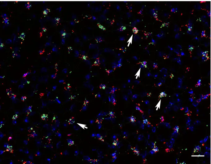
The first aim of this study was to clearly identify and characterize Cox-2 expressing cells in the kidney. Specific emphasize was placed on finding markers for the medullary Cox-2 expressing cells. This task was in part performed to establish a selective mouse model in the course of this work. To this end it was necessary to ensure all Cox-2 expressing cells of the inner medulla were included in the potential mouse model.
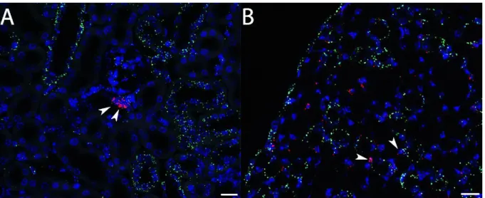
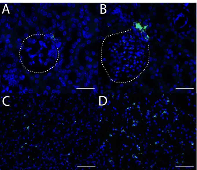
Therefore, I wanted to determine which cells express Cox-2 in a stimulated setting and how it is regulated in adult wildtype animals. Three conditions were to be evaluated: a high salt diet, water deprivation for 24h and genetically induced hypoxia signaling. The number, cell type markers and distribution of Cox-2 expressing cells across the kidney was characterized in detail under these conditions with the novel RNAscope
®in-situ hybridization technique.
Secondly, after establishing a suitable mouse model, the role of Cox-2 in the adult kidney and the involvement of Cox-2 during nephrogenesis were to be investigated. In the global knockout model of Cox-2 as well as human fetuses exposed to NSAIDS in the third trimester a severe renal phenotype was reported. The phenotype presents with tubular hyperplasia, a large number of sclerotic glomeruli in the subcapsular region and interstitial fibrosis. Due to the contribution of these cells to the phenotype, I investigated whether Cox-2 expression during nephrogenesis in the cells of the FoxD1
+stromal progenitor compartment is necessary for normal kidney development and function. These stromal progenitor cells differentiate into interstitial cells, renin and mesangial cells as well as vascular smooth muscle cells. The role of this cell population was investigated with the help of a targeted deletion of floxed Cox-2 alleles combined with a cell specific Cre-recombinase for the FoxD1 compartment (FoxD1
+/CreCox2
fl/flmice).
The third part of this study was to distinguish and clarify the functional relevance of
Cox-2 expression in medullary interstitial cells from the previously described function
of Cox-2 in the macula densa of adult kidneys. To evaluate the function of Cox-2 in
medullary interstitial cells I used a selective deletion of the Cox-2 gene in interstitial
cells of the kidney with an inducible Cre-recombinase under control of the PDGFR-β
promotor (PDGFR-β
+/iCreCox2
fl/flmice). As parameters for kidney function systolic
blood pressure, GFR, water intake, urinary ion concentrations, -volume and key mRNA
targets were determined and analyzed.
19
The concluding part of this study was concerned with investigating the role of resident
Cox-2 expression in the kidney as an inflammatory mediator in the model of adenine
induced fibrosis and unilateral ureter obstruction. Contradicting reports for the role of
Cox-2 in pathological settings have been published to date
89–92. But the source for the
potentially disease progression relevant Cox-2 expression has not been identified due
to the lack of an appropriate animal model
91. Using a cell specific deletion, I
investigated whether the absence of Cox-2 in PDGFR-β
+cells has any effect on the
progression of renal fibrosis. The evaluation of disease progression was investigated
through functional parameters, mRNA expression of fibrotic markers with qPCR and
RNAscope
®in-situ hybridization.
20
2 Material and Methods
2.1 Material 2.1.1 Instruments
Instrument Manufacturer
Anthos 2010 Microplate reader anthos Mikrosysteme GmbH, Friesoythe, Germany
Cameras AxioCam MRm, Zeiss, Jena
AxioCam 105 color, Zeiss, Jena Centrifuges Haematokrit 210, Hettich, Tuttlingen
Tischzentrifuge, neoLab, Heidelberg Z300, Hermle, Wehingen
Zentrifuge 5417R, Eppendorf, Hamburg Chemidoc™ Touch Imaging System Biorad, München, Germany
Filter sets for microscopy:
TRITC-Filter Cy2-Filter Cy3-Filter DAPI-Filter
Filter set 43 DsRed, Zeiss, Jena Filter set 38 HE, Zeiss, Jena Filter set 50, Zeiss, Jena Filter set 49, Zeiss, Jena Fluorescent lamp Colibri.2, Zeiss Jena
Gel Electrophorese System Compact M. Biometra, Göttingen Heating blocks Thermomixer, Eppendorf, Hamburg
Thermomixer 5436, Eppendorf, Hamburg Heating plate HI 1220, Leica, Wetzlar
Homogenizer Ultra-Turrax T25, Janke &Kunkel, Staufen Ice machine Ziegra Eismaschinen, Isernhagen
Incubator Memmert, Schwabach
Inhalation Device UniVet Groppler, Deggendorf, Germany Invitrogen™Qubit™3.0 Fluorometer Thermo Fisher Scientific, UK Magnetic mixer MR 80, Heidolph, Schwabach
MR 3001 K, Heidolph, Schwabach Metabolic cages Tecniplast, Hohenpeißenberg, Germany Microscope Axio Observer.Z1, Zeiss, Jena
Microtome Rotationsmikrotom RM2165, Leica, Wetzlar Rotationsmikrotom RM2265, Leica, Wetzlar
Microwave Sharp, Osaka
Osmomat 030 Gonotec, Germany
PCR machine Labcycler, SensoQuest, Göttingen Lightcycler LC480, Roche, Mannheim Peristaltic pump 323, Watson Marlow, Wilmington, USA
21 2.1.2 Consumables
Personal Computer Dell, Intel Core i7, NVIDIA GeForce GTX1080 8 GB
PH meter Hanna Instruments, Vöhringen
Photometer NanoDrop 1000, Peqlab, Erlangen
Pipettes Pipetman P2, P10, P20, P100, P200, P1000, Gilson, Middleton, USA
RNAscope® oven HybEZ Oven, Advanced Cell Diagnostics, Hayward, USA
Scales Feinwaage ABT 120-5DM, Kern, Balingen-Frommern EMS, Kern, Balingen-Frommern
Shaker GFL, Burgwedel
Rotamax, Heidolph, Schwabach
Tail cuff BP measurement system Non-Invasive Blood Pressure Monitoring System, TSE Systems, Bad Homburg
Vortex USA REAX1, Heidolph, Schwabach
Water bath Aqualine AL12, Lauda, Lauda-Königshofen 1083, GFL, Burgwedel
Water purification MilliQ Plus PF, Millipore, Schwalbach XP flame photometer BWB Technologies, UK
Product Manufacturer
FINE-JECT 12mm, gauge 30mm VWR International, Ismaning
Cannula 27G for hematocrit Becton Dickinson, Franklin Lakes, USA Minicaps end to end, na-heparinized, 5µl Hirschmann Laborgeräte GmbH & Co. KG,
Eberstadt
Minicaps end to end, na-heparinized, 0,5µl Hirschmann Laborgeräte GmbH & Co. KG, Eberstadt
Cover for Pasteur pipettes Roth, Karlsruhe
Cover slip Roth, Karlsruhe
Dissecting tools Fine Science Tools, Heidelberg Filter paper Schleicher & Schuell, Dassel
Glassware Roth, Karlsruhe
Schott, Mainz
Gloves Roth, Karlsruhe
Hematocrit capillaries Sanguis Counting, Nümbrecht Hematocrit sealing kit Brand, Wertheim
ImmEdge Pen Vector Laboratories, Burlingame, USA
Microscope slides, SuperFrost® Plus Menzel, Braunschweig
22 2.1.3 Commercial kits, enzymes and chemicals
Parafilm Bemis, Neenah, USA
Pasteur pipettes VWR, Darmstadt
Pipette controller accu-jet pro Brand, Wertheim Pipette tips with and without filter Nerbe, Winsen
Sarstedt, Nümbrecht
Biozym Scientific, Hessisch Oldendorf Plates, 96 well for qPCR Sarstedt, Nümbrecht
Serological pipettes 5ml, 10ml, 25 ml Sarstedt, Nümbrecht
Silicon forms for embedding Roth, Karlsruhe
Syringes Becton Dickinson, Franklin Lakes, USA
Tissue embedding cassettes Roth, Karlsruhe Tubes 0.5ml, 1ml, 2ml Sarstedt, Nümbrecht
Tubes 15ml, 50ml Sarstedt, Nümbrecht
Aspiration tube assemblies for calibrated microcapillary pipettes
Sigma-Aldrich, München
Multipurpose container 30ml Greiner Bio-One GmbH, Frickenhausen surgical suture Vicryl Ethicon, Norderstedt, Germany
Product Manufacturer
Adenine Chow (0,2%) Altromin Spezialfutter GmbH & Co. KG, Lage, Germany
Agarose Sigma-Aldrich, München
Bovine Serum Albumin (BSA) Sigma-Aldrich, München
Chloroform Merck, Darmstadt
Dulbecco´s PBS Sigma-Aldrich, München
Elisa kit ANG I IBL International, Hamburg
Enalapril maleate Sigma-Aldrich, München
Ethanol p.a Honeywell, Morris Plains, USA
Ethylenediaminetetraacetic acid Merck, Darmstadt First Strand Buffer, 5x Invitrogen, Karlsruhe
FITC-Sinistrin Fresenius Kabi, Bad Homburg
Formaldehyde solution (min. 37%) Merck, Darmstadt
Gene Ruler™ 100bp plus DNA ladder Thermo Scientific, Waltham, USA Glycergel mounting medium (IF) Dako Cytomation, Glostrup, Dänemark
Glycerin 87% Merck, Darmstadt
Glycogen Invitrogen, Karlsruhe
23
GoTaq DNA Polymerase, 5 U/μl Promega, Mannheim GoTaq Reaction Buffer Green Promega, Mannheim Hematoxylin solution, Gill Nr. 1 Sigma-Aldrich, München
HCl 1N Merck, Darmstadt
HCl fuming Merck, Darmstadt
HEPES(4-(2-hydroxyethyl)-1- piperazineethanesulfonic acid)
Sigma-Aldrich, München
High Salt chow (4% NaCl) Ssniff, Soest, Germany
Horse Serum (HS) Gibco, Life technologies, Grand Island, USA
Isoflurane Baxter, Unterschleißheim
Isopropanol Merck, Darmstadt
Isopropanol p.a AnalaR Normapur, VWR, Radnor, USA
Isotonic NaCl Solution, sterile 0,9% B. Braun, Melsungen K2HPO4 x 3 H2O Merck, Darmstadt
Ketamine, 10% bela-pharm, Vechta
KH2PO4 Merck, Darmstadt
Lightcycler480®SYBR-Green-Master-PCR Kit Roche, Mannheim Low Salt chow (0,03% NaCl) Ssniff, Soest, Germany
Methanol Merck, Darmstadt
M-MLV Reverse Transcriptase, 200 U/μl Invitrogen, Karlsruhe
Na2HPO4 Sigma-Aldrich, München
NaCl Merck, Darmstadt
NaH2PO4 Sigma-Aldrich, München
NaOH 1N Merck, Darmstadt
Oligo(dT)15 Primer, 0,5μg/μl Thermo Scientific, Waltham, USA
Paraformaldehyde Roth, Karlsruhe
Paraplast-Plus Paraffin Roth, Karlsruhe PCR Nucleotide Mix
(dATP, dCTP, dGTP, dTTP, 10 mM each)
Promega, Mannheim
RNAscope® 2.5 HD Detectionreagents – brown
Advanced Cell Diagnostics, Hayward, USA
RNAscope® 2.5 HD Detectionreagents – duplex
Advanced Cell Diagnostics, Hayward, USA
RNAscope® H2O2 & Protease Plus Reagents Advanced Cell Diagnostics, Hayward, USA RNAscope® Target Retrieval Reagent Advanced Cell Diagnostics, Hayward, USA RNAscope® Wash Buffer Advanced Cell Diagnostics, Hayward, USA RNAscope® Multiplex Fluorescent Assay Advanced Cell Diagnostics, Hayward, USA Roti®-Safe GelStain Roth, Karlsruhe
24 2.1.4 Buffers and Solutions
Unless stated otherwise, all Buffers and Solutions were prepared with purified water (MilliQ-Water).
PBS (Phosphate Buffered Saline)-Otto-Buffer, pH 7,4
NaCl 140 mM
K2HPO4 x 3 H2O 10 mM
KH2PO4 2,5 mM
Paraformaldehyde fixation solution for immunofluorescence (IF), pH 7,4
Dulbecco´s PBS
Paraformaldehyde 3%
Tris-EDTA Buffer, (epitope unmasking) pH 8,5
Tris 9,9 mM
EDTA 1,3 mM
Stock-Solution for IF
PBS-Otto
BSA 1%
Tamoxifen Chow (400mg tamoxifen citrate/kg)
Harlan Laboratories, NM Horst, Niederlande
Tris(hydroxymethyl)-aminomethan (Tris) Affymetrix, Cleveland, USA TRIsure®-Reagent Bioline, Luckenwalde
VectaMount™ mounting medium (ISH) Vector Laboratories, Burlingame, USA
Xylazine, 2% Serumwerk, Bernburg
Xylol Merck, Darmstadt
Xylol (for RNAscope®) AppliChem, Darmstadt
Acidic acid 100% Sigma-Aldrich, München
TSA Plus Cyanine 3 and Cyanine 5 System Perkin Elmer, Rehbach, Germany
Maleic acid Sigma-Aldrich, München
25 Blocking-Solution for IF
PBS-Otto
BSA 1%
Horse Serum 10%
NBF fixation solution for ISH, pH 7,0
Formalin (37-40% stock solution) 10%
NaH2PO4 33,3 mM
Na2HPO4 45,8 mM
TAE (Tris-Acetate-EDTA) Buffer, pH 8,5
Tris 40 mM
Acidic acid 20 mM
EDTA 1 mM
Agarose Gel for genotyping
TAE
Agarose 2%
Roti®-GelStain 0,02%
HEPES Buffer for GFR, pH 7,4
HEPES 0,5 M
FITC injection solution for GFR
NaCl solution 0,9%, sterile
FITC 1%
NaOH for genotyping
NaOH 25 mM
Tris-HCl, pH 8,0, for genotyping
Tris 1 M
26 Maleate Buffer, pH 6,0
Tris 33,5 mM
maleic acid 50 mM
EDTA 10,0 mM
2.1.5 Primer for genotyping and real time pcr
All primers were produced by Eurofins Genomics GmbH (Ebersberg) according to the sequences provided by us. Primers were shipped as lyophilized powder and dissolved in purified water to a final concentration of 100pmol/µl for all uses.
Primer for genotyping of mice
Construct Sequence (5´-3´) product size (bp)
Cox-2del s AATTACTGCTGAAGCCCACC
as GAATCTCCTAGAACTGACTGG 1054 = del
Cox-2flox s AATTACTGCTGAAGCCCACC
as AGAAGGCTTCCCAGCTTTTGTAACC
1058 = flox 823 = wt FoxD1+/Cre s1 CTCCTCCGTGTCCTCGTC
s2 TCTGGTCCAAGAATCCGAAG as GGGAGGATTGGGAAGACAAT
450 = Cre 237 = wt PDGFR-β+/iCre s1 GAA CTG TCA CCG GCA GGA
as AGG CAA ATT TTG GTG TAC GG c1 CAA ATG TTG CTT GTC TGG TG c2 CTC AGT CGA GTG CAC AGT TT
400 = Cre
200 = internal conrtol
CAG+/iCre s CAGAACCTGAAGATGTTC
as CCAGATTACGTATATCC 286 = Cre
Vhlflox s CTAGGCACCGAGCTTAGAGGTTTGCG as CTGACTTCCACTGATGCTTGTCACAG
450 = flox 270 = wt
Primer for quantitative Real-Time-PCR
Gene Sequence (5´-3´) product size (bp)
AQ2 s CTGGCTGTCAATGCTCTCCAC
as TTGTCACTGCGGCGCTCATC 122
AR s GCAATTGCAGCCAAGTACAA
as ATGGTGTCACCGACTTGG 97
BGT1 s GGTCCTTTTGGTCACAGAG
as GCTGGAGGCGTAGTAGTCAAA 65
27
Col1a1 s CTGACGCATGGCCAAGAAGA
as ATACCTCGGGTTTCCACGTC 91
Cox-2 s AGC CAT TTC CTT CTC TCC TG
as ACA ACA ACT CCA TCC TCC TG 894 Cx3CR1 s CCTGCCTCTGAGAAATGGAG
as ATCTCTCCAGCCCCTGAAAT 332
ENACα s TACGCGACAACAATCCCCAAGTGG
as ATGGAAGACATCCAGAGATTGGAG 334
GAPDH s CACCAGGGCTGCCATTTGCA
as GCTCCACCCTTCAAGTGG 294
HSP70.1 s ACCATGAAGAAGACTTTAAATAACCTTGAC
as CCCGGTGTGGTCTAGAAAACA 90
PDGFR-β s GAG GCT TAT CCG ATG CCT TCT
as AAA CTA ACT CGC CAG CGC C 79
Renin s ATGAAGGGGGTGTCTGTGGGGTC
as ATGTCGGGGAGGGTGGGCACCTG 194
RPL32 s TTA AGC GAA ACT GGC GGA AAC
as TTG TTG CTC CCA TAA CCG ATG 100 SMIT1 s AGTGGCTTCCATGGGTCAT
as TGCCCATTGGAATAAGCATC 60
UTA-1 s TGAGACGCAGTGAAGAGGAGAA
as ATGACCACTCGATGTGGAACAC 141
α-SMA s ACTGGGACGACATGGAAAAG
as CATCTCCAGAGTCCAGCACA 240
2.1.6 Probes for in-situ hybridization and antibodies Primary antibodies
Antibody Manufacturer Dilution
Chicken-anti-Renin Davids Biotech., Regensburg 1 : 400 Mouse-anti-α-SMA Abcam, Cambridge, UK 1 : 600
Secondary antibodies
Antibody Conjugation Manufacturer Dilution
Donkey-anti-chicken Cy 2 Dianova, Hamburg 1 : 400 Donkey-anti-mouse Cy 5 Dianova, Hamburg 1 : 400
28 Probes for in-situ hybridization with RNAscope®
All probes were manufactured by Advanced Cell Diagnostics (Hayward, USA).
Probe
RNAscope® Negative Control Probe – DapB RNAscope® Positive Control Probe – mM-PPIB RNAscope® Probe – mM-Col1a1 C1
RNAscope® Probe – mM-CSPG4 C1 & C2 RNAscope® Probe – mM-Cx3CR1 C1 RNAscope® Probe – mM-EPO C1& C2 RNAscope® Probe – mM-PDGFRb C1 & C2 RNAscope® Probe – mM-Ptgs1-O1 C1 RNAscope® Probe – mM-Ptgs2-O1 C1& C2 RNAscope® Probe – mM-Ren C1 & C2 RNAscope® Probe – mM-TNC C1 & C2 RNAscope® Probe – Rn –Ptgs2 C1
2.1.7 Software and internet services
This work was written with Microsoft Office 2016. Graphs were prepared using
GraphPad prism 5. For microscopy, the Zen Blue software was used. Pictures were
edited using Zen Blue and Adobe Photoshop CS5. The PubMeb site of the NCBI (The
National Center for Biotechnology; Information: http://www.ncbi.nlm.nih.gov/pubmed/)
was used for literature research. Citations were cataloged and organized with the
Zotero software (version 5.0.59).
29 2.2 Methods
2.2.1 Animals
2.2.1.1 Mouse strains and animal breeding
All animal experiments were conducted according to the “National Institutes of Health guidelines for the care and use of animals in research” and were approved by the local ethics committee. All mice were kept under optimal conditions, consisting of constant 23°C room temperature, relative humidity of 55 ± 5% and a constant 12h dark/light cycle. Standard rodent chow (0,6% NaCl, Ssniff, Deutschland) and tab water were provided ad libitum unless stated otherwise. All mice were bred in a C57/Bl6 background.
For this work, several genetically modified mouse lines were used:
PDGFR-β-CreER
T2(PDGFR-β
+/iCre) transgenic mice expressing a tamoxifen inducible Cre-recombinase under control of the PDGFR-β promoter. The PDGFR-β-CreER
T2is a fusion protein consisting of the Cre-recombinase and a modified estrogen binding site (CreER
T2)
14,93. Mice were kindly provided by Ralf H Adams, Max-Planck-Institut.
FoxD1
tm1(GFP/cre)Amc(FoxD1
+/Cre) knock-in mice express a fusion protein of Cre- recombinase and eGFP under the endogenous FoxD1 promotor. Mice were purchased from Jackson Laboratories, Bar Harbor, ME, USA (Stock No:
012463)
94,95 CAGG-CreER
T2(CAG
+/iCre) mice express a tamoxifen inducible Cre- recombinase under control of the beta actin promoter/enhancer coupled with the cytomegalovirus (CMV) immediate-early enhancer. Mice were kindly provided by Andreas Ludwig, Pharmacology, Universität Erlangen.
96 B6;129S4-Ptgs2
tm1Hahe/J (Cox2
flox) mice have loxP sites targeting exon 4 and 5 of the Cox-2 gene. After Cre-recombination a stop codon is created in exon 6, disrupting gene transcription
88. Mice were purchased from Jackson Laboratories, Bar Harbor, ME, USA (Stock No: 030785)
Vhl
floxmice contain loxP sites, targeting the sequence coding for the promotor
and exon 1 of von-Hippel-Lindau protein (Vhl)
14,97. After Cre-recombination a
null allele is created and transcription does not take place. Deletion of Vhl
inhibits the proteasomal degradation of hypoxia inducible factors, stably
inducing hypoxia signaling in the affected cells. Mice were provided by Volker
H. Haase, Vanderbilt University, Nashville, USA.
30
For the comparison between the newly generated genotypes and Cox-2
-/-mice, paraffin section generated in the course of previous work at the institute for physiology at Regensburg, were used
87. No live Cox-2
-/-mice were used for this work. Tissue of young non-albuminuric MWF rats was kindly provided by AG Castrop, institute for physiology, Regensburg.
For experiments mice of both genders were used, except for the model of adenine- induced fibrosis, where only males were used
98. Animals had an average age of 15 weeks at the time of the experiments. All newly generated genotypes were born in mendelian rations.
2.2.1.2 Genotyping of mice
Routine genotyping of mice was performed according to our laboratory standard protocol as follows by extracting genomic DNA and performing PCR.
At the age of 4 weeks mice were labeled with ear tags and ear punch out tissue of approximately 3mm diameter was taken in the same step. Ear tissue was digested in 50µl NaOH (25mM) at 96°C for 45 minutes in a heating block. After vortexing and adding 10µl Tris-HCl (1M, pH 8,0), samples were centrifuged 6 min at 10000 rpm. For PCR 0,5µl of the supernatant was used.
25 µl PCR mixture
5 µl GoTaq Buffer
0,75 µl for each Primer (10pmol/µl)
2 µl dNTPs (2,5mM)
0,2 µl GoTaq
0,5 µl gDNA
ad 25 µl MilliQ-H2O
31 PCR-running-programm
number of cycles
Duration Temperature Phase
1 5 min 94 °C activation
┌ 30-60 s 94 °C denaturation
36 30-60 s x °C annealing
└ 30-50 s 72 °C elongation
1 10 min 72 °C elongation
1 ∞ 4 °C storage
Primer specific annealing temperatures
PCR Temperature
Cox2del 56 °C
Cox2flox 61 °C
FoxD1+/Cre 58 °C
CAG+/iCre 54 °C
PDGFR-β+/iCre 56 °C
Vhlflox 62 °C
For separation of the resulting products, 10µl PCR mixture were applied to a 2%
Agarose gel and separated by horizontal electrophoresis with 130V in TAE running buffer.
2.2.1.3 Induction of Cre-Recombinase activity with tamoxifen
To activate the inducible Cre-recombinases used in this work, namely PDGFR-β
+/iCreand CAG
+/iCre, mice aged 8 weeks, were fed a tamoxifen containing diet (400mg/kg
tamoxifen citrate) for 4 weeks. This was followed by a 3 week rest period on normal
rodent diet (0,6% NaCl) to ensure that any observed effects were not caused by
tamoxifen.
32 2.2.1.4 High salt diet
To test the reaction of the generated mouse strain to a salt stimulus, after induction of Cre-recombinase activity and the following rest period, mice were fed a diet containing 4% NaCl for 14 days. Experiments were performed on day 14 unless stated otherwise.
2.2.1.5 Treatment with low salt diet and Enalapril
In order to stimulate the RAAS, animals were treated with a low salt diet containing 0,03% NaCl for 10 days. Additionally 50 mg/L of the ACE-inhibitor enalapril was added to the drinking water for the same period. Experiments were performed on day 10 unless stated otherwise.
2.2.1.6 Adenine-induced kidney fibrosis
For studying the effects of the targeted deletion of Cox-2 in the kidney on the progression of fibrosis, the model of adenine-induced fibrosis was used
98–100. After tamoxifen induction of Cre-recombinase and the following rest period, male mice were fed adenine containing diet (0,2%) for 3 weeks. Experiments were performed after exactly 3 weeks (3wks adenine) or after an additional 3 weeks in which the mice were again feed a normal diet (3wks adenine + 3wks).
2.2.1.7 Water deprivation for 24h
To induce Cox-2 expression in the inner medulla and evaluate the osmotic stress response of the mice, they were deprived of drinking water for 24h and killed immediately after this time period.
2.2.1.8 Unilateral ureteral obstruction
Mice were anesthetized with 2% isoflurane inhalation and placed on a 37°C warming plate. An anesthetic mask was placed over the nose of the mouse to ensure a continuous application of isoflurane for the duration of the procedure.
To ligate the right ureter a small incision was made over the approximate location of the ureter. The skin was carefully retracted and the ureter located with a soft Q-tip to avoid internal damage. A double knotted ligature was placed on the ureter and the skin was sewed up twice to prevent an opening of the wound. Mice were kept under observation for 2h after the operation and then daily checked to ensure wellbeing.
2.2.1.9 GFR measurement
To measure the glomerular filtration rate (GFR) in conscious mice, a slightly modified
protocol utilizing the clearance of FITC-Sinistrin by the kidney
101was used. This
33
technique makes it possible to repeatedly measure the GFR of the same mouse. Thus changes in GFR in the same mouse between different treatments could be evaluated.
Briefly, mice were immobilized for two minutes in 4% isoflurane inhalation anesthesia.
While the mice were unconscious a single bolus injection of FITC-Sinistrin, adjusted to the bodyweight (BW), was given in the tail vein. Mice were then placed in a heated chamber, leaving access to the tail. Mice were awake and active within one minute after the bolus injection. Blood samples were collected 3,7,10,15,35,55 and 75 min after the injection into 5µl capillaries from the tail vein. After collection, blood samples were centrifuged at 12000 rpm for 7min. 0,5µl of the resulting plasma were diluted in HEPES-Buffer and the FITC-Sinistrin fluorescence in the samples was measured with a Qubit™ 3.0 Fluorometer. GFR of mice was calculated by applying the two- compartment model. To get a better comparison between mice, especially when of different gender, GFR/BW was calculated and used for analysis.
2.2.2 Retrograde arterial perfusion of mice
To fix kidney tissue for histological evaluation, mice were perfused with either 3%
paraformaldehyde (pH 7,4) for immunofluorescence, or with 10% NBF solution (pH 7,0) if the tissue was used for in-situ hybridization with the RNAscope
®technology.
Mice were first anesthetized with an i.p injection of Ketamine (80mg/kg BW) and Xylazine (16mg/kg BW). After opening the abdominal cavity and exposing the abdominal aorta the left kidney was ligated to preserve the tissue for mRNA extraction.
A clamp was placed on the aorta just below the branching of the kidney arteries
(Arteriae renales) to preserve the kidney perfusion. The aorta abdominalis was
carefully opened below the clamp with scissors and a perfusion catheter was inserted
into the aorta with the help of a vessel dilator and fixed with a clamp. After removing
the upper clamp the vena cava inferior is opened below the catheter to equilibrate the
pressure. Continous perfusion of the kidney with a steady flow of 13ml/min and a total
volume of 40ml fixation solution was started. After the perfusion the ligated left kidney
was removed, cut in half and snap frozen with liquid nitrogen. Tissue was stored at -
80°C until used. The perfused kidney was removed, cut in half and stored either in
70% Methanol (for IF) at 4°C or in NBF (ISH) at RT for 16-24h before embedding in
paraffin.
34 2.2.3 Zonal dissection of kidneys
To determine the zonal distribution of different mRNA targets, kidneys were dissected into three zones after removal from the mouse. The kidneys were halved and thin slices were prepared with a razorblade and immediately transferred into petri dish containing 10ml ice-cold sterile NaCl 0,9% solution. Under a microscope, the thin kidney slices were then separated with a razorblade into cortex, outer medulla and inner medulla.
At least four slices per kidney were dissected, the tissue was then pooled by zone into a cup and snap frozen in liquid nitrogen. Tissue was stored the same as complete kidneys at -80°C for mRNA extraction.
2.2.4 Tail cuff blood pressure measurement
Blood pressure measurements were performed on conscious mice with tail cuff manometry. Briefly, mice were placed in a restraining chamber, giving free access to the tail, the measurement probe and inflatable pressure cuff were placed over the tail and the mouse was left for 5 min to rest. Performing this procedure at the same time each day the mice were acclimatized to the measurement procedure for five days.
Then on each day for the duration stated in the experiments, five measurements were performed per mouse and a mean of these measurements was used for evaluation of the results.
2.2.5 Metabolic cages
For determination of 24h urine volume and the corresponding water intake, mice were housed in individual metabolic cages. Each cage was pre-treated with a thin silicon film in the lower part to facilitate a better separation of urine and fecal matter into separate containers. The mice were given a house for better acclimatization and feed sufficient for one day along with a weighted water bottle. Each morning mice were transferred to a fresh cage, urine was collected and water intake calculated by weighing the water bottle again. Mice were placed in the cages for two days before the measurement period, undergoing the entire procedure to get them acclimatized.
Unless stated otherwise, 5 days of urine collection and water intake were measured
for each mouse per condition.
35 2.2.6 Spot urine collection and analysis
Mice were placed in a clean plastic box with a lid each day for 3 consecutive days to collect urine samples. If the mice did not urinate spontaneously (spot urine) after 15min in the box, they were placed back into the cage and the procedure was repeated the next day. Samples were frozen at -80°C until analysis.
2.2.6.1 Osmolality measurement
Urine osmolality was determined by freezing point depression. 5µl urine were diluted in 45µl Millipore-H
2O, vortexed and measured in the Osmomat 030. Resulting values were multiplied with the dilution and converted to mOsmol/l for evaluation.
2.2.6.2 Sodium and potassium measurement in urine
The concentration of sodium and potassium in urine was determined simultaneously by flame photometry. Before the measurement, the instrument was calibrated with standard dilutions (5 point, linear dilution) made from commercial stocks for both ions.
10µl urine were diluted in 10ml MilliQ-H
2O, vortexed and then measured in duplicates.
The mean values were multiplied with the dilution and converted to mMol/l for evaluation of the results.
2.2.7 Histological methods
2.2.7.1 Embedding of tissue in paraffin
For embedding the perfused tissue in paraffin, the halved kidneys were transferred to embedding cassettes and dehydrated in an ascending alcohol series.
Time, temperature in-situ hybridization immunofluorescence
1 x 30 min, RT 70% Ethanol p.a. 70% Methanol 2 x 30 min, RT 80% Ethanol p.a. 80% Methanol 2 x 30 min, RT 90% Ethanol p.a. 90% Methanol 2 x 30 min, RT 100% Ethanol p.a. 100% Methanol 2 x 30 min, RT 100% Isopropanol p.a. 100% Isopropanol 1 x 30 min, 45 °C 100% Isopropanol p.a. 100% Isopropanol 1 x 30 min, 60 °C 1 : 1 Isopropanol/paraffin 1 : 1 Isopropanol/paraffin
2 x 24h, 60 °C Paraffin Paraffin
Kidneys were then placed in silicon forms with liquid paraffin, the cut surface facing
down. Paraffin was left to solidify overnight, blocks were then removed from the silicon
forms and stored at RT.
36 2.2.7.2 Section of paraffin embedded tissue
Tissue blocks were roughly trimmed with a razorblade before placing them in the microtome holder and firmly screwed into place. Sections of 5µm thickness were prepared from the tissue and carefully placed in a 41°C water bath to stretch out wrinkles in the tissue. Sections were then placed on SuperFrost
®Plus slides with a brush and dried overnight at room temperature.
2.2.7.3 Immunofluorescence staining
Immunofluorescence staining was used to visualize protein structures in the kidney tissue studied. For this aim indirect immunofluorescence with two antibodies (AB) was used, the primary antibody binding to the target epitope in the tissue and the secondary antibody reacting specifically with the primary AB. To make the target visible for microscopy the secondary AB is conjugated to a fluorescent dye to show a strong fluorescence when exited with light at distinct wavelengths. Before the incubation of the primary AB the paraffin was removed and the tissue rehydrated by a decreasing alcohol series.
Time Solution
2 x 10 min 100% Xylol 2 x 5 min 100% Isopropanol 1 x 5 min 90% Isopropanol 1 x 5 min 80% Isopropanol 1 x 5 min 70% Isopropanol 1 x 5 min MilliQ-H2O


