THE ROLE OF MAST CELLS
IN THE MICROENVIRONMENT OF TUMORS
Inaugural Dissertation
zur
Erlangung des Doktorgrades Dr. nat. med.
der Medizinischen Fakultät und
der Mathematisch‐Naturwissenschaftlichen Fakultät der Universität zu Köln
vorgelegt von
Dipl. Biol. Anja Rabenhorst aus Wismar
Köln, 2012
Berichterstatter/-in: PD Dr. Roswitha Nischt PD Dr. Hildegard Büning
Tag der letzten mündlichen Prüfung: 23.01.2013
Table of content
Table of content
Summary 01
Zusammenfassung 02
Abbreviations 03
1. Introduction 07
1.1. Mast cells 07
1.2. Models for investigating mast cell functions in vivo 08
1.3. Hallmarks of cancer 11
1.3.1. Sustaining proliferative signaling 12
1.3.2. Evading growth suppressors 12
1.3.3. Avoiding immune destruction 12
1.3.4. Enabling replicative immortality 12
1.3.5. Tumor-promoting inflammation 13
1.3.6. Activating invasion and metastasis 13
1.3.7. Inducing angiogenesis 13
1.3.8. Genome instability and mutation 14
1.3.9. Resisting cell death 14
1.3.10. Deregulating cellular energetics by reprogramming the energy metabolism 14
1.4. Mast cell mediators and their potential role in tumorigenesis 15
1.5. Mast cells in tumors 17
1.6. Primary cutaneous lymphoma 18
2. Aims 20
Table of content
3. Material and Methods 22
3.1. Material 22
3.1.1. Chemicals and solutions 22
3.1.2. Buffers 23
3.1.3. Single-stranded oligonucleotides 24
3.1.3.1. Genotyping 24
3.1.3.2. Real-time PCR 24
3.1.4. Laboratory equipment 25
3.1.5. Software 25
3.2. Methods 26
3.2.1. Collection of cutaneous biopsies from patients with primary cutaneous lymphoma 26
3.2.2. Histology 27
3.2.2.1. Human samples 27
3.2.2.2. Murine samples 28
3.2.3. In vitro experiments 29
3.2.3.1. Cell isolation 29
3.2.3.2. Cell culture 29
3.2.3.3. Inhibition and stimulation of mediator release from mast cells 31
3.2.3.4. Cytometric bead array in cell culture supernatants 31
3.2.3.5. Quantitative real-time PCR 32
3.2.3.5.1. RNA isolation and reverse transcription into cDNA 32
3.2.3.5.2. RNA and cDNA quantification 32
3.2.3.5.3. Real-time PCR 32
3.2.3.6. Cell proliferation measurement 33
3.2.4. In vivo experiments 34
3.2.4.1. Mouse strains 34
3.2.4.2. Genotyping 34
3.2.4.2.1. DNA isolation 34
3.2.4.2.2. Polymerase chain reaction (PCR) 35
3.2.4.2.3. Gel electrophoresis 35
3.2.4.3. Administration of diphtheria toxin 36
Table of content
3.2.4.4. Subcutaneous injection of tumor cell lines
and measuring tumor growth 36
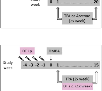
3.2.4.5. Two-step model of chemically induced skin carcinogenesis 37
3.2.5. Statistical analysis 38
4. Results 39
4.1. Mast cells in the microenvironment of human PCL 39
4.1.1. Clinical characteristics of PCL patients participating in the study 39
4.1.2. Mast cell numbers are increased in primary cutaneous lymphoma 42
4.1.3. Mast cell numbers correlate with progression of primary cutaneous lymphoma 45
4.1.4. Mast cell degranulation is increased in primary cutaneous lymphoma 47
4.1.5. Microvessel density is increased in mycosis fungoides 49
4.1.6. Mast cells release mediators that promote tumor growth 52
4.1.7. Unstimulated PCL cells produce proinflammatory cytokines 53
4.1.8. Mast cell supernatant induces changes in cytokine production of primary cutaneous lymphoma cells 55
4.1.9. Mast cell supernatant induces proliferation of primary cutaneous lymphoma cells 56
4.2. Mast cells in the microenvironment of murine tumors 58
4.2.1. Murine mast cell supernatant induces cytokine release and proliferation of the mouse T-cell lymphoma cell line EL4 58
4.2.2. Mast cell-deficient mouse models for the investigation of tumor growth 60
4.2.3. Mast cell-deficient mice show decreased growth of subcutaneously injected tumors 62
4.2.4. Mast cell-deficient mice show decreased
chemically induced carcinogenesis 65
Table of content
5. Discussion 67
5.1. Increased mast cell number and microvessel density in primary cutaneous lymphoma 67
5.2. Stimulation of tumor growth by mast cells in vitro and in vivo 69
5.2.1. Potential effects of mast cells on different hallmarks of cancer 70
5.2.1.1. Sustaining proliferative signaling 72
5.2.1.2. Avoiding immune destruction 72
5.2.1.3. Activating invasion and metastasis 73
5.2.1.4. Inducing angiogenesis 74
5.2.2. Decreased growth of subcutaneously injected tumors and decreased chemically induced carcinogenesis in mast cell-deficient mice 75
6. References 77
Figure Index 89
Table Index 91
Acknowledgments 92
Erklärung 93
Curriculum vitae 95
Summary
1
Summary
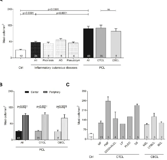
Mast cells exert important functions in innate and adaptive immunity and are therefore strategically located at inner and outer body surfaces, such as skin, gastrointestinal tract and blood vessels. Once mast cells are activated, they release a broad array of prestored or newly synthesized mediators, including a series of cytokines and chemokines. Recent studies showed that mast cells also infiltrate many types of solid cancers and hematologic malignancies. Here, mast cell products can participate in creating a microenvironment that either promotes or inhibits tumor growth. However, the role of mast cells in tumor biology is still controversial and underlying mechanisms remain largely undefined. In the present study, we show for the first time that mast cell numbers are significantly increased in skin biopsies from patients with primary cutaneous lymphoma. Mast cell infiltration is most prominent in the periphery, at lymphoma rims. Also, degranulation of mast cells is significantly increased.
Interestingly, patients with advanced stages of the disease show higher mast
cell counts than stable patients. Similarly, numbers of mast cells correlate with
disease progression. In addition, mast cell numbers correlate with the density of
microvessels. Supernatant of cultured mast cells induces in vitro production of
cytokines and proliferation in primary cutaneous lymphoma cells. To further
elucidate the contribution of mast cells to tumor biology, we use new transgenic
mouse models of inducible or constitutive selective deficiency of connective
tissue mast cells and show that growth of subcutaneously injected tumors and
chemically induced carcinogenesis is significantly decreased in mast cell-
deficient mice. Taken together, these experiments demonstrate that mast cells
play a tumor-promoting role in human primary cutaneous lymphoma and
different murine tumor models. Moreover, our data provide a rationale for
exploiting tumor-associated mast cells as prognostic marker and therapeutic
target in primary cutaneous lymphoma and other tumors.
Zusammenfassung
Zusammenfassung
Mastzellen üben entscheidende Funktionen in der angeborenen und adaptiven Immunantwort aus. Sie kommen deshalb in besonders großer Zahl an inneren und äußeren Körperoberflächen vor, wie z.B. in der Haut, im Magen-Darm-Trakt und in der Nähe von Blutgefäßen. Wenn Mastzellen aktiviert werden, sezernieren sie viele verschiedene gespeicherte oder neu synthetisierte Mediatoren, z.B. zahlreiche Zytokine und Chemokine. Infiltrate aus Mastzellen finden sich auch in vielen soliden Tumoren und hämatologischen Neoplasien.
Die Produkte der Mastzellen können hier eine Umgebung schaffen, die entweder das Tumorwachstum fördert oder hemmt. Die genaue Funktion der Mastzellen in der Tumorbiologie wird jedoch noch kontrovers diskutiert und die zugrunde liegenden Mechanismen sind weitgehend unbekannt. Die vorliegende Arbeit zeigt erstmals, dass Mastzellen auch in Hautbiopsien von Patienten mit primären kutanen Lymphomen signifikant vermehrt sind. Die Infiltrate aus Mastzellen finden sich vor allem im Randbereich der Lymphome. Die Degranulation von Mastzellen ist ebenfalls deutlich gesteigert.
Interessanterweise zeigen Patienten mit fortgeschrittenen Krankheitsstadien
höhere Mastzellzahlen als stabile Patienten. Vergleichbar korreliert die
Mastzellzahl auch mit dem Fortschreiten der Krankheit. Weiterhin korreliert die
Zahl der Mastzellen mit der Dichte der Gefäße. In vitro induziert der Überstand
von kultivierten Mastzellen die Produktion von Zytokinen und Proliferation von
primären kutanen Lymphomzellen. Um den Beitrag von Mastzellen zur
Tumorbiologie weiter aufzuklären, haben wir neue transgene Mausmodelle
verwendet, die eine induzierbare oder konstitutive Defizienz von
Bindegewebsmastzellen aufweisen, und zeigen, dass das Wachstum von
subkutan injizierten Tumoren und chemisch induzierten Papillomen in diesen
Tieren signifikant erniedrigt ist. Zusammenfassend belegen diese Experimente,
dass Mastzellen eine Tumor-fördernde Funktion bei primären kutanen
Lymphomen und verschiedenen murinen Tumormodellen besitzen. Die Daten
liefern zudem die Basis für weitere Untersuchungen zur Nutzung von Tumor-
assoziierten Mastzellen als prognostischer Marker und therapeutisches Target
in primären kutanen Lymphomen und anderen Tumoren.
Abbreviations
3
Abbreviations
% percentage
° degree
µg mikrogram
µl mikroliter
µM mikromolar
A tumor area
AD atopic dermatitis
Ang angiopoietin
BAC bacterial artificial chromosome
BCL primary cutaneous diffuse large B-cell lymphoma BMMC bone marrow-derived mast cells
bp base pairs
C celsius
Ca I calcium ionophore
CBA cytometric bead array
CBCL cutaneous B-cell lymphoma
CCL2 MCP-1
CCL5 RANTES
CD cluster of differentiation
CD30+ALCL CD30 positive primary cutaneous anaplastic large cell lymphoma
cDNA complementary DNA
CFBCL primary cutaneous follicle center lymphoma
COX cyclooxygenase
CSF colony stimulating factor
CTCL cutaneous T-cell lymphoma CTMC connective tissue mast cell
Ctrl control
CXCL10 IP-10
CXCL8 IL-8
CXCL9 MIG
DC dendritic cell
Abbreviations
DMBA 7,12-Dimethylbenz(a)anthracene DMEM dulbecco´s modified eagle medium
DMSO dimethyl sulfoxide
DNA deoxyribonucleic acid
dNTP deoxynucleoside triphosphate
DT diphtheria toxin
DTR diphtheria toxin receptor
ED extensively degranulated
EDTA ethylenediaminetetraacetic acid ELISA enzyme-linked immunosorbent assay EORTC European Organization of Research and
Treatment of Cancer
FACS fluorescence-activated cell sorting
FBS fetal bovine serum
FGF fibroblast growth factor
FMF folliculotropic mycosis fungoides
Fwd forward
g gram
GAPDH glyceraldehyde 3-phosphate dehydrogenase G-CSF granulocyte-colony stimulating factor
GM-CSF granulocyte-macrophage colony-stimulating factor
h hours
H&E hematoxylin/eosin
HBSS hank´s balanced salt solution
Hetero heterozygous
Homo homozygous
HPLC high-performance liquid chromatography
HSC hematopoietic stem cell
i.p. intraperitoneal
IFN interferon
Ig immunoglobulin
IL interleukin
IMDM iscove´s modified dulbecco´s medium
ISCL International Society of Cutaneous Lymphoma
Abbreviations
5
kb kilo-base pair
LP lymphomatoid papulosis
LT leukotriene
M molar
MC mast cell
MCP mast cell progenitor
Mcpt5 mast cell protease-5
MD moderately degranulated
MDSC myeloid-derived suppressor cells
MF mycosis fungoides
mill million
min minute
MIP macrophage inflammatory protein
ml milliliter
mm millimeter
mM millimolar
MMC mucosal mast cell
MMP matrix metalloproteinase
mRNA messenger RNA
MTS 3-(4,5-dimethyl-2-yl)-5-(3-carboxymethoxyphenyl)- 2-(4-sulfophenyl)-2H-tetrazolium
MZL primary cutaneous marginal zone B-cell lymphoma
n number
ND not degranulated
NEA non-essential amino acid solution
NGF nerve growth factor
NHL non-Hodgkin lymphoma
nm nanometer
nM nanomolar
NO nitric oxide
ns not significant
PAF platelet-activating factor
PCL primary cutaneous lymphoma
PCR polymerase chain reaction
Abbreviations
PDGF platelet-derived growth factor
pg picogram
PG prostaglandin
pH negative logarithm of the hydrogen ion concentration PLEO primary cutaneous small/medium-sized pleomorphic
T-cell lymphoma
Pseudolym pseudolymphoma
Rev reverse
RNA ribonucleic acid
rpm rounds per minute
RPMI roswell park memorial institute medium
s.c. subcutaneous
SCF stem cell factor
SD standard deviation
SDS sodium dodecyl sulfate
sec seconds
SEM standard error of the mean
SS Sézary syndrome
TGF transforming growth factor
TNF tumor necrosis factor
TPA 12-O-tetradecanoylphorbol 13-acetate
U unit
UV ultraviolet
V volt
VEGF vascular endothelial growth factor
Vol tumor volume
w/v weight per volume
WHO World Health Organization
WT wildtype
x fold
Introduction
7
1. Introduction
1.1. Mast cells
In 1878, Paul Ehrlich discovered cells with large cytoplasmic granules that he named "mast cells".
1First, mast cells were believed to be a component of the connective tissue, derived from undifferentiated mesenchymal cells.
2,3Today, we know that mast cells are bone marrow-derived hematopoietic cells.
Hematopoietic stem cells give rise to mast cell progenitors, which circulate in the blood and enter the tissues, where differentiation and maturation into mature mast cells takes place (Figure 1).
4However, it is still controversially discussed whether the mast cell progenitor is derived directly from a pluripotent precursor or from the myeloid lineage.
Figure 1. Mast cell development. Mast cells originate from hematopoietic stem cells (HSC) in the bone marrow that give rise to mast cell progenitors (MCP). These mast cell progenitors are released into the blood and circulate until they migrate into the tissues, where they finally differentiate into mature mast cells. MMC, mucosal mast cells; CTMC, connective tissue mast cells.
The phenotype of mature mast cells varies depending on the tissue
microenvironment. Human mast cell subpopulations are divided into MC
TCcells,
designated to their tryptase and chymase content, and MC
Tcells that only
contain tryptase. In analogy to human mast cells, also different types of murine
mast cells exist. In mice, one can distinguish between mucosal mast cells
(MMC), located within mucosal epithelia and connective tissue mast cells
(CTMC), located in skin, peritoneal cavity and submucosa.
5Because human
MC
Tcells predominantly appear in the alveolar septa of the lung and in the
small intestinal mucosa, they most closely correspond to mouse MMC, whereas
human MC
TCcells resemble mouse CTMC.
6Introduction
Due to the preferred location of mast cells in tissues exposed to the environment such as skin, airways and gastrointestinal tract
7-9, they are one of the first immune cells to interact with invading pathogens
10,11or antigens.
12,13Thus, mast cells are predestined for performing important functions in innate
10,14,15and adaptive
14,16-18immunity. Furthermore, several studies reported on a possible role of mast cells in wound healing, tissue remodeling and transplant tolerance.
19-21Additionally, mast cells seem to contribute to diet- induced obesity and diabetes.
22Moreover, a pathogenic role of mast cells was described for rheumatoid arthritis, multiple sclerosis and atherosclerosis
23-26as well as for different human malignancies, as described in detail in section 1.5.
Mast cells are long-lived cells that can re-enter the cell cycle and proliferate after appropriate stimulation.
27Furthermore, the increased recruitment and/or retention, and local maturation of mast cell progenitors can contribute to the expansion of mast cell populations.
6,28The best-characterized activation pathway in mast cells is their activation through immunoglobulin E (IgE), which binds to the high affinity IgE receptor Fcε-receptor I (FcεRI).
29In mice, activation through IgG1 and FcγRIII is also possible. Besides, a large variety of other immunological and non-immunological signals can induce mast cell activation.
27Depending on the type and concentration of the activation signal, mast cells can release either all classes of mediators at high levels via degranulation or may release certain mediators more selectively by secretion.
14This offers the possibility to respond to the requirements of the specific biological process with large variation. In addition, being rechargeable, mast cells can also participate in multiple cycles of mediator release. Furthermore, mast cells can functionally interact with other cell types, including T-cells, B-cells, dendritic cells (DC) and eosinophils, both by releasing mediators and by cell-cell interactions.
1.2. Models for investigating mast cell functions in vivo
Over the past 30 years, Kit-mutant mice like WBBF1-Kit
W/W-vand C57BL/6 Kit
W-sh/W-sh
have usually been used for the investigation of mast cell biology in
vivo.
4,30Kit
Wcontains a point mutation that encodes for a truncated Kit protein,
which lacks the transmembrane domain and is therefore not expressed on the
cell surface, Kit
W-vshows a mutation in the Kit tyrosine kinase domain that
decreases the kinase activity of the receptor and Kit
W-shcontains an inversion
Introduction
9
mutation of the transcriptional regulatory elements upstream of the Kit transcription start site on mouse chromosome 5.
14,17,27Adult WBBF1-Kit
W/W-vand C57BL/6 Kit
W-sh/W-shmice are deficient in mast cells and melanocytes.
4In the WBBF1-Kit
W/W-vstrain, the number of mast cells in the skin, stomach, cecum and mesentery is reduced to less than 1% compared to congeneic controls.
Additionally, WBBF1-Kit
W/W-vmice have several other abnormalities, such as macrocytic anemia, a decrease in the number of bone marrow and blood neutrophils, sterility and a decreased number of intestinal cells of Cajal.
4,28,30The mast cell-deficient strain C57BL/6 Kit
W-sh/W-shis fertile and not anemic.
30,31C57BL/6 Kit
W-sh/W-shanimals are largely devoid of mast cells in the tongue, trachea, lung, stomach, spleen, small intestine, mesentery, peritoneum and inguinal lymph nodes. In the skin, 1.2% of the normal number of mast cells are still present.
30However, C57BL/6 Kit
W-sh/W-shmice also show abnormalities like splenomegaly, thrombocytosis, neutrophilia, and decreased numbers of F4/80- positive cells in the bone marrow.
32In contrast to the Kit-mutant lines used up to now, recently developed mouse models of inducible (Mcpt5-Cre/iDTR) and constitutive (Mcpt5-Cre/R-DTA) mast cell deficiency
33,34show an otherwise normal immune system. For creating these new mouse models, bacterial artificial chromosome (BAC) transgene technology was employed to express Cre recombinase under the control of the mast cell-specific promoter mast cell protease-5 (Mcpt5).
33The constructs were injected into C57BL/6 oocytes for random integration into the mouse genome.
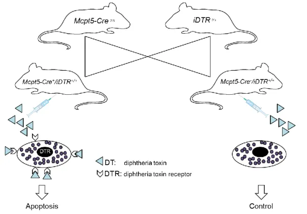
To develop a mouse model of inducible mast cell deficiency, Mcpt5- Cre/iDTR
33,34mice were generated by breeding the mast cell-specific transgenic line Mcpt5-Cre to the iDTR line, which is characterized by expression of a simian diphtheria toxin receptor (DTR) in cells that have deleted a loxP-flanked stop cassette by Cre-mediated recombination (Figure 2).
35Mcpt5-Cre+/iDTR+
mice show efficient and specific depletion of connective tissue mast cells upon
intraperitoneal injections with diphtheria toxin (DT). A mouse model of
constitutive mast cell deficiency, was obtained by crossing the mast cell-specific
transgenic line Mcpt5-Cre to the R-DTA line
36(Figure 3). These mice express
diphtheria toxin (DT) under the control of a loxP-flanked stop cassette. The
loxP-flanked cassette prevents DT expression in the absence of Cre activity.
Introduction
Figure 2. Mcpt5-Cre/iDTR – A mouse model of inducible mast cell deficiency.
Intraperitoneally injected diphtheria toxin (DT) binds to the receptors on the surface of connective tissue mast cells, which leads to apoptosis of these cells. DTR, diphtheria toxin receptor; DT, diphtheria toxin
Figure 3. Mcpt5-Cre/R-DTA – A mouse model of constitutive mast cell deficiency. Upon Cre activity connective tissue mast cells express diphtheria toxin (DT), which leads to apoptosis of these cells. DT, diphtheria toxin
Introduction
11
1.3. Hallmarks of cancer
The hallmarks of cancer (Figure 4) include, according to Hanahan and Weinberg
37, ten different mechanisms as shown below:
Figure 4. Hallmarks of cancer. Adapted from Hanahan and Weinberg, 201137
Introduction
1.3.1. Sustaining proliferative signaling
Whereas normal tissue controls the production and release of growth-promoting signals to ensure constant cell numbers and intact tissue architecture, tumor cells deregulate these signals. The enabling signals are conveyed by growth factors that bind cell surface receptors. Sources of these proliferative signals are still largely unknown.
1.3.2. Evading growth suppressors
Besides limitless growing, tumor cells are able to circumvent programs that negatively regulate cell proliferation, which are mostly dependent on tumor suppressors. Additionally, contact inhibition which, in dense cell populations, also yields to a suppressed cell proliferation is abolished in many cancers.
Furthermore, deactivating the TGF-β pathway, known for its antiproliferative effects, promotes malignancy.
1.3.3. Avoiding immune destruction
Immune surveillance is usually responsible for recognizing and eliminating the majority of abnormal cells. Tumor cells are often able to avoid detection by the immune system or can limit the extent of being killed. For example, tumor cells can "paralyze" infiltrating cells of the immune system by secreting TGF-β or other immunosuppressive factors. Moreover, they can also actively recruit immunosuppressive cells, such as regulatory T-cells and myeloid-derived suppressor cells.
1.3.4. Enabling replicative immortality
In order to generate tumors, tumor cells aquire unlimited replicative potential. In contrast, normal cells can only pass through a limited number of successive cell growth and cell divison cycles. In the end, normal cells become senescent, meaning they enter a nonproliferative, but viable state. It may occur that some of these cells escape from this state and acquire unlimited replicative potential.
Cells that undergo this transition are then called "immortalized". They may
proceed to form tumors due to their ability to maintain telomeric DNA at lengths
sufficient to avoid undergoing senescence or apoptosis, achieved by
upregulating the expression of telomerase.
Introduction
13
1.3.5. Tumor-promoting inflammation
Tumors are infiltrated by cells of the innate and adaptive immune system and mirror inflammatory conditions. Historically, such immune responses were thought to reflect an attempt of the immune system to eradicate tumors. Today, it is well recognized that the tumor-associated inflammatory response can also have the opposite effect of enhancing tumorigenesis and progression.
Inflammation can contribute to different hallmarks of cancer by supplying a large variety of molecules to the tumor microenvironment that promote tumors, for example growth factors to sustain proliferative signaling, survival factors that limit cell death, proangiogenic factors and extracellular matrix-modifying enzymes that facilitate angiogenesis, invasion and metastasis.
1.3.6. Activating invasion and metastasis
Invasion and metastasis is a multistep process, starting with local invasion, followed by intravasation of tumor cells into nearby blood and lymphatic vessels, transition of tumor cells through the lymphatic and hematogenous systems, extravasation of tumor cells from the lumina of these vessels into the parenchyma of distant tissues, formation of micrometastasis and finally by growth of micrometastatic lesions into macroscopic tumors.
1.3.7. Inducing angiogenesis
Tumors need nutrients and oxygen and can eliminate metabolic waste and carbon dioxide. Therefore, angiogenesis, which means sprouting of new vessels from existing ones, is necessary to generate a tumor-associated vasculature. During tumor progression, an "angiogenic switch" causes continuous sprouting of normal vasculature. Tumor vasculature is marked by early capillary sprouting, convoluted and excessive vessel branching, distorted and enlarged vessels, erratic blood flow, leakiness and abnormal levels of endothelial cell proliferation and apoptosis.
38The best-studied inducer of angiogenesis is vascular endothelial growth factor (VEGF). VEGF gene expression is upregulated by hypoxia as well as by oncogene signaling.
Furthermore, VEGF ligands are sequestered in the extracellular matrix in latent
forms that are subject to release and activation by extracellular matrix-
degrading proteases, like for example matrix metalloproteinase-9 (MMP-9).
Introduction
In addition, other proangiogenic signals, like fibroblast growth factor (FGF), have been implicated in sustaining tumor angiogenesis.
1.3.8. Genome instability and mutation
As tumor progression is a multistep process, every step offers the opportunity for mutations and deregulating gene expression. Due to the inherent ability to detect and resolve DNA defects, the rate of spontaneous mutations is usually very low. Tumor cells often show increased rates of mutations. This mutability is also achieved through increased sensitivity to mutagenic agents, through a breakdown in one or several components of the genomic maintenance machinery, or through both mechanisms. Additionally, the accumulation of mutations is accelerated by comprising the surveillance systems that normally monitors genomic integrity and transfers genetically damaged cells into senescence or apoptosis.
1.3.9. Resisting cell death
Programmed cell death by apoptosis is a natural barrier to cancer development.
Tumor cells have developed many different strategies to limit or circumvent apoptosis, for example loss of tumor suppressor functions, increased expression of antiapoptotic regulators (Bcl-2, Bcl-x
L) or survival signals (Igf1/2) and downregulation of proapoptotic factors (Bax, Bim, Puma).
1.3.10. Deregulating cellular energetics by reprogramming the energy metabolism
Under aerobic conditions, normal cells process glucose via glycolysis into
pyruvate and thereafter into carbon dioxide through mitochondrial oxidative
phosphorylation. Tumor cells can reprogram their glucose metabolism by
shifting their energy production largely to glycolysis. Besides, reliance on
glycolysis is of particular importance under hypoxic conditions within many
tumors. Furthermore, increased glycolysis allows the distribution of glycolytic
intermediates into various biosynthetic pathways, which facilitate the
biosynthesis of macromolecules required for assembling new cells.
Introduction
15
1.4. Mast cell mediators and their potential role in tumorigenesis
Upon activation, mast cells exert their biological functions by releasing preformed as well as de novo synthesized mediators. Mast cells produce a broad array of such mediators and cell-cell signaling molecules. About thirty different cytokines, chemokines as well as other metabolites have been shown to be produced by mast cells.
39They can be grouped into three major classes.
12First, preformed and granule-associated mediators are produced like histamine, serotonin, proteases like tryptase and chymase as well as TNF, VEGF and FGF. Second, newly synthesized lipid mediators are released, namely the leukotrienes LTC
4and LTB
4and the prostaglandins PGD
2and PGE
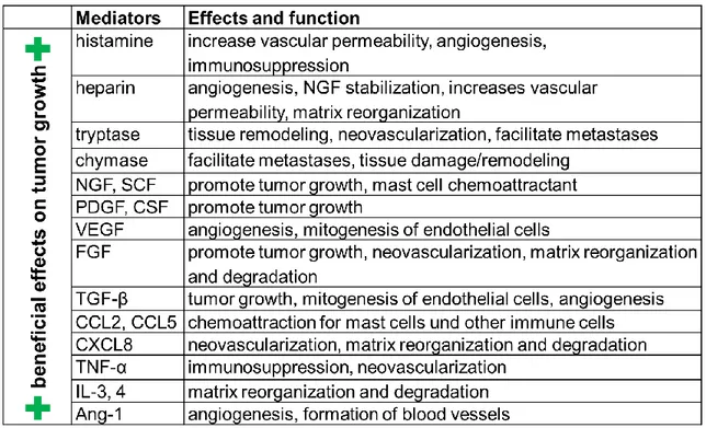
2. Third, de novo synthesized cytokines and chemokines are produced. In tumor biology, these mediators can either have "beneficial" effects on tumor growth, causing progression and spread of the tumor, or "detrimental" effects, limiting the growth of the tumor. To mention just some of these mediators, histamine and growth factors like NGF, SCF, PDGF and TGF-β promote tumor development, whereas heparin, VEGF, FGF and CXCL8 (IL-8) are known to induce angiogenesis, which is an essential feature for tumorigenesis. Some of these growth factors can also serve as mast cell chemoattractants in a feedback loop. On the other hand, mast cell-produced cytokines may also participate in anti-tumor responses and tumor rejection by promoting inflammation (IL-1, IL-2, IL-6, IL- 10, TNF-α, IFN-γ) or by inducing tumor cell apoptosis (IL-4, IL-13). Table 1 and 2 summarize mast cell mediators and their functions concerning tumor growth.
However, investigations on the role of specific mast cell products in tumor
development, especially concerning the balance between pro- and anti-
inflammatory mechanisms, are still at the very beginning.
Introduction
Table 1. Mediators produced by mast cells exerting beneficial effects on tumor growth.
Table 2. Mediators produced by mast cells exerting detrimental effects on tumor growth.
Introduction
17
1.5. Mast cells in tumors
In a large variety of cancers, immune cells of the tumor microenvironment have been demonstrated to play crucial roles in tumor biology.
37,40In particular, tumor-associated macrophages, myeloid-derived suppressor cells (MDSC) and T regulatory cells (T
regs) participate in the development of tumors by regulating various hallmarks of cancer, like proliferation, angiogenesis, immunomodulation and tissue remodeling.
The accumulation of mast cells especially in the periphery of tumors was first reported by Westphal in 1891.
41But also recent studies demonstrated impressively that mast cells serve as critical regulators of the tumor microenvironment.
41-43Many types of solid cancers and hematologic malignancies show an increased number of mast cells.
44-55Mast cell counts often also correlate with tumor stage, prognosis and invasiveness, suggesting a protumorigenic role of mast cells in these malignancies.
44-51More precisely, increased numbers of mast cells have been reported to correlate with poor prognosis in lymphoid neoplasms such as Hodgkin´s lymphoma
47, B-cell non- Hodgkin’s lymphoma
50and multiple myeloma
56. Similar data have been obtained in solid cancers, such as pancreatic cancer
57, hepatocarcinoma and cholangiocarcinoma
49, prostate cancer
44, neurofibroma
48and melanoma
46,58. In summary, the previous studies demonstrated that: (1) Mast cells participate in creating a microenvironment that promotes tumor growth. (2) Mast cell- deficient mice show an impaired carcinogenesis and reduced tumor neo- angiogenesis.
59,60(3) Pharmacological inhibition of mast cell degranulation can inhibit tumor growth.
60In contrast to the protumorigenic role of mast cells described for a large number
of tumors, mast cells were also reported to exert antitumorigenic effects in
certain malignancies, for example by supporting cancer rejection.
52-54In
colorectal cancer, infiltrates of mast cells have been associated with lower rates
of lymph node metastasis and distant metastasis.
52Also, in breast cancer,
stromal mast cells were found to correlate with a favorable prognosis.
53,54Additionally, mast cells even showed plasticity by exerting both detrimental and
beneficial effects in the same cancer entity depending on the tumor stage, as
for example elegantly demonstrated for prostate adenocarcinoma.
44Introduction
It has been speculated that mast cells remodel the tumor microenvironment through release of cytokines and chemokines upon degranulation as well as through interactions with other cell types.
61Aggregation of eosinophils together with mast cells has also been described to correlate with poor prognosis, for example in Hodgkin´s lymphoma.
62Apart from the interaction of mast cells with other immune cells of the tumor microenvironment, it is also conceivable that mast cells modulate other stromal structures such as blood vessels or extracellular matrix. As an example, mast cells may indirectly participate in tumor development by producing VEGF that in turn stimulates tumor growth through enhanced tumor neo-angiogenesis. Supporting this hypothesis, in lung cancer
63and squamous cell carcinomas of the esophagus
64and cervix
65, mast cell infiltrates correlated with both microvessel density and tumor progression.
The complex functions of mast cells in tumor biology may in part be related to the differential release of mediators, but specific effects on tumor growth are still poorly understood.
1.6. Primary cutaneous lymphoma
Cutaneous lymphomas are clonal lymphoid neoplasms, which belong to the group of extranodal non-Hodgkin´s lymphomas (NHL).
66-69They usually reflect a clonal proliferation of lymphocytes in the skin. The annual incidence is estimated at 1/100.000.
67By definition one has to distinguish between secondary cutaneous lymphomas, as manifestations of nodal or other extranodal lymphomas also involving the skin, and primary cutaneous lymphomas (PCL).
As an example for a skin tumor in which the role of mast cells had not been
described yet, we investigated primary cutaneous lymphomas (PCL). In PCL,
functional interactions between neoplastic cells and their microenvironment are
largely unknown. PCL are a clinical and histological distinct group of T- or B-cell
malignancies that originate in the skin and usually remain localized to the skin
for a longer period of time. In Europe, cutaneous T-cell lymphomas (CTCL)
constitute about 75-80% of all PCLs, cutaneous B-cell lymphomas (CBCL) 20-
25%, but different distributions have been observed in other parts of the world.
66Diagnosis and classification of primary cutaneous lymphomas into CTCL and
CBCL and their subtypes is based on a combination of clinical, histological and
Introduction
19
immunophenotypical data. The most common subtypes of CTCL include mycosis fungoides (MF), Sézary syndrome (SS) and lymphomatoid papulosis (LP), whereas CBCL are subdivided into primary cutaneous marginal zone B- cell lymphoma (MZL), primary cutaneous follicle center lymphoma (CFBCL) and primary cutaneous diffuse large B-cell lymphoma (BCL).
Clinical manifestation and prognosis of PCL are highly variable and depend on the subtype and stage of the disease.
70,71For example, early stages of MF, characterized by eczematous skin lesions resulting from inflammation associated with proliferation of malignant cells in the epidermis, typically run an indolent course.
70More advanced stages of MF show intradermal tumors, which arise from poorly differentiated subclones of malignant cells that spread into deeper layers of the skin and later into peripheral blood, lymph nodes and internal organs. In these advanced stages, conventional therapies can achieve only short-term clinical responses and median survival is less than three years.
The prognosis is even worse in patients with SS, the leukemic variant of CTCL.
In contrast, most patients with CBCL show stable nodular lesions associated with an indolent course.
72Involvement of immune cells is suggested by elevated levels of inflammatory
cytokines in plasma and skin sections of CTCL patients, such as IL-1β, IL-7, IL-
15, IL-17, IL-18 and IL-23.
73-75Recent investigations reported on significant
expression of IL-17 in cells and tissues from patients with MF and SS.
73,74IL-17
is known to participate in proinflammatory responses by initiating the production
of various cytokines (IL-6, G-CSF, GM-CSF, IL-1β, TGF-β1, TNF), chemokines
(CXCL8 and CCL2), and prostaglandins (PGE
2) from other cell types such as
epithelial cells, keratinocytes, endothelial cells, fibroblasts, macrophages and
potentially mast cells. In CTCL, it has been shown that IL-2 and IL-15 are able
to up-regulate the expression of IL-17 in neoplastic T-cells.
74CTCL lesions are
known to exhibit increased angiogenesis and several angiogenic and
inflammatory proteins that are induced by IL-17 (for example TNF, CCL20,
MMP-9, COX-2, VEGF and CXCL8) are also expressed in CTCL lesions.
76-78Interestingly, a recent study using a mouse model of CTCL reported on
increased numbers of macrophages and neutrophils and showed that tumor
growth is reduced upon depletion of macrophages.
79Aims
2. Aims
Mast cells infiltrate many types of cancers. Recent studies demonstrated that mast cells actively participate in modulating tumor growth, either by directly affecting growth and invasiveness of tumor cells or by indirectly regulating the tumor microenvironment, e.g. by interacting with other immune cells, inducing neo-angiogenesis or degrading extracellular matrix, which then promotes tumor growth. However, the precise role of mast cells in tumors remains largely unknown. Therefore, the present study aims to clarify specific functions of mast cells in tumor biology addressing the following four goals:
1. To explore numbers of mast cells and their correlation with tumor growth, we decided to initially investigate mast cell counts in primary cutaneous lymphoma (PCL) as an example of a tumor entity, in which mast cells have not been addressed to date. More specifically, we will analyze the number and distribution of mast cells in different subtypes of PCL compared to normal skin and inflammatory cutaneous diseases, such as psoriasis and atopic dermatitis. In addition, hypothesizing that mast cells are activated in the tumor microenvironment of PCL, we will explore mast cell degranulation in PCL. Furthermore, we will correlate mast cell numbers and degranulation with the course and prognosis of different PCL subtypes.
2. To further elucidate the contribution of mast cells to tumor growth, we will
use new transgenic mouse models with inducible or constitutive selective
deficiency of connective tissue mast cells and analyze tumorigenesis in
these models compared to control mice. Here, we will investigate different
subcutaneously injected tumors, namely the murine lymphoma cell line EL4,
the pancreatic adenocarcinoma cell line Pan02 and the Lewis lung
carcinoma cell line LLC, as well as chemically induced carcinogenesis.
Aims
21
3. To clarify the effect of specific mast cell products on tumor growth, we will first analyze whether supernatant of human or murine mast cells stimulates cytokine production and proliferation of primary PCL cells and human or murine lymphoma cell lines. Next, we will investigate levels of different cytokines in supernatants of human and murine mast cells using a cytometric bead array approach.
4. To address the role of mast cells in tumor neo-angiogenesis, we will
analyze vessel density in PCL and in the murine EL4 tumor model. We will
also investigate growth of EL4 tumors in mast cell-specific VEGF knockout
mice.
Material and Methods
3. Material and Methods
3.1. Material
3.1.1. Chemicals and solutions
Standard chemicals and solutions were purchased from:
Biochrom AG, Berlin, Germany Brenntag, Mülheim, Germany
Gibco/Invitrogen, Karlsruhe, Germany Merck, Darmstadt, Germany
Roth, Karlsruhe, Germany Serva, Heidelberg, Germany
Sigma-Aldrich, Taufkirchen, Germany StarLab, Hamburg, Germany
Product Company
Acetone Roth
Agarose StarPure StarLab
Calcium ionophore A23187 Sigma-Aldrich
Cromolyn sodium salt Sigma-Aldrich
Dimethyl sulphoxide (DMSO) Sigma-Aldrich
Disodium hydrogen phosphate (Na
2HPO
4) Merck
Dulbecco´s Modified Eagle Medium (DMEM, 1x) Gibco/Invitrogen EDTA (1% (w/v) in PBS, cell culture) Biochrom AG
Ethanol (≥ 99.5%) Roth
Ethylenediaminetetraacetic acid (EDTA) Roth
Fetal bovine serum (FBS) Biochrom AG
Hanks´ Balanced Salt Solution (HBSS) Sigma-Aldrich
Hydrochloric acid (HCl) Roth
Iscove´s Modified Dulbecco´s Medium (IMDM, 1x) Gibco/Invitrogen
Isopropanol Brenntag
L-glutamine (200 mM) Biochrom AG
Non-essential amino acid solution (NEA, 100x) Sigma-Aldrich Penicillin/Streptomycin (10.000 U/ml / 10.000 µg/ml) Biochrom AG
Potassium chloride (KCl) Roth
Potassium dihydrogen phosphate (KH
2PO
4) Roth
Roswell Park Memorial Institute (RPMI) medium 1640 (1x) Gibco/Invitrogen
Material and Methods
23
Roti®-Histofix (4%) Roth
Sodium acetate (C
2H
3NaO
2) Merck
Sodium chloride (NaCl) Roth
Sodium dodecyl sulfate (SDS) Serva
Sodium pyruvate (100 mM) Gibco/Invitrogen
Tris (HOCH
2)
3CNH
2) Roth
Trypsin (2.5%, 10x) Gibco/Invitrogen
β-Mercaptoethanol (50 mM) Gibco/Invitrogen
Table 3. Frequently used chemicals and solutions.
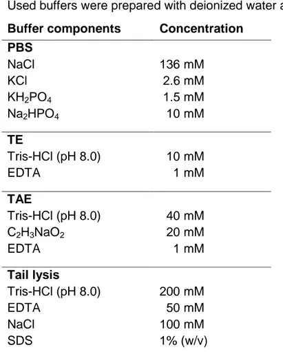
3.1.2. Buffers
Used buffers were prepared with deionized water at room temperature.
Buffer components Concentration PBS
NaCl KCl KH
2PO
4Na
2HPO
4136 mM 2.6 mM 1.5 mM
010 mMTE
Tris-HCl (pH 8.0) EDTA
010 mM 001 mM
TAE
Tris-HCl (pH 8.0) C
2H
3NaO
2EDTA
040 mM 020 mM 001 mM
Tail lysis
Tris-HCl (pH 8.0) EDTA
NaCl SDS
200 mM
050 mM100 mM 1% (w/v)
Table 4. Chemical composition of frequently used buffers.
Material and Methods
3.1.3. Single-stranded oligonucleotides
All single-stranded oligonucleotides were purchased lyophilized, in HPLC quality, from metabion international AG, Martinsried, Germany. 100
µM stocksolutions were prepared according to the manufacturer´s instructions in ddH
2O.
For use in PCR, stocks were diluted 1:10.
3.1.3.1. Genotyping
Name Sequence Bands
Mcpt5-Cre Mcpt5-CreUP CreSeq1b-DO Mcpt5-Ex1-DO3
5’ACAGTGGTATTCCCGGGGAGTGT3’
5’GTCAGTGCGTTCAAAGGCCA3’
5’GCTTTGGTGCTGGAACCCAGGA3’
WT 224 bp Cre+ 224 bp 554 bp
iDTR Mutant Common WT Rev
5’CATCAAGGAAACCCTGGACTACTG3’
5’AAAGTCGCTCTGAGTTGTTAT3’
5’GGAGCGGGAGAAATGGATATG3’
WT 603 bp Homo 242 bp Hetero 242 bp 603 bp
R-DTA
Fwd Rev
5’AAAGTCGCTCTGAGTTGTTAT3’
5’AAGAACGGAGCCGGTTGGCG3’
WT no R-DTA+ 592 bp
VEGF VEGF Rev VEGF Fwd
5’TCCGTACGACGCATTTCTAG3’
5’CCTGGCCCTCAAGTACACCTT3’
WT 100 bp Homo 150 bp Hetero 100 bp 150 bp Table 5. Primer used for animal genotyping.
3.1.3.2. Real-time PCR
Name Sequence Reference
IL-6 IL-6 Fwd IL-6 Rev
5’GGTACATCCTCGACGGCATCTC3’
5’GTTGGGTCAGGGGTGGTTATTG3’
80
TGF-β1 TGF b1 Fwd TGF b1 Rev
5’CAGAAATACAGCAACAATTCCTGG3’
5’TTGCAGTGTGTTATCCGTGCTGTC3’
81
GAPDH
GAPDH sense GAPDH antisense
5’CGGAGTCAACGGATTTGGTCGTAT3’
5’AGCCTTCTCCATGGTGGTGAAGAC3’
82
Table 6. Primer used for quantitative real-time PCR.
Material and Methods
25
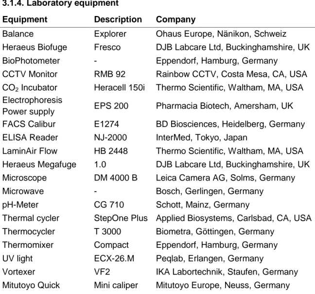
3.1.4. Laboratory equipment
Equipment Description Company
Balance Explorer Ohaus Europe, Nänikon, Schweiz Heraeus Biofuge Fresco DJB Labcare Ltd, Buckinghamshire, UK
BioPhotometer - Eppendorf, Hamburg, Germany
CCTV Monitor RMB 92 Rainbow CCTV, Costa Mesa, CA, USA CO
2Incubator Heracell 150i Thermo Scientific, Waltham, MA, USA Electrophoresis
Power supply EPS 200 Pharmacia Biotech, Amersham, UK FACS Calibur E1274 BD Biosciences, Heidelberg, Germany ELISA Reader NJ-2000 InterMed, Tokyo, Japan
LaminAir Flow HB 2448 Thermo Scientific, Waltham, MA, USA Heraeus Megafuge 1.0 DJB Labcare Ltd, Buckinghamshire, UK Microscope DM 4000 B Leica Camera AG, Solms, Germany
Microwave - Bosch, Gerlingen, Germany
pH-Meter CG 710 Schott, Mainz, Germany
Thermal cycler StepOne Plus Applied Biosystems, Carlsbad, CA, USA Thermocycler T 3000 Biometra, Göttingen, Germany
Thermomixer Compact Eppendorf, Hamburg, Germany UV light ECX-26.M Peqlab, Erlangen, Germany
Vortexer VF2 IKA Labortechnik, Staufen, Germany
Mitutoyo Quick Mini caliper Mitutoyo Europe, Neuss, Germany
Table 7. Frequently used laboratory equipment.
3.1.5. Software
Software Version Company
Adobe Photoshop 7.0 Adobe Systems Inc., Dublin, Ireland CellQuest Pro 5.2.1 BD Biosciences, Heidelberg, Germany Diskus 4.50.1638 Diskus, Königswinter, Germany
EndNote X4 Bld 4845 Thomson Reuters, New York, NY, USA FCAP
TMArray 1.0 BD Biosciences, Heidelberg, Germany GraphPad Prism 5.01 GraphPad Inc., San Diego, CA, USA Microsoft Excel 2010 Microsoft, Redmond, WA, USA Pannoramic Viewer 1.15 3DHistech Kft., Budapest, Hungary StepOne Software 2.1 Applied Biosystems, Carlsbad, CA, USA
Table 8. Software used for data collection and analysis.
Material and Methods
3.2. Methods
3.2.1. Collection of cutaneous biopsies from patients with primary cutaneous lymphoma
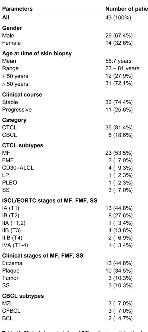
Cutaneous biopsies were obtained from 43 patients with PCL for diagnostic purposes and kindly provided by Dr. Max Schlaak, PD Dr. Peter Kurschat and Prof. Dr. Dr. Cornelia Mauch within the framework of the Z2 project of the SFB829 at the University of Cologne. All patients had attended the Hauttumorzentrum of the Department of Dermatology, University Hospital of Cologne between 1995 and 2010 (heads of the Hauttumorzentrum: Prof. Dr. Dr.
Cornelia Mauch, PD Dr. Peter Kurschat; physicians: Dr. Max Schlaak, Prof. Dr.
Christine Neumann). The diagnosis of PCL and assignment to disease
categories and subtypes according to the WHO-EORTC classification were
performed by Dr. Max Schlaak and PD Dr. Peter Kurschat and were based on
established criteria.
66,68In addition, assignment to subtypes of PCL was
confirmed by a histopathological reference center for PCL (Kempf and Pfaltz,
Laboratory for Histological Diagnostics, Zurich, Switzerland). Out of the 43
patients, 35 patients (81.4%) were diagnosed with CTCL and 8 patients (18.6%)
with CBCL. Detailed clinical characteristics of all patients are listed in Table 10
in the results section. For control, 12 biopsies from subjects with normal skin
were kindly provided by Dr. Lukas Heukamp and Prof. Dr. Reinhard Büttner
(Institute of Pathology, University Hospital of Cologne) and 24 biopsies from
patients with inflammatory cutaneous diseases, namely 8 patients with
psoriasis, 8 patients with atopic dermatitis and 8 patients with
pseudolymphoma, obtained for diagnostic purposes were used. All procedures
were approved by the Institutional Ethics Committee of the University of
Cologne, Cologne, Germany, under written, informed patient consent and
adherence to the declaration of Helsinki (AZ 08-144). Patients with CTCL were
grouped in cooperation with Dr. Max Schlaak according to the ISCL/EORTC
classification.
71,83Additionally, to also reflect the clinical course of PCL, patients
with CTCL were divided in cooperation with Dr. Max Schlaak into those with
stable or progressive disease, defining stable disease as maximally two topical
treatments (e.g., UV irradiation, topical corticosteroids) and progressive disease
as three or more topical and systemic treatments (e.g,. UV irradiation, topical
corticosteroids, surgery, systemic therapy such as bexarotene or interferon
Material and Methods
27
alpha). Progression-free survival was defined in cooperation with Dr. Sebastian Theurich and PD Dr. Dr. Michael von Bergwelt-Baildon (Department I of Internal Medicine, University Hospital of Cologne) as time span from initial diagnosis until first change of systemic therapy due to disease progression and recorded over 30 years. For Kaplan-Meier curves, the event criterion was defined as
>100 mast cells/mm
2.
3.2.2. Histology
Staining procedures were performed in cooperation with Dr. Lukas Heukamp and Prof. Dr. Reinhard Büttner (Institute of Pathology, University Hospital of Cologne) within the framework of the Z1 project of the SFB832 at the University of Cologne.
3.2.2.1. Human samples
Histological analysis of mast cells in human PCL was initiated by Dr. Max Schlaak and PD Dr. Karin Hartmann. These initial studies suggested an increase of mast cells in PCL. Therefore, sections of paraffin-embedded cutaneous biopsies from 43 patients with PCL and control subjects with normal skin and inflammatory cutaneous diseases were systematically evaluated by immunohistochemistry with antibodies against mast cell tryptase (pretreatment with 0.1% protease, staining with 1:3.000 dilution of antibody Dako 7052, Dako, Hamburg, Germany) and CD117 (Kit; pretreatment with citrate buffer pH 6.0, staining with 1:100 dilution of antibody Dako A4502, Dako, Hamburg, Germany). Evaluation of skin sections was performed by counting the number of tryptase- and CD117-positive mast cells under a Leica microscope with Diskus software at 200x magnification in 5 high power fields. Counts were used to calculate the mean number of mast cells per mm
2. A comparison between stainings with antibodies against tryptase and CD117 showed highly consistent results. Results were, therefore, only expressed as tryptase-positive mast cells.
For control, sections were also counted by Silke Leja (MTA, Department of Dermatology), as a second independent reviewer.
To grade degranulation of mast cells, skin sections were assessed at 200x
magnification and mast cells were semiquantitatively classified into 3 groups of
not degranulated, moderately degranulated or extensively degranulated mast
Material and Methods
cells.
34Sections were also stained by hematoxylin/eosin (H&E) following standard protocols in a routine histology laboratory.
For analysis of microvessel density, sections of paraffin-embedded cutaneous biopsies from patients with MF and control patients with inflammatory cutaneous diseases were evaluated by immunohistochemistry with antibodies against CD31 (pretreatment with citrate buffer pH 6.0, staining with 1:500 dilution of antibody Dako M0823, Dako, Hamburg, Germany) and CD34 (pretreatment with citrate buffer pH 6.0, staining with 1:2000 dilution of antibody Cell Marque 134M-1, Cell Marque, Sierra College Blvd., Rocklin, CA, USA).
Evaluation of skin sections was performed by recording the distribution of CD31- and CD34-positive microvessels with a minimal lumen size of 50 µm
2and by counting their number under a Leica microscope with Diskus software at 100x magnification in 5 high power fields. Counts were used to calculate the mean number of microvessels per mm
2. A comparison between stainings with antibodies against CD31 and CD34 showed highly consistent results. Results were, therefore, only expressed as CD31-positive microvessels.
3.2.2.2. Murine samples
In order to detect mast cells in the different mouse models (as described below), punch biopsies of back skin were fixed in 4% formalin (Roti®-Histofix) over night and embedded in paraffin. Sections were stained by Giemsa stain and toluidine blue following standard protocols in a routine histology laboratory.
For evaluation of microvessel density in the different mouse models (as
described below), punch biopsies of back skin were embedded in paraffin and
CD31 staining (pretreatment with citrate buffer pH 6.0, staining with 1:50 dilution
of antibody Histonova DIA-310, Dianova, Hamburg, Germany) was performed.
Material and Methods
29
3.2.3. In vitro experiments 3.2.3.1. Cell isolation
To obtain primary Sézary cells, CD4+ T-cells from a patient with Sézary syndrome were isolated from freshly collected blood, kindly provided by Dr. Max Schlaak, using the Macs Whole Blood Column Kit for CD4+ T-cells (Miltenyi Biotech, Bergisch Gladbach, Germany) and generated by culturing as described in section 3.2.3.2. Additionally, CD4+ T-cells from the blood of healthy donors, kindly provided by PD Dr. Karin Hartmann, were isolated as controls.
Primary murine bone marrow-derived mast cells (BMMC) were isolated under sterile conditions from the femoral lavage of WT C57BL/6 mice.
303.2.3.2. Cell culture
All cell cultures were maintained at 37°C in 5% CO
2in a humidified atmosphere.
The human CTCL cell line Mac2B, originally derived from a patient with CD30+ALCL
84, was kindly provided by Dr. Marshall Kadin (Department of Dermatology and Skin Surgery, Boston University School of Medicine, Roger Williams Medical Center, Providence, USA) and maintained in RPMI 1640 medium containing 20% FBS, 2 mM L-glutamine, 100 U/ml penicillin and 100 μg/ml streptomycin.
Human CTCL cell lines MyLa, derived from a patient with MF
85, and SeAx, derived from a patient with SS
86, as well as the human B-cell lymphoma cell line BJAB, derived from African Burkitt-like lymphoma
87, were kindly provided by Dr.
Maria Karpova (Department of Dermatology, University Hospital Zurich, Zurich, Switzerland). All three cell lines were cultured in RPMI 1640 medium containing 10% FBS, 2 mM L-glutamine, 1 mM sodium pyruvate, 100 U/ml penicillin and 100 μg/ml streptomycin.
The cell line Jurkat (originally called JM), a pseudodiploid human cell line that originated from a patient with acute T-cell leukemia
88, was cultured in RPMI 1640 medium containing 10% FBS, 4 mM L-glutamine, 100 U/ml penicillin and 100 μg/ml streptomycin.
Primary Sézary cells were cultured as described
89and kept in RPMI 1640
medium containing 20% FBS, 2 mM L-glutamine, 100 U/ml penicillin and 100
μg/ml streptomycin. Isolated CD4+ T-cells were maintained in RPMI 1640
Material and Methods