Novel tools for mast cell research:
Mast cell-specific Cre-mediated gene inactivation and inducible ablation of mast cells in vivo
Inaugural-Dissertation
zur
Erlangung des Doktorgrades
der Mathematisch-Naturwissenschaftlichen Fakultät
der Universität zu Köln
vorgelegt von
Julia Scholten aus Duisburg
Köln, 2009
Berichterstatter: Prof. Dr. Dr. Thomas Krieg Prof. Dr. Manolis Pasparakis
Tag der mündlichen Prüfung: 5. Februar 2009
Mast cells are well known effector cells in allergic disorders. In the recent years, however, mast cells were demonstrated to play pivotal roles in initiating and modulating innate and adaptive immune responses. The animal models available until today for the investigation of mast cell functions are primarily the Kit-mutant KitW/Wv and KitWsh/Wsh mouse strains.
These mice are devoid of mast cells and can be reconstituted with mast cells from gene deficient mice. The phenotypic abnormalities, apart from mast cell deficiency, e.g. the profound perturbation of the hematopoietic system, however, limit the utility of these strains. Aiming at new models of mast cell-specific gene inactivation in vivo and of mast cell-deficiency, the Cre/loxP system was applied to allow Cre-mediated mutagenesis selectively in mast cells. BAC (bacterial artificial chromosome) transgenic animals were successfully generated that display Cre expression under the control of either the Mcpt5 or Mcpt6 promoter. Cre-mediated recombination as assessed by reporter gene expression in Mcpt5-Cre or Mcpt6-Cre R26R-EYFP double positive mice was found to be highly efficient in peritoneal and skin mast cells, but was not found in hematopoietic cell populations other than mast cells. Thus, the new mast cell-specific Cre transgenic mouse lines will provide useful tools for the investigation of mast cell-specific functions of individual genes.
Furthermore, the Mcpt5-Cre strain was used to establish a new model for inducible ablation of mast cells in adult mice. By crossing this line to the iDTR strain, which expresses the high affinity diphtheria toxin receptor (DTR) after Cre-mediated deletion of a stop element, mast cells were rendered diphtheria toxin (DT) sensitive. DT treatment of Mcpt5-Cre iDTR double positive mice resulted in successful ablation of connective tissue type mast cells (CTMC), but not of mucosal mast cells (MMC). The depletion of CTMC resulted in significant reduction of a passive, systemic anaphylactic response demonstrating that this system for mast cell ablation represents a useful model for the investigation of mast cell functions during immune responses. Interestingly, the repopulation of the peritoneal cavity and the skin with mast cells after DT-mediated mast cell depletion followed a very slow kinetic with a noteworthy recurrence of peritoneal mast cells three months after DT treatment. Skin mast cells do not return significantly within a period of four months. Thus, the long persistence of the mast cell-depleted state allows the application of this system for long-term experiments and should provide new insights into the mechanisms of mast cell homeostasis and recruitment.
Mastzellen sind vor allem bekannt als Effektorzellen allergischer Erkrankungen. In den letzten Jahren wurde jedoch gezeigt, dass Mastzellen entscheidend zu der Mobilisierung und Modellierung von angeborenen und adaptiven Immunantworten beitragen. Die derzeitig verfügbaren Modelle für die in vivo-Analyse von Mastzellfunktionen, sind vorwiegend die Kit-mutierten, Mastzell-defizienten KitW/Wv und KitWsh/Wsh Mäuse, die mit in vitro differenzierten Mastzellen von Gen-defizienten Mäusen rekonstituiert werden können. Phänotypische Abnormitäten zusätzlich zu der Mastzell-Defizienz, z. B.
Störungen des hämatopoietischen Systems, erschweren jedoch das Experimentieren und die Interpretation der Daten. Aus diesem Grund wurde hier das Cre/loxP-System herangezogen, um in vivo Gene gezielt in Mastzellen zu inaktivieren und um neue Modelle für Mastzell-Defizienz zu generieren. BAC (bacterial artificial chromosome) transgene Mauslinien wurden erfolgreich hergestellt, die die Cre-Rekombinase unter der Kontrolle des Mcpt5- bzw. Mcpt6-Promotors ausprägen. In Mcpt5-Cre bzw. Mcpt6-Cre R26R-EYFP doppelt positiven Mäusen konnte eine höchst effiziente, Cre-mediierte Rekombination in Mastzellen der Peritonealhöhle und der Haut, jedoch nicht in anderen Zellpopulationen, nachgewiesen werden. Die neuen Mastzell-spezifischen Cre- transgenen Mauslinien werden daher in Zukunft die Untersuchung Mastzell-spezifischer Funktionen einzelner Gene erheblich erleichtern.
Des Weiteren wurde die Mcpt5-Cre Linie für die Etablierung eines neuen Models für induzierbare Ablation von Mastzellen in vivo verwendet. Die Kreuzung dieser Linie mit dem iDTR Mausstamm, der den hoch affinen Diphtherietoxin Rezeptor (DTR) nach Cre- mediierter Deletion eines Stop-Elements ausprägt, führt zu einer Sensibilisierung von Mastzellen gegenüber dem Diphtherietoxin (DT) in Mcpt5-Cre iDTR doppelt positiven Tieren. Die Behandlung mit DT resultierte in einer effizienten Depletion von Bindegewebs- mastzellen (CTMC), jedoch nicht von mukosalen Mastzellen (MMC). Die Ablation der CTMC reichte aus, eine passive, systemische anaphylaktische Reaktion signifikant zu reduzieren, was demonstriert, dass das System der induzierbaren Mastzell-Ablation ein geeignetes Modell für die Erforschung von Mastzell-Funktionen darstellt. Interessanter- weise folgte die Repopulation der Bauchhöhle und der Haut mit Mastzellen einer höchst langsamen Kinetik. Ein signifikanter Anstieg der Mastzell-Zahlen in der Bauchhöhle zeigte sich erst drei Monate nach Depletion. Eine deutliche Rückkehr der Hautmastzellen war auch nach vier Monaten noch nicht zu beobachten. Folglich erlaubt die langfristige Depletion der Mastzellen die Anwendung dieses Systems in Langzeitexperimenten und kann, des Weiteren, neue Einblicke in die Mechanismen der Homöostase und Rekrutierung von Mastzellpopulationen gewähren.
Table of contents
1 Introduction ... 5
1.1 Mast cells ... 5
1.1.1 Mast cell development and heterogeneity ... 5
1.1.2 Mast cell granules and proteases ... 7
1.1.3 From mast cell activation to effector functions ... 9
1.1.4 Mast cell associated disorders ... 12
1.1.5 Models for the investigation of mast cells ... 13
1.2 Generation of mouse models ... 16
1.2.1 Classical gene “Knock out” ... 16
1.2.2 Conditional gene targeting ... 16
1.2.2.1 Site specific recombination systems ... 16
1.2.2.2 Applications of the Cre/loxP system ... 17
1.2.3 BAC transgene technology ... 18
1.3 Objectives ... 20
2 Results ... 21
2.1 Generation and analysis of mast cell-specific Cre transgenic mice ... 21
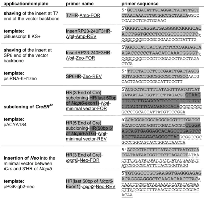
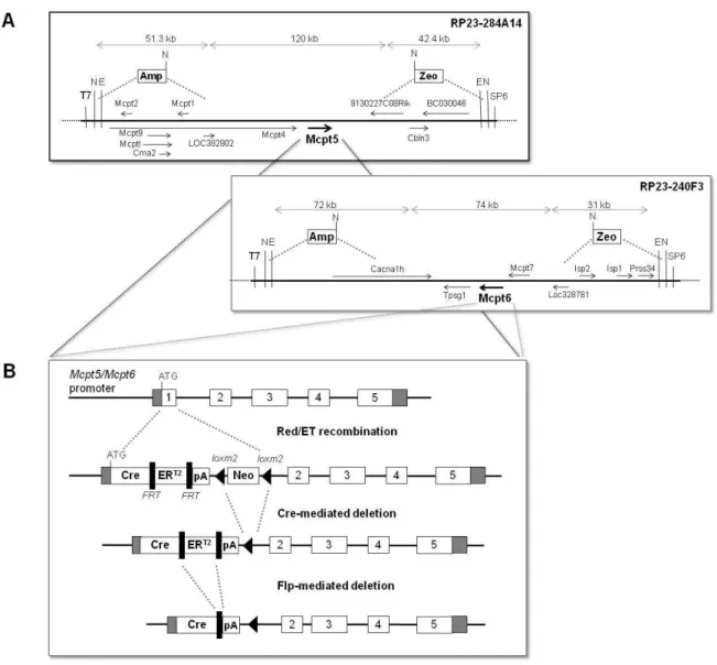
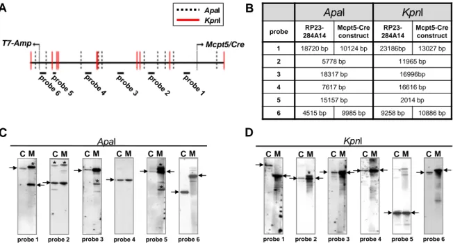
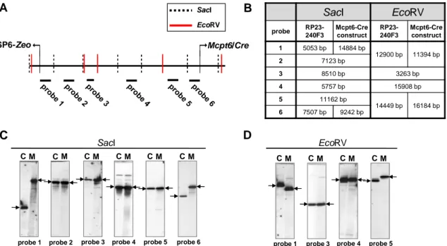
2.1.1 Generation of the Mcpt5-Cre(ERT2) and Mcpt6-Cre gene constructs ... 21
2.1.1.1 Shortening (“shaving”) of the BAC inserts ... 25
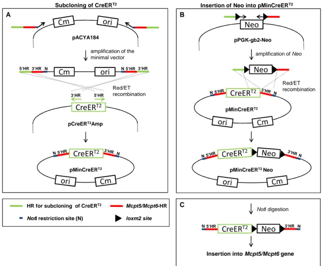
2.1.1.2 Assembly of CreERT2-Neo and insertion into the Mcpt5 and Mcpt6 genes ... 25
2.1.1.3 Test for precision and fidelity of BAC modification ... 26
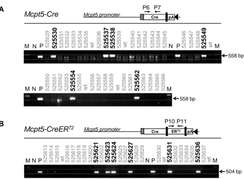
2.1.2 Generation of transgenic mice ... 29
2.1.3 Screening of Mcpt5-Cre founder lines for Cre-mediated recombination ... 31
2.1.4 Detailed analysis of A-Mcpt5-Cre and X-Mcpt5-Cre transgenic mice ... 32
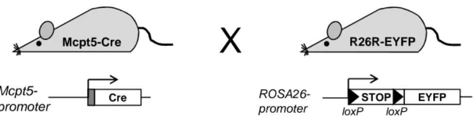
2.1.4.1 Efficient Cre-mediated recombination in mast cells ... 32
2.1.4.2 Investigation of mast cell-specificity of Cre-mediated recombination ... 34
2.1.4.3 Exclusion of Cre-mediated genotoxicity in Mcpt5-Cre mice ... 37
2.1.5 Screening of Mcpt6-Cre transgenic mice for Cre-mediated recombination ... 38
2.1.6 Test for Cre-mediated EYFP expression in BMMCs ... 40
2.1.7 Screening of Mcpt5-CreERT2 transgenic mice for Cre-mediated recombination .... 42
2.2 A new model for inducible ablation of mast cells in adult mice ... 43
2.2.1 Ablation of peritoneal and skin mast cells ... 43
2.2.2 Depletion of gastrointestinal mast cells ... 49
2.2.3 Effect of mast cell depletion on passive systemic anaphylaxis ... 51
2.2.4 Repopulation of mast cells after DT-induced ablation ... 52
3 Discussion ... 55
3.1 Novel transgenic strains for mast cell-specific Cre-mediated mutagenesis in vivo ... 56
3.1.1 Design and construction of the transgene ... 56
3.1.2 The Mcpt5 and Mcpt6 promoters allow efficient Cre expression in mast cells ... 57
3.1.3 Cre-mediated deletion is restricted to mast cells ... 59
3.1.4 Absence of Cre-mediated genotoxicity in Mcpt5-Cre mice ... 60
3.1.5 Attempt to generate mice with inducible, mast cell-specific Cre activity ... 61
3.2 A new model for inducible ablation of mast cells in vivo ... 61
3.2.1 Efficient ablation of connective tissue mast cells in adult mice ... 62
3.2.2 Reduced anaphylactic response in mast cell-depleted animals ... 63
3.2.3 Slow recovery of mast cell populations after induced ablation ... 64
4 Materials and Methods ... 67
4.1 Chemicals and enzymes ... 67
4.2 Bacterial cell culture ... 67
4.2.1 LB (Luria Broth) medium and agar ... 67
4.2.2 Antibiotics ... 67
4.2.3 Escherichia coli (E. coli) strains ... 67
4.3 Oligonucleotides ... 68
4.4 Standard molecular biology methods ... 69
4.4.1 Polymerase chain reaction and colony PCR ... 69
4.4.2 DNA sequencing ... 70
4.4.3 Gel purification of DNA fragments ... 70
4.4.4 Small and intermediate scale preparation of plasmid DNA ... 70
4.4.5 Quantification of DNA ... 70
4.4.6 Restriction digest ... 71
4.4.7 Southern blot ... 71
4.4.7.1 Buffers ... 71
4.4.7.2 Blotting ... 71
4.4.7.3 Probe labeling ... 72
4.4.7.4 Hybridization and detection ... 72
4.5 Molecular biology methods for modification of BAC vectors ... 72
4.5.1 BAC clones ... 72
4.5.2 BAC recombineering ... 73
4.5.2.1 Templates and sources of DNA cassettes ... 74
4.5.2.2 Primer design and generation of the PCR product containing the HRs ... 74
4.5.2.3 BAC modification protocol ... 75
4.5.3 Cre- and Flp-mediated recombination in E. coli ... 76
4.5.4 Small scale preparation of BAC DNA... 77
4.5.5 Purification of BAC DNA for pronucleus injection ... 77
4.5.5.1 Buffers and reagents ... 77
4.5.5.2 Maxi preparation of BAC DNA ... 78
4.5.5.3 Separation of the BAC insert from the vector backbone ... 78
4.5.5.4 Electroelution ... 79
4.5.5.5 Drop dialysis ... 80
4.5.5.6 Pulsed-field gel electrophoresis ... 80
4.5.6 Generation of transgenic mice by pronucleus injection ... 80
4.6 Mice ... 80
4.6.1 Mouse strains ... 80
4.6.2 Genotyping ... 81
4.6.2.1 Isolation of genomic tail DNA ... 81
4.6.2.2 Genotyping protocols ... 81
4.6.3 Procedures of animal experimentation... 82
4.6.3.1 Administration of tamoxifen ... 82
4.6.3.2 Administration of diphtheria toxin ... 82
4.6.3.3 Back skin biopsies ... 82
4.6.3.4 Passive systemic anaphylaxis ... 82
4.7 Cell culture ... 83
4.7.1 Cell culture media and reagents ... 83
4.7.2 Cultivation of BMMCs ... 83
4.7.3 Cultivation of PCMCs ... 84
4.8 Flow cytometry ... 84
4.8.1 Buffers and reagents ... 84
4.8.2 Antibodies for flow cytometry ... 85
4.8.3 FACS staining ... 85
4.8.4 Single cell suspensions ... 85
4.8.4.1 Peritoneal lavage ... 85
4.8.4.2 Skin cell suspension ... 86
4.8.4.3 Spleen cell suspension... 86
4.8.4.4 Enrichment of splenic granulocytes ... 86
4.9 Histology ... 87
5 Abbreviations ... 88
6 References ... 90
1 Introduction
1.1 Mast cells
Mast cells are highly granulated immune cells that reside in tissues and serosal cavities throughout the body but predominantly at inner and outer body surfaces, like the skin, the airways and the intestinal tract and also in close vicinity to blood vessels and nerves (Galli, 1990). At these locations they are well placed to serve as the first line of defense.
Mast cells were first described by Paul Ehrlich in the late 19th century based on the metachromatic staining properties of the large cytoplasmic granules (Ehrlich, 1878). The
“well-fed appearance” led him to designate these cells as mast cells. Today we know that the granule content does not originate from an uptake but from the synthesis and storage of high amounts of proteoglycans, proteases and other mediators.
Mast cells are widely recognized as major effectors in allergic disorders and in responses to parasites. But in the last decade, it turned out that mast cells also serve other important functions. The essential role of mast cells in the immune system is also reflected by the fact that no human individual has been identified until today that lacks mast cells (Stevens and Adachi, 2007).
1.1.1 Mast cell development and heterogeneity
Mast cells originate from multipotent hematopoietic precursors in the bone marrow.
Differentiated to committed mast cell progenitors (MCP), they are released to the blood and circulate until they extravasate into the tissue and serosal cavities where they finally differentiate to the mature tissue resident mast cells (Hallgren and Gurish, 2007;
Rodewald et al., 1996). The spleen has also been demonstrated to be a source of MCPs in the adult mouse (Arinobu et al., 2005; Khalil et al., 1996). The earliest murine committed MCP has been identified in fetal blood at day 15.5 of gestation (Rodewald et al., 1996). In contrast, the direct identification of MCPs in the circulation of adult mice has not been accomplished by reason of their low frequency, i.e. 0.001% of mononuclear cells as determined by mast cell colony formation by blood cells ex vivo (Hallgren and Gurish, 2007). A sequential increase of MCPs first in the bone marrow, then in the blood and finally in the intestine, could be observed in worm infected mice demonstrating the recruitment of mast cells from the bone marrow to the site of infection (Pennock and Grencis, 2004). The development of MCPs to mature mast cells is strongly dependent on the microenvironment and, thus, on the production of growth factors at the site of differentiation. The most important factor for mast cell growth, differentiation, proliferation and survival is the stem cell factor (SCF) that binds to the receptor tyrosine kinase c-Kit and is expressed by stromal cells as a soluble or a membrane bound molecule in various
tissues (Galli et al., 1994; Longley et al., 1997). The Kit gene is a proto-oncogene originally discovered as the cellular homolog (c-Kit) of the feline sarcoma virus oncogene v-Kit (Besmer et al., 1986). The Kit receptor is expressed on hematopoietic progenitor cells but is down regulated upon differentiation in all lineages except mast cells which express Kit and remain SCF responsive throughout their entire differentiation and life span (Metcalfe et al., 1997). The critical role of c-Kit/SCF interaction was demonstrated in mice with spontaneous mutations in the c-Kit locus (also known as white spotting locus (W)), e.g. in KitW/W-v and KitWsh/Wsh mice (Grimbaldeston et al., 2005; Kitamura et al., 1978) or in mice with mutations of the SCF locus, (SI/SId mice (Kitamura and Go, 1979), see also section 1.1.5). In addition to SCF, the mediators Interleukin (IL)-3, IL-4, IL-9, IL-10 and nerve growth factor (NGF) contribute to mast cell growth and differentiation (Okayama and Kawakami, 2006).
Mast cells that reside in various tissues differ in their phenotypes. This heterogeneity among mast cells is due to the diverse microenvironments and the presence of various combinations of the above mentioned growth factors at these sites where the precursor cells differentiate into mature mast cells (Okayama and Kawakami, 2006). Histochemical differences among mast cells described by Enerbäck et al. led to the classification of two distinct mast cell subpopulations in rodents, i.e. connective tissue mast cells (CTMC) and mucosal mast cells (MMC) (Enerbäck et al., 1985). These mast cell phenotypes differ in their localization and secretory granule content. CTMCs are found predominantly in the subepithelial strata of the skin and the peritoneal cavity but also in the submucosal layers of the intestine. Characteristically, CTMCs store abundant heparin sulfate proteoglycan, chymases, tryptases and mast cell carboxypeptidase A (MC-CPA). In contrast, MMCs reside predominantly within the epithelia in the intestinal and respiratory tract, i.e. they are located above the basement membrane between the epithelial cells. They are smaller than CTMCs and have fewer granules. The MMC granules contain chonodroitin sulfate proteoglycan and chymases, but not tryptases (Metcalfe et al., 1997). An important finding was that the MMC population remarkably expands in T cell-dependent responses to certain intestinal parasites and that, in athymic mice, MMC numbers are reduced, while CTMC numbers are not, revealing a T cell dependence of MMCs, but not of CTMCs (Metcalfe et al., 1997). The intestine harbors a large reservoir of mast cell progenitors.
Helminth infections cause a rapid expansion of the MMC population, likely facilitated by the presence of these intestinal precursors and by de novo production of mast cell progenitors in the bone marrow that are committed to enter the intestine from the blood (Gurish and Boyce, 2006; Pennock and Grencis, 2004). Within three weeks after expulsion of the pathogen, the number of intestinal mast cells decreases again suggesting a life span of most MMCs of only one to two weeks. On the contrary, an extraordinarily
long life span, i.e. probably more than one year, has been reported for CTMCs in the skin and peritoneal cavity (Kitamura, 1989).
Also in humans, two mast cell subpopulations, distinguished by their granule content, were described. The MCT subpopulation contains only tryptases, whereas the MCTC contain tryptase and chymase as well as cathepsin G-like protease and MC-CPA (Metcalfe et al., 1997). In terms of localization, MCT correspond most closely to the MMC phenotype in rodents as they reside predominantly in the lung septa and small intestine mucosa. In contrast, the MCTC, localized particularly in the skin and intestinal submucosa, resemble most closely the rodent CTMC population (Metcalfe et al., 1997).
1.1.2 Mast cell granules and proteases
The mast cell granules predominantly contain proteoglycans and proteases. The serglycin proteoglycans with heparin and chonodroitin sulfate site chains in the secretory granules are the most negatively charged molecules in the body (Huang et al., 1998b) and are the reason for the metachromatic staining properties of mast cells that can readily be detected with cationic dyes like Toluidine blue or Giemsa. The neutral proteases, which are active at a neutral pH, are synthesized as inactive zymogens containing a hydrophobic signal peptide for the translation into the endoplasmic reticulum followed by a propeptide. In the mature secretory granules the proteases are stored in their enzymatically active form that results from proteolytic cleavage of the propeptide and are ionically bound to the serglycin proteoglycans (Huang et al., 1998b). About 50% of the weight of a mature mast cell consists of the proteases/serglycin complexes (Thakurdas et al., 2007). The three major families of mast cell proteases are tryptases, chymases and mast cell carboxypeptidase A (MC-CPA) (Reynolds et al., 1989). Both, tryptases and chymases, belong to the family of serine proteases with endopeptidase activity whereas CPA is a metalloproteinase with exopeptidase activity.
In mice, two mast cell tryptases, mMCP-6 and mMCP-7 (McNeil et al., 1992b; Reynolds et al., 1991), and five chymases, mMCP-1, -2, -4, -5 and -9 (Hunt et al., 1997; McNeil et al., 1991; Serafin et al., 1990; Serafin et al., 1991; Trong et al., 1989), were described. In contrast to the MC-CPA, encoded on chromosome 3, the genes for the tryptases, Mcpt6 and Mcpt7, are located in a 1.5 Mb complex on chromosome 17, which is a cluster of genes encoding for 13 mouse proteases all belonging to the serine protease superfamily.
Among these, mMCP-6 and mMCP-7 are selectively expressed in mast cells (Wong et al., 2004). Like the tryptase genes, the genes encoding for the chymases, Mcpt1, 2, 4, 5 and 9, are organized in a gene cluster which is located on chromosome 14 (Gurish et al., 1993). This cluster also includes the genes that encode cathepsin G and numerous granzymes (Huang et al., 1998b).
The mast cell proteases are differentially expressed in the mast cell subpopulations.
CTMCs display abundant mMCP-4, -5 and -6 as well as MC-CPA expression whereas MMC express preferentially mMCP-1 and -2 (Pejler et al., 2007). The expression pattern of mast cell proteases shows that even the subpopulation of MMCs and CTMCs are not homogenous. MMCP-9, for instance, is preferentially expressed in uterine CTMCs (Hunt et al., 1997). Stevens and coworkers demonstrated that perivascular mast cells in BALB/c mice express mMCP-2 and high steady state levels of mMCP-7 in addition to mMCP-4, - 5, -6 and MC-CPA (Stevens et al., 1994). Furthermore, a strain specific difference has been detected for mMCP-7. This protease is not expressed in C57BL/6 mice due to a point mutation leading to differential splicing and, thus, to an introduction of on additional stop codon (Hunt et al., 1996). The mast cell protease phenotype seems to be plastic during immune responses since Friend and coworkers showed that in Trichinella spiralis- infected BALB/c mice the phenotype of mast cells changed in respect to their protease content. Dependent on the time after infection and on the intestinal tissue layer they detected, in addition to mast cells expressing either mMCP-5 or mMCP-1/mMCP-2, transitional phenotypes that express either mMCP-5/mMCP-2 without mMCP-1, or only mMCP-2 (Friend et al., 1996).
The functions of mast cell proteases are versatile. Results of in vitro studies implicated mast cell proteases in the activation of matrix metalloproteases, cleavage of angiotensin I to angiotensin II and extracellular matrix remodeling by cleavage of fibronectin (Pejler et al., 2007). In vivo, the tryptase mMCP-6 was demonstrated to be essential for the clearance of a Klebsiella pneumonia infection. By inducing IL-8 production from endothelial cells mMCP-6 indirectly mediates neutrophil extravasation and accumulation in the tissue (Thakurdas et al., 2007). The disruption of the Mcpt1 gene, usually expressed in MMCs, leads to an impaired expulsion of the nematode Trichinella spiralis (Knight et al., 2000). Using mMCP-5 deficient mice, Abonia et al. demonstrated a critical role of mMCP-5 in ischemic tissue injury (Abonia et al., 2005). Furthermore, the CTMC proteases mMCP-4 and -6 were implicated in mast cell-mediated upregulation of angiogenesis in a model of epithelial cancer (Coussens et al., 1999). Interestingly, chymase-heparin complexes exocytosed into the dermal tissue seem to regulate mast cell numbers in the skin by converting membrane bound SCF expressed by keratinocytes to its soluble form. In addition these complexes mediate the accumulation of eosinophils in a model of dermatitis (Tomimori et al., 2002a; Tomimori et al., 2002b). Finally, using an mc- cpa mutant Schneider and coworkers could demonstrate the critical role of MC-CPA in the regulation of endothelin effects and in the protection against snake venom sarafotoxins (Schneider et al., 2007).
1.1.3 From mast cell activation to effector functions
Mast cells were widely recognized as key effector cells in type I hypersensitivity reactions or in responses to parasites. Especially in countries where parasite infections play a minor role, mast cells were primarily regarded as potentially harmful cells causing allergic disorders. Today it is well accepted that mast cells exert various other physiological functions. The strategical location close to the inner and outer body surfaces and their location close to blood vessels enable mast cells to quickly react to invading pathogens and to initiate immune responses by producing and secreting a broad spectrum of factors and by recruiting other effector cells from the circulation.
Mast cells are equipped with a multitude of receptors serving directly or indirectly as sensors of pathogen invasion. Many of these belong to the large family of pattern recognition receptors (PRR) that bind directly to pathogen associated molecular patterns (PAMPs) shared by distinct groups of pathogens. Toll-like receptors (TLR) are an important group of the PRR family. Via the TLR2/6 heterodimer, the TLR4 and via the intracellular TLR9 they are able to recognize gram positive bacteria (peptidoglycans), gram negative bacteria (lipopolysaccharide, LPS) and bacterial DNA containing CpG motives, respectively (Matsushima et al., 2004; McCurdy et al., 2001; Supajatura et al., 2002). Furthermore, mast cells release proinflammatory cytokines like type I interferons (IFN) when activated by dsRNA (Poly I:C, a mimic of viral infection) via TLR3 and ssRNA via TLR7 indicating a role of mast cells in anti-viral responses (Kulka et al., 2004;
Matsushima et al., 2004).
The activation of mast cells via FcεRI was investigated extensively (Kinet, 1999). This receptor is constitutively expressed on mast cells and constantly loaded with IgE due to its high affinity to the invariable chain of this immunoglobulin class (Benoist and Mathis, 2002). The crosslinking of FcεRI bound IgE by multivalent antigens results in degranulation. In the presence of SCF and IFNγ mast cells additionally express the FcγRIII and FcγRI, respectively (Tkaczyk et al., 2004). The crosslinking of these receptors by IgG immune complexes can also induce degranulation. Beside these activating receptors, mast cells also express the inhibitory receptor FcγRIIB. Coaggregation of FcγRIIB with FcεRI or FcγRIII by IgG immune complexes results in downregulation of the secretory response (Ott and Cambier, 2000; Tkaczyk et al., 2004). Also the interaction of complement components or their cleavage products (C3a and C5a) with complement receptors on mast cells results in activation and release of mast cell mediators (Marshall, 2004).
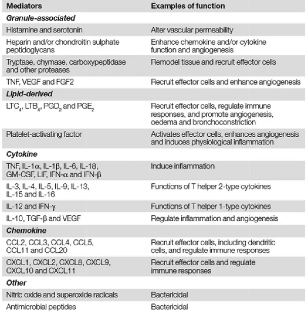
Mast cells can be regarded as factories of a huge variety of mediators. Three major classes of mediators are produced by mast cells (Marshall, 2004): (1) The preformed and granule-associated mediators like histamine, serotonin, mast cell proteases as well as the
factors TNF, VEGF and FGF2; (2) the newly synthesized lipid mediators like leukotriene (LT)C4, LTB4, prostaglandin
chemokines. The cytokines and immunomodulatory cytokines.
potential functions are listed in T
Responses To Pathogens” by Jean S. Marshall
Table 1.1 Mediators synthesized by mast cells. Table is taken from the review of Marshall, 2004
“Mast-Cell Responses To Pathogens”. CCL, CC ligand; FGF2, fibroblast growth factor 2; GM
factor; IFN, interferon; IL, interleukin; LIF, leukemia inhibitory factor; LT, leukotriene; PG, prostaglandin; TGF-β, transforming growth factor
endothelial growth factor.
Dependent on the mode of activation, mast cell
mediator profiles thereby enabling mast cells to initiate and modulate immune responses in a way appropriate to meet the pathogenic challenge
factors TNF, VEGF and FGF2; (2) the newly synthesized lipid mediators like leukotriene , prostaglandin (PG)D2 and PGE2 and (3) do novo-synthesized cytokines and
The cytokines produced by mast cells can be grouped in
immunomodulatory cytokines. The huge variety of mast cell-mediators as well as their sted in Table 1.1 which was taken from the review “Mast Responses To Pathogens” by Jean S. Marshall (Marshall, 2004).
Mediators synthesized by mast cells. Table is taken from the review of Marshall, 2004 Cell Responses To Pathogens”. CCL, CC-chemokine ligand; CXCL,
ligand; FGF2, fibroblast growth factor 2; GM-CSF, granulocyte/macrophage colony
factor; IFN, interferon; IL, interleukin; LIF, leukemia inhibitory factor; LT, leukotriene; PG, , transforming growth factor-β; TNF, tumor-necrosis factor; VEGF, vascular
Dependent on the mode of activation, mast cells can release distinct
mediator profiles thereby enabling mast cells to initiate and modulate immune responses riate to meet the pathogenic challenge.
factors TNF, VEGF and FGF2; (2) the newly synthesized lipid mediators like leukotriene synthesized cytokines and by mast cells can be grouped in pro-inflammatory mediators as well as their the review “Mast-Cell
Mediators synthesized by mast cells. Table is taken from the review of Marshall, 2004 chemokine ligand; CXCL, CXC-chemokine CSF, granulocyte/macrophage colony-stimulating factor; IFN, interferon; IL, interleukin; LIF, leukemia inhibitory factor; LT, leukotriene; PG, necrosis factor; VEGF, vascular
distinct cytokine and mediator profiles thereby enabling mast cells to initiate and modulate immune responses
The importance of mast cells for host defense against bacterial infection was demonstrated by studies of Klebsiella pneumonia infection and of a peritonitis model of cecal ligation and puncture (CLP) (Echtenacher et al., 1996; Malaviya et al., 1996). Using genetically mast cell-deficient mice (Kit-mutant mice, see below) it was shown that the rapid release of TNF from mast cells facilitated neutrophil recruitment necessary for clearance of infection. Mast cells are the only cells known to prestore TNF in their secretory granules, which can be immediately released upon activation (Gordon and Galli, 1990a), stressing the potential role of mast cell-derived TNF in the initiation of inflammatory responses. In addition to TNF, other mediators secreted by mast cells promote the recruitment of effector cells. These mediators include histamine that increases vascular permeability and enhances the expression of adhesion molecules on endothelial cells (Marshall and Jawdat, 2004), as well as mast cell proteases which play a critical role in the recruitment of neutrophils and eosinophils (Huang et al., 1998a;
Schmidlin et al., 2002). In vitro studies showed that mast cells are capable of phagocytosing pathogens and that mast cells produce antimicrobial peptides suggesting also direct effector functions of mast cells in host defense (Abraham and Malaviya, 1997;
Di Nardo et al., 2003). These data were confirmed by a report that revealed a mechanism used by mast cells to kill bacteria extracellularly. This study demonstrated in vitro that, upon exposure to Streptococcus pyogenes, mast cells released extracellular structures composed of DNA, histones, tryptase and antimicrobial peptides. This process was mediated by reactive oxygen species (ROS)-dependent cell death (von Köckritz- Blickwede et al., 2008). In addition to their critical role in host defense, several reports implicated mast cells in wound healing and tissue remodeling (Trautmann et al., 2000;
Weller et al., 2006).
The data discussed so far illustrates that mast cells are the cells responsible for the immediate innate response to invading pathogens. There are many indications that mast cells also set the stage for adaptive immune responses and exert modulatory functions on T and B cell responses. Accordingly, mast cells can influence the migration, maturation and function of dendritic cells and in this line indirectly induce the polarization of naïve T cells towards TH2 effector cells (Galli et al., 2005). McLachlan et al. could show that the T cell recruitment to lymph nodes draining at site of infection and the hypertrophy of these lymph nodes was substantially increased by mast cell-derived TNF (McLachlan et al., 2003). In addition, a recent study by McLachlan and coworkers suggested mast cells as the sensory arm of the adaptive immune system. They could show that small-molecule mast cell activators, administered together with vaccine antigen, evoked substantial increases of antigen-specific serum IgG, highlighting mast cell activators as a new class of adjuvants (McLachlan et al., 2008). Surprisingly, mast cells, best known as
immunostimulatory cells and promoters of inflammation, have recently been implicated in immunosuppression, based on experiments with Kit-mutant, mast cell-deficient mice. The immunosuppressive function of regulatory T cells in a model of transplant tolerance was found to be dependent on mast cells (Lu et al., 2006). Another study demonstrated that mast cell-derived IL-10 limits skin pathology during allergic contact dermatitis and chronic irradiation with UVB (Grimbaldeston et al., 2007).
1.1.4 Mast cell associated disorders
The most extensively studied disorder associated with mast cells is the type I hypersensitivity. Because of the extremely high affinity of the Fcreceptor for IgE (FcεRI), mast cells are constantly coated with IgE (Benoist and Mathis, 2002). Crosslinking of the surface IgE molecules by multivalent antigens leads to degranulation and release of preformed mediators like histamine, prostaglandins, leukotrienes and TNF causing the immediate allergic response. Furthermore, mast cells have an important role in the manifestation of chronic allergic disorders like asthma, allergic rhinitis and atopic dermatitis. In mice, it was shown that the development of the typical features of asthma, i.e. excessive production of mucus, airway hyperresponsiveness, bronchoconstriction, chronic inflammation of airway mucosa and recruitment of eosinophils and other leukocytes, was dependent on mast cells expressing the γ-chain of the FcεRI and FcγRIII receptors (Yu et al., 2006). Anaphylaxis is a life-threatening condition caused by the sudden release of mast cell- and basophil-derived mediators into the circulation. This severe form of an immediate hypersensitivity reaction is classically mediated by IgE and FcεRI. In rodents, however, an alternative mode of mast cell activation via IgG and FcγRIII was described (Grimbaldeston et al., 2006).
In addition to antigen-specific IgE-mediated responses, antigen-nonspecific IgE can modulate mast cell functions as well. Mast cells and antigen-nonspecific IgE were both required for optimal sensitization in a model of contact hypersensitivity (CHS), a T cell- mediated immune response. The TNF released by mast cells in this model was important for the optimal migration of dendritic cells to the draining lymph nodes which present the contact allergen to T cells (Grimbaldeston et al., 2006).
Beside the IgE/FcεRI-mediated allergic disorders, a pathogenic role was suggested for mast cells in various other human diseases like multiple sclerosis, bullous pemphigoid and rheumatoid arthritis. Enhanced degranulation and accumulation of mast cells and their products at the affected sites in those patients indicated a contribution of mast cells to these diseases (Benoist and Mathis, 2002). The observations were supported by results obtained in mouse models for these disorders. Multiple sclerosis and the corresponding mouse model of experimental autoimmune encephalomyelitis (EAE) are both critically
dependent on proinflammatory TH1 cells. Interestingly, mast cell-deficient (Kit-mutant) mice develop an only milder form of the disease (Secor et al., 2000). Rheumatoid arthritis induced by antibodies against the enzyme glucose-6-phosphate isomerase (GPI) was also shown to be dependent on mast cells as mast cell-deficient mice were resistant to arthritis induction (Lee et al., 2002). The same was found in another model of collagen- induced arthritis. The results have to be validated further since contradictory results were obtained with different mast cell-deficient mouse strains (see also section 1.1.5) (Zhou et al., 2007). The strong therapeutic effect of a β2-adrenergic agonist, that prevents mast cell degranulation, on experimental arthritis, however, supports a contribution of mast cells to this autoimmune disease (Malfait et al., 1999). Likewise, in a mouse model for bullous pemphigoid, mast cells were demonstrated to play a crucial role by recruiting neutrophils to the developing lesion (Chen et al., 2001).
Recently, the involvement of mast cells in tumorigenesis is gaining increasing attention.
Mast cells accumulate in the microenvironment of many tumors (Theoharides and Conti, 2004). As mast cells can release mediators that are beneficial as well as mediators that are detrimental for the tumor, the overall effect of mast cells on tumorigenesis was not clear. Interestingly, in a model of adenomatous polyposis coli (APC), Khazaie and coworkers could show that the development of the polyps was mast cell dependent (Gounaris et al., 2007). Consistent with these results, mast cells accumulate around Myc- induced pancreatic islet tumors. Reduced tumor growth after administration of inhibitors of mast cell degranulation and impaired tumor progression in Kit-mutant, mast cell-deficient mice provided evidence for an important function of mast cells in tumor progression (Soucek et al., 2007). In a model of epithelial carcinogenesis involving mast cell- competent and mast cell-deficient (Kit-mutant) HPV16 transgenic mice, Coussens et al.
demonstrated an implication of mast cells in the induction of premalignant neovascularization (Coussens et al., 1999).
1.1.5 Models for the investigation of mast cells
To investigate biochemistry and functions of mast cells ex vivo it is necessary to obtain pure suspensions of mast cells. The cultivation of primary mast cells isolated from mice was often hampered by the scarcity of mast cells in the various tissues. For in vitro studies, mast cell lines like the murine, growth factor-independent C57 or growth factor- dependent MC/9 and PT18 lines (Burd et al., 1989) and human mast cell lines like the immature HMC-1 line (Butterfield et al., 1988) are available. These lines, however, might not reflect the mast cell phenotypes found in vivo. An improvement was the finding that mast cells can be derived from bone marrow cells in the presence of IL-3 (Kitamura, 1989). These bone marrow-derived mast cells (BMMC) have a rather immature phenotype as judged by their production of chonodroitin sulfate and of the proteases mMCP-5 and -6
and MC-CPA but lack of mMCP-4 (Lunderius et al., 2000). The isolation and cultivation of fetal skin-derived mast cells (FSMC (Yamada et al., 2003)) and peritoneal cell-derived mast cells (PCMC (Malbec et al., 2007)) provided useful tools for the investigation of mast cells in vitro as these cultured mast cells closely resemble connective tissue type mast cells in vivo.
To investigate mast cell functions in vivo, researchers utilized mouse strains carrying spontaneous mutations of either the c-Kit gene (white spotting locus, W) or of the steel locus (sl) encoding for the Kit ligand (stem cell factor, SCF) which result, among other phenotypic abnormalities, in profound mast cell-deficiency (Galli and Kitamura, 1987). The most commonly used mouse model for mast cell investigation is the WBB6F1-KitW/Wv mouse. W represents a point mutation at an exon-intron junction that causes an altered splicing of the mRNA, resulting in abrogation of surface Kit expression (Galli et al., 2005).
Mice homozygous for the W allele are white and die before postnatal day ten. The W mutation is fixed to the WB background (W-spotted line B) due to a breeding program of continuous brother-sister matings of KitW/+ mice (Waskow et al., 2004). Wv is a loss-of- function point mutation in the tyrosine kinase domain of c-Kit, that significantly reduce (but does not abolish) kinase activity upon ligand binding. The F1-hybrid of the KitW/+ and KitWv/+ mice (WBBL/6F1-KitW/Wv) is viable and profoundly lacks all mast cells with less than 1% of wild-type levels of skin mast cells (Kitamura et al., 1978). In addition, these mice suffer from several other phenotypic abnormalities like macrocytic anemia, sterility, lack of melanocytes and interstitial cells of Cajal and a high incidence of spontaneous dermatitis, squamous papillomas of the fore-stomach, gastric ulcera and dilatation of the duodenum (Grimbaldeston et al., 2005). The same phenotype is present in Sl/Sld mice in which a mutation of the gene encoding for SCF results in mast cell deficiency (Galli and Kitamura, 1987). Another mouse model that is gaining increasing popularity is the KitWsh/Wsh mouse.
The “Wsash” (Wsh) mutation is an inversion mutation spanning 2.8 to 3.3 Mb (Nigrovic et al., 2008) in the region upstream of the transcription start of c-Kit that arose spontaneously in a cross between two inbred strains (C3H/HeH x 101/H) 23 years ago (Berrozpe et al., 2006; Grimbaldeston et al., 2005). Mast cells are present in newborn mice but their numbers decrease with age until, at the age of ten weeks, the mice are virtually mast cell-deficient (Wolters et al., 2005; Yamazaki et al., 1994). Grimbaldeston et al. detected in the back skin of ten to twelve week-old KitWsh/Wsh mice mast cell numbers that were about 1.2 to 7.2% of those observed in wild-type littermates. Surprisingly, it has been shown that after induction of chronic inflammatory responses, mast cell populations developed in KitWsh/Wsh but also in KitW/Wv mice at the affected sites (Metz et al., 2007). In contrast to the KitW/Wv mice, KitWsh/Wsh mice were considered to display less phenotypic abnormalities. They are viable and fertile but also lack the interstitial cells of Cajal and
demonstrated significant bile reflux into the stomach (Grimbaldeston et al., 2005).
However, new data generated by Nigrovic et al. demonstrated hematopoietic abnormalities in KitWsh/Wsh mice (Nigrovic et al., 2008). Consistent with our own unpublished observations, they described significant splenomegaly in 12 week old mice along with expansion of the myeloid and megakaryocyte populations. In addition, the inversion mutation in KitWsh/Wsh mice disrupted the corin gene encoding for a serine protease that activates pro-atrial natriuretic peptide resulting in cardiac hypertrophy (Nigrovic et al., 2008).
In order to demonstrate that phenotypic differences between mast cell-deficient and congenic wild-type mice are indeed due to the lack of mast cells, mast cell-deficient mice were reconstituted with either bone marrow cells or with BMMCs derived from congenic wild-type or gene deficient mice. While the adoptive transfer of hematopoietic cells into KitW/Wv and KitWsh/Wsh mice is possible, it is not in Sl/Sld mice because SCF is expressed predominantly by the microenvironment and not by the hematopoietic cells them self (Galli and Kitamura, 1987). For the reconstitution, the in vitro derived mast cells can be administered by intravenous, intraperitoneal, intradermal or by injection into the anterior wall of the stomach. The reconstituted mice were called “mast cell knock in mice” (Metz et al., 2007). Dependent on the anatomical site, the transferred BMMCs were reported to differentiate into mast cells resembling native mast cell populations at the corresponding site in wild type mice (Nakano et al., 1985). The reconstitution of mast cells in the different tissues is, dependent on the rout of administration, variable and mast cell numbers can differ from numbers in wild type mice. For example, more mast cells appear in the gastrointestinal tract when injected intravenously.
These reconstitution models yielded important insights into functions of mast cells but inherent technical problems limit experimentation and interpretation of data obtained with these systems. These vagaries became especially apparent when Zhou et al. revealed a profound difference in the development of antibody-mediated arthritis in KitW/Wv and KitWsh/Wsh mice. While KitW/Wv mice were resistant, KitWsh/Wsh mice developed full arthritis (Zhou et al., 2007). These discrepant results illustrate that Kit-mutant mice are not ideal models for the study of mast cell functions and that new models of mast cell-deficiency are urgently needed, which are independent of Kit mutations.
1.2 Generation of mouse models
The investigation of systems as complex as mammalian immune responses as well as the elucidation of the contributions of individual cell types to host defense, requires models that reflect in vivo situations. The mouse is such a suitable model since on the one hand murine immunology largely resembles the human situation and on the other hand there are enormous possibilities to manipulate the mouse genome. Gene-deficient mice as well as mice displaying a cell type specific gene knock out provide powerful tools to gain insights into immune functions.
1.2.1 Classical gene “Knock out”
To investigate gene functions in vivo, targeted mutations were introduced into the germline of the mouse in order to achieve inactivation of a gene of interest in all cell lineages in the mouse. For this purpose, the genome of embryonic stem (ES) cells was manipulated by homologous recombination. Injected into blastocysts, these ES cells can contribute to all cell lineages including germ cells, thus, resulting in heritable mutations (Capecchi, 1989; Koller and Smithies, 1992; Thomas and Capecchi, 1987).
This classical gene knock out methodology is a valuable tool that provided important information. Germline mutations, however, can be lethal and complete gene inactivation in the entire mouse often does not allow conclusions on cell type-specific functions.
Therefore, spatial and temporal control of mutagenesis in the mouse was desired.
1.2.2 Conditional gene targeting
1.2.2.1 Site specific recombination systems
Conditional, i.e. cell type-specific or inducible mutagenesis in vivo, became possible by the introduction of site specific recombination (SSR) systems into the mouse. SSR relies on bacterial or yeast recombinases that cleave DNA at specific target sequences and ligate it to a second site (Kilby et al., 1993). The Cre/loxP recombination system proved particularly useful. The Cre recombinase (causes recombination) originates from the P1 bacteriophage and catalyzes recombination between two recognition sites, designated loxP (locus of crossing (X)-over of P1). This 34 bp sequence contains an eight bp core spacer region that determines the orientation of the loxP site, and two flanking 13 bp palindromes, the binding sites for the recombinase. The Cre enzyme deletes sequences flanked by loxP sites of identical orientation leaving a single recognition site behind. In case of sites with opposite orientation, the loxP-flanked sequence is inverted. In addition, translocation between loxP sites located on different chromosomes can occur (Nagy, 2000).
To achieve Cre-mediated mutagenesis in vivo, Cre transgenic lines are crossed to mouse strains containing loxP-flanked sequences. As shown by Vooijs et al., the success of Cre- mediated recombination in vivo is dependent on the position of the loxP sites in the genome and on the cell type in which SSR is desired (Vooijs et al., 2001). Furthermore, the level of Cre expression is critical for efficiency of recombination. As the Cre recombinase is derived from a prokaryotic organism its expression is not optimal in eukaryotic cells. The high frequency of CpG motives, for instance, can lead to epigenetic silencing during mammalian development. In order to adapt Cre cDNA to the transcription and translation system in eukaryotic cells, Shimshek et al. generated a codon-improved Cre recombinase (iCre (Shimshek et al., 2002)). This mutated Cre sequence contains silent base mutations corresponding to human codon-usage preferences and a minimized CpG content. Putative cryptic splice sites were eliminated and an optimal Kozak consensus sequence was included.
The temporal control of SSR in vivo was accomplished either using inducible promoters to drive Cre expression (Kuhn et al., 1995; St-Onge et al., 1996) or by chemically induced activation of an inactive form of the Cre protein. In the latter case, the Cre open reading frame was fused to the human estrogen receptor ligand binding domain (ER). The CreER fusion protein is trapped in the cytoplasm in an inactive state bound to the heat shock protein 90 (HSP 90) complex. Upon ligand binding, CreER is released from the complex and free to translocate into the nucleus where it can catalyze recombination. The estrogen binding domains used in this system are mutated (Cre-EBD (G521R) (Schwenk et al., 1998), CreERT (G521R) (Feil et al., 1996)) and have a high affinity to the synthetic compound 4-OH-Tamoxifen (OHT), but not to endogenous 17β-estradiol. The affinity to OHT was further improved by mutation resulting in the CreERT1 and CreERT2 fusion proteins (Feil et al., 1997).
Another site specific recombination system that has successfully been applied in mouse ES cells and gene targeting is the Flp/frt system derived from the yeast Saccharomyces cerevisiae (Dymecki, 1996b). The principle of Flp-mediated recombination resembles that of the Cre/loxP-system, although it is not as widely used as the Cre recombinase. The Flp recombinase is largely applied for the deletion of the selectable marker in ES cells after successful gene targeting (Kwan, 2002) or in vivo in combination with the Cre/loxP system.
1.2.2.2 Applications of the Cre/loxP system
To apply the Cre/loxP system to the mouse, genomic gene segments are flanked with loxP sites by classical gene targeting in ES cells. Conditional mutagenesis is then achieved by crossing the so called “floxed” mouse to a strain expressing the Cre recombinase under the control of a cell type-specific promoter. This binary system
represents a powerful tool to investigate gene functions by gene inactivation selectively in a cell population of interest. Since the first description of in vivo gene inactivation using the Cre/loxP system (Gu et al., 1994), an impressive number of floxed mouse lines have been generated as well as numerous tissue and cell type-specific Cre recombinase expressing strains, many of them listed on the webpage http://www.mshri.on.ca/nagy/.
Furthermore, the Cre/loxP system can be used to activate gene expression in selected cell types. For this purpose, the open reading frame of a gene of interest is separated from a promoter by a loxP-flanked stop element containing polyadenylation sequences (Lakso et al., 1992). The expression of the gene is inhibited until Cre-mediated excision of the stop cassette. This principle of cell type-specific gene activation has found multiple applications. It is largely used in Cre excision reporter strains in order to demonstrate the functionality of a Cre transgenic line (Soriano, 1999; Srinivas et al., 2001). In these strains, the expression of indicator proteins, like β-glalactosidase or fluorescent proteins, under the control of a ubiquitously active promoter (e.g. ROSA26) indicates successful Cre-mediated deletion of the stop element. The same was applied with success for depletion of individual cell populations in vivo by Cre-mediated derepression of a diphtheria toxin fragment A (DTA) gene (Ivanova et al., 2005; Voehringer et al., 2008).
Temporally controlled ablation of cell populations was achieved by Cre-mediated control of expression of the simian diphtheria toxin receptor (DTR) resulting in sensitization of the DTR-expressing cells to exogenous diphtheria toxin (Buch et al., 2005). Furthermore, the conditional expression of oncogenes (Lakso et al., 1992) and of mutated versions of genes (Forlino et al., 1999; Gerbaulet et al., submitted), allowed insides into tumorigenesis and the effect of loss or gain of function mutations on a given cell type.
1.2.3 BAC transgene technology
The introduction of foreign genes into the germ line of mice was achieved either by homologous recombination in ES cells resulting in the expression of the foreign gene under the control of an endogenous promoter (i.e. knock-in) or by pronucleus injection resulting in random integration of the construct into the genome. Thus, the construct for random integration must include the complete promoter in addition to the open reading frame.
In the past, transgenes were limited in size and usually contained the cDNA of a gene of interest and a minimal promoter sequence. This restriction was due to the limited availability of DNA-engineering methods for large DNA molecules. Consequently, the lack of cis-regulatory elements in the promoter or intronic sequences could affect the expression level. Furthermore, small transgenes are notably prone to position effects exerted by regulatory elements present at the site of random integration. Effects of promoters, enhancers and silencers at the integration site may override those of the