Generation of inducible Cre systems for
conditional gene inactivation in mice
Inauguraldissertation zur
Erlangung des Doktorgrades
der Mathematisch-Naturwissenschaftlichen Fakultät der Universität zu Köln
vorgelegt von
Frank Thomas Wunderlich aus
Freiburg i. Br.
Berichterstatter: Prof. Dr. Klaus Rajewsky Prof. Dr. Jens Brüning
Tag der mündlichen Prüfung: ____________________________
TABLE OF CONTENTS I
ABBREVIATIONS IV
1. INTRODUCTION 1
1.1 Genetic engineering by site specific recombinases 1
1.2 Conditional gene targeting 5
1.3 Inducible gene targeting 10
1.3.1 Transcriptional control of Cre activity 11 1.3.2 Posttranslational control of Cre activity 14
1.4 Objectives 21
2. MATERIALS AND METODS 22
2.1 Chemicals and antibodies 22
2.2 Molecular biology 22
2.2.1 Competent E. coli and isolation of plasmid DNA 22 2.2.2 Constructions of expression plasmids and targeting vectors 23
2.2.2.1 CrePR expression plasmids 25
2.2.2.2 ROSA26 Cre indicator targeting vector 27
2.2.2.3 ROSA26 Cre*PR targeting vector 28
2.2.2.4 HPRT CAGGS Cre*PR targeting vector 28
2.2.2.5 HPRT EF1α Cre*PR targeting vector 30
2.2.2.6 HPRT CAGGS tet-on targeting vector 30
2.2.2.7 HPRT EF1α tet-on targeting vector 32
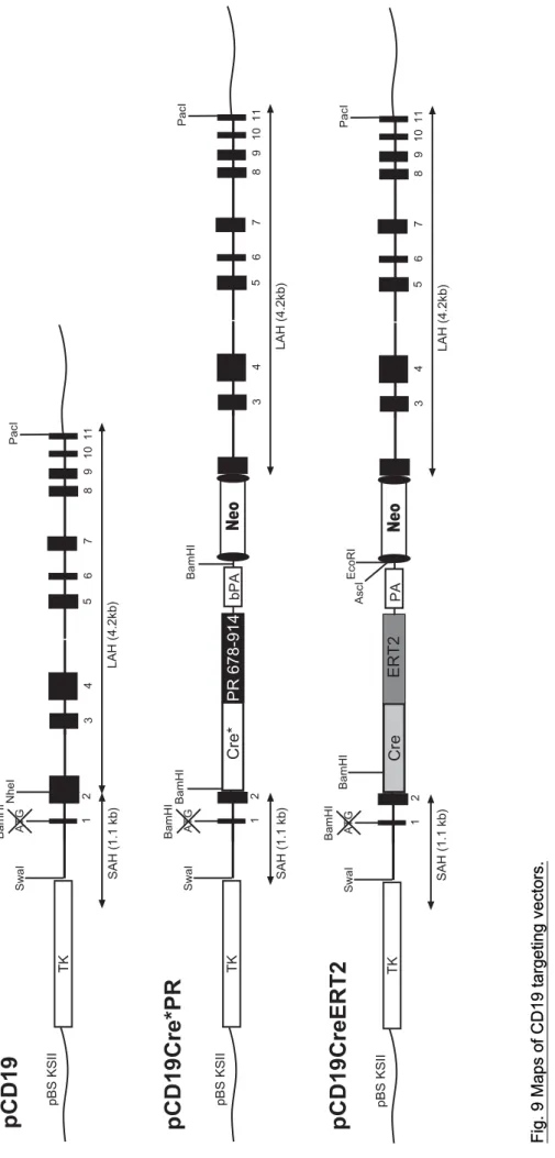
2.2.2.8 CD19 Cre*PR targeting vector 32
2.2.2.9 CD19 CreERT2 targeting vector 32
2.2.3 Isolation of genomic DNA 34
2.2.4 RT-PCR 34
2.2.5 DNA electrophoresis 35
2.2.6 DNA sequencing 35
2.2.7 Quantification of RNA and DNA 35
2.2.8 PCR 35
2.3 Cell biology 38
2.3.1 Fibroblast cell culture 38
2.3.2 Embryonic stem cell culture 38
2.3.3 Culture of ex vivo splenocytes 39
2.3.4 HTNC treatment 40
2.3.5 Induction of Cre activity by ligand addition 40 2.3.6 Preparation of cell suspensions from lymphoid organs 41
2.3.7 Flow cytometry 41
2.4 Biochemistry 41
2.4.1 Protein extracts preparation 41
2.4.2 Western blot 42
2.5 Mouse experiments 42
2.5.1 Mice 42
2.5.2 Activation of CrePR in vivo 42
3. RESULTS 44
3.1 A strategy for rapid screening of inducible Cre constructs 44 3.2 An attempt to generate an improved Cre indicator mouse strain 45
3.3 The inducible Cre*PR system 57
3.4 Targeted insertion of Cre*PR into ubiquitously expressed loci 65 3.4.1 Knock-in of CAGGS- and EF1α-driven Cre*PR into the HPRT
locus of RRDR ES cells
65 3.4.1.1 Analysis of chimeras generated from
RRDR/CAGGS Cre*PR ES cells
71 3.4.1.2 Analysis of the CAGGS Cre*PR system in vivo 72 3.4.2 Knock-in of Cre*PR into the ROSA26 locus 74 3.4.2.1 Analysis of the ROSACre*PR system in vivo 76 3.4.2.2 Analysis of ROSACre*PR in mouse embryonic fibroblasts 78 3.5 B lymphocyte restricted expression of Cre*PR 79 3.6 An attempt to generate a tet-inducible system
controlling Cre activity
85
4. DISCUSSION 89
4.1 RRDR as a new indicator for Cre activity 89
4.2 The inducibility of Cre*PR in fibroblasts 94
4.3 The Cre*PR system in ES cells 95
4.4 The Cre*PR system in mice 98
4.5 B cell specific inducible Cre systems 100
4.6 Doxycycline inducible systems 102
5. SUMMARY 104
5.1 Summary 104
5.2 Zusammenfassung 105
6. REFERENCES 106
7. ACKNOWLEDGEMENTS 133
8. VERSICHERUNG 134
9. LEBENSLAUF 135
10. SUPPLEMENTARY DATA 136
aa amino acid
AP alkaline phosphatase
BAC bacterial artifical chromosome β-Gal β-galactosidase
β-Geo fusion protein between β-Gal and neo
bm bone marrow cells
bp base pair
bPA bovine polyadenylation signal BSA bovine serum albumin
CAGGS chicken β-actin promoter coupled to the CMV enhancer CAT chloramphenicol acetyltransferase gene
CCP CAGGS Cre*PR
CMV cytomegalovirus
cps counts per second
Cre causes recombination, recombinase from phage PI
CreLBD fusion between Cre and the LBD of steroid hormone receptors DMEM Dulbecco’s modified Eagle medium
dox doxycycline
DTA diphtheria toxin A
EF mouse embryonic fibroblasts
EF1α promoter of the elongation factor 1α gene eGFP enhanced green fluorescent protein
ER estrogen receptor
ER-LBD LBD of the ER
ERT1 LBD of the ER containing mutations G400V, L539A and L540A ERT2 LBD of the ER containing mutations G400V, M543A and L544A ES cells embryonic stem cells
E
217-β-estradiol
FACS fluorescence assisted cell sorting FCS fetal calf serum
floxed loxP sites flanked
Flp Flp recombinase from yeast FRT Flp recognition target site
GANC gancyclovir
GLVP Gal4 LBD VP16
HAT hypoxanthine, aminopterine, thymidine
hCre humanized Cre gene
hER human ER
hPR human PR
HPRT hypoxanthine-phospho-ribosyl-transferase
HR homologous recombination
HRP horse-radish peroxidase hsp heat shock protein
HTNC His-TAT-NLS-Cre
kb kilo base pairs
kDa kilo Dalton
lacZ lacZ gene from E. coli encoding for β-Gal LAH long arm of homology
LBD ligand binding domain
loxP locus of X over in PI, recognition sequence of Cre MCS multiple cloning site
mER murine estrogen receptor neo neomycin resistance gene NLS nuclear localisation signal
ORF open reading frame
pA polyadenylation signal PBS phosphate buffered saline
PR progesterone receptor
PR891 LBD of the progesterone receptor ending with aa 891 PR914 LBD of the progesterone receptor ending with aa 914 PR-LBD LBD of the PR
RRDR ROSA26 double reporter
RRDR-DsRed RRDR containing the DsRed-stop cassette RRDR-eGFP RRDR with excised DsRed-stop cassette rtTA reverse tet-transactivator
RU486 mifepristone
SA splice acceptor
SAH short arm of homology
SD splice donor
SV40 pA pA of simian virus 40
tetO tet-operator sequences
tet-off tet-controllable system using the tTA tet-on tet-controllable system using the rtTA TK thymidine kinase of HSV
tTA tet-transactivator
V336A mutation V336A in the hCre gene 4-OHT 4-hydroxy-tamoxifen
VP16 transactivation domain of VP16 WSS Westphal stop sequence
wt wild type
The genomes of man (Lander et al., 2001), mouse (Waterston et al., 2002), Drosophila (Adams et al., 2000) and several other organisms have been completely sequenced in recent years. This progress tremendously facilitated the analysis of gene functions in vivo. Gene targeting by homologous recombination is a powerful technique to explore the function of genes in mice (for review, see e. g. Capecchi, 1989). First described in 1987 by Thomas and Capecchi, it became a standard technique during the last years. The gene of interest is modified in embryonic stem cells (ES cells) by inserting a selection marker into the open reading frame (ORF) of the gene via homologous recombination (Thomas & Capecchi, 1987). The targeted pluripotent ES cells are kept in culture under non-differentiating conditions and are subsequently injected into donor blastocysts, which then become implanted into pseudo-pregnant foster mice. The resulting chimeric animal, consisting of wild type and gene targeted cells, is then backcrossed to wild type mice. If the chimeras contain germ cells originating from the gene targeted cells the genetic alteration can be transmitted through the germ line. This method allows the production of animals which carry the mutation of the gene in all cells of the body (for review, see e. g.
Müller, 1999; Cohen-Tannoudji & Babinet, 1998). However, this technique has several inherent limitations such as embryonic lethality, compensatory effects by redundant genes, and side effects due to the leftover of the selection marker in the genome. These problems can be circumvented by more sophisticated methods of gene manipulation. The technology of conditional gene targeting allows for modification of genes in defined cell types, in spatially and temporally controlled manners (for review, see e. g. Rajewsky et al., 1996; Kühn & Schwenk, 1997). Thus, the function of a given target gene can be studied in individual cell types at different developmental stages or after administration of specific inducers.
1.1 Genetic engineering by sequence specific recombinases
The field of gene targeting was revolutionized through the application of sequence specific recombinases as tools for genomic engineering. A number of bacterial and yeast recombinase enzymes are able to rearrange DNA at specific target sequences.
This simple, but elegant reaction results in a precisely defined recombination event
between two appropriate DNA target sequences. The hitherto available site specific
recombinase systems belong to two major groups: The resolvase/invertase family and the λ-integrase family. Members of the resolvase/invertase family comprise phiC31 integrase, β-recombinase, HK022 integrase or γδ-resolvase, which catalyse site specific DNA rearrangements via a conserved serine residue (for review, see e.
g. Smith & Thorpe, 2002). The two most famous λ-integrase members are the Flp and Cre recombinases which use a tyrosine located in the C-terminal part of the respective enzyme as catalytic residue. Some of these recombinase systems have been found to be suited for genetic engineering in eukaryotic cells (for review, see e.
g. Kilby et al., 1993).
The complexity of these recombination systems considerably varies both in their requirement for cofactors or accessory molecules and in the size of the DNA target sites involved. However, λ-integrases such as Cre from bacteriophage PI and Flp from yeast catalyse recombinations between specific target sites of 34 bp without further need for cofactors. Since the only other requirement for the reaction is the expression of the appropriate recombinase enzyme, the concomitant presence of target sites and appropriate recombinase is sufficient for specific gene modification in many different species.
The Cre/loxP system
Gene modification employing site specific recombinases was pioneered using the Cre/loxP recombination technology (for review, see e. g. Rajewsky et al., 1996). The site specific DNA recombinase Cre (causes recombination) of the bacteriophage PI recognizes specifically 34 bp long sequences, called loxP (locus of X over in PI) (Sternberg & Hamilton, 1981; Sternberg et al., 1981). Cre excises DNA segments flanked by directly repeated loxP sites as a circular molecule, leaving a single loxP site in the genome (Sauer et al., 1988; Gu et al., 1993). When the loxP sites are arranged in opposite directions, Cre catalyses inversion of the intervening DNA (Kano et al., 1998; Lam & Rajewsky, 1998). Also, intramolecular recombinations can be performed, when the loxP sites are on different strands of DNA (van Deursen et al., 1995). Insertion of a DNA segment into a loxP site is also possible, though the excision reaction is favoured over the integration event (Fukushige & Sauer 1992).
The usage of mutant loxP sites was shown to be useful in insertion strategies (Kolb,
2001). Since Cre is one of the few recombinases which does not require cofactors or
1990), Cre is the first choice for introducing gene modifications into the mouse genome. Modification of the ES cell genome by the Cre/loxP system is a two step process. The desired genetic modification including the loxP sites is introduced into the genome of ES cells via homologous recombination. Subsequently, the targeted allele is generated by site specific recombination of the loxP sites through expression of Cre by transient transfections of Cre-containing plasmids or other means. The removal of positive selection markers is one important application of Cre-mediated recombination in ES cells, since the presence of the selection marker gene can interfere with expression of the targeted gene or adjacent genes (Pham et al., 1996).
Therefore, the selection marker is flanked by two loxP sites in the same orientation, which allows its deletion by Cre-mediated recombination leaving a single loxP site in the genome. Gene targeting provides the possibility to introduce deletions, point mutations, and insertions in ES cells. For instance, it has been used to delete a small region of the coding region of the Igα gene (Torres et al., 1996), to introduce specific point mutations into the cytoplasmic tail of the Igα gene (Kraus et al., 2001), and to insert rearranged variable regions into the Ig κ locus (Pelanda et al., 1996) and Ig λ locus (Oberdoerffer et al., 2003). Large genomic deletions or inversions can be introduced into ES cells using the Cre/loxP technology. This requires two independent successive gene targetings, but due to the lower frequency of Cre- mediated recombination at large distances between loxP sites, a positive selection strategy is recommended (Milstone et al., 1999). This approach has been used to generate deletions of several centimorgans (Ramirez-Solis et al., 1995).
The Flp/FRT system
Another system for the modification of genes was developed using the Saccharomyces cerevisiae derived 43 kDa Flp recombinase (for review, see e. g.
Chen & Rice, 2003). The Flp recombinase is encoded by the 2µ circular plasmid of the budding yeast and mediates inversion between two 34 bp Flp recognition target (FRT) sites arranged in opposite direction, which are located on the same plasmid.
Recombination between these FRT sites results in inversion of half of the 2µ plasmid
with respect to the other and provides a mechanism for producing multiple plasmid
copies from a single replication initiation by flipping the direction of the replication
fork. Initial applications of the Flp/FRT recombination system at stably integrated
chromosomal targets of eukaryotic cells displayed only low recombination
frequencies. Recently, the efficiency of this system has been considerably enhanced by specific modifications. The first improvement concerned the recognition sequence of Flp. The minimal 34 bp FRT site consists of two 13 bp inverted repeated binding sequences for Flp flanking the 8 bp asymmetric core region, which defines the directionality of the target site and which is the site of crossing over in the recombination event. In yeast, the 34 bp FRT recognition sequence is flanked by a third binding site for Flp in which a single nucleotide is exchanged resulting in a 48 bp wild type FRT site. Two 48 bp FRT sites can be much more efficiently recombined than two minimal 34 bp recognition sequences. The second improvement was achieved through the generation of novel Flp recombinase variants with enhanced thermostability isolated by protein evolution. Three versions of Flp recombinase are available: Flp wild type (wt), Flp-L and Flpe. In Flp-L, a point mutation changes the encoded amino acid 70 from phenylalanine to leucine, which causes an enhanced thermostability of Flp-L at 37°C, but, coincidently, there is a loss of approximately 80% activity in comparison to Flp-wt in vitro (Buchholz et al., 1996). Flpe harbours four amino acid exchanges (P2S, L33S, Y108N and S294P), which collectively improve the in vitro recombination activity 4-fold at 37°C and 10-fold at 40°C compared to Flp-wt (Buchholz et al., 1998). Mice expressing Flpe either in the germ line (Flp deleter) (Rodriguez et al., 2000) or constitutively in every organ (Flipper) (Farley et al., 2000) were shown to efficiently excise DNA segments between two head to tail arranged FRT sites. These mouse strains can be used to delete FRT site flanked positive selection marker required for gene targeting in vivo.
The phiC31/att system
At present, the Cre/loxP and the Flp/FRT recombination systems are most commonly
used to manipulate the genomes of eukaryotes in heterologous environments in a
site specific manner. However, additional recombinase systems have to be
established in order to improve the technique of gene targeting for more
sophisticated strategies. Recently, the Streptomyces phage derived phiC31 integrase
has been described to mediate recombination efficiently in eukaryotic cells when a
nuclear import signal (NLS) was located at the C-terminus of the enzyme (Andreas et
al., 2002). The phiC31 system displayed deletion efficiencies of up to 50% at stably
integrated chromosomal targets in a side by side comparison to the well defined
Cre/loxP system. In contrast to the Cre and Flp systems, however, the phiC31
integrase catalyses both integration and excision events irreversibly, since the sites
generated by the recombination cannot serve as substrates for the enzyme (Belteki
et al., 2003).
Conditional gene targeting is defined as a gene modification, which is restricted to specific cell types and/or developmental stages of the mouse. The usage of the Cre/loxP system in conditional gene targeting requires both a mouse strain containing a loxP-flanked modification of the target gene and an additional mouse strain, which expresses the Cre recombinase in specific cell types. Crossing these two strains results in the generation of a conditional mouse mutant, which carries the mutation restricted to those cell types, in which the Cre recombinase is expressed.
Such a conditional mouse mutant can be used to study the function of ubiquitously expressed genes in individual defined cell types and to circumvent lethality at early embryonic stages due to inactivation of a putative essential gene.
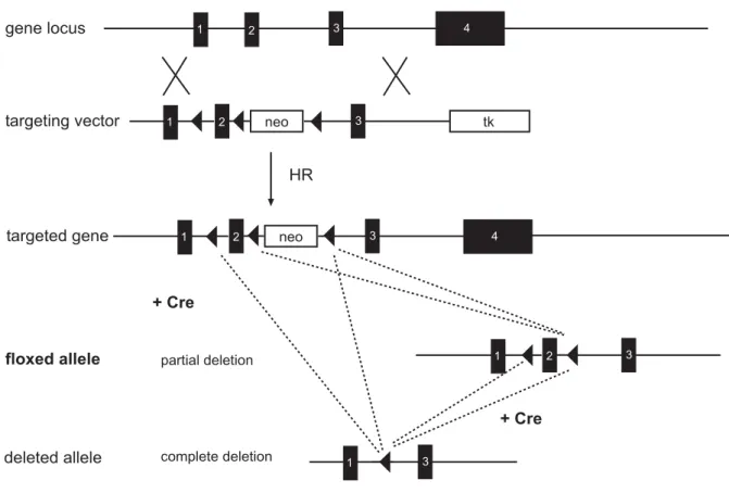
Generation of loxP-flanked alleles
For the removal of a gene segment by conditional gene targeting, the region of interest has to be flanked by loxP sites of the same orientation. This can be achieved by the ‘flox and delete’ approach, which requires construction of a gene targeting vector with three loxP sites pointing in the same orientation, two of which flank the positive selection marker and the third loxP site is placed within a homology arm (Fig.1). Transient expression of Cre in targeted ES cell clones results in two types of clones which have lost the selection marker. Recombination between the two external loxP sites leads to deletion of the selection marker and the gene segment whereas recombination between the loxP sites flanking the selection marker generates the loxP-flanked version of the gene (Fig.1). ES cell clones harbouring the loxP-flanked, or conditional, allele can be used to generate mice used in conditional gene targeting experiments. The positioning of the two remaining loxP sites within the locus of the target gene is critical for conditional gene targeting. They have to be placed in non-coding sequences around critical sequences such as exons so that gene expression is not disturbed by their presence. The desired deletion of the gene segment is achieved upon Cre-mediated recombination of the loxP-flanked region.
This ‘flox and delete’ strategy was first demonstrated to function in an approach to
conditionally inactivate the DNA polymerase β (Gu et al. 1994). The promoter and the
first exon of the DNA polymerase β gene were flanked by loxP sites and the
generated pol-β flox mouse strain was crossed with the lck-cre mouse strain (Orban
et al., 1992), in which Cre is expressed only in thymocytes. The DNA polymerase
was thus inactivated exclusively in T lineage cells circumventing the embryonic
lethality caused by ablation of pol-β in the entire mouse by conventional knock-out.
1 2 neo 3 4 tk
1 2 3 4
1 2 neo 3 4 4
1 2 3 4
4
1 3 4
+ Cre
partial deletion
+ Cre
complete deletion
HR gene locus
t argeting vector
targeted gene
floxed allele
deleted allele
Fig. 1 Generation of a loxP-flanked allele.
The conditional targeting vector harbours a loxP (filled triangles) flanked neo cassette (neo) and a third loxP site which is inserted into intron 1. Homologous recombination (HR) in ES cells generates targeted clones which become subjected to transient Cre expression in a second transfection round. Two types of clones have lost the positive selection marker (neo) by this strategy. In one, recombination between the two external loxP sites leads to complete deletion of the selection marker and the gene segment, whereas the other recombination between the loxP sites flanking the selection marker generates the loxP-flanked allele (floxed allele).
Cre transgenic mice
At present, a large number of Cre transgenic mouse strains are available, enabling researchers to restrict conditional genetic modifications to B cells (Rickert et al., 1997), to T cells (Lee et al., 2001), to different areas of the brain (Tsien et al., 1996;
Casanova et al., 2001) and to various other tissues of the mouse (Tonks et al., 2003;
Wen et al., 2003; Barlow et al., 1997; more Cre-transgenic mouse strains in Genesis
2000, 26, 99-166 or under http://www.mshri.on.ca/nagy/cre-pub.html). Such Cre
expressing mouse strains can be generated either by using conventional random
transgenesis, or by targeted insertion into a gene (knock-in), or by using a bacterial
artificial chromosome (BAC) strategy. In any case, the expression pattern of the
promoter used to express Cre determines onset and cell type specificity of the Cre-
mediated gene modification. The technique of conventional random transgenesis has
several disadvantages: the limited understanding of promoter regions used to
elements present at the integration site on transgene expression and the necessity to analyze many different founder lines which may contain multiple copies, tandem repeats, broken and differently integrated transgenic constructs. The method of targeted insertion of Cre into a gene can be applied, when the inactivation of one allele of the endogenous gene can be tolerated. This has been shown to work by Rickert and coworkers, who generated a knock-in mouse strain by targeted insertion of the Cre gene into the second exon of the CD19 locus (Rickert et al., 1995). In this CD19-cre mouse strain, the Cre recombinase is exclusively expressed in B lymphocytes and mediates 75-80% excision of loxP-flanked targets in B cells in the bone marrow, whereas 90-95% deletion is observed in splenic B cells due to continuous expression of Cre (Rickert et al., 1997). Another technique, which is currently used to generate Cre expressing mouse strains, is the insertion of the Cre gene into bacterial artifical chromosomes (BAC) and the subsequent generation of BAC transgenic mice. BACs carry large genomic fragments from mouse or human of up to 400 kb and can be obtained commercially. The Cre gene can be introduced into defined genes encoded by the BAC via homologous recombination in bacteria (Testa et al., 2003). The large size of the BAC renders it likely that most if not all of the transcriptional control elements of the gene, into which Cre has been introduced, are present. This should result in a Cre expression pattern closely related to the expression of the endogenous gene. This strategy was successfully used by M.
Alimzhanov (unpublished data) to express Cre solely in mature B cells by inserting the Cre gene in the CD21 gene of a 250 kb BAC.
Indicators for Cre activity
The success of Cre-mediated conditional gene targeting critically depends on
stringent regulation of Cre expressing mouse strains, which have to be intercrossed
with mice containing loxP-flanked target genes. If expressed from a transgenic
construct, Cre-mediated deletion may occur in a mosaic or leaky fashion rendering
excision of the loxP-flanked target alleles incomplete or unspecific, respectively. A
general complication of Cre transgenes is due to the irreversibility of Cre-mediated
deletion. Any transient expression of Cre during mouse development caused by the
control elements of the gene into which Cre has been inserted, or alternatively
caused by artefacts of transgenesis, will lead to deletion of loxP-flanked targets in the
cells expressing Cre. These cells and all cells arising from them through division will
A B
STOP
Promoter Indicator gene
Promoter Indicator gene
Indicator protein
Indicator 1
Promoter Indicator 2
Indicator 2 Promoter Indicator 2
Indicator 1
+ Cre + Cre
Fig. 2 Indicator constructs for Cre activity.
(A) Expression of the indicator gene is prevented by a transcriptional stop sequence (STOP) which is flanked by loxP sites (closed triangles). Cre-mediated excision of the STOP results in expression of the indicator gene which can be monitored, for example, by enzymatic reactions.
(B) Shut down/activation indicator construct. The promoter expresses indicator 1, whereas expression of indicator 2 is interrupted by the presence of the indicator 1 gene. Cre-mediated excision of the indicator 1 gene results in shut down of indicator 1 expression and activation of indicator 2 expression.
contain the modified alleles, regardless whether Cre expression is continued or not.
Cre expressing mouse strains can be characterized with the help of reporter mice that permit in vivo monitoring of Cre-mediated recombination events in diverse tissues and at different developmental stages (Fig. 2A). For instance, a reporter strain carrying a loxP-flanked stop sequence preceding the β-galactosidase (β-Gal) gene driven by the chicken β-actin promoter has been used to score excision events in mice (Tsien et al., 1996). Cre-mediated excision of the loxP-flanked stop sequence leads to β-Gal expression, which can be monitored by enzymatic reactions for β-Gal.
However, a limitation of this Cre reporter mouse strain became apparent, when characterizing the expression pattern of the β-galactosidase gene upon Cre-mediated excision of the loxP-flanked stop sequence in all tissues. The indicator gene was expressed in a mosaic fashion in some organs such as brain. This mosaic expression pattern of the β-galactosidase gene may result from artefacts of the conventional random transgenesis, which was used to generate this mouse strain.
Artefacts of transgenesis can be caused by integration of multiple copies, tandem
repeats, breakage of the indicator constructs, unknown multiple integration sites,
inactivation of the indicator gene by methylation, influences of enhancers/silencers
used to express the transgene.
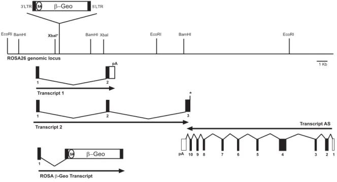
A more convenient strategy for the generation of a Cre reporter strain would be to target a single reporter construct into a gene which is expressed ubiquitously throughout all developmental stages of the mouse. An excellent candidate for an appropriate locus is the ROSA26 locus, which was originally identified by gene trap (Zambrowicz et al., 1997). In the initially reported mouse line, retroviral sequences and a β-Gal neomycin resistance fusion gene (β-Geo) were integrated into the locus ROSA26. The wild type ROSA26 locus expresses two transcripts with no significant open reading frames (ORF) and a third in antisense orientation, which putatively encodes a protein, and which is not affected by the β-Geo insertion. Expression of the β-Geo gene is initiated from exon 1 of the ROSA26 allele and is ubiquitous during embryonic development and also in adult tissues. Mao and colleagues (1999) subsequently engineered the gene trapped proviral ROSA26 locus in ES cells by introducing a three loxP sites containing cassette, in which two loxP sites flanked the selection marker and one external loxP site flanking a transcriptional stop sequence to interrupt the expression of the β-Geo gene. This mouse strain expressed β-Gal only after Cre-mediated excision of the loxP-flanked region. This was verified by breeding the ROSA26 reporter strain to a Cre transgenic mouse strain which expresses Cre at early embryonic stages (Schwenk et al., 1995), i.e. Cre mediated excision was disseminated to all tissues of the adult mouse. Enzymatic reactions for β-Gal with the substrates X-Gal and fluoresceine-di-β-D-galactopyranoside (FDG) were carried out to detect expression of β-Gal at the single cell level. The expression of β-Gal upon Cre-mediated excision of the three loxP-flanked cassette in this mouse strain was shown to be ubiquitous (Wunderlich & Rajewsky, unpublished results).
Later on, however, several limitations of this reporter strain became apparent. For
instance, recombination between two adjacent loxP sites by partial Cre-mediated
deletion results in retention of either the selection marker or the stop fragment in the
locus, which therefore still lacks β-Geo gene expression despite Cre-mediated
recombination. The excision event remains undetectable by X-Gal staining. In
addition, loxP-flanked targets, which are integrated into the ROSA26 locus, were
shown to be excised with low efficiency by inducible Cre constructs for reasons not
yet understood (Voojis et al., 2001; Seibler et al., 2003). Therefore, mouse strains
carrying a reporter construct for Cre in the ROSA26 locus are not suited for the characterization of inducible Cre mouse strains. Moreover, until recently, enzymatic reactions had to be performed to detect β−galactosidase expression in Cre reporter mouse strains. Such enzymatic reactions are time consuming and difficult to perform in some organs.
A more sophisticated reporter gene, which can be detected without any enzymatic reaction just by its presence, is the eGFP gene (enhanced Green Fluorescent Protein). The eGFP protein, originally derived from the jellyfish Aequorea victoria, can be visualized by its inherent fluorescence, even in living cells (for review, see e. g.
Cubitt et al., 1995). EGFP owes its visible absorbance and fluorescence to a p- hydroxybenzylideneimidazolinone chromophore formed by cyclisation of Thr65, Tyr66 and Gly67 and 1, 2-dehydrogenation of the tyrosine. Site directed mutagenesis of the amino acids forming the chromophore leads to versions of the fluorescent protein whose emission spectrum are shifted to yellow or blue such as eYFP (Yellow) and eCFP (Cyan). The only limitation of using these fluorescent proteins as reporter genes is the large amount of molecules (8x10
5), which have to be expressed in a single cell to detect fluorescence by fluorescence microscopy (Rizzuto et al., 1995).
Therefore, strong promoters have to be used in order to generate a Cre reporter strain which expresses eGFP after Cre-mediated recombination. Another possibility is to use a loxP-flanked indicator 1 gene, which serves in a dual function to prevent expression of the indicator 2 gene before Cre-mediated deletion and, in addition, to control for expression from the reporter construct. In this case, Cre-mediated recombination leads to inactivation of indicator 1 gene and to expression of the indicator 2 gene. (Fig. 2B). This shut down/activation theme has been employed in the generation of the Z/EG and Z/AP Cre reporter strains, which switch their indicator genes either from lacZ to eGFP (Novak et al., 2000) or from lacZ to alkaline phosphatase (Lobe et al., 1999) upon Cre recombinase activity.
1.3 Inducible gene targeting
One step forward in techniques of conditional gene targeting is the temporally
controlled expression of Cre, by which the onset of the conditional genetic alteration
can be chosen at specific developmental stages (for review, see e. g. Lewandowski,
2001). A pioneering breakthrough in this field was the generation of the Mx-Cre
mouse strain (Kühn et al., 1995). Here, the Cre gene was placed under the control of
α/β leads to Cre expression and, thus, to highly efficient deletion of the loxP-flanked target genes such as the DNA pol-β gene segment (Gu et al., 1994) in a wide variety of tissues in mice. Deletion of loxP-flanked sequences using the Mx-Cre system is not restricted to lymphocytes only, since many cell types in the mouse are responsive to interferon. At present, Cre activity is tried to be more tightly regulated at both the transcriptional and the posttranslational level by inducible systems.
1.3.1 Transcriptional control of Cre activity
Cell type specific transcriptional control of Cre can be achieved by systems, in which the activity of almost silent minimal promoters can be enhanced manifold through binding of certain inducer proteins. These inducer proteins are in turn regulated by small molecules such as tetracycline (Gossen & Bujard, 1992), RU486 (Wang et al., 1997a) or ecdysone (No et al., 1996). By placing the Cre expression under the control of a minimal promoter and directing the expression of the inducer protein (transactivator) to certain cell types, transcription of Cre in mice can be activated by administration of the above mentioned small molecules. A main disadvantage of these regulatory systems is the separate insertion of the gene encoding the transactivator and that of the controllable Cre into the genome leading to large numbers of transgenic mice, which have to be intercrossed and tested for the desired combination of the transgenes.
The GLVP system
In the GLVP system, the inducer protein consists of the Gal4 DNA binding motif, the
Ligand binding domain (LBD) of the progesterone receptor and the VP16
transactivation domain. This chimeric transactivating unit specifically binds to the 17-
mer Gal4 target sequence fused to a minimal promoter upon administration of RU486
and activates expression of genes of interest placed under control of the 17-mer Gal4
target sequence/minimal promoter hybrid (Wang et al., 1997a). Thereby, DNA
specific binding to the 17-mer sequences is mediated by the GAL4 DNA binding
motif, which in turn enables the transactivation domain of VP16 (Triezenberg et al.,
1988) to activate transcription from the minimal promoter only in the presence of
RU486, since the LBD of the progesterone receptor regulates the temporal inducer
dependent activity of the synthetic molecule. Tight regulation of gene expression by
this system has been reported in tissue cultures (Burcin et al., 1998), but rare and
ineffective in vivo applications minimize the value for the temporal control of the Cre recombinase (Pierson et al., 2000; Chaisson et al., 2002). The main obstacle of this system seems to be the inefficient activation of the transactivator molecule by the steroid compound, since stably expressing cell types in mice mediate only a low extent of transcription upon induction from the minimal promoter.
The ecdysone inducible system
In an alternative approach for the inducible regulation of Cre, Cre could be placed under control of a system involving the insect molting hormone ecdysone as an inducer. The effect of ecdysone is mediated through the functional ecdysone receptor, which is a heterodimer of the ecdysone receptor and the gene product of the ultraspiracle gene. The functional ecdysone receptor binds to specific promoter elements of the target gene in order to activate transcription. Mutagenesis of the transactivation domain and the DNA binding domain of the natural occurring receptors resulted in a tightly controllable system in mice (No et al., 1996). However, the binary character of the heterodimeric receptor complicates the usage in vivo, since both partners must be present at equal amounts. Otherwise, homodimeric complexes are formed, which cannot activate the promoter and may function in a competitive way. Furthermore, the inducer ecdysone or its synthetic counterpart muristerone have to be available at non-physiological micromolar concentrations to sufficiently activate the ecdysone heterodimeric receptor complex (Saez et al., 2000).
The tet-system
Tetracycline controlled expression systems are widely used to regulate genes in cell
lines (Resnitzky et al., 1994), plants (Weinmann et al., 1994), yeast (Nagahashi et
al., 1997), Drosophila (Bello et al., 1998), and mice (Hasan et al., 2001; Schönig et
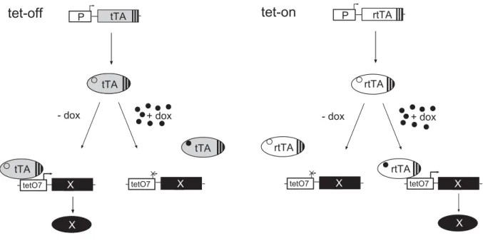
al., 2003). Fig. 3 schematically outlines the function of the original tet-off system and
the modified tet-on system. The tet-off system uses a synthetic transcription factor
tTA (tetracycline controlled transcriptional activator), which is a fusion between a
bacterial tet-repressor (tetR) and the transcriptional activating domain of VP16. tTA
activates a minimal promoter which is fused downstream to an array of tet-operator
(tetO) sequences (Gossen & Bujard, 1992), the cognate binding sites of tetR and,
thus, of tTA. In the tet-off system, transcription is abrogated by administration of
doxycycline, which upon binding to the tTA prevents binding of tTA to the tet-
responsive promoter. Despite of its extensive use, however, the tet-off system has
tTA
+ dox - dox
tTA
X
tetO7
tTA
X X
tetO7
rtTA
rtTA
X
tetO7
X X
tetO7
rtTA
+ dox - dox
Fig. 3 Transcriptional regulation of gene expression by the tet-systems.
The tet-off system. The promoter (P) drives the expression of the tet-transactivator (tTA), which binds in the absence of doxycycline (dox) to the tet-responsive promoter (tetO7) and activates expression of the gene of interest (X). Administration of dox causes dissocation of tTA from tetO7 and transcription is terminated.
The tet-on system. Only in the presence of dox, the reverse tet-transactivator (rtTA) can bind to its promoter and activate expression of the gene of interest.
some disadvantages for applications in mice. Doxycycline has to be continuously fed to prevent transcription of the target gene, until expression of the target gene can be initiated by removal of doxycyline (Kistner et al., 1996). Such long periods of doxycycline application results in storage of the antibiotic into cartilage and bones of mice (Bocker et al., 1984; Sande & Mandell, 1990). This may result in the slow release of doxycycline from its storage places after discontinuation of doxycycline administration and therefore may interfere with activation of the target genes by tTA.
In the complementary tet-on system, transcription of the target gene will only be activated in the presence of doxycycline. This system is based on rtTA (reverse tetracycline controlled activator), which contains mutations in the tetR part resulting in requirement of doxycycline binding in order to bind to tetO (Gossen et al., 1995).
However, the affinity of rtTA to doxycycline is three magnitudes lower than that of the
tTA and, therefore, rtTA requires more inducer to become activated (Baron et al.,
1997). A recent report addressing this problem generated new rtTA mutants termed
rtTA-M2 and rtTA-S2, which could already be activated by the low dose of 2 ng/ml
doxycycline in tissue culture resulting in a 50-fold improved affinity of these new
mutants to doxycycline (Urlinger et al., 2000). In both the tet-off and tet-on system,
the genomic integration site of the tetO promoter driving the transgene of interest is
critical. When endogenous enhancers are located in close environment to the tetO
promoter, the minimal promoter though silent in principle is activated, thus resulting in leaky expression of the transgene. A recently described integration site for the tetO promoter offers an alternative approach. A bidirectional tetO promoter (Baron et al., 1995) in transgenic LC-1 mice (Schönig et al., 2003) drives the expression of Cre and luciferase, respectively, and can be tightly regulated by transactivating units of both the tet-on and tet-off system. Hitherto, a general disadvantage of both tet- systems is their binary character with the consequence of large breeding effort to obtain the desired combination of the transgenes, which is time consuming and expensive. Recently, however, this problem was overcome by Utomo et al. (1999) who placed both the rtTA with its own promoter and the tetO driving expression of Cre on the same fragment of DNA.
The lac system
The lac system from E.coli can also be applied to regulate gene expression in an inducible manner in mice. The principle of the lac system constitutes of a tetrameric lac repressor complex, which binds to operator sequences in the lac promoter thus blocking the docking sites for the RNA polymerase in the absence of inducer.
Administration of allolactose or isopropyl-beta-D-thiogalactopyranoside (IPTG) induces conformational changes of the lac repressor proteins, which subsequently dissociate from the promoter thus allowing for initiation of transcription of the desired gene. However, also the lac system has its problems. The major limitation is the positioning of the operator sequences into the eukaryotic promoter which should be regulated without disturbing its activity. Another limitation is the occurrence of CpG islands in the lac repressor gene which causes its silencing in mice. However, this limitation was recently overcome by eliminating the naturally occurring CpG islands and the mutation of cryptic splice sites in the lac repressor gene (Cronin et al., 2001).
This modified lac gene was indeed suited for heterologous expression in mammals.
1.3.2 Posttranslational control of Cre activity
Steroid hormones such as estrogen, progesterone, androgen and glucocorticoid regulate gene expression directly through classical intracellular steroid hormone receptors and possibly indirectly through novel unconventional membrane bound receptors belonging to the G protein coupled receptor superfamily (for review, see e.
g. Wehling, 1997; Picard, 1998). The intracellular steroid hormone receptors possess
a well characterized ligand binding domain (LBD), which can be used for
can be expressed from any given cell type specific promoter. In the absence of steroid hormone, the LBDs are bound by heat shock proteins, which inactivate recombinase activity presumably by sterical hindrance. Only after addition of the steroid hormones as inducers, the fusion protein becomes activated and can mediate recombination of loxP sites flanked sequences in the nucleus (Fig. 4). This posttranslational inducible system was extensively used in exploring the regulation of transcription factors (Littlewood et al., 1995; Jackson et al., 1993).
hsp90
Cre
LBD
nucleus
ligand
active
recombination
Cre hsp90 LBD
nucleus
target inactive
complex
no recombination
target
A
B
Fig. 4 Posttranslational control of Cre activity.
(A) Cre is fused to the ligand binding domain (LBD) of a steroid hormone receptor and kept as an inactive complex in the cytoplasm by bound heat shock proteins (hsp90).
(B) The complex dissociates upon ligand binding and CreLBD translocates to the nucleus and mediates recombination.
The family of intracellular steroid receptors share similarities in their structure,
function and signal transduction capacity (reviewed in Beato et al., 1995). After
binding the hormone, the receptors translocate from the cytoplasm to nucleus and
activate or repress transcription of their target genes, which is mediated through
binding to special palindromic HRE sequences (hormone responsive element) in the
promoter. Fig. 5 shows the functional domains of steroid hormone receptors: the non- conserved transactivation domain (A/B), the DNA binding domain (C), the hinge domain (D) and the ligand binding domain (E/F) (reviewed in Evans, 1988).
A/B C D E F
F
934hPR
680 921 633
567
D LBD
Kellendonk et al., 1996
D LBD
Wang et al., 1997 PR914
PR891
641 680 891
640 680 914
D LBD
F
595 551 311
250 185
hER
ERT2
M543AL544A G400V
F
595551 311
282
D LBD
ED4
G521R
F
595551 311
303
D LBD
Feil et al., 1997 Schwenk et al., 1998
D LBD
In the absence of hormone, complexes of heatshock proteins retain the receptors in Fig. 5 Modular structure of steroid hormone receptors.
All steroid hormone receptors consist of a non-conserved transactivation domain (A/B), a DNA binding domain (C), a hinge domain (D) and a ligand binding domain (E/F). Numbers indicate the position of amino acids in the human progesterone receptor (PR) and the human estrogen receptor (ER), respectively. Ligand binding domains (LBD) which are marked were used by the cited groups to inducible regulate fusion proteins at the posttranslational level.
the cytoplasm (Passinen et al., 1999). Binding of hsp90 and other factors like the
immunophilins hsp56 and hsp70 to the LBD maintain the receptors in an inactive but
ligand-friendly conformation. Hormone binding causes dissociation of this large
multiprotein complex and subsequent import of the receptors into the nucleus. The
nuclear import is achieved by specific amino acid sequences termed NLS (nuclear
localisation signal) which are masked by the binding of the hsp chaperones in the
cytoplasm (Guiochon-Mantel et al., 1989; Tyagi et al., 1998). Once in the nucleus,
the steroid receptors dimerize, bind as homodimers to specific HRE in the promoter
activating or repressing transcription.
The LBDs of steroid hormone receptors contain (i) the binding pocket for the hormone, (ii) the internal NLS, (iii) the hsp90-binding sites and, (iv) the domains necessary for dimerization of the monomeric receptors (for review, see e. g. Moras &
Gronemeyer, 1998). The LBDs of all steroid hormone receptors exhibit a common structural feature. They consist of 12 α-helices which form an antiparallel sandwich (Williams & Sigler, 1998; Brzozowski et al., 1997). Ligand binding causes sequential conformational changes of helices H11, H10, H3 and H12, whereas H12 functions as a lid of the binding core. These conformational changes lead to dissocation of hsp from the receptor and to its translocation into the nucleus (Allan et al., 1992a).
Until recently, posttranslationally inducible recombinase systems used wild type LBDs of steroid hormone receptors fused to Flp or Cre (Logie & Stewart, 1995;
Metzger et al., 1995). However, these chimeric recombinases are activated by endogenous steroids of the LBD and are therefore of limited use for applications in mice. Consequently, mutations were introduced into the LBDs of chimeric Cre recombinases, that abolish the activation by natural ligands while retaining the ability to be activated by synthetic hormone antagonists (Fig. 5). Mutated LBDs of the glucocorticoid receptor (Brocard et al., 1998), the androgen receptor (Kaczmarczyk &
Green, 2003), the progesterone receptor (Kellendonk et al., 1999) and the estrogen receptor (Schwenk et al., 1998) have been used to regulate Cre activity at the posttranslational level. Fig. 4 shows a general scheme for the regulation of Cre through LBDs. The fusion protein between Cre and the LBD is kept as an inactive complex in the cytoplasm by the bound hsp90 proteins. Ligand binding causes dissociation of this complex and the CreLBD fusion protein enters the nucleus and mediates recombination of loxP-flanked targets (for review, see e. g. Picard, 1994).
Several limitations of this system became evident in initial experiments, and the
system has to be further optimized to control Cre activity. For instance, there is still
significant background recombination in the absence of inducer. Several reasons
may contribute to the background activity of the fusion protein. First, the addition of
strong NLS domains as that from the SV40 large T antigen to the fusion protein
results in a higher background activity. Second, the hinge domain between Cre and
the LBD could be cleaved by intracellular proteases resulting in uncontrolled Cre
recombinase activity. Third, unexpected processing of CreLBD mRNAs, as e.g. by cryptic splicing, could also result in background activity.
An additional limitation of the system is the lower deletion efficiencies obtained with CreLBD fusion proteins in comparison to Cre (Kellendonk et al., 1996, Zhang et al., 1996). This results in only partial deletion of loxP-flanked targets after ligand administration in most of the available CreLBD transgenic mouse strains (Schwenk et al., 1998). The lower efficiencies can be explained by the insufficient availability of the inducer, i.e. the inducer concentration is not sufficient in certain organs to activate CreLBD. Alternatively, Cre-mediated catalysis could be impaired by sterical hindrance of the fused LBD. In addition, recombinase activity could be impeded by unfavourable chromatin configurations of the loxP-flanked DNA segment at the time period of nuclear localisation of the fusion protein.
Fusions between Cre and the LBD of the estrogen receptor
In order to generate a posttranslational system of Cre fused to the LBD of the estrogen receptor (ER), single amino acid substitutions have to be introduced in the ER-LBD to prevent its activation by the endogenous hormone 17 β−estradiol (E
2) but, coincidently, to maintain its binding capacities for synthetic steroids. In 1989, a natural ER mutant was isolated from chicken, which exhibited diminished affinity for E
2due to an amino acid exchange from glycine to valine at position 400 (G400V) (Tora et al., 1989). Another ER mutant was described for the murine ER (mER) with a glycine for arginine substitution at position 525 (G525R), which displayed only 1/1000 of the affinity for E
2of the WT-ER (Danielian et al., 1993). However, this G525R mutant could be still activated by the antiestrogen 4-hydroxy-tamoxifen (4- OHT) at the physiological concentration of 200 nM as well as by the ER-antagonists raloxifene and ICI 182,780 (Van Den Bemd et al., 1999). These ER-antagonists are known to bind at the same binding pocket of the ER as E
2, but they induce different conformational changes in the LBD (Brzozowski et al., 1997). However, these conformational changes still induce translocation of the ER to the nucleus, but once inside the nucleus, the antagonist-ER complex induces a completely different activation and suppression pattern of genes in comparison to the E
2-ER complex.
Besides the LBD of the mutant mER, LBDs of the mutant human ER (hER) have also
been used in posttranslational inducible systems. In particular, the mutant human ER
(G521R) (Feil et al., 1996), harbouring a homologous mutation to the mER (G525R),
mice. This mouse strain was shown to regulate Cre activity in every organ, except the thymus, in a tamoxifen dependent manner (Brocard et al., 1997; Metzger &
Chambon, 2001). Tamoxifen injections led to heterogenous excision efficiencies of loxP-flanked target genes with highest efficiencies of approximately 40-50% in tail, skin, kidney and spleen. Schwenk et al. (1998) have described a similar CreER construct expressed exclusively in B cells since the transgene was controlled by the heavy chain enhancer combined with the SV40 minimal promoter. Intra-peritoneal injections of 8 mg 4-OHT in mice carrying both the B lymphocyte restricted CreER allele and the loxP-flanked region of the DNA pol-β gene caused up to 65% excision of the loxP-flanked gene segment solely in B cells.
The homologous mutations G521R (hER) and G525R (mER) possess not only reduced affinity to E
2, but also to 4-OHT. In consequence, nearly lethal doses of 4- OHT have to be injected to achieve activation of the CreER fusion protein (Danielian et al., 1998).
Feil et al. (1997) significantly improved the affinity of the hER LBD to synthetic ligands. They developed two different mutant hER LBDs, termed ERT1 and ERT2, each containing three different amino acid substitutions, which result in unresponsiveness to E
2but in a 10-fold higher affinity to synthetic ligands as compared to the former single mutants of the ER-LBD (mERG525R and hERG521R, respectively). A fusion protein between Cre and the triple mutant ERT1 with the mutations G400V, L539A and L540A is activated at nanomolar concentrations of ICI 182,780 in tissue culture. Also, a chimeric molecule between Cre and the triple mutant ERT2 (G400V, M543A, L544A) can be activated by 10 nM 4-OHT. The activation characteristics of the CreERT2 in vivo were confirmed by recent publications. A transgenic mouse strain with CreERT2 expression restricted to keratinocytes (K14 CreERT2) requires 10-times lower doses of tamoxifen in order to activate Cre activity in comparison with a mouse strain, which expresses the CreERT (G525R) single mutant controlled by the same promoter (Indra et al., 1999;
Vasioukhin et al., 1999). During the last years, numerous CreERT2 transgenic
mouse strains were generated, which express the fusion protein ubiquitously (Seibler
et al., 2003), or restricted to muscle (Kühbandner et al., 2002), to brain (Casanova et
al., 2002), and to several other tissues (Forde et al., 2002; Leone et al., 2003). In
most of these strains, highly efficient deletion of loxP-flanked targets can be induced
by oral application or i. p. injection of tamoxifen in the range of 2-5 mg daily for 5
days. The reduced doses of tamoxifen, which have to be injected and subsequently block endogenous estrogen receptor mediated signalling, do not interfere with most experimental applications. Coincidently, the background activity of the CreERT2 fusion protein in the absence of inducer is ranging at tolerable levels between 3-10%
in mice. A disadvantage of tamoxifen inducible systems is that they cannot be applied in the brain, since the hormone does not pass the blood brain barrier at sufficient concentrations to activate CreERT2 fusion proteins. Seibler et al. (2003) generated a ubiquitously expressed CreERT2 mouse strain by knock-in into the ROSA26 locus, which yielded almost complete deletion of a loxP-flanked gene segment of the ect2 gene in all tissues upon tamoxifen injection, except in the brain, though the CreERT2 protein was expressed in the brain as revealed by Western blotting.
Fusions between Cre and the LBD of the progesterone receptor
Posttranslational control of Cre activity has also been achieved by chimeric
molecules between Cre and the LBD of the progesterone receptor (PR), which are
expressed under a constitutive active cell type specific promoter. Mutants of the LBD
of the CrePR fusion protein result in a CrePR that is unresponsive to progesterone,
but can still be activated by synthetic derivatives of progesterone. These mutations
are located at the C-terminus of the PR, which is known to undergo conformational
changes upon binding of progesterone (Allan et al., 1992b). Indeed, C-terminal
truncations of the PR-LBD are sufficient to mediate unresponsiveness to
progesterone, but not to the antiprogesterone RU486 (Vegeto et al., 1992; Xu et al.,
1996). Two carboxy-terminal truncations are presently in usage, a 42 amino acid
truncation (PR891) and a 19 amino acid truncation (PR914). The latter keeps a
higher affinity to RU486 and is activated at nanomolar concentrations of RU486
(Wang et al., 1997b). Kellendonk et al. (1996) demonstrated for the first time that Cre
activity of CrePR fusion proteins is dependent on the presence of RU486 in tissue
culture. These authors reported that expansion of the hinge domain between Cre and
PR enhanced the maximal activity of CrePR in the presence of RU486. However,
expansion of the hinge domain is also accompanied with a progressive increase of
background activity. A fusion between Cre and the PR-LBD ranging from amino acid
641-891 (CrePR1) displayed the best properties in their assay and had therefore
been tested in mice by either targeted or random transgenesis. Usage of cell type
specific promoters restricted CrePR1 expression to different areas of the brain
(Minamino et al., 2001), and to the skin (Zhou et al., 2002; Cao et al., 2001). A common feature of all these CrePR1 mouse strains was the appearance of the background activity with prolonged age of the mice, whereas RU486 administration led to high extents of inducible Cre mediated recombination.
1.5 Objectives
The aim of this study is to generate novel mouse strains allowing the inducible
expression of Cre and, therefore, the inducible modification of genes by the Cre/loxP-
technology. I chose two methods to achieve this aim, an inducible CrePR system and
a tet-on system for control of Cre-expression. These systems were first tested in vitro
and the most promising constructs were subsequently transmitted into the mouse
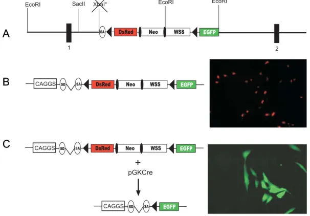
germ line to investigate their performance in vivo. As a first step, a reporter construct
allowing for quantitative monitoring of Cre activity was introduced into the ROSA26
locus by homologous recombination in HM-1 ES cells. This Cre reporter consists of a
loxP-flanked DsRed gene followed downstream by an eGFP gene. Cre-mediated
excision of DsRed turns on the eGFP gene which can be visualized by a change of
fluorescence from red to green. Second, various inducible Cre constructs were tested
in these Cre reporter containing ES cells. In order to avoid differences caused by
genomic integration site and variations in copy numbers of the transgene, these
constructs were targeted by knock-in into two defined genomic loci: the HPRT locus
and the ROSA26 locus. ES cell clones displaying favourable characteristics, i.e. high
inducibility of Cre paired with low background activity can be immediately injected
into blastocysts for the generation of chimeras. The in vivo characteristics of the
respective inducible Cre system can immediately be tested in vivo in chimeras, since
ES cell derived cells of the chimeras contain both the Cre reporter and the inducible
Cre construct. This test can yield quantitative results due to the nature of the Cre
reporter, which allows to distinguish between ES cell derived tissues in which Cre-
mediated recombination occurs (eGFP positive), and those in which no
recombination occurred (DsRed positive). This experimental set-up allows for the
rapid screening of inducible Cre systems in ES cells and in chimeras and
subsequently for the immediate generation of mouse strains from constructs
displaying favourable characteristics. In addition, I generated a posttranslational
inducible CrePR system restricted to B cells by targeted insertion into the CD19
locus.
2. MATERIALS AND METHODS
2.1 Chemicals and antibodies
All chemicals were obtained from Sigma (Steinheim, Germany) or Merck (Darmstadt, Germany), if not otherwise stated. Enzymes were delivered from the following companies: Boehringer (Mannheim, Germany), Gibco (Karlsruhe, Germany), NEB (Schwalbach, Germany), Stratagene (Heidelberg, Germany), and Takara (over Boehringer, Ingelheim, Germany). Size markers for agarose gel electrophoresis were provided from Gibco (1 kb marker, λ-HindIII Marker, 100 bp ladder).
Table1. List of antibodies
Specificity Clone Reference Supplier
B220/CD45R RA3-6B2 Coffman et al., 1982 Home made/
Pharmingen
CD4 GK.1.5/4 Dialynas et al., 1983 Pharmingen
CD5 53-7.3 Ledbetter & Herzenberg, 1979 Pharmingen CD8 53-6.7 Ledbetter & Herzenberg, 1979 Pharmingen
CD19 1D3 Krop et al., 1996 Pharmingen
Cre Polyclonal serum
Kellendonk et al., 1996 Babco Cre Polyclonal
serum
Novagen
Actin AC-15 Sigma
GFP Polyclonal serum
Clontech
TCR-β H57-597 Kubo et al., 1989 Pharmingen
2.2 Molecular biology
Standard methods of molecular biology were performed - if not otherwise stated - according to protocols described in Sambrook et al. (1989).
2.2.1 Competent E.coli and isolation of plasmid DNA
Competent Escherichia coli DH5α cells or Stbl-2 cells were prepared according to the
protocol of Inoue et al. (1990) and used in heat shock transformations of plasmid
instructions.
Plasmid DNA was isolated from transformed E. coli DH5α with an alkaline lysis method (QIAGEN, Hilden, Germany) according to the protocol of Zhou et al. (1990).
Plasmid DNA of a higher purity was obtained using QIAGEN columns (QIAGEN, Hilden, Germany) following the supplier’s instructions.
2.2.2 Constructions of expression plasmids and targeting vectors
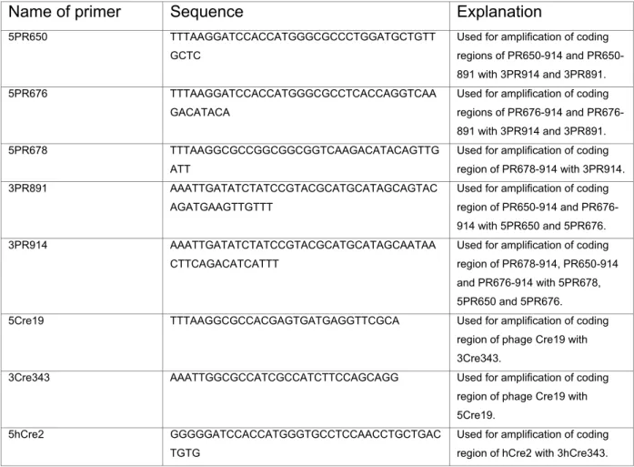
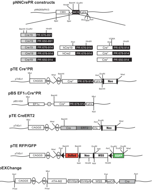
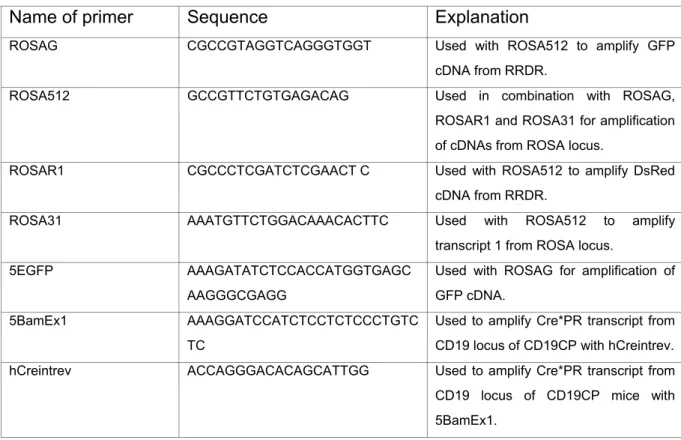
Primers for the amplification of cDNAs used in plasmid constructions are depicted in Table 2. Schematic overviews of the generated expression plasmids and targeting vectors together with major restriction sites are shown in Fig. 6-9. All constructs used for plasmid generation were confirmed by sequencing. Sequences of the generated plasmids and targeting vectors are available in chapter 10 supplementary data as pdf formats.
Table 2. Primers used for cloning
Sequences of oligonucleotides are shown in 5’ to 3’ direction. Explanation indicates the usage of the primer in a PCR reaction.
Name of primer Sequence Explanation
5PR650 TTTAAGGATCCACCATGGGCGCCCTGGATGCTGTT GCTC
Used for amplification of coding regions of PR650-914 and PR650- 891 with 3PR914 and 3PR891.
5PR676 TTTAAGGATCCACCATGGGCGCCTCACCAGGTCAA GACATACA
Used for amplification of coding regions of PR676-914 and PR676- 891 with 3PR914 and 3PR891.
5PR678 TTTAAGGCGCCGGCGGCGGTCAAGACATACAGTTG ATT
Used for amplification of coding region of PR678-914 with 3PR914.
3PR891 AAATTGATATCTATCCGTACGCATGCATAGCAGTAC AGATGAAGTTGTTT
Used for amplification of coding region of PR650-914 and PR676- 914 with 5PR650 and 5PR676.
3PR914 AAATTGATATCTATCCGTACGCATGCATAGCAATAA CTTCAGACATCATTT
Used for amplification of coding region of PR678-914, PR650-914 and PR676-914 with 5PR678, 5PR650 and 5PR676.
5Cre19 TTTAAGGCGCCACGAGTGATGAGGTTCGCA Used for amplification of coding region of phage Cre19 with 3Cre343.
3Cre343 AAATTGGCGCCATCGCCATCTTCCAGCAGG Used for amplification of coding region of phage Cre19 with 5Cre19.
5hCre2 GGGGGATCCACCATGGGTGCCTCCAACCTGCTGAC TGTG
Used for amplification of coding region of hCre2 with 3hCre343.
5hCre19 TTTAAGGATCCACCATGGGTGCCACGAGTGATGAG GTTCGCA
Used for amplification of coding region of hCre19 and Cre* with 3hCre343 and 3hCreV336A.
3hCre343 CCCTTGGCGCCGTCCCCATCCTCGAGCAG Used for amplification of coding region of hCre2 and hCre19 with 5hCre2 and 5hCre19.
3hCreV336A AAATTGGCGCCGTCCCCATCCTCGAGCAGCCTCGC CATGGCCCC
Used for amplification of coding region of Cre* with 5hCre19.
3PacloxBNco CACCATGGTGGATCCATAACTTCGTATAATGTATGC TATACGAAGTTATTTAATTAACCACTGGAAAGACCG CGAA
Cloning of RRDR
3loxAscEVEINheMsc TTTTGGCCAGCTAGCGAATTCGATATCGGCGCGCC GATAACTTCGTATAATGTATGCTATACGAAGTTATAA GCTTACTTACCATGTCAGAT
Cloning of RRDR
3BstEIIRFP TTTGGTCACCTTCAGCTTCACGG Cloning of RRDR 3EGFP TTTGATATCTAAGATACATTGATGAGTTTGGA Cloning of RRDR 3NotBambPA TTTGCGGCCGCGGATCCCCCCAGCTGGTTCTTTC Cloning of RRDR 3XhoFRTneo TTTCTCGAGGAAGTTCCTATACTTTCTAGAGAATAG
GAACTTCGGAATAGGAACTTCCCCCAGCTGGTTCTT TC
Cloning of RRDR
3RedStu TTTTAGGCCTCCCAGCCCATCGTCTTCTTCTGCATT ACGGGGCCGTCGGAGGGGAAGTTCACGCCGATGA ACTTCACCTTGTAGATGAAGCAGCCGTCTTGCAGTG ACGAGTCTTGGGTCACAGTCACGAC
Cloning of RRDR
3SacIIROSA TTTCCGCGGCCGCCGGATCCCCGCAAACGCACCAA GC
Cloning of pROSA26w/oATG
5BstEIIROSA AAAGGTGACCCTTCTTTCCCCC Cloning of pROSA26w/oATG 5RedBstEII AAAAGGTGACCAAGGGCGGCCCCCTGCCCTTCGC
CTGGGACATACTGTCGCCGCAATTCCAATAC
Cloning of RRDR
5PacloxRFP AAATTAATTAAATAACTTCGTATAGCATACATTATAC GAAGTTATCTGCAGGGATCCACCATGGGCAAGAAG AAGAGGAAGGTGGCCTCCTCCGAGAACGTCATCAC C
Cloning of RRDR
5SalWSS AAAGTCGACTCGGGGACACCAAATATGGC Cloning of RRDR 5EGFP AAAGATATCTCCACCATGGTGAGCAAGGGCGAGG Cloning of RRDR 5NotbPA AAAGCGGCCGCGGGGATCAATTCTCTAGAGC Cloning of RRDR 5SalFRTneo AAAGTCGACGAAGTTCCTATTCCGAAGTTCCTATTC
TCTAGAAAGTATAGGAACTTCTACCGGGTAGGGGA GGCG
Cloning of RRDR
XbaNheSA AAATCTAGAGCCTCGCTAGCATCTGTAGGGCGCAG TAGTC
Cloning of RRDR
3AscWSS AAAGCGGCCGCGGCGCGCCGATATCCTGCAGACTT ACCTTCCATGTCAGATCCAGACATGATAAG
Cloning of RRDR
3BamCAGGS TTTGGATCCGAATTCACGCGTGTTTAAACTTGCCCA GGAGCTGTAGGAA
Cloning of pExChange
3BamSV40 TTTGGATCCTACCACATTTGTAGAGGTTTTAC Cloning of pExChange 3EFPme TTTGTTTAAACCACGACACCTGAAATGGAAGAAAAA
AAC
Cloning of pExChange
5BamWSSmod AAAGGATCCTCGGGGACACCAAATATGGC Cloning of pExChange 5BglIISV40 AAAAGATCTCAGACATGATAAGATACATTGA Cloning of pExChange
5SpeCAGGS AAAACTAGTGGCGCGCCGTTTAAACTATTAATAGTA ATCAATTACGGG
Cloning of pExChange
3BamEx1 TTTGGATCCGGTAGCCAGGCTCCCTGGG Cloning of pCD19 3PacEx11 TTTTTAATTAACAGCTGTGGAAGAGAGAGC G Cloning of pCD19
3probeB GGATCCCTCTCATATCTTCATA Amplification of probe B for CD19 3XbaEx2 TTTTCTAGATTAATTAAGCTAGCACTAGTGGGGAGC
CCCAGCCCCTCT
Cloning of pCD19
5NheLA AAAGCTAGCCCTCCCGGACTCCTCACCT Cloning of pCD19 5NotEX1e AAAGCGGCCGCATTTAAATCTAAGCTAGAATGAAAC
CTA
Cloning of pCD19
5BamEx1 AAAGGATCCATCTCCTCTCCCTGTCTC Cloning of pCD19
5probeB CAGCTGAGTCTTATGAAAATGCA Amplification of probe B for CD19