zur
Erlangung der Doktorwürde der
Naturwissenschaftlich-Mathematischen Gesamtfakultät der
Ruprecht-Karls-Universität Heidelberg
vorgelegt von Ana Milosevic
aus
Uzice, Serbien und Montenegro Tag der mündlichen Prüfung:
Charakterisierung des csgA-Gens und des Einflusses seiner Inaktivierung auf die Entwicklung
Gutachter:Prof. Dr. Hans Ulrich Schairer Prof. Dr. Richard Herrmann
of the Rupertus Carola University of Heidelberg, Germany
for the degree of Doctor of Natural Sciencies
Presented by Ana Milosevic
born in Uzice, Serbia and Montenegro Heidelberg, 2003
Characterization of the csgA gene and influence of csgA inactivation on development
Examiners: Prof. Dr. Hans Ulrich Schairer Prof. Dr. Richard Herrmann
Dr. Hans Ulrich Schairer for giving me the opportunity to prepare this thesis in fascinating field of microbiology. I greatly appreciate his scientific supervision during my work, encouragement and help, and his valuable suggestions for improvement of the manuscript.
My grateful thanks goes to all my colleges, especially to Susanne Müller for her excellent collaboration during all three years, help in many practical matters during my work and reviewing the thesis manuscript, to Wulf Plaga for critical discussions, his patiently attendance to any questions and useful suggestions to overcome the experimental problems, to Esther Duperchy for her help and encouragemen, to Hong Wan and Andreas Leclerque for many useful suggestions and collaboration and to Diana Hofmann for all her advices concerning my work and great hospitality during my staying in her flat at the first days in Germany.
I am very grateful to Prof. Dr. Richard Herrmann for his scientific discussions and valuable advices. I am also very grateful for his continuous interest in my work.
Many thanks for all help and support I received from the people of the ZMBH.
My warmest thanks to my parents and my sister for their support and enthusiasm over the years, and for always believing in me.
1.2. Gliding motility ...4
1.2.1. Gliding motility of myxobacteria ...4
1.2.1.1. A system ...5
1.2.1.2. S system...5
1.2.1.3. The mgl locus...6
1.2.1.4. The frz locus ...6
1.2.1.5. Rippling ...7
1.3. Fruiting body formation...7
1.3.1. Pheromone activity in S.!aurantiaca...8
1.3.2. Artificially induced sporulation...9
1.3.3. Genes involved in S.!aurantiaca fruiting body formation ...9
1.4. Intercellular signalling and communication in bacteria ...11
1.4.1. Intercellular signalling in Gram-negative bacteria ...12
1.4.2. Intercellular signalling in Gram-positive bacteria ...12
1.4.3. Intercellular signalling in M. xanthus...13
1.4.3.1. A signalling...13
1.4.3.2. B signalling...13
1.4.3.3. C signalling...14
1.4.3.4. D signalling...16
1.4.3.5. E signalling ...17
1.4.4. Intercellular signalling in S.!aurantiaca...17
1.5. The aims of this work ...17
II. Results...19
2.1. Molecular cloning and sequence analysis of the csgA locus from S.!aurantiaca 20 2.1.1. Cloning of the csgA locus from S. aurantiaca...21
2.1.1.1. Preparation and screening of a S. aurantiaca genomic library...22
2.1.1.2. Subcloning of the 3 kbp XhoI fragment harbouring the S. aurantiaca csgA gene...23
2.1.1.3. Subcloning of the XhoI csgA fragment from l11 into the plasmid pACYC!177...24
2.1.2. Sequence analysis of the csgA locus from S. aurantiaca...24
2.1.2.1. Analysis of the upstream and downstream sequences of csgA ...29
2.2. Investigation of the physiological function of CsgA in vivo;...30
Disruption of the csgA gene in S.!aurantiaca...30
2.2.1. Construction of plasmid pAM8 ...30
2.2.2. Construction of the csgA insertion mutant AM8 ...31
2.2.3. Developmental phenotype of the csgA insertion mutant strain ...34
2.2.4. The ability of AM8 myxospores to germinate ...37
2.2.5. Ability of the wild type to restore developmental phenotype of mutant AM8 ...37
2.2.6. Interaction between WP120 and AM8 mutant cells ...38
2.3. Transcription of csgA in S.!aurantiaca...40
2.3.1. Determination of the csgA expression in the merodiploid mutant AM14....40
2.3.1.1.Construction of plasmid pAM14 ...40
2.3.1.2.Constructions of the merodiploid mutant strain AM14 ...41
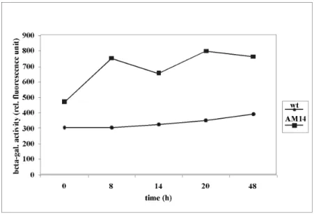
2.3.1.3. Determination of the b-galactosidase activity in AM14 ...42
2.3.1.4. Detection of the b-galactosidase activity in situ...44
2.3.1.5. Developmental phenotype of AM14...45
2.3.2. Detection of csgA expression by RT-PCR ...46
2.4. Production of CsgA in S.!aurantiaca...46
2.4.1. Heterologous expression of the fragment encoding antigenic determinants of CsgA ...47
2.4.1.1. Cloning the DNA fragment encoding the CsgA antigens into the pQE42 expression vector...47
2.4.1.2. Purification of the recombinant protein, 6xHis-DHFR-CsgA, under denaturing conditions ...49
2.4.1.3. Production of CsgA in S!aurantiaca...50
2.4.1.5. Production of anti-peptide antibodies ...52
III. Discussion...57
3.1. S.!aurantiaca csgA gene locus ...58
3.2. CsgA protein and similarity with members of the SRD family...61
3.3. Physiological function of CsgA ...62
3.4. Expression of the csgA gene in S.!aurantiaca...67
3.5. Immunological identification of CsgA in S.!aurantiaca...69
3.6. Perspectives...70
IV. Materials and Methods ...72
4.1. Materials ...73
4.1.1. Specified chemicals, consumables and equipments ...73
4.1.1.1. Chemicals ...73
4.1.1.2. Consumables...74
4.1.1.3. Equipment...74
4.1.2. Protein ...74
4.1.2.1. Antibodies...74
4.1.2.2. Protein weight standards ...74
4.1.3. Reagent kits for methods in molecular biology and enzymes...75
4.1.3.1. Reagent kits for methods in molecular biology...75
4.1.3.2. Enzymes ...75
4.1.4. Nucleic acids ...75
4.1.4.1. Plasmid ...75
4.1.4.2. Primers ...75
4.1.4.3. Oligonucleotides ...76
4.1.4.4. DNA and RNA molecular weight markers ...76
4.1.5. Bacterial strains ...76
4.1.5.1. Escherichia coli...76
4.1.5.2. S. aurantiaca strain ...77
4.1.6. Media and stocks solutions ...77
4.1.6.1. Media...77
4.1.6.2. Buffers and stock solutions ...77
4.2. Methods ...78
4.2.1. Microbiologic techniques...78
4.2.1.1. Growth of E.!coli...78
4.2.1.2. Growth of S.!aurantiaca...78
4.2.1.3. Indol induced sporulation of S. aurantiaca (Gerth and Reichenbach, 1994) ...78
4.2.1.4. Heat shock of S.!aurantiaca...78
4.2.1.5. S. aurantiaca fruiting body formation assay ...79
4.2.1.8. Preservation of S.!aurantiaca cultures ...79
4.2.1.9. Electroporation...79
4.2.1.9.1. Electroporation of E.!coli...80
4.2.1.9.2. Electroporation of S.!aurantiaca (Stamm et al., 1999) ...80
4.2.1.10. Blue white colony screening selection of E.!coli...80
4.2.1.11. Preparation of bacteriophage host E.!coli cells...81
4.2.1.12. Infection of E.!coli with the bacteriophages...81
4.2.1.13. Tittering of the phage library...81
4.2.1.14. Amplification of the S.!aurantiaca genomic phage library...82
4.2.1.15. Plaque lifts ...82
4.2.1.16. Purification of bacteriophage clones...82
4.2.2. Isolation and manipulation of DNA ...83
4.2.2.1. Isolation of plasmid DNA from E. coli...83
4.2.2.1.1. Isolation of plasmid DNA from E.!coli cells with alkaline lyses ...83
4.2.2.1.2. Isolation of plasmid DNA from E. coli cells with anion exchange columns...83
4.2.2.2. Isolation of chromosomal DNA from S. aurantiaca (Neumann et al., 1992) ...83
4.2.2.3. Isolation of lambda DNA ...84
4.2.2.4. Phenol/chloroform extraction of DNA (Sambrook et al., 1989) ...84
4.2.2.5. Alcohol precipitation of DNA ...84
4.2.2.6. Quantitation of DNA...84
4.2.2.7. DNA restriction...84
4.2.2.8. Partial digestion of DNA...85
4.2.2.9. DNA ligation ...85
4.2.2.10. Construction of the S. aurantiaca phage library...85
4.2.2.11. Filling of 5` overhanging ends of DNA ...85
4.2.2.12. Removing of 3` overhanging ends of DNA ...86
4.2.2.13. Dephosphorylation of DNA fragments ...86
4.2.2.14. Amplification of DNA fragments via PCR (polymerase chain reaction) ...86
4.2.2.15. Purification of PCR products...86
4.2.2.16. DNA sequencing...87
4.2.3. Electrophoresis of DNA...87
4.2.3.1. Agarose gel electrophoresis of DNA ...87
4.2.3.2. Recovery of DNA fragments from low-melting agarose gel ...87
4.2.4. DNA hybridisation...87
4.2.4.1. Dot blot analysis ...87
4.2.4.2. Southern blot analysis (Southern, 1975) ...88
4.2.4.3. Hybrisation and detection with biotin-labelled probes ...88
4.2.4.4. Nonradioactive labelling of DNA probes...89
4.2.5. Isolation and manipulation of RNA...89
4.2.5.1. Isolation of RNA from S.!aurantiaca cells...89
4.2.5.2. RNA electrophoresis ...90
4.2.5.3. RT-PCR ...90
4.2.6. Protein purification and analysis ...90
4.2.6.1. Isolation of total protein extract from S.!aurantiaca...90
4.2.6.2. Determination of protein concentration (Bradford, 1976) ...91
4.2.6.3. SDS-Polyacrilamide gel electrophoresis (Laemmli, 1970)...91
4.2.6.4. Coomassie blue staining of gels ...91
4.2.6.5. Immunobloting ...92
4.2.6.5.1. Protein transfer from the gel to the membrane ...92
4.2.6.5.2. Immunodetection ...92
4.2.6.5.2.1. Detection of the HRP conjugated secondary antibody-ECL ...92
4.2.6.5.2.2. Detection of the AP conjugated secondary antibody ...93
4.2.6.6. Anti-peptide antibodies ...93
4.2.6.6.1. Synthesis of the peptides ...93
4.2.6.6.2. Coupling of the peptides to a carrier protein ...93
4.2.6.6.3. Immunisation ...93
4.2.6.6.4. Purification of serum on peptide columns...93
4.2.6.7. Antibody against fusion protein...94
4.2.6.7.1. Preparation of E. coli cell lysate ...94
4.2.6.7.2. Purification of fusion protein on Ni-column ...94
4.2.6.7.3. Immunisation with fusion protein ...95
4.2.6.7.4. Concentrating protein solutions ...95
4.2.6.8. Determination of b-galactosidase activity (Ruan et al., 1993) ...95
4.3. Software ...96
V. Summary...97
VI. References ... 100
VII. Appendices ... 107
The myxobacteria have a remarkable life cycle that includes intercellular communication, cell differentiation and multicellular organisation. As a response to starvation myxobacterial cells undergo a specific developmental process leading to the formation of spores that are enclosed in fruiting bodies. Their development represents a model to investigate the flow of information between cells, signal transduction pathways and differential gene expression.
In higher organisms, complex morphological processes include differentiation of the cells from the same progeny into physiologically specialised tissues. Prokaryotic development includes changes in cell function and cell form in order to achieve benefit to the bacterial population to changes in environmental conditions. One aim of prokaryotic development is the formation of spores. In some bacterial species, as Bacillus subtilis, sporulation leads to the asymmetric division of the mother cell into two compartments (Piggot and Coote, 1976; Errington 1993). The small compartmen called the forespore, maturates to the metabolically quiescent endospore. The large compartment resembles the mother cell that lyses after spore maturation to set the spore free. In myxobacteria, differentiation of the vegetative cells into spores takes place at the end of the complex developmental cycle. As in higher organisms cell differentiation in myxobacteria is preceded by extensive cell movements and the formation of multicellular structures. In order to build up these multicellular structures myxobacterial cells coordinate their behaviour by intercellular signalling and direct cell-to-cell contacts.
The formation of fruiting bodies of myxobacteria shows great similarities to the life cycle of the cellular slime mould Dictyostelium discoideum (Raman, Hashimoto et al., 1976). During starvation, these unicellular amoebae form multicellular structures from which spores are formed that germinate when the conditions become more favourable.
The experimental accessibility of myxobacteria along with features mentioned above, represent them as a valuable prokaryotic model to study morhogenesis and development.
Myxobacteria are Gram-negative bacteria classified in the order Myxococcales that belongs into the delta-branch of the Proteobacteria. Stigmatella aurantiaca and the closely related Myxococcus xanthus are the best studied species of the myxobacterial group.
From the time of their detailed description by Ronald Thaxter in 1892 (Pfister, 1984) until now, myxobacteria fascinate scientist with their complex life cycle. The life cycle of the myxobacteria is bipartite. It is composed of a vegetative growth cycle and the developmental cycle, which is triggered by starvation. Upon nutrient depletion, the cells migrate into aggregations centres, from which fruiting bodies containing the myxospores arise. When nutrients become available, the myxospores germinate and the vegetative cycle starts again.
Myxobacteria grow on insoluble organic substrates such as decaying wood or leaves.
Vegetative cells are rod shaped and about three times longer than E.coli cells. Since they do not have a flagella, the cells move by gliding, a special way of moving on a solid surface. Myxobacterial cells interact with each other forming a swarming community. The cells secrete slime containing lytic enzymes: lysozymes, proteases and also cellulases that degrade biopolymers. This way of feeding can be achieved only at high cells density, the so-called "wolf pack effect" (Dworkin, 1963).
Myxobacterial cells communicate with each other by direct cell-to-cell contact and by exchanging different signal molecules in the swarming community as well as during development. They represent so-called social prokaryotes.
Myxobacteria have a very large genome in comparison to other bacterial species (about two times larger than the genome of E. coli). The size of the S. aurantiaca genome is about 9,35 Mbp and is approximately equal to that of the myxobacterium M. xanthus (Chen et al., 1990; Neumann et al., 1992). The extremely large size of the myxobacterial genome reflects the potential to build multicellular structures during
development and the capability of these bacteria to produce a broad range of secondary metabolites (Schairer, 1993).
As a group myxobacteria produce a large spectrum of secondary metabolites like epothilon (Gerth et al., 1996), myxothiazol (Gerth et al., 1980), myxalamid (Gerth et al., 1996), stigmatellin (Kunze et al., 1984), soraphen (Gerth et al., 1994), TA (Rosenberg et al., 1973). Some of them are proven to be clinically very important.
1.2. Gliding motility
Myxobacteria move by gliding, a special form of locomotion that requires a solid surface (Burchard, 1984). Gliding cells move in the direction of their long axis, with stop intervals between and the reversal of the gliding direction. Many different classes of bacteria move by gliding. Recent studies suggest that bacterial gliding motility cannot be explained by only one model system. It is more likely that different types of motors are involved in gliding motility in different classes of bacteria. Some bacteria use type IV pilus extension and retraction powered by ATP hydrolysis to move over the surface (Merz et al., 2000). Gliding of some filamentous cyanobacteria depend on the polysaccharide extrusion (Hoiczyk and Baumeister, 1998). Speculation about gliding in the myxoplasma group suggests involvement of the cytoskeleton and the surface adhesion proteins (Korolev et al., 1994; Lünsdorf and Schairer, 2001).
1.2.1. Gliding motility of myxobacteria
In M. xanthus, gliding motility is controlled by two separated multigene systems known as A (adventurous) and S (social) system (Hodgkin and Kaiser, 1979). The A system controls gilding of single cells, the S system is responsible for gliding of cells in groups. These two systems contribute equally to the wild-type gliding phenotype.
The A system includes a minimum of 37 genes whose products control the interaction of the cell with the solid surface (Hodgkin and Kaider, 1979; MacNeil et al., 1994). Mutants defective in A motility are divided in to two classes cgl (conditional gliding) and agl (adventurous gliding) (Hodgkin and Kaiser, 1979). There are several hypotheses about the mechanism of A motility.
One hypothesis suggests that import and export of macromolecules may be the direct force that move the cells, something like propulsion of the cells. This hypothesis is mostly based on the finding that the AglU lipoprotein has similarities to the TolB protein of E. coli (White and Hartzell, 2000). The TolB protein is part of a large protein complex, which uses proton motive force to transport molecules across the outer membrane.
Another hypothesis suggests the involvement of some structures of the cell wall in A motillity. The involvement of the specific surface structures in gliding was observed for the first time from the scanning electron micrographs of four different gliding bacteria species including S. aurantiaca and M. xanthus (Lünsdorf and Schairer, 2001). These structures are described as chain-like strands that associate with each other and form bands, which are wrapped, helically around the cell. The helical bands were not observed on the surface of cells treated with sodium azide or potassium cyanide. These two chemicals blocked the respiratory chain so that the cells were frozen and gliding motility was stopped.
1.2.1.2. S system
The S motility relies on the type IV pili. The S motility mutants lack polar pili and also removal of the pili from the wild type cells leads to defects in S motility (Kaiser, 1979). The pil gene cluster whose products are involved in pilus biogenesis are identified. PilA is the primary pilin protein, PilB is the putative NTPase functioning in pilus biogenesis, PilT is the putative NTPase acting in pilus retraction. PilG,-H and I are suggested to form an ATP-binding transporter involved in the transport of proteins
required in the pilus biogenesis (Wall et al., 1999; Wu et al., 1997; Wu et al., 1998). In addition to the pil genes, one other gene tgl (transient gliding) is required for S motility.
Tlg is thought to be a lipoprotein whose function may be to facilitate pilus protrusion or pilus retraction (Rodriquez-Soto and Kaiser, 1997).
A model system that suggests the mechanism of the S motility proposes that pili are extended from the leading pole of the gliding cell. Contact between pili and surface induces pilus retraction, which results in cell movement (Kaiser, 2000; Sun et al., 2000).
1.2.1.3. The mgl locus
Another locus in M. xanthus with an important role in gliding motility is designated as mgl (mutual gliding). Mutations in the mgl locus abolish gliding motility of the cells.
Two cotranscribed genes mglA and mglB have been identified (Stephens et al., 1989).
The predicted amino acid sequence of MglA shows homology to the members of the GTP-binding protein class. MglA might have an important role to control expression of genes whose products are required for gliding motility (Hartzell, 1997). The predicted sequence of MglB exhibits similarities to one of the calcium binding sites of the yeast calmodulin (Hartzell and Kaiser, 1991).
The S. aurantiaca mgl genes were identified with a sequence homology of about 90% to the mglA and mglB genes of M. xanthus. Insertional mutagenesis showed that the mgl genes in S. aurantiaca are required for the motile phenotype of the cells (Schairer, 1993).
1.2.1.4. The frz locus
A genetic locus involved in the control of the frequency of reversal movements in M. xanthus is called `frizzy` (frz). Cells with mutations in the frz genes either reverse direction much less frequently or much more frequently than wild type cells. The frz mutants showed impaired aggregation but produced normal spores. Six genes were identified frzA,-B,-CD,-E,-G and –F with homology to the chemotaxis genes of
Zusman, 1999).
1.2.1.5. Rippling
Rippling is a rhythmical movement of cells. The cells start to move synchronically to form a series of equidistant parallel ridges, which move in a pulsating manner.
Myxobacteria appear to be the only procaryotes with this specific rhythmic behaviour (Reichenbach, 1986). Rippling precedes fruiting body formation but is not required for it. Rippling is induced by peptidoglycan. Thus, presence of rippling cells appears to be a sensitive indicator for the presence of extracellular peptidoglycan components. So it is more likely that rippling is incidental with fruiting body formation because rippling is induced by releasing peptidoglycan during development. Rippling requires the CsgA protein which is an extracellular polypeptide essential for M. xanthus development (Shimkets and Kaiser, 1982).
1.3. Fruiting body formation
As mentioned above, myxobacteria have a complex life cycle. During the vegetative growth phase, cells divide by transverse fission. Upon starvation, cells start to glide into aggregation centers from which the fruiting bodies arise. In the fruiting bodies vegetative cells differentiate into spherical, dormant myxospores. Each fruiting body encloses 105 myxospores, respectively. The shape of the fruiting body is species specific. Whereas M.!xanthus fruiting bodies are simple mounds filled with spores, S.!aurantiaca forms morphologically complex structures, resembling a small tree, with a branched stalk harbouring several sporangioles.
The morphological changes occur in a defined temporal order during development.
In S.!aurantiaca the whole process takes about 24 h. Different morphological stages during development are defined as early aggregates, early stalk (morel-like structure), late stalk (champignon-like structures) and mature fruiting bodies that are visible about 9, 12, 15 and 24 h after the beginning of starvation (Qualls et al., 1978a). Fruiting body
formation of S.!aurantiaca is stimulated by incandescent light and requires the production of a pheromone (Qualls et al., 1978b).
Fig.1.1. Diagram of the myxobacterial life cycle (Dworkin, 1985). Fruiting body of M. xanthus and S. aurantiaca are illustrated.
1.3.1. Pheromone activity in S. aurantiaca
S. aurantiaca cells secrete and respond to a pheromone that is necessary for fruiting body formation. The pheromone was eluted from cells assayed for fruiting body formation on filter paper and purified by steam distillation followed by reversed-phase and normal-phase HPLC (Plaga et al., 1998). It is a branched aliphatic hydroxy ketone, 2,5,8-trimethyl-8-hidroxy-nonan-4-one, named stigmolone (Hull et al., 1998).
Stigmolone is a new type of a pheromone molecule in prokaryotes since elucidation of its chemical structure showed that it does not belong to any known class of pheromones up to now. It acts in concentrations of about 1 nM to shorten the time of aggregation on the beginning of development in a bioassay (Plaga et al., 1998). Addition of purified stigmolone accelerated the rate of aggregation when added to 5x10⁷ cells. The aggregation rate was comparable to that observed in a population of 2x10⁸ cells as
correlates with the number of cells. Therefore, stigmolone may have a role in “quorum sensing” at the beginning of the developmental cycle. Species-specifity is indicated by the fact that M. xanthus does not respond to stigmolone by accelerating fruiting body formation in a bioassay (Plaga et al., 1998).
Cells have to be in contact with each other or with a solid surface to secrete stigmolone. The stigmolone biosynthetic pathway and the putative pheromone receptor are still unknown. The structure of the stigmolone suggests the involvement of some metabolites from the catabolism of leucine or from a biosynthetic pathway leading to terpentoids, fatty acids or polyketides (Plaga et al., 1998).
1.3.2. Artificially induced sporulation
Sporulation can be induced independently from fruiting body formation by addition of various chemicals. In S. aurantiaca indol and some indol derivates are the most potent inducers of sporulation (Dworkin, 1994; Gerth and Reichenbach, 1978).
The starvation dependent and starvation independent sporulation follows a time scale and has different nutritional requirements and different inducers. There are also structural differences between the two types of spores. The starvation-independent spores of M. xanthus lack the fruiting body spore protein S (Komano et al., 1980), the coat is thinner (Zusman, 1980) and they contain more ribosomes. Both kinds of spores contain protein U (Komano et al., 1980) and both pathways of sporulation induce a beta-lactamase activity (O´Connor and Zusman, 1997).
1.3.3. Genes involved in S. aurantiaca fruiting body formation
To identify developmentally regulated genes in S. aurantiaca Tn5lacZ transposon mutagenesis was performed (Pospiech et al., 1993). Three different classes of mutants impaired in fruiting body formation were detected. Members of the first class form abnormal fruiting bodies, those of the second-class aggregate into clumps, and those of the third class of mutants are not able to aggregate at all (Pospiech et al., 1993).
Further analysis of the transposon induced mutant AP182 led to the identification of the fbfB gene involved in fruiting. Analysis of the upstream and downstream regions of fbfB showed the existence of further fbf genes, fbfA, fbfC, fbfD (Müller, 2002;
Silakowski et al., 1996) that are arranged in the same orientation. fbfC and fbfD form an operon and more or less, the whole fbfA sequence is needed for the correct expression of fbfCD. The gene fbfB is located upstream of fbfA in a divergent orientation (Silakowski et al., 1998). The gene product of fbfA shows a homology of about 30% to the N-acetylglucoseamine transferase (NodC) of Rhizobium meliloti and the chitin synthase of Saccharomyces cerevisiae. These enzymes are involved in the synthesis of extracellular polysaccharides. FbfA therefore might be an enzyme catalying the synthesis of extracellular polysacharides that are involved in signalling. FbfB encodes a putative protein that shows homology to the galactose oxidase of Dactylium dendroides (Silakowski et al., 1998). A putative function of FbfB could be the oxidation of primary alcohols to aldehydes (Silakowski et al., 1998). The putative FbfC polypeptide has no homology to known proteins. FbfD shows homology to an ORF with unknown function of M. xanthus (Müller, 2002). Insertion of the neo gene into each of the fbf genes led to mutants that form just clumps during starvation. Mixing of fbfA mutant cells with the nonagreggating transposon mutant AP191 led to a partial phenotypic complementation, the formation of a morel-like structure (Silakowski et al., 1996). Mixing of the fbfB mutant cells with AP191 led to the formation of a champignon-like structure.
Analysis of the fbf gene expression in merodiploid strains containing various large upstream regions of the analysed fbf gene 3'truncated and fused to the DtrpA-lacZ reporter gene revealed that each gene from the cluster is expressed during development.
The fbfA gene is transcribed about 8 h after the start of development, fbfB is expressed about 14 h after induction of the fruiting body formation. The genes fbfC and fbfD are both expressed about 8 h after induction of starvation. Downstream of the fbfB gene the mta gene cluster was detected encoding polyketide synthases and nonribosomal peptide synthetases. These two mta cluster products are involved in the synthesis and modification of the secondary metabolite myxothiazol and not in fruiting body formation (Silakowski et al., 1998; Silakowski et al., 1999).
Bacteria use sophisticated chemical communication systems in order to coordinate the behaviour of their populations. This capability is important to improve access to different nutrient sources, to achieve rapid colonisation of a new ecological niche.
Bacterial communication also allows survival of the population by differentiation into morphologically more resistant forms or defence against competitive microorganisms or the eukaryotic immune system (Shapiro, 1988). Bacterial cells are able to respond to different molecules produced by bacteria but also by plants or animals cells, and the other way round. Cell density dependent conjugal transfer of Ti plasmids between Agrobacterium tumefaciens cells is triggered by opines produced by the plant host (Zhang et al., 1993). The homoserine lactone which is the density sensing molecule in Pseudomonas aeruginosa can also influence the host immune response (Telford et al., 1998). Some pathogenic bacterial species produce molecules that can bind to hormone receptors and in this way bacteria may manipulate eukaryotic host cell signal transduction pathways.
Information transfer between cells determines differentiation and morphogenesis in a wide variety of bacterial systems: induction of luminescence in Vibrio by homoserine lactones, sporulation in Bacillus, erection of aerial hyphae by Streptomyces, fruiting body formation in myxobacteria.
Signalling molecules can be small diffusible molecules and secreted polypeptides as well as surface associated macromolecules. They are also called bacterial "hormones",
"pheromones" or "autoinducers" (Wirth et al., 1996). Bacteria use signalling molecules to monitor the state of other cells in the population. The so-called "quorum sensing", a cell density sensing mechanism, enables bacteria to function as multicellular organisms.
This cell density sensing mechanism depends on the activation of a response regulator by a self-generated diffusible signal molecule.
1.4.1. Intercellular signalling in Gram-negative bacteria
Gram-negative bacteria use homoserine lactones as small signalling molecules that diffuse across the outer and inner membrane to reach their target protein in the cytoplasma. One of the first described autoinducers was the N-acyl-homoserine-lactone autoinducer (AHL) from the marine bacterium Vibrio fisheri involved in the control of bioluminiscence. Luminescence operons consist of several genes (Engebrecht et al., 1983). The luxR gene encodes an autoinducer dependent transcriptional activator of the luxI-G operon. LuxI is an autoinducer synthase. LuxC, D and E form a complex that generates long-chain fatty aldehyde, actual substrates of the luciferase reaction. LuxA and B are two subunits of the luciferase. The function of LuxG is unknown. Cellular and enviromental concentration of this signal molecule (AHL) is identical, since it diffuses freely through membrane (Kaplan and Greenberg, 1985). At high cell densities the concentration of the autoinducer increases and reaches a sufficient high concentration to bind to LuxR. LuxR in turn activates the transcription of the lux operon.
Beside the homoserine lactone mediated "quorum sensing" other molecules with signalling function have been identified. The gama-butyrolactones in Streptomyces are involved in the control of antibiotic biosynthesis, resistance and differentiation (Horinouchi and Beppu, 1992). Butyrolactones have antifungal activity in Pseudomonas aureofaciens (Gamard et al., 1997) and the 3-hydroxypalmitic acid methyl ester is involved in regulation of virulence in the plant pathogen Ralstonia solanacearum (Flavier et al., 1997).
1.4.2. Intercellular signalling in Gram-positive bacteria
Gram-positive bacteria, use small modified peptides as signalling molecules that can interact with two-component histidine kinase signal trasduction systems (Wirth et al., 1996). Small octapeptides act as signalling molecules to regulate cell density dependent virulence gene expression in Staphylococcus aureus (Ji et al., 1995). Extracellular signal peptides are involved in the initiation of sporulation in Bacillus subtilis.
Streptococcus pneumoniae (Pestova et al., 1996).
1.4.3. Intercellular signalling in M. xanthus
Myxobacterial development strictly depends on signalling between cells. These signals coordinate temporal gene expression in the course of development. The cell-cell signal mutants are unable to complete development by themselves, but they can overcome this developmental block when they are mixed with wild type cells. The result of the complementation studies indicate that cell-cell signalling mutants can be placed into several different classes. Mutants from the same class fail to complement each other. Mutant cells are defective in producing a signal but they retain the ability to respond to the signal (Hagen et al., 1978; Janssen and Dworkin, 1985; LaRossa et al., 1983; Shimkets and Dworkin, 1981). At least five different signalling pathways have been identified in M. xanthus.
1.4.3.1. A signalling
Mutants defective in producing the A signal arrest at about 1 to 2 hours after initiation of development in the preaggregation stage as a flat film of cells. Five genes known as asgA, asgB, asgC (Kuspa and Kaiser, 1989; Plamann et al., 1994; Plamann et al., 1995; Shimkets, 1999), asgD (Cho and Zusman, 1999) and asgE (Garrza et al., 2000) have been identified to function together in order to produce the active A signal.
The A signal is proposed to be a mixture of amino acids and peptides generated in amounts proportional to the cell density by extracellular proteolysis (Kuspa et al, 1992;
Plamann et al., 1992).
1.4.3.2. B signalling
The B signal acts early in development. All of the bsg mutations fall into one single gene named bsgA (Gill and Cull, 1986). Mutants fail to aggregate, sporulate, and are unable to express developmentally regulated genes. The bsgA gene product is an ATP- dependent protease with homology to the Lon protease of E. coli (Gill et al., 1993). The
suggested role of the BsgA protease is that it is involved in the regulation of the initiation of the developmental phase.
1.4.3.3. C signalling
The C signalling pathway is the most intensively studied in M. xanthus. The C signal acts about 6 h after the beginning of development. All csg mutations fall into a single genetic locus named csgA, formerly known as spoC (Shimkets et al., 1983). Mutants unable to synthesized CsgA fail to ripple, aggregation and sporulation are severely impaired and expression of developmental genes that is normally induced 6 hours after the beginning of starvation is reduced or abolished (Shimkets et al., 1983; Kroos and Kaiser, 1987). Overproduction of CsgA leads to premature aggregation and sporulation as well to the formation of small fruiting bodies (Kruse et al., 2001). In contrast, reduced synthesis of CsgA causes a delay in aggregation, reduces the ability to sporulate and causes the formation of large fruiting bodies (Kruse et al., 2001). csgA expression slowly increases during development and reaches a peak at the sporulation stage (Hagen and Shimkets, 1990).
The csgA mutant phenotype can be restored by adding the purified CsgA from immature wild type fruiting bodies or the MalE-CsgA fusion protein produced in E. coli (Kim and Kaiser, 1990; Lee et al., 1995).
The predicted amino acid sequence of CsgA shows homology to the members of the short-chain alcohol dehydrogenases family. These enzymes use NAD(H) or NADP(H) to catalyze the interconversion of secondary alcohols and ketones or mediate decarboxylation (Persson et al, 1991). The CsgA protein with a single mutation at the N terminus was unable to bind radiolabeled NAD+ in vitro and to rescue the csgA mutant phenotype (Lee et al., 1995). A single amino acid substitution in the putative substrate binding domain of the CsgA protein leads to a mutant unable to develop (Lee, et al., 1995).
The CsgA is an extracellular protein associated with cell surface (Kim and Kaiser, 1990; Shimkets and Rafiee, 1990). It is still unclear if CsgA has an extracellular
whether CsgA acts as a signal itself.
The reported importance of the putative coenzyme binding site and the putative substrate binding site for CsgA function support the first model (Lee, et al., 1995).
Additionally, overproduction of SocE, another member of the short-chain alcohol dehydrogenase family, rescues the developmental phenotype in the csgA mutant (Crawford and Shimkets, 2000). The putative CsgA substrate is unknown and it is hard to predict the structure of the substrate since members of the short alcohol dehydrogenase family have a large spectrum of substrates. The second model is based on the findings that the 17 kDa protein isolated from wild type cells during development can restore the csgA mutant phenotype (Kim and Kaiser, 1990). Two forms of the CsgA protein have been identified in extracts of developmental cells. A large form of 25 kDa that corresponds to the full-length protein encoded by the csgA gene and a smaller form of 17 kDa (Kruse et al., 2001). The large form might represent a precursor protein that becomes proteolytically cleaved to a polypeptide of 17 kDa that has C signalling activity.
Despite unclear nature of the C signal, the cellular responses to C signalling are known. As mentioned above it induces rippling, aggregation, sporulation and expression of many genes including csgA itself. It was shown by addition of the purifed 17 kDa protein to csgA mutant cells (Kim and Kaiser, 1990) and by reducing the transcription of csgA in vivo by nested deletions of the upstream region (Li et al., 1992) that a low concentration of the C signal is required for rippling and aggregation. A higher concentration induces sporulation and C signal dependent gene expression including csgA itself. Therefore the model of the C signalling pathway indicates two branches.
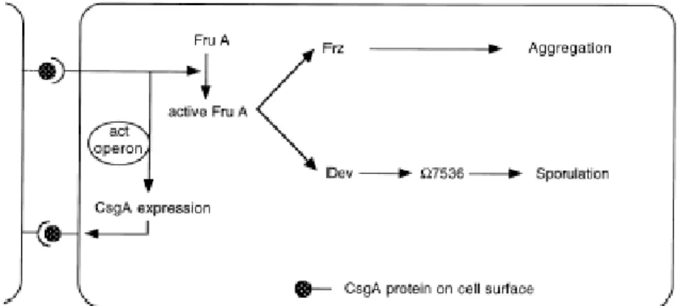
One branch leads to the regulation of the movement responds of cells and the other branch controls sporulation and expression of the late developmental genes (Fig.1.2.).
Fig. 1.2. A model for the C-signal transduction pathway (Gronewold and Kaiser, 2001).
Upstream in the C signalling pathway, the act operon controls the level and the time course of CsgA production (Gronewold and Kaiser, 2001). The CsgA activates FruA, which is a transcriptional regulator protein with a putative helix-turn-helix DNA binding domain (Ellehauge et al., 1998). Synthesis of FruA is regulated on the transcriptional level and does not depend on C signal (Ellehauge et al., 1998). More likely C signal transmission induces activation of FruA presumably by phosphorylation (Ellehauge et al., 1998). Downstream from FruA the C signal pathway branches. One branch leads to the regulation of rippling and aggregation. Active FruA induces methylation of FrzCD protein, a homolog to bacterial chemotaxis proteins. Frz proteins are important for rippling and aggregation and control of specific gliding parameters in response to the C signal (Zusman 1982; Jelsbak and Sogaard-Andersen, 1999).
Increased methylation of the FrzCD correlates with a decreased reversal frequency, allowing the cells to move in chains into the aggregation center. When cells aggregate, the level of C-signal increases. In the presence of high level of a C signal and active FruA, the dev operon and other late developmental genes are expressed which are required for sporulation (Thöny-Meyer and Kaiser, 1993).
1.4.3.4. D signalling
The D signal acts between 1 and 2 h after beginning of development. Only one gene is identified, designated as dsgA (Chang and Kaiser, 1989). Mutations in dsgA delay aggregation and reduce the sporulation efficiency. The dsgA gene encodes a protein with 50 % similarity to the translation initiation factor IF3 of E. coli (Chang and Dworkin, 1994).
1.4.3.5. E signalling
The E signal acts about 3-5 h after the beginning of development. The E signal mutant fails to aggregate or sporulate normally. Two genes were identified esgA and esgB that encode the E1a and E1 b subunits of the branched-chain a-keto acid dehydrogenases involved in amino acid and fatty acid metabolism (Downard and Toal, 1995; Toal et al., 1995). Downard and Toal (1995) assumed that long branched-chain keto acids are incorporated into the phospholipid membrane during vegetative growth.
During development they are released and act as the actual E signal.
1.4.4. Intercellular signalling in S. aurantiaca
S. aurantiaca forms morphologically more complex fruiting bodies in comparison to M. xanthus and therefore represents a better prokaryotic model to study genetic determination of morphogenesis. Formation of the complex fruiting bodies of S. aurantiaca consist of a branched stalk with sporangioles requires specific communication between the cells in order to coordinate their behaviour. One type of the signalling molecule was isolated from S. aurantiaca cells (Plaga et al., 1998). As mentioned above it is a novel type of pheromone, which acts to help cells to stay together in the aggregation phase.
Another gene product involved in intercellular signalling was identifed in S. aurantiaca, the csgA homolog of M. xanthus. Inactivation of the csgA gene in S. aurantiaca was reported to affect fruiting body formation (Butterfass, 1992).
1.5. The aims of this work
As mentioned above S. aurantiaca is a social prokaryote. Cells communicate with each other by direct contact or by exchanging various signal molecules. Isolation and characterization of these signalling molecules would contribute to the understanding of the complex life cycle of S. aurantiaca.
The M. xanthus csgA gene homolog was identified in S. aurantiaca. Inactivation of the gene was shown to impaire fruiting body formation. Because of the strong evidences supporting the role of the CsgA protein in intracellular signalling in M. xanthus, an initial characterisation of the csgA gene in S. aurantiaca was undertaken.
The work of this thesis includes cloning of a DNA fragment harbouring the csgA gene flanked by its upstream and downstream sequences; a detailed sequence analysis of the csgA upstream region; elucidation of the CsgA function in vivo by disrupting the csgA gene and observation of the mutant phenotype during starvation; investigation of csgA transcription in a merodiploid strain that contains the upstream region of csgA fused to a lacZ reporter gene; immunological identification of the CsgA protein during S. aurantiaca growth and development.
2.1. Molecular cloning and sequence analysis of the csgA locus from S. aurantiaca
In M. xanthus the csgA encoded polypeptide is a molecular timer for the developmental program. It constitutes the C signalling pathway that is required for regulation of the correct temporal order of the three morphological stages known as rippling, aggregation and sporulation during development. It is also involved in the regulation of the expression of developmental genes that are expressed about 6 h after the beginning of starvation. According to the important role of CsgA in the M. xanthus development and because of the close phylogenetic relation to S. aurantiaca, the question arised if there is a similar C signalling pathway in S. aurantiaca involved in intercellular signalling during fruiting body formation.
To address this question it was first necessary to identify the homologous gene in S. aurantiaca. Previously the S. aurantiaca csgA homolog was identified by Southern blot analysis using a M. xanthus csgA gene probe. An EcoRI fragment (12 kbp) harbouring the csgA gene was subcloned from a S. aurantiaca lambda gene library into the plasmid pUC18. Sequence analysis revealed that an EcoRI fragment contained the coding part of the csgA gene flanked by a 148 bp upstream sequence. The S. aurantiaca csgA gene sequence was added into the EMBL/GenBank data base, accession number M95300 (Butterfass, 1992). The csgA gene encodes a protein of 173 amino acids with a predicted molecular mass of about 19 kDa. The gene sequence shows about 70 % homology to the M. xanthus csgA gene. The deduced amino acid sequence of the S. aurantiaca csgA revealed an identity of about 54% to the M. xanthus CsgA.
A csgA S. aurantiaca mutant phenotype was reported previously (Butterfass, 1992).
A merodiploid mutant strain was constructed in which 2 truncated copies of the gene were present, separated by vector sequences. One csgA allele was lacking the 3´part and another csgA allele was disrupted by the insertion of a kanamycin cassette, so that the merodiploid mutant did not contain a functional csgA gene. Under starvation conditions this mutant formed a bulk of sporangioles without a differentiated stalk and a delay of 12 hours in the time course of the mutant development as compared to the wild type was reported (Butterfass, 1992).
2.1.1. Cloning of the csgA locus from S. aurantiaca
On the basis of this data, the putative role of the csgA gene product during S. aurantiaca development should be analysed in detail. To characterize csgA expression during development and to construct a csgA insertional mutant strain (double recombination) it was first necessary to isolate a larger DNA fragment harbouring the csgA coding region flanked by the whole upstream and by long downstream sequences.
A 465 bp internal fragment of the csgA gene was amplified by PCR using the chromosomal DNA of the S. aurantiaca wild type as a template and the primers csgA 7/csgA 8. The purified PCR product was biotin labelled and used as a probe in a Southern analysis of S. aurantiaca wild type DNA digested with various restriction enzymes. The SalI-, XbaI-, NotI-, HindIII-, SacI- chromosomal DNA fragments which hybridized with the probe were too large (more than 7,7 kbp) for subcloning in standard cloning vectors (Fig.2.1.). Restriction with BamHI resulted in a fragment of about 1,7 kbp which was not large enough to contain the upstream and downstream regions of the csgA gene. A 3 kbp XhoI fragment that hybridized with the probe had a suitable size for further cloning.
Fig.2.1. Southern analysis of restricted chromosomal DNA of the S.aurantiaca wild type using the biotin labelled S. aurantiaca csgA gene as a probe. 10 mg of DNA (lanes: 2-8) were digested with SalI, BamHI, XbaI, NotI, HindIII, SacI, XhoI and separated on a 0,9 % agarose gel. The size of the fragments were estimated using the DNA Molecular Weight Marker IV (Roche Diagnostics)-lane 1.
2.1.1.1. Preparation and screening of a S. aurantiaca genomic library
In order to facilitate cloning of S. aurantiaca csgA gene locus, a genomic library was constructed. The bacteriophage vector Lambda DASH II (Stratagene) predigested with restriction enzyme BamHI, which accomodates inserts ranging from 9 to 23 kbp, was used as the vector to construct the genomic library of S. aurantiaca. To obtaine a truly random library, high molecular weight chromosomal DNA from S. aurantiaca was fractionated by partial digestion with the restriction endonuclease Sau3AI which produces BamHI-compatible cohesive ends. Serial dilutions of Sau3AI were prepared to determine the optimal concentration of the enzyme necessary for generating DNA fragments with a size of 9 to 23 kbp. Partial digestion was accomplished using 0,025 U of Sau3AI per microgram of chromosomal DNA in a reaction incubated at 37°C for 40 min. Obtained DNA fragments were cut out from the gel and purified with the gel extraction kit (Qiagen). Library construction was performed following the instruction manual of lambda DASH II/ BamHI vector kit (Stratagene) and the detailed steps are described in Materials and Methods. The size of the constructed genomic library was determined to be 1x10⁵ plaque forming unit (pfu)/mg of genomic DNA, which offers more than 99% probability of isolating a particular gene from a bacterial genome.
The 465 bp csgA PCR fragment from the S. aurantiaca wild type (see 2.1.1) was used as biotin labbeled probe to screen the S. aurantiaca phage library. Six independent plaques that hybridized with the probe were detected. The DNA of the positive phages was isolated and Southern analysis was performed using the same probe as for the screening of the library. One positive clone designated l11, respectively, was chosen for further work. In the Southern analysis (Fig.2.2.) of XhoI digested l11 DNA one fragment of about 3 kbp was detected. Double digestion with XhoI/EcoRI revealed one fragment of about 1,5 kbp which hybridized with the csgA probe. According to previous data, l11 probably harboures about 1,5 kbp of the upstream region of csgA. Digestion of lambda 11 with SalI revealed a fragment of about 11 kbp that hybridized with the probe.
Fig.2.2. Southern analysis of restricted l11 phage DNA using the csgA gene as a probe. Lane 1- 2: l11 DNA (2 mg) digested with XhoI; XhoI/EcoRI and SalI separated on 0,9 % agarose gel.
The size of the fragments was estimated using DNA Molecular Weight Marker IV (Roche Diagnostics).
2.1.1.2. Subcloning of the 3 kbp XhoI fragment harbouring the S. aurantiaca csgA gene
The 3 kbp XhoI fragment harbouring the csgA gene was purified from l11 phage DNA, restricted with XhoI and separated on a 0,8 % low-melting agarose gel using the gel extraction kit (Qiagen). Isolated csgA fragment was used in subsequent ligation reactions with different vectors: Litmus 28, pBSSK-, pBCSK- and pUC18, pre-digested with XhoI. Ligation reactions were used for the transformation of E. coli DH5a. After white-blue screening and restrictions analysis positive transformants were not identified.
Since subcloning of the XhoI fragment in the previously described vectors did not leads to sucess when the E. coli DH5a host strain was used, therefore special restriction minus E. coli competent cells, XL1 - Blue MRF´, which make possible the cloning of the highly methylated DNA were used in subsequent transformation reactions. Clones
obtained after the transformations were restricted with XhoI but only religated plasmids were detected.
Another special E. coli strains ABLE C and ABLE K (Stratagene) in which the copy number of cloning vectors per cell, is reduced thus increasing the probability to clone DNA encoding toxic proteins, were used as host cells. Again no positive transformants were detected by restriction analysis.
In total, more than three hundred different clones were screened by restriction analysis. Therefore it was concluded that cloning of the XhoI csgA fragment from S. aurantiaca was not possible in high or medium -copy number plasmids.
2.1.1.3. Subcloning of the XhoI csgA fragment from l11 into the plasmid pACYC 177
Final approach for subcloning of the XhoI fragment from l11 the low copy number vector pACYC 177 (New England Biolabs) was used. The 3 kbp csgA fragment from l 11 was purified as previously described and inserted into pACYC 177 digested with XhoI. The recombinant plasmid was used to transform E. coli XL1-Blue MRF`. The insertion of DNA fragment into the XhoI site of the plasmid pACYC 177 leads to a disruption of the neo gene. This was used for screening of positive clones by replica plating. A total of 192 clones were transferred after growth in LB medium supplemented with tetracycline and ampicillin on LB plates containing additionally kanamycin. Only clones which were not able to grow on LB plates with kanamycin were further analysed. One plasmid designated pAM5 showed a correct restriction pattern after digestion with XhoI. Southern analysis of the XhoI digested pAM5 with biotin labelled PCR product of the csgA gene of S. aurantiaca revealed a fragment of about 3 kbp.
2.1.2. Sequence analysis of the csgA locus from S. aurantiaca
The nucleotide sequences of both strands of the 3 kbp XhoI fragment were determined and analysed further. The sequence data confirmed that the XhoI fragment
downstream region with approximately the same size. Sequences data were compared with those already published (Butterfass, 1992) and no differences were found.
As previously reported (Butterfass, 1992) a 840 bp fragment harbouring csgA gene was sequenced. Sequence analysis revealed a GTG codon at position 148 as the putative start codon and a TAG codon at position 679 as stop codon of the csgA gene. No Shine- Dalgarno sequence was found in front of the putative start codon. The gene encodes a protein of about 173 amino acids with a calculated molecular mass of about 19 kDa.
Isolation and sequencing of the much larger fragment of 3 kbp harbouring the csgA gene made it possible to further analyse 1,5 kbp of the csgA upstream sequence.
Upstream from the proposed csgA start codon GTG (located at position 1533 on the 3 kbp XhoI fragment) two additional putative start codons were found in the correct reading frame (Fig.2.3.). The first ATG codon (located at position 1185) is precedes by a Shine-Dalgarno sequence (GGAGG) in an unfavourable distance from the start codon (2 bp from ATG). The second ATG codon (located at position 1344) has no Shine- Dalgarno sequence. The size of the csgA gene starting with a ATG (bp 1185) is 870 bp and encodes a putative polypeptide of 289 amino acids with a calculated molecular mass of about 32 kDa. The size of the gene starting with the second ATG (bp 1344) is 711 bp long and encodes a putative polypeptide of 236 amino acids with predicted molecular mass of about 26 kDa. Those two putative start codons located upstream of the first proposed GTG start codon cannot be excluded as initiation sites of the csgA- encoded protein.
Fig.2.3. Nucleotide sequence of the csgA locus (part of the 3kbp sequence from 1120 bp to 2080 bp is shown). Putative start codons ATG (1185 bp), ATG (1344 bp) and GTG (1533) of the csgA gene (are underlined) and deduced amino acid sequences of CsgA protein. The serine, tyrosine and lysine residues in the putative catalitic site of CsgA protein are underlined.
The alignment of the S. aurantiaca csgA gene with M. xanthus csgA is shown in Fig.
2.4. All three putative start codons of the S. aurantiaca csgA were indicated on the alignment report. The comparation of the three possible S. aurantiaca csgA genes with the csgA sequence of M. xanthus was performed using the program Multiple Sequence Alignment of the Lasergene program package. The alignment revealed a homology of about 63% when S. aurantiaca csgA translation start codon is ATG located at position
sequence of S. aurantiaca (Butterfass, 1992) and M. xanthus is about 61%.
Fig.2.4. Alignment of the S. aurantiaca csgA gene that of M. xanthus. The putative start codons of the S. aurantiaca csgA are shown in coloured boxes: potential start codon ATG (bp 1185) is shown in a blue box; potential start codon ATG (bp 1344) is shown in a red box; proposed start codon GTG (bp 1534) is shown in a green box. The start codon of the M. xanthus csgA gene is shown in a red box.
The alignment of the deduced amino acid sequence of the S. aurantiaca csgA coding region and the amino acid sequence of the CsgA from M. xanthus is shown in Fig.2.5.
The homology between the putative CsgA polypeptide of 289 amino acids with that of M. xanthus is about 56%. The same homology was found between the S. aurantiaca putative CsgA of 236 amino acids and CsgA from M. xanthus. The smallest CsgA form from S. aurantiaca (173 amino acids) showed a homology of about 52% to CsgA of M. xanthus.
Fig.2.5. Alignment of the amino acid residues of the CsgA from S. aurantiaca with CsgA from M. xanthus.The amino acid residues are numbered as indicated. Identical amino acid residues are boxed. The starts of the three different predicted CsgA versions from S. aurantiaca are shown in colored boxes: Largest size of the putativ CsgA in a blue box; medium size of the putativ CsgA in a red box; small size of the putative CsgA in a green box. First amino acid residue of the M. xanthus CsgA is shown in a red box.
A search for conserved domains in CsgA revealed that the protein contains the putative conserved domain of the short chain dehydrogenase family. The CsgA has the conserved motif YXXXK in the putative catalytic site (Fig.2.3.). Additionally CsgA has also a serine residue positioned near this consensus motif (Fig.2.3.). The tyrosine, serine and lysine residues are supposed to be the catalytic triad. The similarity between CsgA and many members of the family is about 50%, respectively. Similarity between CsgA and an oxidoreductase from Vibrio parahaemolyticus is about 57%. CsgA shows about 58% similarity with an oxidoreductase from Coxiella burnetii. The CsgA is about 52%
similar to the 3-oxo-acyl-carrier protein reductase involved in the fatty-acid biosynthesis in Leptospira interrogans.
Analysis of the sequences flanking csgA revealed three putative ORFs. The analysis was performed by setting up the minimal number of amino acids for the ORFs to 170.
Two of these ORFs were identified upstream and one downstream of csgA (Fig.2.6.). A part of the ORF1 (bp 1-1102) with a putative start codon ATG and no Shine-Dalgarno sequence is located upstream of csgA in a divergent orientation. The stop codon of ORF1 could not be identified on the XhoI fragment. The nucleotide sequence shows a homology of about 60% to a gene that codes for a protoporphyrinogen oxidase in M. xanthus. Due to the high similarity ORF1 was designated as protoporphyrinogen oxidase gene. ORF2 (bp 350-1093) overlaps with the 5` part of the protoporphyrinogen oxidase and encodes a putative polypeptide of 247 amino acids (ca 27 kDa). No suitable Shine-Dalgarno sequence was found in front of the putative start codon ATG. However, no sequence has been found to be homologous to the deduced amino acid sequence of ORF2. The third open reading frame, ORF3, (bp 1996-2667) is located downstream from csgA in a divergent orientation. It codes for a polypeptide of 223 amino acids (ca 25 kDa). The 3`part of ORF3 (55 bp) overlaps with the 3`terminus of csgA. The nucleotide sequence of ORF3 shows homology of about 48% to fprA of M. xanthus.
The deduced amino acid sequence of ORF3 showed homology of about 43% to FprA, flavin associated protein, from M. xanthus. Due to the similarity between ORF3 and the fprA gene of M. xanthus ORF3 was designated as fprA.
Fig. 2.6. Map of the putative ORFs located on the 3 kbp XhoI fragment of S. aurantiaca
2.2. Investigation of the physiological function of CsgA in vivo;
Disruption of the csgA gene in S. aurantiaca
To analyse the role of CsgA in S. aurantiaca development, the csgA gene was disrupted by insertional mutagenesis. An internal fragment of the csgA gene was replaced by a tetracycline resistance cassette resulting in a CsgA null mutant strain. This csgA insertion mutant strain, along with its isogenic parent, was assayed for the ability to form fruiting bodies in response to amino acid starvation.
2.2.1. Construction of plasmid pAM8
In order to inactivate the wild type csgA gene in the chromosome of S. aurantiaca, plasmid pAM8, harbouring the disrupted csgA allele was constructed. Plasmid pAM8 was generated by digesting pAM5 (plasmid that contains the csgA locus) with the restriction enzymes SacI and SphI in order to remove the internal part (340 bp) of the csgA gene. After the separation of the restriction mixture by electrophoresis the plasmid fragment of about 6,6 kbp was isolated and purified. Both restriction enzymes generated 5’-overhanging ends that were filled using T4 DNA polymerase to be able to clone the tetracycline resistance cassette into the plasmid. The tetracycline resistance gene from pBR322 was amplified by PCR with the primer pair TcfwXba and TcrvXba. Deep Vent Polymerase was used in order to produce blunt ends. Insertion of the tetracycline resistance gene into the 6,6 kbp vector was done according to the standard protocol and E. coli strain DH5a was used for transformation. After selection on LB plates containing tetracycline several colonies were selected, plasmid DNA was purified and used for a restriction analysis. Digestion with the enzymes XhoI, or XbaI, or XhoI/SalI revealed one correct construct. This recombinant plasmid designated pAM8 carried the disrupted csgA gene with the tetracycline resistance cassette in the opposite transcriptional orientation of csgA. This was verified by sequencing of pAM8 using an internal primer.
To construct a csgA insertion mutant plasmid pAM8 was linearized with ScaI to enhance a double recombination event and transferred into S. aurantiaca wild type by electroporation. Tryptone plates containing oxytetracycline were used for selection.
There were two possibilities of integration of the disrupted csgA allele from pAM8 into the chromosome of S. aurantiaca. The disrupted csgA gene may replace the wild type allele by a double recombination event leading to a null CsgA strain. Another possibility is the single homologous crossover between pAM8, carrying disrupted csgA allele, and the chromosomal csgA locus leading to the integration of the entire plasmid into the genome. Single crossover may occur at the 3`or 5` part of the csgA gene resulting in two different merodiploid strains. In both cases two copies of the csgA gene are present in tandem separated by the plasmid sequence. One copy of the gene is intact, another one is truncated (Fig 2.7.).
Fig 2.7. Construction of the csgA insertion mutant (AM8) a) pAM8 plasmid map; b) S. aurantiaca wild type csgA locus; c) double recombinant – AM8, csgA insertion mutant; d) single recombination event at the 5`end of the gene – merodiploid strain; e) single recombination event at the 3`end of the gene – merodiploid strain.
To distinguish between this two recombination events, Southern hybridisation was performed using different probes. After selection on oxytetracyline six recombinants were obtained and used for further analysis. The biotin labelled plasmid pACYC 177 was used for hybridisation. In the case of a double recombination event no signal should be visible. Chromosomal DNA from the recombinant clones was digested with XhoI.
Five recombinants showed a positive signal with pACYC 177 leading to the conclusion that all of them were merodiploid mutants with the plasmid pAM8 integrated into the genome. One recombinant clone gave no positive signal with pACYC 177, indicating a double recombination event.
hybridisation was preformed using XhoI digested DNA and the biotin labelled tetracyline resistance gene. A 4,1 kbp fragment was detected confirming the integration of the tetracycline resistance cassette into the genome of this recombinant mutant.
Using the 3 kpb XhoI fragment (isolated from pAM5, containing the csgA gene) as a probe for hybridisation with XhoI digested chromosomal DNA of this putative double recombination mutant, a unique 4,1 kbp fragment was detected. This result verified the assumption that csgA is indeed inactivated by the tetracycline resistance gene in the strain designated as AM8 (Fig.2.8.).
Fig 2.8. Southern hybridisation of Xho I digested chromosomal DNA of S. aurantiaca wild type and AM8. The 3 kbp Xho I fragment from pAM5 was used as a probe.
Lane 1- DNA Molecular Weight Marker IV (Roche Diagnostics).
Lane 2- S. aurantiaca wild type DNA
Lane 3- AM8 (csgA insertional mutant strain) DNA.
2.2.3. Developmental phenotype of the csgA insertion mutant strain
In order to investigate the developmental phenotype of the csgA insertion mutant strain fruiting body assay was performed. The same number of the mutant and wild type cells were placed on water agar plates and additionally also on filter papers. Images were taken at different time points after the beginning of starvation (Fig.2.9.).
After 8 hours of development no differences between mutant and wild type cell behaviour were detectable. The spots of mutant and wild type cells had the same size, cell density and the edges of the spots were similar. With progression of the development from 8 to 12 h the first differences between the two strains could be observed. During the indicated time period mutant cells migrated to the outer part of the spot forming a circle with high cell density. Unlike mutant cells, wild type cells concentrated mostly in the inner part of the spot and the edge of the spot was transparent as at the beginning of the development. From 12 to 20 h after the beginning of starvation mutant cells continued to accumulate in an outer circle. They formed aggregation centres very close to each other in the outer ring. Importantly specific rippling trails were not observed with the mutant cells during this time period. Wild type cells forme many aggregation centres between 12 and 20 h after the beginning of development from which fruiting bodies will arise in later stages of the development.
Aggregation of the wild type cells was also visible in the inner part of the spot. This is not the case during aggregation of the mutant cells. The rippling that precedes aggregation and overlaps with the early stages of aggregation was well visible when analysing development of wild type cells. After 20 to 26 h mutant as well as wild type cells formed fruiting bodies. No changes in the appearance of the fruiting bodies was observed after 48 h. The mutant fruiting bodies were located in the outer ring of the circle whereas the wild type ones were also present in the inner part of the circle.
Fig.2.9. Developmental phenotype of csgA insertional mutant versus wild type. Cells were exposed to starvation on water agar plates for indicated period of time. Spots were viewed from above.
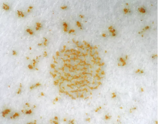
Fruiting body assay performed on filter papers placed on water agar showed even more clear differences in the behaviour of the mutant cells versus the wild type ones during development. Wild type cells preferentially stayed in the centre of the circle so that mature wild type fruiting bodies were located in the circle. The mutant cells migrated from the centre of the spot to the outer part during development so that mature fruiting bodies were located more dispersed around the circle (Fig.2.10.).
Fig.2.10. Developmental phenotype of csgA insertional mutant versus wild type. Cells were placed on filter paper located on water agar plates. Spots were viewed from above after 48 h.
The phenotype of the mature mutant fruiting body was the same as the wild type one.
Mutant AM8 formed wild type fruiting bodies consisting of a branched stalk bearing several sporangioles (Fig.2.11.).
Fig.2.11. Side-view of the representative fruiting body formed by the csgA insertional mutant (AM8) and the wild type.
The germination assay was performed as described in Materials and Methods.
Swarming cells of AM8 or wild type were visible after few days of incubation. This result clearly indicates that myxospores formed by the csgA insertional mutant are able to germinate but efficiency of germination is not known.
2.2.5. Ability of the wild type to restore developmental phenotype of mutant AM8
The csgA insertional mutant cells do not ripple and show a different migration and aggregation pattern as compared with the wild type cells during development. At that point the question rises if it is possible to restore the developmental phenotype of the mutant by mixing it with the wild type. The experiment was performed by mixing equal amounts of the wild type cells with mutant cells prior to starvation.
Rippling that precedes fruiting body formation was detected in the mixed cell population. Also the aggregation pattern of the mixed population was more or less similar to the wild type when the test was performed on the agar surface. The fruiting body formation testing of the mixed population on filter papers showed different patterns of organisation of the mature fruiting bodies in comparison to the wild type.
Wild type as shown previously formed fruits mostly concentrated in the inner part of the circle. The mixed population of the cells formed fruiting bodies concentrated in the circle with additionally some fruits that were dispersed around the circle (Fig.2.12).
Further, rippling waves are detectable in mixed population of the cells and some of the fruits are dispersed around in a mutant like manner.