Functional characterization of prohibitins by conditional inactivation in the mouse
I n a u g u r a l - D i s s e r t a t i o n
zur
Erlangung des Doktorgrades
der Mathematisch-Naturwissenschaftlichen Fakultät
der Universität zu Köln
vorgelegt von Carsten Merkwirth
aus Kassel
Köln, 2008
Berichterstatter:
Professor Dr. Thomas Langer Professor Dr. Jens C. Brüning Professor Dr. Ari Waisman
Tag der mündlichen Prüfung: 28. Mai 2008
Für meine Oma
“Wissenschaft ist wie Fussball –
Beide brauchen Kopf, Herz und Leidenschaft.”
Jens Falta (Physiker)
Abstract
Prohibitins comprise an evolutionary conserved and ubiquitously expressed family of
membrane proteins with poorly described functions. Large assemblies of PHB1 and PHB2
subunits are localized in the inner membrane of mitochondria, but various roles in other
cellular compartments have also been proposed for both proteins. To determine physiological
functions of mammalian prohibitins, a conditional mouse model for the analysis of the murine
Phb2 gene was established, which allows a tissue-restricted and time-controlled Phb2 gene
deletion. Mouse embryonic fibroblasts (MEFs) isolated from genetically modified Phb2
fl/flembryos were generated to define cellular activities of prohibitins. The presented experiments
restrict the function of prohibitins to mitochondria and identify the processing of the dynamin-
like GTPase OPA1, an essential component of the mitochondrial fusion machinery, as the
central cellular process controlled by prohibitins. Cre-mediated deletion of Phb2 in MEFs
leads to the selective loss of long isoforms of OPA1. This results in fragmentation of the
mitochondrial network accompanied by an aberrant cristae morphogenesis in prohibitin-
deficient cells. Furthermore, loss of PHB2 is characterized by an impaired cellular
proliferation and resistance towards apoptosis. Expression of a long OPA1 isoform in PHB2-
deficient cells suppresses these defects identifying impaired OPA1 processing as the primary
cellular defect in the absence of prohibitins. In conclusion, these results assign an essential
function to prohibitins in the formation of mitochondrial cristae and suggest a coupling of cell
proliferation to mitochondrial morphogenesis.
Table of Contents
Abstract ... 5 1 Introduction ... 8
1.1 Organization of biological membranes 8
1.2 Lipid microdomains 9
1.3 SPFH-domain containing proteins 11
1.3.1 Stomatins and stomatin-like proteins 14 1.3.2 Flotillin/reggie proteins 17
1.3.3 HflK/C proteins 18
1.4 Prohibitins 21
1.4.1 Nomenclature of prohibitins 21
1.4.2 Prohibitin distribution 21
1.4.3 Protein organization of prohibitins 22 1.4.4 Cellular localization of prohibitins 23 1.4.4.1 Complex assembly of prohibitins in mitochondria 23 1.4.4.2 Regulatory roles of the mitochondrial prohibitin complex 25 1.4.4.3 Regulation of gene expression by nuclear localized prohibitins 28 1.4.4.4 Functions of plasma membrane-localized prohibitins 30
1.5 Mitochondrial fusion 31
1.5.1 Mitofusins and Charcot-Marie-Tooth Disease 33 1.5.2 OPA1 and dominant optic atrophy 35 1.5.3 Regulation of mitochondrial dynamics 37
1.6 Objectives of the thesis 39
2 Material and Methods ... 40
2.1 Molecular Biology 40
2.1.1 Cloning procedures 40
2.1.1.1 Generation of the Phb2 gene targeting vector 40 2.1.1.2 Generation of stable Phb2 expression plasmids 40 2.1.2 Competent cells and isolation of plasmid DNA 41 2.1.3 Isolation of genomic DNA from cells and tissues 42 2.1.4 Polymerase Chain Reaction (PCR) 43 2.1.5 RNA isolation and RT-PCR 44
2.1.6 DNA sequencing 44
2.1.7 Southern Blotting 45
2.2 Cell Biology 46
2.2.1 ES cell culture 46
2.2.2 MEF cell culture 46
2.2.3 Cre protein transduction in vitro 47
2.2.4 Assessment of cell proliferation 47
2.2.5 Cell death analysis 48
2.2.6 Flow cytometry 48
2.2.7 Fluorescence microcopy 48
2.2.8 Transmission electron microscopy 49 2.2.9 Isolation of mitochondria from MEFs 49
2.3 Biochemistry 50
2.3.1 Preparation of protein extracts 50
2.3.2 Immunoblotting 50
2.3.3 Blue native polyacrylamide gel electrophoresis (BN-PAGE) 51 2.3.4 Respiratory chain function 52
2.4 Mouse analysis 53
2.4.1 Animal Care 53
2.4.2 Mouse handling and breeding 53
2.4.3 Mice 53
3 Results ... 54
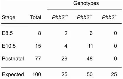
3.1 Conditional gene targeting of the murine Phb2 gene 54 3.1.1 Homologous recombination of the targeting construct in murine ES cells 55 3.1.2 Germline transmission of the targeted Phb2 allele 56 3.1.3 Embryonic lethality of PHB2-deficient mice 56 3.1.4 Embryonic lethality of brain-specific PHB2-deficient mice 57 3.2 Generation of in vitro systems for Phb2 ablation 58 3.2.1 Mouse embryonic fibroblasts 58 3.2.2 Complementation of PHB2 deficiency in vitro 59 3.3 Functional analysis of prohibitin-deficient MEFs 61 3.3.1 Loss of prohibitins does not lead to spontaneous apoptosis 61 3.3.2 Cell proliferation arrest of prohibitin-deficient MEFs 62 3.3.3 Cell proliferation depends on mitochondrially targeted PHB2 64 3.3.4 Mutations in predicted nuclear localization signals and receptor boxes of PHB2 do not interfere with cell proliferation 66 3.3.5 Mitochondrial fragmentation in prohibitin-deficient MEFs 67 3.3.6 Defective cristae morphogenesis in PHB2-deficient mitochondria 68 3.4 Prohibitins control OPA1 cleavage in mammalian mitochondria 71 3.4.1 Impaired OPA1 processing in prohibitin-deficient MEFs 71 3.4.2 Maintenance of respiratory activities in PHB2-deficient mitochondria 72 3.4.3 Expression of a long OPA1 isoform restores mitochondrial morphology in prohibitin- deficient MEFs 76 3.4.4 L-OPA1∆ expression restores cristae morphogenesis in Phb2-/- cells 78 3.4.5 Partial restoration of cell proliferation of Phb2-/- cells upon expression of L-OPA1∆ 79 4 Discussion ... 80
4.1 Mitochondria-localized prohibitins are indispensable for cell proliferation 80 4.2 Prohibitins are required for mitochondrial cristae morphogenesis 83 4.3 Prohibitins control OPA1 cleavage in the mitochondrial inner membrane 84 4.4 Anti-apoptotic function of prohibitins 85 4.5 Prohibitins are essential for embryonic development 86 4.6 Prohibitins may serve as scaffolds in the organization of the mitochondrial inner membrane 88 4.7 Perspectives 90 5 Summary... 92
6 Zusammenfassung ... 93
7 References... 94
8 Figure index...117
9 Table index ...118
10 List of Abbreviations ...119
11 Appendix...121
Acknowledgement...122
Eidesstattliche Erklärung...123
Lebenslauf...124
1 Introduction
1.1 Organization of biological membranes
Cellular membranes represent essential barriers that define the boundary of individual cells (Engelman, 2005). Moreover, they compartmentalize organelles within a cell by segregating them from the cytosol thus providing permeability barriers that surround aqueous interiors and allow for the separation of ion and solute concentrations (Maxfield and Tabas, 2005). The biological membrane is a liquid-like structure representing a homogenous fluid lipid bilayer (Singer and Nicolson, 1972). This traditional view of a biological membrane has been extended over the last years to a more complex structure. Thousands of different lipids arranged with a multiplicity of membrane proteins give rise to a highly dynamic lipid-protein composite. The plasma membrane of eukaryotic cells, like other biological membranes, contains more lipid species than required for establishing a lipid bilayer. This diversity of lipids in eukaryotic membranes raised the question, whether specific lipids could serve to organize biological membranes into discrete domains with different properties (Karnovsky et al., 1982; Thompson and Tillack, 1985). Indeed, an asymmetric distribution of phospholipids was observed in the plasma membrane of erythrocytes suggesting a lateral heterogeneity of lipids and proteins in biological membranes (van Meer et al., 1980). This finding was supported by the discovery that glycospingolipids cluster in the Golgi apparatus before being sorted to the apical surface of polarized cells (van Meer et al., 1987). Based on these initial observations, a spatial organization of membranes into discrete microdomains has been proposed, which thereby provides the rationale for the compartmentalization of important biological processes including signal transduction pathways, apoptosis, cell adhesion and migration, synaptic transmission, organization of the cytoskeleton and membrane fusion during both exocytosis and endocytosis (Brown and London, 1998; Harris and Siu, 2002;
Simons and Toomre, 2000; Tsui-Pierchala et al., 2002). The concept of lateral lipid
assemblies within the plasma membrane finally lead to the hypothesis of lipid rafts, a
particular type of microdomain in the plasma membrane (Simons and Ikonen, 1997).
1.2 Lipid microdomains
Lipid rafts were hypothesized to be lateral dynamic assemblies of lipids which constitute a non-random organization and partitioning of the plasma membrane into microdomains (Harder et al., 1998; Simons and Ikonen, 1997). In particular, these microdomains are enriched in cholesterol and sphingolipids and thought to provide a platform for the concentration of specific proteins and to spatially segregate molecules. Due to different biophysical properties of the enriched lipids, microdomains are present in the liquid- ordered phase separated from the surrounding lipid bilayer (Brown and London, 1998). The basic concept of lipid rafts is to facilitate specific protein-protein interactions by the selective exclusion or inclusion of proteins (Hancock, 2006). The lipid-based separation and fusion of such domains and their associated proteins would therefore provide a dynamic spatial and temporal regulation of signalling cascades according to the requirement of the cell (Simons and Toomre, 2000).
This concept was experimentally supported by the isolation of lipid microdomains based on their insolubility in cold non-ionic Triton X-100 detergent (Yu et al., 1973). These detergent-resistant membranes (DRM) were found to be enriched in both cholesterol and glycophosphatidylinositol (GPI)-anchored proteins and showed an altered density on sucrose- gradients (Brown and Rose, 1992; Varma and Mayor, 1998). On the basis of biophysical experiments, the size of lipid rafts was proposed to be in the range of 10 – 200 nm (Pralle et al., 2000). Small rafts tend to form larger platforms via protein-protein and protein-lipid interactions (Pike, 2006). This suggests that membrane microdomains contribute to the integrity of membranes by providing a scaffolding function. The association of the cytoskeleton with biological membranes has also been proposed to influence membrane organization (Janmey and Lindberg, 2004). In addition to the binding of membrane proximate cytoskeletal adaptors to membrane proteins, cytoskeletal proteins also interact with specific lipids which is likely to influence the organization of membrane microdomains through both protein and lipid anchorage points on the inner leaflet (Babiychuk and Draeger, 2000;
Holowka et al., 2000; Vereb et al., 2003). These observations shed new light on possible
regulatory mechanisms controlling the membrane lateral heterogeneity in the execution of
important cellular functions like signal transduction and membrane trafficking (Simons and
Toomre, 2000). In this context, rafts have been appreciated to serve as platforms allowing the
recruitment of signalling molecules to defined patches. Most importantly, signalling by T cell
receptors (Janes et al., 2000), B cell receptors (Cheng et al., 1999), growth factor receptors
(Waugh et al., 1999), interleukins and insulin (Mastick et al., 1995) were proposed to be associated with lipid rafts (Bromley et al., 2001; Paratcha and Ibanez, 2002). For individual receptors, rafts may form a concentrating platform, activated by ligand binding (Zajchowski and Robbins, 2002). It has been argued that the local restriction of signalling pathways in rafts would on the one hand allow activated receptors enhanced access to downstream effector molecules and, on the other hand, protect these signalling complexes from non-raft molecules that otherwise could negatively affect the transduction process (Simons and Toomre, 2000).
In addition, it has been suggested that receptor clustering might in turn lead to the organization of smaller rafts into larger microdomains and hence increase the spatial concentration of signalling components (Anderson and Jacobson, 2002; Harris and Siu, 2002;
Pike, 2003; Simons and Toomre, 2000; Subczynski and Kusumi, 2003). Interestingly, the partitioning in microdomains might also have a role in the transduction of signalling via lipids themselves or through lipid-modified proteins anchored to the membrane (Varma and Mayor, 1998). Among those, the GPI-modification of cell adhesion molecules like cadherins, NCAM120 and ephrins presumably drives sorting into special microdomains through their lipid anchorage (Bruckner et al., 1999; Doyle et al., 1998; Olive et al., 1995). Similarly, the Ras signalling cascade seems to be compartmentalized into membrane microdomains by the selective palmitoylation of the H-Ras isoform, thereby conferring a partition into lipid rafts (Hancock et al., 1990). An accumulation in lipid rafts has also been observed for lipid- modified Src kinases and G-subunits of heterotrimeric G proteins (Oh and Schnitzer, 2001;
Resh, 1999). Phosphatidylinositol and its phosphorylated derivatives are associated with a wide variety of cellular functions including signalling (Berridge and Irvine, 1989), ion channel activation (Suh and Hille, 2005) and membrane trafficking (Simonsen et al., 2001). In addition to its role as a second messenger in signal transduction processes (Berridge and Irvine, 1984), phosphatidylinositol-4,5-bisphosphate (PIP
2) anchors proteins to the plasma membrane through pleckstrin homology (PH) domains and therefore potentially contributes to the organization of membrane microdomains (DiNitto et al., 2003; Lemmon, 2003). Recent evidence suggests that lipid micro-environments also contribute to the spatial segregation of components required for membrane docking and fusion at particular sites of the plasma membrane (Chamberlain et al., 2001; Ikonen, 2001; Lang et al., 2001). Moreover, PIP
2links lipid microdomains to clathrin-mediated endocytosis and synaptic vesicle trafficking (Di Paolo et al., 2004; Honing et al., 2005; Wenk and De Camilli, 2004).
Although membrane lateral homogeneity is accepted as a requirement for the function
of biological membranes and its incorporation into the lipid raft hypothesis confers specificity
to this broad concept, direct evidence for the existence of raft microdomains is still missing (Jacobson et al., 2007; Munro, 2003). On the one hand, this might be due to technical difficulties to prove the existence of lipid rafts. On the other hand, however, recent experiments have raised potential concerns about the lipid raft hypothesis (Munro, 2003).
Critical points about the raft microdomains are mainly based on the fact that most of the evidence for their existence and function relies on indirect methods (Lai, 2003). Critics were corroborated further by an asymmetry in the lipid composition of the exo- and endoplasmic leaflets of cell membranes and a lack of evidence for the formation of lipid domains in the inner leaflet (Munro, 2003). Since the vast majority of findings concerning lipid rafts have been obtained for plasma membranes, the notion of lipid rafts as a generalized principle present in endoplasmic membranes of cell organelles has to be considered critically (Mukherjee and Maxfield, 2004). Notably, an emerging view favours the idea of a protein- assisted establishment of microdomains in biological membranes.
1.3 SPFH-domain containing proteins
Using stomatin sequences for bioinformatic analyses, a protein family associated with lipid microdomains was identified (Tavernarakis et al., 1999) which contains a domain within its central region bearing a high similarity to prohibitins, the flotillins/reggie-proteins and the bacterial plasma membrane proteins HflK and HflC (Figure 1). The conserved region was termed SPFH-domain (after Stomatin, Prohibitin, Flotillin and HflK/C) (Tavernarakis et al., 1999). The family of SPFH-domain containing proteins is also termed PHB (prohibitin) domain family or PID (after Proliferation, Ion and Death) (Morrow and Parton, 2005;
Nadimpalli et al., 2000). Members of the SPFH protein family share a ~200 amino acid N-
terminal core motif present in eukaryotes as well as archaea and prokaryotes suggesting an
ancient origin (Rivera-Milla et al., 2006) (Figure 1). Although the alignment of prohibitins
reveals a sequence similarity of 60% and an identity of 47%, the similarity of both proteins to
mouse flotillin-2 is only 4 and 7%, respectively. Phylogenetic analyses of SPFH-domains
from different proteins revealed only ambiguous relationships within this superfamily,
indicating independent origins for the individual members and convergent evolution of the
PHB domain (Rivera-Milla et al., 2006) (Figure 2). SPFH family members are integral or
membrane-associated proteins present in various cellular membranes, including the plasma
membrane (Lang et al., 1998; Snyers et al., 1999), Golgi (Glebov et al., 2006), endoplasmatic
reticulum (Browman et al., 2006) and mitochondrial inner membrane (Ikonen et al., 1995)
exposing their SPFH-domain to a hydrophilic environment (Tavernarakis et al., 1999).
Interestingly, various SPFH-domain-containing proteins are enriched in detergent-resistant membranes (DRM) suggesting an association with lipid microdomains in diverse membranes of cellular compartments (Browman et al., 2007; Langhorst et al., 2005). Subcellular targeting of SPFH members to plasma membrane microdomains occurs via different mechanisms. The SPFH-domain of stomatin homologues contains sequences driving the localization to the plasma membrane and lipid rafts (Salzer and Prohaska, 2001). Dependent on two hydrophobic regions within the SPFH-domain, flotillin-1/reggie-2 proteins are targeted to the plasma membrane (Liu et al., 2005) (Figure 1).
Lipid modification is an additional common mechanism for proper targeting of proteins to DRMs and lipid microdomains (Melkonian et al., 1999). Palmitoylation of several SPFH protein members has been shown to be involved in the sorting to plasma membrane microdomains. A cysteine residue (C43) in the SPFH-domain of flotillin-1/reggie-2 is modified by palmitoylation and functions together with hydrophobic regions in plasma membrane association (Morrow et al., 2002). Mutational exchange of this cysteine residue to alanine prevents the association of flotillin-1/reggie-2 with the plasma membrane (Morrow et al., 2002). Similarly, palmitoylation of a cysteine residue (C29) in stomatin targets the protein to lipid rafts suggesting a crucial role for palmitoylation in subcellular sorting (Snyers et al., 1999; Wang et al., 1991). Mutations in the SPFH-domain of podocin disrupt the proper localization to the plasma membrane and cause retention in the endoplasmic reticulum (Roselli et al., 2004). The sorting of endoplasmic reticulum- and mitochondria-localized SPFH proteins depends mostly on N-terminal regions. The N-termini of prohibitin-1 (PHB1) and prohibitin-2 (PHB2), erlins and stomatin are sufficient to target a heterologous protein to mitochondria (Kasashima et al., 2006; Tatsuta et al., 2005), to the endoplasmic reticulum (Browman et al., 2006) and to cytoplasmic vesicular structures, respectively (Umlauf et al., 2004). While a mitochondrial targeting signal is predicted for PHB2 (Kasashima et al., 2006;
Tatsuta et al., 2005), specific sorting signals have not been identified in other SPFH-domain- containing proteins (Browman et al., 2007).
On a structural level, the SPFH-domain functions as a mediator of protein-protein
interactions in stomatin proteins. The SPFH-domain of MEC-2, the Caenorhabditis elegans
homologue of human stomatin, mediates interaction with MEC-4 and MEC-10 subunits of the
degenerin channel in the plasma membrane of touch-sensory neurons (Huang et al., 1995). In
this scenario, the SPFH-domain generates close proximity between the regulatory N- and C-
termini of MEC-2 and the degenerin channel subunits (Zhang et al., 2004). Moreover, biochemical data suggest a role of the SPFH-domain in self-oligomerization.
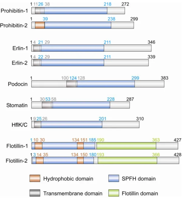
Figure 1. Domain structure of SPFH-domain containing proteins.
Schematic representation of the protein domain structure of mammalian prohibitins, erlins, stomatin, podocin, flotillins and bacterial HflK/C. Gray boxes indicate transmembrane domains; brown, hydrophobic domains; blue, SPFH-domains; green, flotillin domains. Numbers in corresponding colours indicate the amino acid residues marking the boundaries of domains. Modified from (Browman et al., 2007).
In human stomatin, the SPFH-domain is required for the correct positioning of the C- terminus for self-assembly which is disrupted in C-terminal truncated versions (Snyers et al., 1998). Genetic studies support the notion that homo-oligomerization of stomatins is required for their function (Gu et al., 1996; Tavernarakis and Driscoll, 1997). Recently obtained structural data from a archaebacterial stomatin orthologue further strengthen the findings on oligomerization (Yokoyama et al., 2008). In stomatin and podocin, the SPFH-domain forms a hairpin conformation which mediates the proper localization of N- and C-termini to the cytoplasm where interactions with accessory molecules take place (Roselli et al., 2002;
Snyers et al., 1999). These findings suggest that the SPFH-domain also serves as a structural scaffold (Goodman et al., 2002; Zhang et al., 2004).
1.3.1 Stomatins and stomatin-like proteins
Stomatin is the most representative member of SPFH-domain proteins. The 31-kDa
integral membrane protein belongs to the family of band 7.2b proteins and is widely
expressed from prokaryotes to eukaryotes (Gallagher and Forget, 1995; Hiebl-Dirschmied et
al., 1991). Human stomatin is highly expressed in erythrocytes and was originally thought to
be associated with overhydrated hereditary stomatocytosis (OHSt), a form of haemolytic
anaemia characterized by increased erythrocyte permeability to monovalent cations (Fricke et
al., 2003). Erythrocytes isolated from OHSt patients lacked the stomatin protein (Stewart et
al., 1993). However, further studies disproved an involvement of stomatin in the disease
(Delaunay et al., 1999; Innes et al., 1999). Notably, stomatin-deficient mice show an
apparently normal erythrocyte function (Zhu et al., 1999). Stomatin localizes predominantly
to the plasma membrane and intracellular vesicles of the endocytic pathway (Snyers et al.,
1999). In epithelial cells, stomatin has been shown to concentrate in plasma membrane
protrusions and late endocytic compartments. At these subcellular localizations, stomatin is
present in high-order oligomers consisting of 9-12 monomers (Snyers et al., 1998). At the
plasma membrane, stomatin was detected in clusters in electron microscopy studies, where it
colocalizes with the actin cytoskeleton (Snyers et al., 1997). These observations led to the
notion that stomatin oligomers might serve a role in membrane compartmentalization by
providing a microdomain scaffold (Snyers et al., 1998). Interestingly, stomatin has been found
in lipid microdomains in numerous cell types (Foster et al., 2003; Garin et al., 2001) and was
shown to be associated with lipid bodies (Umlauf et al., 2004). Further studies implied a role
of stomatin in insulin-dependent glucose transport, since stomatin has been shown to interact
directly with the glucose transporter GLUT-1, thereby regulating its trafficking and transport activity (Zhang et al., 2001; Zhang et al., 1999). The analysis of stomatin-like proteins in various organisms suggested a role in ion channel regulation. The C. elegans homologue of stomatin, MEC-2, is exclusively expressed in specialized touch receptor neurons responsible for mechanosensation. Mutational loss of MEC-2 results in decreased touch sensitivity (Gillespie and Walker, 2001). The protein was shown to interact with and regulate the activity of members of the mechanosensitive degenerin-epithelial sodium channels (DEG-ENaC) (Ernstrom and Chalfie, 2002). The stomatin homologue exists in a multiprotein complex with the degenerin-type channel proteins MEC-4 and MEC-10 which is thought to link the MEC transduction channel to microtubules (Goodman et al., 2002; Huang et al., 1995). Functional conservation of this function is indicated by a recent report demonstrating that the mammalian stomatin homologue SLP-3 is also involved in mechanosensation in mice (Wetzel et al., 2007). In contrast to the ubiquitous expression of stomatin, SLP-3 expression is restricted to neuronal tissue. SLP-3-deficient mice exhibit a markedly reduced touch sensitivity due to loss of function of a subset of mechanoreceptors in the skin, acid-sensing ion channels (ASICs) (Wetzel et al., 2007). In a heterologous system, SLP-3 and stomatin were shown to interact with and modulate the activity of different ASICs (Price et al., 2004). These results suggest that SLP-3 might mediate touch sensitivity by regulating ASICs in mechanosensory axons in the skin similar to the regulation of MEC-4 and MEC-10 by MEC-2 (Wetzel et al., 2007).
Another specialized SPFH family member and mammalian stomatin homologue is
podocin (NPHS2), a 42-kDa integral membrane protein which shows 47% identity to human
stomatin (Figure 1). Podocin is exclusively expressed in podocytes, a subset of highly
specialized kidney epithelial cells, which are involved in plasma ultrafiltration during primary
urine formation (Roselli et al., 2002). The protein localizes to the slit diaphragm, a specialized
intercellular junction in the mammalian kidney (Huber and Benzing, 2005; Roselli et al.,
2002). Podocin was found in high order-oligomers that are constituents of detergent-insoluble
microdomains in the plasma membrane of podocytes (Huber et al., 2003). In this respect,
interactions of podocin with other microdomain-associated podocyte proteins, CD2-associated
protein (CD2AP) and nephrin, have been reported (Schwarz et al., 2001). Podocin has been
suggested to potentiate nephrin signalling by localizing nephrin to lipid raft domains (Huber
et al., 2003). Inefficient nephrin signalling via the nephrin/CD2AP/podocin complex is
thought to contribute to the development of podocyte dysfunction (Huber et al., 2001; Shih et
al., 1999). Recently, the non-selective cation channel transient receptor potential canonical 6
(TRPC6) was identified as an additional component of the slit diaphragm protein complex.
Podocin was shown to interact with TRPC6 and to enhance its ion channel activity (Huber et al., 2006). Thus, podocin might be involved in mechanosensation at the kidney filtration barrier by regulating TRPC6 channel activity (Huber et al., 2007). Strikingly, mutations in podocin were associated with autosomal recessive steroid-resistant nephritic syndrome, a progressive disorder leading to end-stage renal disease (Boute et al., 2000; Fuchshuber et al., 1995). Interestingly, ion channel regulation by both MEC-2 and podocin is dependent on their ability to bind cholesterol (Huber et al., 2006). Thus, the feature of stomatin proteins to form large multimeric complexes in cellular membranes connected with their cholesterol binding ability could lead to localized changes in the membrane lipid composition and might provide the basis for a molecular understanding of their role in ion channel regulation. Ion channels might become activated by altered membrane properties caused by locally elevated cholesterol levels (Huber et al., 2006). The putative role in ion channel regulation and mechanosensation has only been shown for members of the stomatin family, suggesting that these functions might be unique to these proteins. However, the involvement of other SPFH- domain-containing proteins in ion-channel regulation during mechanosensation remains to be investigated (Browman et al., 2007).
In addition to SLP-1 and SLP-3, another protein of the stomatin family has been
reported. Stomatin-like protein 2 (SLP-2) was initially discovered in erythrocytes (Wang and
Morrow, 2000). Interestingly, more recent findings suggest a mitochondrial localization of
SLP-2. SLP-2 forms a complex in the mitochondrial inner membrane and was shown to
interact specifically with mitofusin-2 (Hajek et al., 2007), an essential dynamin-like GTPase
in the outer membrane of mitochondria required for mitochondrial fusion (Santel and Fuller,
2001). These observations suggest that SLP-2 links the inner to the outer mitochondrial
membrane and might regulate the activity of mitofusin-2 via a direct protein-protein
interaction. However, the molecular mechanism of SLP-2 function and its putative
contribution to inner membrane organization remain elusive and require to be addressed in
further detail. Another striking observation is the interaction of SLP-2 with prohibitins in the
mitochondrial inner membrane. RNAi-mediated depletion of SLP-2 affects the steady-state
level of PHB1 and PHB2 suggesting a role for SLP-2 in the regulation of mitochondrial
proteolysis (Da Cruz, 2008).
1.3.2 Flotillin/reggie proteins
Flotillin/reggie proteins are additional members of the SPFH-domain-containing protein family (Tavernarakis et al., 1999) (Figure 1). Reggie-1 and -2 are highly conserved proteins which were characterized initially in goldfish and rats as plasma membrane- associated proteins (Malaga-Trillo et al., 2002). In this context, reggie proteins were identified and named due to their upregulation during axonal regeneration of retinal ganglion cells upon optical nerve transsection (Lang et al., 1998; Schulte et al., 1997). The independent identification of reggie homologues in mouse and fruitfly led to the alternative name flotillin- 1 and -2 (corresponding to reggie-2 and -1, respectively) which were associated with the floating lipid fraction isolated from murine lung tissue (Bickel et al., 1997; Galbiati et al., 1998). Mammalian flotillins are widely expressed in various tissues and cell types (Lang et al., 1998; Salzer and Prohaska, 2001; Solomon et al., 2002; Stuermer et al., 2001; Volonte et al., 1999). On a cellular level, flotillins are mainly localized at the plasma membrane and are considered lipid raft components due to their presence in detergent-resistant membranes (DRMs) (Stuermer et al., 2001). In addition, localizations of both flotillin-1 and -2 to endosomes, lipid droplets and phagosomes have been reported (Dermine et al., 2001; Gagescu et al., 2000; Morrow and Parton, 2005). Flotillin-1, but not flotillin-2, was also found to be associated with the trans-Golgi network (TGN) (Gkantiragas et al., 2001). Moreover, a cell- cycle dependent translocation of flotillin-1 to the nucleus has been observed (Santamaria et al., 2005).
Plasma membrane association of flotillins occurs in the absence of typical
transmembrane domains suggesting alternative modes of membrane binding. Two conserved
hydrophobic membrane-associating domains in the N-terminus of flotillins are considered to
mediate the interaction with the inner leaflet of the plasma membrane (Morrow et al., 2002)
(Figure 1). Post-translational modifications like palmitoylation and myristoylation of both
flotillins may assist plasma membrane anchoring and targeting to lipid microdomains (Liu et
al., 2005; Morrow et al., 2002; Neumann-Giesen et al., 2004). Both flotillin-1 and -2 purified
from erythrocyte membrane fractions have been shown to form high-order homo-oligomers
(Salzer and Prohaska, 2001). Moreover, hetero-oligomerization of flotillins into tetramer
units, which depends on C-terminal coiled-coil domains, has been suggested (Neumann-
Giesen et al., 2004; Solis et al., 2007). Whether hetero-oligomerization of mammalian
flotillins is connected to a functional interdependence is discussed controversially. In contrast
to a RNAi-mediated depletion of flotillin-2, which leads to a reduction in flotillin-1 protein
levels, the reciprocal experiments do not support this notion (Solis et al., 2007). Confocal and electron microscopy analyses revealed the presence of flotillin clusters at the plasma membrane, indicative of small, uniform microdomains with a diameter of 100 nm (Kokubo et al., 2003; Stuermer et al., 2001). A co-clustering of flotillins with various GPI-anchored cell surface proteins (Stuermer et al., 2001), cell adhesion molecules (Stuermer et al., 2004), intracellular signalling components (Rajendran et al., 2003; Slaughter et al., 2003) and the cytoskeleton (Langhorst et al., 2007) was observed suggesting that flotillins contribute structurally to the formation of membrane microdomains (Frick et al., 2007; Stuermer and Plattner, 2005). It has been further proposed that flotillin clusters form lipid raft-like membrane scaffolds with important roles in cell-cell adhesion and signal transduction (Langhorst et al., 2005; Simons and Toomre, 2000; Stuermer et al., 2001). It has recently been reported that mammalian flotillins are involved in the regulation of clathrin-independent endocytosis, supporting the idea that flotillin scaffolds provide functional dynamic microdomains (Frick et al., 2007; Glebov et al., 2006). Interestingly, a role for the secretion of signalling components has been demonstrated for Drosophila melanogaster flotillin-2 indicating a crucial role for intracellular trafficking and the generation of morphogen gradients during fruitfly development (Katanaev et al., 2008). A clinical relevance of flotillins is unclear, since natural mutations have not been discovered to date. However, increased flotillin-1 expression levels have been associated to type II diabetes (James et al., 2001) and neuropathological disorders such as Parkinson´s disease (Jacobowitz and Kallarakal, 2004) and Alzheimer´s disease (Girardot et al., 2003; Kokubo et al., 2000).
1.3.3 HflK/C proteins
The Escherichia coli HflK and HflC proteins are bacterial members of the SPFH protein family (Tavernarakis et al., 1999) (Figure 1). Both are transmembrane proteins consisting of approximately 310 amino acids encoded by the hflA (high frequency of lysogenization) operon that regulates the lysogenic decision during bacteriophage λ infection (Banuett and Herskowitz, 1987; Herskowitz and Hagen, 1980; Kihara et al., 1997). HflK and HflC proteins form a high molecular weight complex (HflK/C) which is anchored to the bacterial plasma membrane by N-terminal transmembrane segments and exposes C-terminal coiled-coil domains to the periplasmic side (Kihara et al., 1997; Kihara and Ito, 1998).
Furthermore, the C-terminal domains are involved in hetero-oligomerization of the proteins
(Briere and Dunn, 2006). HflK and HflC subunits exhibit functional interdependence which is
reflected by decreased stability of one subunit in the absence of the other (Banuett and
Herskowitz, 1987). Notably, the hflA locus encodes an additional protein, FtsH, which is
categorized as an ATP-dependent metalloprotease of the AAA family (ATPases associated
with a variety of cellular activities) (Noble et al., 1993; Ogura and Wilkinson, 2001). The
HflK/C complex interacts with and assembles into a larger complex with the membrane
protease FtsH thereby modulating its proteolytic activity (Kihara et al., 1996; Saikawa et al.,
2004). The FtsH protease itself controls the decision between lysogenic and lytic cycle growth
during λ-phage infection by modulating the stability of the phage-derived cII protein (Banuett
and Herskowitz, 1987). Additionally, the FtsH protease is involved in the degradation of a
membrane protein translocase. In the absence of HflK/C, the non-assembled subunit SecY of
the SecY-SecE translocase is rapidly degraded suggesting a negative regulation of the
protease by the HflK/C complex (Kihara et al., 1996).
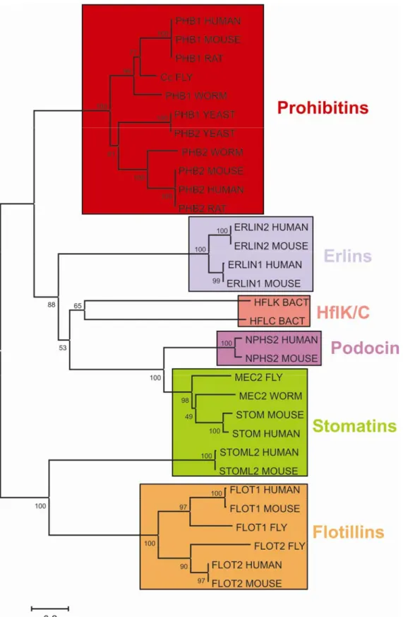
Figure 2. Evolutionary relationship among 31 SPFH-domain containing proteins.
Unrooted dendrogramm depicting the relationship of 31 representative SPFH-domain containing proteins inferred from the Neighbour-Joining method. The consensus tree is based on an amino acid sequence alignment. Protein clusters are coloured. Supporting bootstrap values are indicated at node positions. Phylogenetic analyses were conducted with MEGA4 software.
1.4 Prohibitins
1.4.1 Nomenclature of prohibitins
Prohibitins comprise a highly conserved and ubiquitously expressed protein family in eukaryotic cells (Figure 2). In accordance with their remarkable conservation and widespread distribution, prohibitins have been functionally associated with various cellular functions including cell cycle progression (McClung et al., 1989; Nuell et al., 1991), transcriptional regulation (Montano et al., 1999; Sun et al., 2004), cellular signalling (Terashima et al., 1994), cellular senescence (Coates et al., 1997; Coates et al., 2001; McClung et al., 1992), apoptosis (Fusaro et al., 2003; Vander Heiden et al., 2002), and mitochondrial biogenesis (Artal-Sanz et al., 2003; Berger and Yaffe, 1998; Nijtmans et al., 2000; Steglich et al., 1999).
The family of prohibitin proteins consist of two homologous members, prohibitin-1 (PHB1) and prohibitin-2 (PHB2). Alternative names are B cell-receptor associated protein BAP32 and BAP37 (PHB1 and PHB2, respectively) (Terashima et al., 1994). PHB2 was also designated as prohibitone and REA (repressor of estrogen receptor activity) (Montano et al., 1999).
Prohibitin was initially discovered as an antiproliferative gene by differential hybridization to RNA isolated from regenerating rat liver (McClung et al., 1989).
Subsequently, prohibitin has been proposed as a negative regulator of the cell cycle based on the observation that the microinjection of Phb1 mRNA into mammalian cells blocks cell cycle progression (McClung et al., 1989) whereas injection of antisense oligonucleotides directed against Phb1 mRNA stimulates cell proliferation (Nuell et al., 1991). Notably, the human Phb1 gene was cloned and mapped to a region on chromosome 17 frequently mutated in familial breast cancer leading to the proposal that prohibitin could function as a tumour suppressor gene (Nuell et al., 1991; Sato et al., 1992; White et al., 1991). However, the interpretation of these results has been drawn into doubt, since subsequent studies revealed that these observations can be attributed solely to the 3´ untranslated region (3´UTR) of the prohibitin mRNA (Jupe et al., 1996; Manjeshwar et al., 2003).
1.4.2 Prohibitin distribution
Prohibitin homologues have been detected in various species from prokaryotic to
eukaryotic organisms reflecting their widespread distribution and high degree of conservation
(Mishra et al., 2006; Nijtmans et al., 2002) (Figure 2). The bacterial proteins HflK and HflC are the prokaryotic homologues of prohibitins (Kihara et al., 1996). Two prohibitin members are present and extensively characterized in yeast (Berger and Yaffe, 1998). The Drosophila melanogaster Cc gene appears to be the PHB1 orthologue in flies and is required for normal larvae development (Eveleth and Marsh, 1986). Several prohibitin orthologues have also been identified in plants (De Diego et al., 2007; Snedden and Fromm, 1997). In mammals, the remarkable degree of sequence conservation is evident by the fact that amino acid sequences of rat and mouse prohibitin are identical and differ in only one amino acid from the human sequence. Moreover, yeast prohibitin-1 exhibits an identity of 52% to human PHB1 in its amino acid sequence. In multicellular organisms like C. elegans (Artal-Sanz et al., 2003) and mice (He et al., 2008; Park et al., 2005) prohibitins are also required for embryonic development suggesting an important function of these proteins which has been conserved throughout evolution.
1.4.3 Protein organization of prohibitins
Prohibitin-1 encodes a protein of 30 kDa, whereas prohibitin-2 gives rise to a 37 kDa protein. The PHB domain ranges from amino acids 26-187 in PHB1 and 39-201 in PHB2 (Rivera-Milla et al., 2006) (Figure 1).
The N-terminal domains of both PHB1 and PHB2 are involved in targeting the proteins to their intracellular localization and mediate membrane association (Berger and Yaffe, 1998).
Both prohibitins harbour C-terminal coiled coil domains composed of antiparallel α-helices which are required for hetero-oligomerization (Tatsuta et al., 2005). Biochemical studies suggest complex formation of both PHB1 and PHB2 in various organisms (Coates et al., 1997; Steglich et al., 1999). Coimmunoprecipitation experiments verified an interaction between Phb1p and Phb2p in both yeast (Steglich et al., 1999) and mammalian cells (Coates et al., 1997). Moreover, a quantitative interaction of mammalian prohibitins was inferred from immunodepletion experiments (Coates et al., 2001). The homologous subunits are functionally interdependent in yeast, C. elegans and mammalian cells (Artal-Sanz et al., 2003;
Berger and Yaffe, 1998; Kasashima et al., 2006). Hence, the genetic deletion of one prohibitin
subunit results in the destabilization of the other, strongly suggesting that the prohibitin
complex represents the physiologically active unit.
1.4.4 Cellular localization of prohibitins
The subcellular localization of prohibitins is still a matter of debate and controversial results have been obtained in mammalian cells. Although a mitochondrial localization of prohibitins has been widely accepted, PHB1 and PHB2 have also been identified in the nucleus, the cytosol and the plasma membrane in certain mammalian cell lines (Fusaro et al., 2003; Kasashima et al., 2006; Kurtev et al., 2004). In the nuclear compartment, subunits of prohibitins have been suggested to regulate cell cycle and apoptotic processes by interacting with retinoblastoma tumour suppressor protein, E2F transcription factors and p53 (Kasashima et al., 2006; Nuell et al., 1991; Wang et al., 2002a). It has been further proposed that prohibitins repress the transcriptional activity of estrogen receptors by interacting with histone deacetylases (Kurtev et al., 2004; Montano et al., 1999). Plasma membrane localized prohibitins are thought to modulate epithelial cell adhesion and migration by interacting with c-Raf (Rajalingam et al., 2005) or to function as cell-surface receptors (Kolonin et al., 2004;
Mengwasser et al., 2004; Sharma and Quadri, 2004; Terashima et al., 1994). It should be noted, however, that many of these studies did not consider a mitochondrial localization of prohibitins or addressed only the function of either PHB1 or PHB2.
1.4.4.1 Complex assembly of prohibitins in mitochondria
The mitochondrial localization of prohibitins has been unambiguously demonstrated in the yeast Saccharomyces cerevisiae. In subcellular fractionation experiments, prohibitin subunits were found in association with the inner membrane (Berger and Yaffe, 1998).
Multiple copies of both Phb1p and Phb2p assemble into a high molecular weight complex of 1.2 MDa in the inner membrane of mitochondria (Steglich et al., 1999). Interestingly, a prohibitin complex of similar size has also been detected in the mitochondrial inner membrane of C. elegans (Artal-Sanz et al., 2003) and mammalian cells (Nijtmans et al., 2000). The topology of the yeast prohibitin complex has been investigated by submitochondrial localization and protease treatment of mitoplasts (Steglich et al., 1999).
Sodium carbonate treatment of isolated membrane fractions identified both Phb1p and Phb2p as integral components of the mitochondrial inner membrane (Berger and Yaffe, 1998).
Furthermore, trypsin digestion of mitoplasts generated by osmotic disruption of the
mitochondrial outer membrane revealed protease sensitivity of prohibitin subunits (Steglich et
al., 1999). These findings suggests that the prohibitin complex is anchored to the inner
membrane via N-terminal transmembrane segments and exposes large C-terminal domains into the intermembrane space (IMS) of mitochondria (Steglich et al., 1999). It has been suggested that Phb1p and Phb2p are the only components of the complex since immunodepletion of one subunit results in the absence of the corresponding partner subunit (Coates et al., 2001). This is supported by overexpression studies in yeast demonstrating that only the simultaneous overexpression of both Phb1p and Phb2p leads to an increase in the PHB complex (Nijtmans et al., 2000). Mitochondrial targeting of prohibitins has been analysed by in vitro import studies in yeast (Tatsuta et al., 2005). These results suggest that the N-terminal domains of both Phb1p and Phb2p are required for mitochondrial import.
Sorting of Phb1p to mitochondria is ensured by an unconventional presequence consisting of the first 28 amino acids. In contrast, Phb2p possesses a bipartite N-terminal presequence in amino acids 1-61 composed of the positively charged N-terminal domain followed by a hydrophobic transmembrane segment (Tatsuta et al., 2005). Neither Phb1p nor Phb2p are processed during mitochondrial import (Tatsuta et al., 2005). Crosslinking experiments revealed an association of newly imported Phb1p with the Tim8/Tim13 complex, a soluble, 70 kDa complex in the IMS that functions in the biogenesis of inner and outer membrane proteins (Curran et al., 2002; Davis et al., 2000; Hoppins and Nargang, 2004; Leuenberger et al., 1999; Paschen et al., 2000). The association with the Tim8/Tim13 complex may trap newly imported Phb1p in the IMS and facilitate its transfer to the inner membrane. The subsequent insertion into the inner membrane depends on the membrane potential and is mediated by the Tim23 translocase. In addition, crosslinking revealed that newly imported Phb1p assembles with Phb2p subunits into a subcomplex of ~120 kDa suggesting the presence of membrane-bound assembly intermediates during prohibitin complex formation (Tatsuta et al., 2005) (Figure 3).
Chemical crosslinking experiments and structural models predicted a ring-like assembly of alternating Phb1p and Phb2p subunits (Back et al., 2002). Supported by the finding that crosslinks appeared only within different prohibitin subunits (Back et al., 2002), it has been proposed that heterodimers composed of Phb1p and Phb2p are the building blocks of the prohibitin complex (Figure 3). The native molecular mass of the assembled prohibitin complex of ~ 1 MDa allowed the characterization by single particle electron microscopy.
Large, ring-shaped complexes with an average diameter of ~ 200 Å were detected in images
acquired from purified yeast prohibitin complexes (Tatsuta et al., 2005). These results indicate
that prohibitins form large, ring-shaped complexes in the mitochondrial inner membrane
which are composed of ~ 16-20 alternating subunits of both Phb1p and Phb2p (Back et al., 2002; Tatsuta et al., 2005) (Figure 3).
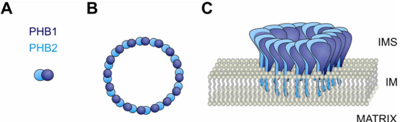
Figure 3. Complex assembly of prohibitin subunits in mitochondria.
Schematic representation of prohibitin subunits PHB1 and PHB2, the ring-shaped prohibitin complex and its topology in the mitochondrial inner membrane. (A) Dimeric assembly intermediates composed of PHB1 and PHB2 constitute the building block of the complex. (B) Circular prohibitin ring complex with alternating subunit composition. The average stoichiometry of the complex is speculative. (C) The prohibitin complex is anchored to the mitochondrial inner membrane via N-terminal transmembrane segments of PHB2. The tightly folded C-terminal coiled-coil domains are exposed to the intermembrane space (IMS). IM = inner membrane.
1.4.4.2 Regulatory roles of the mitochondrial prohibitin complex
To determine the physiological role of prohibitins in vivo, PHB1 and PHB2 have been extensively characterized using genetic approaches in the yeast Saccharomyces cerevisiae.
Notably, the genetic ablation of either PHB1 or PHB2 does not result in apparent phenotypes
(Berger and Yaffe, 1998). Neither single- nor double-mutant phb1-null and phb2-null cells
exhibit growth defects on various carbon sources indicating respiratory competence in the
absence of prohibitins. Moreover, mitochondrial morphology and distribution is not affected
by the loss of prohibitins (Berger and Yaffe, 1998). Interestingly, prohibitins have been linked
to ageing (Jazwinski, 1996; Kirchman et al., 2003). Several reports demonstrated reduced
replicative lifespan, but unaffected chronological lifespan of prohibitin-deficient yeast cells
(Berger and Yaffe, 1998; Coates et al., 1997; Piper and Bringloe, 2002). Furthermore, yeast
lifespan was dramatically reduced in prohibitin-deficient yeast cells lacking the mitochondrial
genome (rho
0) suggesting additive effects which are deleterious for survival (Berger and
Yaffe, 1998; Kirchman et al., 2003). Remarkably, these phenotypes were suppressed by
genetic ablation of RAS2 in these strains suggesting a cooperative function of prohibitins and Ras2p in yeast longevity (Kirchman et al., 2003).
Deletions of prohibitin genes are lethal in combination with a number of mutations indicating strong genetic interactions. For instance, prohibitins genetically interact with components required for mitochondrial inheritance in yeast (Berger and Yaffe, 1998). Genetic ablation of PHB1 or PHB2 combined with mutations in genes encoding the mitochondrial inheritance components Mdm12p, Mdm10p and Mmm1p results in synthetic lethality (Berger and Yaffe, 1998). It has been proposed that Mdm12p, Mdm10p and Mmm1p form a complex which links mitochondria to the actin cytoskeleton thereby ensuring mitochondrial movement and distribution (Boldogh et al., 1998; Boldogh et al., 2003). More recent findings suggested a new role for these components in the biogenesis of mitochondrial outer membrane proteins (Meisinger et al., 2007; Meisinger et al., 2004). Additionally, this complex is required for the assembly and stability of mitochondrial DNA nucleoids (Hanekamp et al., 2002; Hobbs et al., 2001), indicating a function as membrane-embedded machinery for simultaneous inheritance of mitochondria and mitochondrial DNA (mtDNA) (Boldogh et al., 2003). These genetic interactions suggest that prohibitins might function in the regulation of mitochondrial morphology and distribution (Berger and Yaffe, 1998). This is consistent with the finding that the genetic deletion of prohibitins in cells lacking the mitochondrial genome causes mitochondrial fragmentation and disorganization (Berger and Yaffe, 1998). Moreover, synthetic lethality of prohibitins has also been observed with mutations in the phosphatidylethanolamine biosynthetic pathway, pointing to a role of prohibitins in the phospholipid organization of mitochondrial membranes (Birner et al., 2003).
In addition, the simultaneous loss of the prohibitin complex with another integral component of the mitochondrial inner membrane, the m-AAA protease, strongly impairs cell growth (Steglich et al., 1999). The m-AAA protease, a conserved ATP-dependent metalloprotease belonging to the family of AAA
+proteins (ATPases associated with a variety of cellular activities) (Ogura and Wilkinson, 2001), is composed of the homologous subunits Yta10p (Afg3p) and Yta12p (Rca1p) and is part of a protein quality control system in mitochondria (Arlt et al., 1996; Tatsuta and Langer, 2008). The m-AAA protease forms a high molecular mass complex of ~ 850 kDa in the mitochondrial inner membrane exposing catalytic subunits into the matrix (Arlt et al., 1996; Leonhard et al., 1996). Notably, the biochemical characterization revealed the presence of a high molecular mass supercomplex composed of both the prohibitin complex and the m-AAA protease (Steglich et al., 1999).
This supercomplex eluted in fractions corresponding to a molecular mass of approximately 2
MDa in gelfiltration experiments (Steglich et al., 1999). Interestingly, the absence of Phb1p or Phb2p in mitochondria causes an accelerated proteolysis of nonassembled inner membrane proteins by the m-AAA protease suggesting a negative regulatory effect of prohibitins on the m-AAA protease activity (Steglich et al., 1999). A role of the prohibitin complex in the degradation of mitochondrial inner membrane proteins by the m-AAA protease is reminiscent of findings in prokaryotes. In E. coli, the activity of the homologous AAA protease FtsH was found to be negatively regulated by the HflK/C protein complex (Kihara et al., 1996; Kihara et al., 1997). These consistent findings suggest an evolutionary conservation of the regulatory mechanisms controlling AAA protease activities.
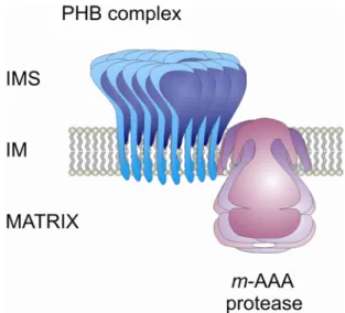
Figure 4. Supercomplex of prohibitins with the ATP-dependent m-AAA protease.
Schematic representation of supercomplex assembly of prohibitins (PHB) with the ATP-dependent m- AAA protease in the mitochondrial inner membrane. See text for details. IMS = intermembrane space, IM = inner membrane.
In further investigations, Nijtmans and coworkers proposed a chaperone-function for
the yeast prohibitin complex. In the absence of prohibitins, mitochondrial translation products
accumulated to a reduced amount as determined by
35S-methionine pulse-chase labelling in
isolated mitochondria (Nijtmans et al., 2000). Consistent with this finding, the overexpression
of prohibitins results in a stabilization of these translation products. This observation was
accompanied by decreased steady-state levels of the mitochondrially encoded proteins Cox2p
and Cox3p in two-dimensional gel electrophoresis. Moreover, the authors could demonstrate a
specific interaction of Cox2p and Cox3p with the prohibitin complex in
coimmunoprecipitations (Nijtmans et al., 2000). These observations suggest that the
prohibitin complex interacts with and stabilizes newly synthesized mitochondrial translation products. Based on initial observations obtained from yeast, prohibitins might have a role in the regulation of mitochondrial morphology. An abnormal mitochondrial morphology was observed in the nematode C. elegans after RNAi-mediated depletion of prohibitins (Artal- Sanz et al., 2003). Indeed, these findings were corroborated in studies with mammalian cells.
RNAi-mediated knockdown of either Phb1 or Phb2 in HeLa cells caused fragmentation of the mitochondrial network and spontaneous apoptosis (Kasashima et al., 2006). Recently, knockdown of Phb1 in epithelial cells has been reported to induce cellular senescence associated with mitochondrial dysfunction (Schleicher et al., 2008). Interestingly, the authors provide the explanation that PHB1 deficiency causes an increased production of reactive oxygen species (ROS) through inhibition of complex I which leads to depolarization of the mitochondrial membrane potential and cellular senescence (Schleicher et al., 2008).
Previously, PHB1 has also been suggested as a component of mitochondrial nucleoids, nucleoprotein complexes associated with mitochondrial DNA (mtDNA) (Bogenhagen et al., 2003). Proteins incorporated in these nucleoid complexes such as mitochondrial transcription factor A (TFAM) and mitochondrial single-strand DNA binding (mtSSB) protein regulate stability, packaging, replication, transcription and maintenance of mtDNA (Chen and Butow, 2005). A very recent study reports a role of prohibitin in the organization and maintenance of mtDNA (Kasashima et al., 2008). Remarkably, downregulation of PHB1 in HeLa cells affects the organization of mitochondrial nucleoids and the steady-state level of TFAM protein.
These findings suggest that PHB1 maintains organization and copy number of mtDNA by regulating TFAM stability (Kasashima et al., 2008).
1.4.4.3 Regulation of gene expression by nuclear localized prohibitins
Despite the general acceptance of a predominant localization of prohibitins to
mitochondria, several reports claim that either PHB1 or PHB2 also localize to the nuclear
compartment (Fusaro et al., 2003; Gamble et al., 2004; Kurtev et al., 2004; Wang et al.,
2002a). Nuclear localization of PHB1 was proposed based on colocalization with
retinoblastoma protein (Rb), p53 and E2F transcription factors in various cell lines (Fusaro et
al., 2003; Wang et al., 2002aa). The majority of human tumours, including breast and prostate
cancer, are associated with mutations directly affecting Rb and p53, or components of
pathways that regulate these proteins. The tumour suppressor protein Rb and its family
members have been demonstrated to modulate the G1-S phase transition during the mammalian cell cycle (Ewen, 1994). E2F transcription factors (E2F1-5) are the major downstream targets of Rb which exerts its growth suppressive effect through E2F inhibition (Weinberg, 1995). In accordance with a putative tumour suppressor function, PHB1 has been shown to interact directly with Rb and p53 to induce their antiproliferative and cell cycle- regulatory activities (Fusaro et al., 2003; Wang et al., 1999a). The association of PHB1 with retinoblastoma family members (Rb, p107 and p130) results in the inhibition of cell proliferation through repression of E2F transcription factors (Wang et al., 1999a).
Consistently, a block in cell proliferation was observed after ectopic expression of PHB1 which inhibited E2F-mediated transcription (Wang et al., 1999b). Remarkably, PHB1 was also found to physically interact with E2F and to inhibit transcription from E2F-responsive promoters suggesting that prohibitin modulates E2F activity also in a Rb-independent manner (Wang et al., 1999b). This is supported by the observation that PHB1 and Rb bind to different regions of E2F (Wang et al., 1999b). Moreover, PHB1-mediated repression of E2F does not respond to signalling activity which reverses Rb-mediated repression (Wang et al., 1999b).
Additional studies demonstrated that transcriptional repression by PHB1 involves the recruitment of the co-repressor N-CoR and chromatin-remodelling complexes like HDAC1 and Brg-1/Brm to promoter elements (Wang et al., 2002a; Wang et al., 2002b; Wang et al., 2004). Interestingly, the opposite effect on PHB1-mediated regulation of transcription was observed for p53. Fusaro and colleagues demonstrated a physical interaction of PHB1 and p53 required for the stimulation of transcription from p53-responsive promoters (Fusaro et al., 2003). The role of PHB1 as a putative modulator of gene transcription raised the intriguing question whether this function can also be attributed to PHB2, since compelling evidence approved the physiological relevance of a mammalian PHB1/2 complex (Coates et al., 2001).
Interestingly, independent studies revealed that PHB2 also acts as a transcriptional regulator in the nucleus (Montano et al., 1999; Sun et al., 2004). PHB2 was identified in a yeast two-hybrid screen as a novel regulator of the estrogen receptor (ER) and subsequently designated as Repressor of estrogen receptor activity (REA) (Montano et al., 1999).
Expression studies demonstrated that PHB2 selectively represses transcription from ER-
responsive promoters through direct binding of nuclear ER and recruitment of HDAC1
(Kurtev et al., 2004). Recent findings also suggest a functional interaction between ER
signalling and PHB1-mediated repression of E2F (Wang et al., 2004). The repressive function
in the estrogen signalling cascade has been substantiated for both PHB1 and PHB2 in vivo
demonstrating the physiological relevance of this regulatory action (He et al., 2008; Mussi et
al., 2006; Park et al., 2005). An additional mode of repression was proposed that involves competitive binding of PHB2 and the transcriptional co-activator SRC-1 to the ligand- activated ER (Delage-Mourroux et al., 2000). It has been reported that, besides associating with the ER, PHB2 binds to additional members of the nuclear receptor family, like COUP- TFI and –TFII, suggesting a role as a general co-repressor (Kurtev et al., 2004). In accordance with these findings, PHB2 also interacted with the myogenic regulatory factors MyoD and MEF, thereby negatively regulating transcription from MyoD- and MEF2-dependent promoters, implying a role for PHB2 in myogenic differentiation (Sun et al., 2004).
Increasing data have emerged that propose a role in transcriptional regulation for both PHB1 and PHB2. Remarkably, both proteins act as transcriptional repressors and might interact with HDAC1 to execute their function. It remains unclear, however, whether both prohibitin proteins act as a complex to mediate transcriptional repression.
1.4.4.4 Functions of plasma membrane-localized prohibitins
Prohibitins have also been localized to the plasma membrane in several studies. An
association of both PHB1 and PHB2 has been identified with the IgM isoform of the B-cell
receptor (BCR) on the plasma membrane of B lymphocytes (Terashima et al., 1994). In this
context, PHB1 and PHB2 were designated BAP32 and BAP37 (B-cell receptor associated
protein), respectively. This association was assumed to influence BCR-mediated signal
transduction and potentially regulate lymphocyte proliferation and differentiation after
stimulation of either the IgM or the IgD antigen receptor (Terashima et al., 1994). More
recent studies provided evidence for a receptor function of prohibitins at the plasma
membrane (Kolonin et al., 2004; Sharma and Quadri, 2004). Kolonin and co-workers
identified a peptide by in vivo phage display that was specifically targeted to the white fat
vasculature in ob/ob mice. Surprisingly, this peptide was found to associate with PHB1
(Kolonin et al., 2004). The covalent attachment of a proapoptotic peptide to this peptide was
selectively targeted to the adipose tissue vasculature and led to induction of apoptosis. A
receptor function for membrane localized PHB1 was inferred by the selective binding of
bead-coupled peptide to in vitro biotinylated PHB1 (Kolonin et al., 2004). Most recently, a
receptor role for Phb1 was reinforced by the finding that a capsular polysaccharide (Vi),
characterized as the virulence antigen of Salmonella typhi, interacts with the prohibitin
complex on the surface of a human intestinal epithelial cell line (Sharma and Quadri, 2004).
In this study, a localization of PHB1 and PHB2 to the plasma membrane of this cell line was demonstrated by surface biotinylation of both proteins and immunofluorescence studies.
Notably, both PHB1 and PHB2 were present in fractions enriched with lipid rafts after sucrose gradient centrifugation (Sharma and Quadri, 2004). Furthermore, both prohibitin homologues were identified in a novel assay that allows detection of tumour derived antigens (Mengwasser et al., 2004). For this purpose human colorectal tumour cells were subcutaneously injected into immuno-incompetent mice. Differential immunization identified PHB1 and PHB2 as serum-borne tumour antigens which were accessible to surface biotinylation and detected in whole cell ELISA assays. Immunofluorescence studies supported plasma membrane localization of PHB1 and PHB2 in colorectal cancer cells. These results suggested that prohibitins are partially membrane localized in colorectal tumour cells and might be released into the serum by secretion or shedding of the components (Mengwasser et al., 2004).
An unexpected role for plasma-membrane localized PHB1 in the activation of the Raf/MEK/ERK pathway has recently been demonstrated (Rajalingam et al., 2005). PHB1 directly interacts with C-Raf and mediates Ras-dependent displacement of 14-3-3 protein from C-Raf. PHB1 thereby facilitates plasma membrane localization of C-Raf and enhances its activation by promoting PP2A-mediated phosphorylation (Rajalingam et al., 2005). The investigators propose that PHB1 might act as plasma membrane scaffold that ensures Ras-Raf interaction in vivo (Rajalingam and Rudel, 2005). These observations suggest a function of PHB1 in the modulation of epithelial cell adhesion and migration, critical events in the development of malignant transformation.
1.5 Mitochondrial fusion
The highly dynamic morphology of mitochondria depends on the tissue, on the
physiological condition of the cell and in particular on the functional status of the organelle
(Detmer and Chan, 2007; Hoppins et al., 2007; McBride et al., 2006; Okamoto and Shaw,
2005). This dynamic behaviour is crucial for a number of cellular processes, such as
apoptosis, the inheritance of mtDNA, defence against oxidative stress and development
through spermatogenesis (Cereghetti and Scorrano, 2006; Chen and Chan, 2005; Chen and
Butow, 2005; Hales and Fuller, 1997). Mitochondrial morphology is regulated by opposing
but balanced fusion and fission events, which maintain the normal mitochondrial network
(Okamoto and Shaw, 2005). Loss of fusion results in mitochondrial fragmentation due to ongoing fission events. Conversely, loss of fission leads to the formation of elongated and highly interconnected mitochondria. The central components of the mitochondrial fusion and fission machineries have been identified, but the molecular mechanisms of both processes are not completely understood. Most proteins involved are conserved in yeast, flies, mice and humans, indicating that the fundamental mechanisms controlling mitochondrial dynamics have been maintained during evolution (Okamoto and Shaw, 2005).
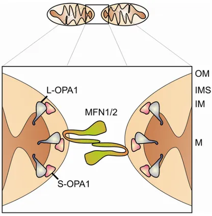
Figure 5. Fusion components of mammalian mitochondria.
Schematic representation of mitochondrial fusion in mammalian cells. Two mitochondria tether through coiled-coil domains of mitofusins (MFN1/2) that are anchored to the mitochondrial outer membrane (OM). L-OPA1 resides in the inner membrane whereas S-OPA1 localizes to the IMS. Both OPA1 isoforms participate in the fusion process. IMS = intermembrane space, IM = inner membrane;
M = matrix. Modified from (Youle and Karbowski, 2005).