Gene Expression Divergence between Subspecies of the House Mouse and the
Contribution to Reproductive Isolation
Inaugural – Dissertation
zur
Erlangung des Doktorgrades
der Mathematisch-Naturwissenschaftlichen Fakultät der Universität zu Köln
vorgelegt von
Ruth Rottscheidt
aus Bad Kreuznach
Köln, 2007
Berichterstatter: Dr. Bettina Harr
Prof. Dr. Diethard Tautz
Tag der letzten mündlichen Prüfung: 19. Februar 2008
Table of Contents
Danksagung... IV Zusammenfassung ... VI Abstract...VIII Declaration...X
1 General Introduction ... 1
1.1 Species, speciation and the evolution of reproductive isolation... 1
1.2 Hybrid incompatibilities associated with gene expression differences ... 6
1.3 The house mouse ... 7
1.4 Aim of the study ... 13
2 Genome wide expression patterns in house mouse subspecies and their F1 hybrids ... 14
2.1 Introduction ... 14
2.2 Materials and Methods... 17
2.2.1 Animals... 17
2.2.2 Sample preparation... 17
2.2.3 Microarray analysis ... 18
2.2.4 Data analysis ... 18
2.3 Results... 24
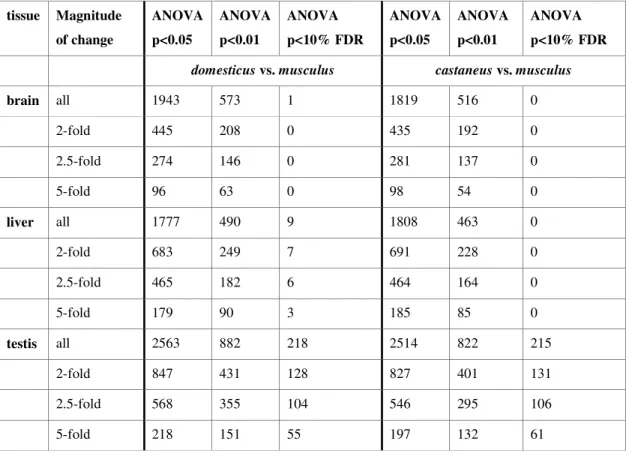
2.3.1 Number of differentially expressed genes... 24
2.3.2 Additivity/nonadditivity of transcripts differentially expressed between the subspecies ... 28
2.3.3 Nonadditive expression in F1 hybrids at transcripts that are not differentially expressed between the parents... 30
2.3.4 Contribution of sequence divergence to expression change... 31
2.3.5 Functional categories of transcripts ... 34
2.4 Discussion ... 35
3 Confirmation of microarray candidate genes: factors affecting expression data in cross-subspecies analyses... 42
3.1 Introduction ... 42
3.2 Materials and Methods... 45
3.2.1 Candidate Genes ... 45
3.2.2 Animals... 46
3.2.3 Sample preparation... 47
3.2.4 cDNA synthesis ... 47
3.2.5 Quantitative real-time PCR (qRT-PCR)... 47
3.2.6 Analysis of qRT-PCR data ... 49
3.2.7 Sequencing of Affymetrix GeneChip ® target regions... 49
3.2.8 Sequencing of amplicon regions ... 51
3.3 Results... 52
3.3.1 qRT-PCR using TaqMan ® gene expression assays... 52
3.3.2 qRT-PCR using Sybr Green fluorescent dye... 54
3.3.3 Sequencing of Affymetrix GeneChip ® target regions... 55
3.3.4 Sequencing of the amplicon regions ... 58
3.3.5 Combining results – evidence for alternative splicing ... 59
3.4 Discussion ... 59
3.4.1 Comparison of two qRT-PCR methods... 60
3.4.2 Sequence differences affecting microarray probe binding ... 63
3.4.3 Alternative splicing as contributor to expression differences... 65
3.4.4 Conclusion ... 66
4 Characterization of candidate genes from a gene expression screen – insights from hybrid zone and population genetic analyses... 67
4.1 Introduction ... 67
4.1.1 Protein and gene expression evolution... 67
4.1.2 Detecting signatures of selection ... 68
4.1.3 Cis- versus trans-regulatory evolution ... 70
4.1.4 Hybrid zone analyses ... 70
4.1.5 Aim of the study... 72
4.2 Materials and Methods... 73
4.2.1 Animals... 73
4.2.2 Sample preparation... 74
4.2.3 cDNA synthesis ... 74
4.2.4 DNA sequencing ... 75
4.2.5 Gene expression analysis... 75
4.2.6 Sequence data analysis ... 76
4.2.7 Correlation of expression-phenotype and genotype – determination of
cis-or trans-acting factors... 79
4.2.8 Assessment of functional consequences for candidate genes from hybrid zone analysis ... 80
4.3 Results... 80
4.3.1 Gene expression analysis using Sybr Green... 80
4.3.2 Sequence analysis... 81
4.3.3 Correlation of expression-phenotype and genotype – determination of cis-or trans-acting factors... 87
4.3.4 Assessment of functional consequences for candidate genes from hybrid zone analysis ... 89
4.4 Discussion ... 92
4.4.1 Protein coding versus regulatory evolution ... 93
4.4.2 Cis- versus trans-regulatory changes ... 99
4.4.3 Functional consequences of candidate genes in a hybrid zone...102
4.4.4 Conclusion ...106
5 Literature ...108
6 Supplement ...127
7 Digital Supplement ...152
Erklärung ...153
Lebenslauf...154
Danksagung
Ich möchte mich bei all jenen bedanken, die mich auf unterschiedliche Art und Weise während der letzten Jahre unterstützt und damit dazu beigetragen haben, dass diese Arbeit nun in dieser Form vorliegt.
Mein erster Dank gilt Dr. Bettina Harr, die mir diese Arbeit ermöglichte. Mit ihren Ideen und ihrer Unterstützung hat sie maβgeblich zum Erfolg dieses Projektes beigetragen. Durch ihre Anregungen und durch viele Diskussionen hat sie mein Verständis für die molekulare Evolution stark erweitert.
Prof. Dr. Diethard Tautz möchte ich für die Übernahme des zweiten Gutachtens und für die Zeit in seiner Arbeitsgruppe danken. Die Möglichkeiten zu forschen, sich auszutauschen und zu informieren hätten idealer nicht sein können.
Weiterhin möchte ich Prof. Dr. Hartmut Arndt für die Übernahme des Prüfungsvorsitzes und Dr. Wim Damen für den Beisitz in meinem Prüfungskomitee danken.
Herzlich danken möchte ich Christian Voolstra, Meike Teschke und Fabian Staubach, die mir mit ihrer ständigen Diskussionsbereitschaft und unzähligen Erklärungen sehr geholfen haben. Vor allem Chris und Meike waren immer bereit, sich mit meinen Fragen auseinander zu setzen, und waren mir auch eine groβe Hilfe beim Korrekturlesen der Arbeit. Till Bayer war immer für mich da, wenn es um Computer- und andere technische Fragen ging. Auβerdem war er der perfekte Reisebegleiter und Mäusefänger in Spanien und Bayern. Birgit Schmitz war eine groβe Unterstützung bei Präparationen, RNA/DNA-Extraktionen, Sequenzierungen und bei der Organisation der mouse-facility. Vielen Dank auch an Christine Pfeiffle und Susanne Kipp für ihren unermüdlichen Einsatz in der mouse-facility und an zahlreiche Mauspfleger und Mauspflegerinnen, deren Arbeit von enormer Wichtigkeit für dieses Projekt war.
Der gesamten Arbeitsgruppe möchte ich für die gute Arbeitsatmosphäre danken.
Neben viel Spaβ hatte ich auch immer das Gefühl einer Zusammengehörigkeit, was
nicht selbstverständlich ist. Besonderer Dank gilt den Mädels Meike Teschke,
Kathryn Stemshorn und Evelyn Schwager für die sportlichen und auch unsportlichen
Mittagspausen und für die vielen guten Gespräche, Till Bayer für alles – Gespräche,
Hilfe, Kuchen, Spaβ, u.v.m. –, Tobias Heinen und Fabian Staubach für das Quäntchen
kölschen Frohsinns im Labor und Maarten Hilbrant für die entspannenden Stunden
mit Tapas, Rotwein und Salsamusik.
Meine Freunde waren immer eine groβe Unterstützung, vor allem Petra, Rebekka und Alex. Vielen Dank für all die kleinen und groβen Dinge, die Ihr im Laufe der Zeit für mich getan habt.
Meiner Familie möchte ich einen ganz besonderen Dank aussprechen: Ihr habt mich
immer unterstützt, ermutigt und Interesse am Fortschritt meiner Arbeit gezeigt. Wann
immer ich Euch gebraucht habe – Ihr wart für mich da!
Zusammenfassung
Dem biologischen Artkonzept zufolge werden Arten als Gruppen von Individuen definiert, die sich miteinander fortpflanzen und von anderen Gruppen reproduktiv isoliert sind. Eine verbreitete Form reproduktiver Isolation im Tierreich ist intrinsische postzygotische Isolation durch Hybridensterilität oder erhöhte Hybridensterblichkeit. Der allgemeinen Überzeugung zufolge werden Fehlfunktionen in Hybriden durch epistatische Interaktionen zwischen inkompatiblen elterlichen Allelen verschiedener Loci (sogenannte Dobzhansky-Muller-Inkompatibilitäten) verursacht. Die Identifizierung von Genen, die zur reproduktiven Isolation von Taxa beitragen, ist wichtig für das Verständnis des Artbildungsprozesses, allerdings hat sich dies in der Vergangenheit als schwierig erwiesen.
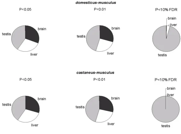
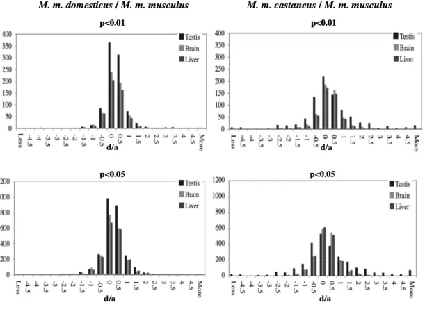
Bei der Entstehung postzygotischer Isolation scheint die Evolution von Genregulation eine wichtige Rolle zu spielen. Deshalb wurden in der vorliegenden Studie mittels Microarrays genomweite Genexpressionsdaten erhoben, um regulatorische Unterschiede zwischen drei Unterarten der Hausmaus, Mus musculus musculus, M. m. domesticus und M. m. castaneus, zu identifizieren. Die Analyse von drei verschiedenen Organen (Gehirn, Leber und Testis) der jeweiligen Unterarten und ihrer reziproken Hybriden erlaubte die Bestimmung des Vererbungsmodus für Genexpressionsunterschiede in den F1-Hybriden. Der gröβte Teil der Transkripte zeigt additive Expression in den Hybriden; nur wenige sind dominant oder überdominant exprimiert, mit Ausnahme einer Kreuzung, die viele missexprimierte Gene im Testis aufweist. Drei verschiedene Analysemethoden sowie Kontrollexperimente bestätigen, dass additive Vererbung von Genexpressions- unterschieden vorherrschend zu sein scheint. Gene, die differentiell exprimierten Transkripten zugrunde liegen, sind vielversprechende Kandidaten, die zu reproduktiver Isolation durch regulatorische Inkompatibilitäten beitragen könnten.
Einge Transkripte mit Expressionsunterschieden zwischen M. m. musculus und
M. m. domesticus wurden für weiterführende Untersuchungen ausgewählt. Die
Validierung der Expressionsniveaus dieser Gene mittels quantitativer Real-Time PCR
hat die Notwendigkeit verdeutlicht, Microarray-Daten durch unabhängige Methoden
zu konfirmieren. Die Ergebnisse zeigen klar, dass Expressionsdaten, auch beim
Vergleich sehr nah miteinander verwandter Taxa, durch Unterschiede in der
Gensequenz beeinflusst werden können, wobei dies für Microarray-Daten gleichermaβen gilt wie für nachfolgende Validierungsmethoden.
Unterschiede in der Evolution von Genexpression zwischen Taxa sind nicht
zwangsläufig funktional bedingt. Aus diesem Grund wurde in der vorliegenden Studie
versucht, funktionale Konsequenzen solcher Unterschiede mittels der Analyse von
Individuen aus der natürlichen musculus-domesticus Hybridzone in Bayern für zwei
ausgewählte Kandidaten-Gene nachzuweisen. Für diese zwei Gene, die einen groβen
Expressionsunterschied zwischen den Elternarten zeigen, konnte kein Anzeichen
dafür festgestellt werden, dass sie einen Beitrag zur reproduktiven Isolation der
beiden Unterarten leisten, da sie in Bezug auf Introgression nicht limitiert zu sein
scheinen. Die Kombination der Hybridzonenuntersuchung mit Ergebnissen aus
populationsgenetischen Analysen lässt vermuten, dass eine adaptive Introgression der
Allele, die das hohe Expressionslevel verursachen, wahrscheinlicher ist. Für beide
Gene konnte nachgewiesen werden, dass der Phänotyp, der mit einem hohem
Expressionsniveau einhergeht, das abgeleitete Merkmal darstellt und mit reduziertem
Nucleotid-Polymorphismus und negativen Tajima’s D-Werten assoziiert ist. Die
Evolution von regulatorischer und Protein-kodierender Sequenz scheint für beide
Gene voneinander entkoppelt und die Expressionsunterschiede ein Resultat von cis-
und nicht trans-regulatorischen Änderungen zu sein.
Abstract
Under the Biological Species Concept species are groups of interbreeding individuals that are reproductively isolated from other such groups. A common form of isolation in animals is intrinsic postzygotic isolation via hybrid sterility/inviability. There is a strong consensus that hybrid dysfunctions are caused by epistatic interactions between incompatible alleles from different loci (Dobzhansky-Muller incompatibilities). The identification of genes that contribute to reproductive isolation between taxa is critical to the understanding of the process of speciation, but identifying such genes has proven to be difficult.
It appears that regulatory evolution might play an important role in postzygotic isolation and the formation of species. The present study employs a whole genome microarray approach to identify genes with regulatory differences between three subspecies of the house mouse, Mus musculus musculus, M. m. domesticus and M. m. castaneus. The within-locus mode of inheritance for gene expression was assessed for three different tissues (brain, liver and testis) by studying the subspecies and their male reciprocal F1 hybrids. The vast majority of transcripts are additively expressed in the hybrids with only few transcripts showing dominance or overdominance in expression except for one direction of one cross, which shows large misexpression in the testis. The reliability of the observed pattern was ensured by three different analysis methods as well as control experiments. The results suggest that additivity is the general mode of inheritance regarding gene expression changes between house mouse subspecies. Differentially expressed transcripts provide promising candidate genes that could be related to reproductive isolation through regulatory incompatibilities.
Several transcripts with expression differences between M. m. musculus and M. m. domesticus were selected for further investigation. A validation approach using quantitative Real-Time PCR strongly emphasizes the need for confirmation of microarray candidate genes. The results show that sequence differences even between closely related taxa have the potential to influence expression data from both microarray and follow-up validation approaches.
As divergent gene expression evolution between taxa may be entirely neutral,
samples from a transect of the natural musculus-domesticus hybrid zone in Bavaria
were analyzed in order to assess functional consequences for two candidate genes.
Both genes show large expression differences between the subspecies. The analysis revealed that it is unlikely that the two genes contribute to reproductive isolation between the subspecies as no sign of limited introgression is evident. Rather, the hybrid zone approach in combination with population genetic analyses suggests adaptive introgression of those alleles that are associated with high expression levels.
In both cases, the high expression phenotype represents the derived state and is
associated with reduced levels of nucleotide polymorphism and a negative Tajima’s
D. For both genes, regulatory and protein-coding evolution is decoupled and the
expression difference results from cis- rather than trans-acting changes.
Declaration
The project was initiated by Bettina Harr. In the course of the project I profited from the work of several other persons, whose contribution I want to acknowledge in the following.
Chapter 2
The inbred-strains of M. m. musculus and M. m. domesticus were provided by J. Pialek from the Department of Population Biology, Institute of Vertebrate Biology, Academy of Sciences of the Czech Republic; M. m. castaneus were donated by A.
Orth and F. Bonhomme from the Laboratory of Genome, Populations, Interactions, and Adaptation in Montpellier, France. The Affymetrix GeneChip ® experiments were conducted by P. Nürnberg and C. Becker from the Cologne Center for Genomics and data analysis was performed by Bettina Harr.
Chapter 3
Meike Teschke provided the German M. m. domesticus mice used for the experiments. Wild-caught M. m. musculus mice were provided by K. Musolf from the Konrad-Lorenz-Institute for Ethology in Vienna.
Chapter 4
The reference data set used for the hybrid zone analysis consists of samples provided
by Bettina Harr, Sonja Ihle, Rick Scavetta, Meike Teschke and myself. Analysis of
these data was performed by Meike Teschke. Population samples of M. m. musculus
and M. m. domesticus used for sequencing analysis were collected and provided by
Sonja Ihle, Rick Scavetta and Meike Teschke. Affymetrix GeneChip ® experiments
were conducted by P. Nürnberg and C. Becker from the Cologne Center for Genomics
and analysis of the GeneChip ® data was performed by Bettina Harr.
1 General Introduction
1.1 Species, speciation and the evolution of reproductive isolation
The definition of a species and the question of how speciation – the splitting of one species into two – takes place has been discussed intensively and controversial since the publishing of Darwin’s “The origin of species” (1859), the book which set the basis for the Modern Synthesis. The founders of the Modern Synthesis substantially changed our understanding of what a species is and therefore what speciation means.
Dobzhansky and Mayr in particular promoted the “Biological Species Concept”
(BSC). Mayr provided the most famous definition of the BSC, which in its latest version states that “species are groups of interbreeding natural populations that are reproductively isolated from other such groups” (Mayr 1995). Thus, species are characterized by reproductive isolation instead of being defined by morphological differences. This species concept is in large parts a result of Dobzhansky’s observation that hybrids between Drosophila sibling species display hybrid sterility, hybrid inviability and assortative mating (Dobzhansky 1937); an observation which laid the foundation for the formulation of his reproductive isolation species concept, which was later incorporated into Mayr’s definition of a species (Mayr 1942).
Reproductive isolation is associated with isolating barriers, “those biological features of organisms that impede the exchange of genes with members of other populations”
(Coyne and Orr 2004, p. 29), which can be divided into premating isolation and postmating isolation, while the latter furthermore discriminates prezygotic and postzygotic barriers. All forms, which include various mechanisms, ensure the genetic distinctness of species and that they can undergo independent evolutionary fates (Orr and Presgraves 2000). Postzygotic isolation refers to sterile or inviable hybrids between two species, while the other forms prevent the occurrence of interspecies hybrids. The BSC has been widely understood as requiring absolute barriers to gene flow between taxa, i.e. no fertile hybrids exist (e.g. Mayr 1963, Barton and Hewitt 1985). Other scientists consider species as entities, which retain their distinctness in sympatry even if occasional hybridization takes place (Grant 1971, Wright 1978).
Coyne and Orr (2004) suggest using a “sliding scale” to define the species status.
Taxa with substantial gene flow (despite morphological distinctness) are not seen as
species, but “as reproductive barriers become stronger, taxa become more and more
“species-like”” until reproductive isolation is complete and taxa are “good species”.
This requires, as they admit themselves, somewhat subjective decisions about the species status. However, the primary aim of speciation studies is to understand the underlying mechanisms of the speciation process. Therefore the question if a species is a “good species” may be of secondary interest.
Since the early 1980’s the interest in speciation studies has shifted towards a genetical perspective of how speciation can be understood (Orr and Presgraves 2000, Orr et al. 2004, Wu and Ting 2004, Mallet 2006). Most studies that are aimed at uncovering genes which are involved in the process of speciation deal with the analysis of intrinsic postzygotic reproductive isolation, i.e. the occurrence of sterile or inviable hybrids between two species. The term “speciation gene” has been widely applied for genes that cause intrinsic postzygotic reproductive isolation and is somewhat unfortunately named, since it strongly implies that genes that reduce hybrid fitness are the cause of speciation (Mallet 2006, Orr 2005). It has been suggested that reproductive isolation evolves as a by-product of differential adaptation of populations (Mayr 1963), which would mean that “speciation genes” are genes that represent aspects of differential adaptation (driven by natural or sexual selection (Albert and Schluter 2005)) which then reduce fitness of hybrids (Wu 2001). Orr and Presgraves (2000) suggest using the term “speciation gene” for any gene that reduces hybrid fitness, as many of the hybrid problems accumulate after the attainment of complete reproductive isolation, since it has been demonstrated that assortative mating, hybrid sterility and inviability increase gradually with genetic distance (Coyne and Orr 1997). Analysis of such genes nevertheless will help to shed light on the “mystery” of speciation.
How is intrinsic postzygotic isolation caused on a genic basis? Dobzhansky
(1937) and Muller (1940) presented experimental evidence that sterility and
inviability in certain crosses of species crosses is caused by incompatibilities between
different loci. Today a large body of evidence has been obtained that shows that
hybrid dysfunctions are indeed due to interactions of different loci (e.g. Orr and
Coyne 1989, Wittbrodt et al. 1989, Gadeau et al. 1999, Presgraves 2003, Sweigart et
al. 2007). The Dobzhansky-Muller model explains how such between-locus
incompatibilities between populations can evolve within populations without
selection acting against any intermediate step. Incompatibilities arise as pleiotropic by-products of the divergence of genomes of geographically separate lineages: alleles from different loci that increase fitness in a pure-species genetic background fail to interact properly when brought together in a hybrid genomic background (Figure 1.1).
In each of the two populations displayed in Figure 1.1, a different mutation occurs and goes to fixation, each yielding a fully viable and fertile genotype. Brought together in a hybrid genome, there is no guarantee that the combination of these alleles functions correctly, as they have never been tested in combination by natural selection. The combination may result in sterility or inviability. A distinct feature of hybrid incompatibilities therefore is that they require epistasis: nonadditive interactions between alleles at different loci (Dobzhansky 1936A, 1937, Muller 1940, 1942). It is important to note that a two-locus incompatibility would be the simplest case: many more loci might be necessary to cause sterility/inviability. Furthermore a genic incompatibility may only have slight effects and complete reproductive isolation may require the cumulative effect of many incompatibilities (Coyne and Orr 2004).
aabb
aaBb Aabb
AAbb aaBB
Hybrid = AaBb aabb
aaBb Aabb
AAbb aaBB
Hybrid = AaBb
Figure 1.1: The Dobzhansky-Muller model. The common ancestor of the two species is shown at the bottom, time runs upwards. The model shows how a genic incompatibility between two loci can evolve unopposed by natural selection. Figure adapted from Coyne and Orr 2004.
To understand the genetic architecture of intrinsic postzygotic isolation, one has
to identify the interacting genes that cause hybrid sterility or inviability. Despite much
effort, only recently genes have been identified. The reason for the difficulties in
identifying such genes is that “the traits of interest, hybrid sterility and inviability, are
by their nature barriers to crossing and thus are refractory to standard genetic
approaches” (Presgraves 2003). Only since actual genes have been identified, it is
possible to address fundamental questions about the factors that cause reproductive
isolation like “which normal function have these genes within species?”, “do they
typically belong to a specific class?”, “are they rapidly evolving?”, “are they
adaptively evolving?”, and so on (Orr et al. 2004). Four of the five identified hybrid incompatibility genes were found in Drosophila (see Orr et al. 2004, Wu and Ting 2004, Orr 2005 for a review and Masly et al. 2006). OdsH, an X-linked homeobox gene (a presumed transcription factor), is misexpressed in the testes of D. simulans x D. mauritiana hybrids and causes male sterility in backcrossed hybrids (Ting et al.
1998, Sun et al. 2004, Ting et al. 2004). Hmr, which encodes a transcriptional regulator of or related to the MYB family, causes male lethality and female sterility in D. melanogaster x D. simulans hybrids, likely due to disrupted gene regulation (Barbash et al. 2003). Lhr has been identified as an interacting partner, but the interaction of two genes alone is insufficient to cause hybrid lethality, additional genes seem to be involved (Brideau et al. 2006). The D. simulans allele of Nup96, a nuclear pore protein, causes lethality when combined with a hemizygous D. melanogaster X-chromosome (Presgraves 2003). JYAlpha (encoding a male fertility-essential Na + -K + ATPase) was recently found to cause sterility in D. melanogaster x D. simulans hybrids (Masly et al. 2006). The only gene causing postzygotic isolation identified in a vertebrate so far is Xmrk-2 in hybrids of Xiphorus maculatus and X. helleri. Xmrk-2 encodes a novel receptor tyrosine kinase, which is, while found as a duplicate gene on the X. maculatus X-chromosome, absent from the X. helleri X-chromosome (Malitschek et al. 1995, Wittbrodt et al. 1989, Schartl et al.
1999). Xmrk-2 is misexpressed in hybrids, causing tumor formation and ultimately lethality. What is missing to date is evidence that the identified “speciation genes” did play a role in the early stages of speciation or if incompatibilities have accumulated subsequent to the species split.
Genes involved in hybrid sterility and inviability are characterized by special
dominance relations. There is good evidence that the genes causing reproductive
isolation are, on average, partially recessive as predicted by the dominance theory of
Haldane’s rule (see for example Orr 1997, Coyne and Orr 2004). For hybrid
inviability the evidence for recessivity is strong (Orr 1993, Presgraves 2003). In an
extensive deficiency mapping experiment for hybrid inviability in Drosophila, it was
shown that recessive-recessive interactions highly outnumber dominant ones
(Presgraves 2003). Also for hybrid sterility evidence for recessive action of genes has
been obtained (Orr 1992, 1997, Hollocher 1996, Sawamura 2000). Recessive
speciation genes can obviously only contribute to the sterility and inviability of F2 or
backcross hybrids (when genes have become homozygous), unless the incompatibilities involve X-linked genes. The dominance theory is now widely accepted to explain the phenomenon of Haldane’s rule (Haldane 1922), the observation that the heterogametic sex suffers from more severe hybrid problems than the homogametic sex. As long as some fraction of the genes causing hybrid problems is recessive and also X-linked genes are involved, hybrid males should fare worse than hybrid females in cases for males are the heterogametic sex. They are afflicted by all X-linked genes involved in hybrid incompatibilities (dominant and recessive) whereas females are affected only by those that are fairly dominant (Coyne and Orr 2004).
Several conclusions can be drawn from the few “speciation genes” identified so
far, although it is hard to generalize from a sample of that size. All these genes have
normal functions within the species. No evidence has been obtained so far that
reproductive isolation involves novel genetic factors like e.g. repetitive DNA
sequences (Orr 2005). Furthermore, genes causing intrinsic postzygotic isolation seem
not to belong to a single functional class. Although some of them are involved in
transcriptional regulation (OdsH, Hmr) – and it remains possible that disruption of
gene regulation is a common cause of hybrid problems – some are not. They are also
not invariably duplicated genes; sometimes they are members of duplicate gene
families (Xmrk-2, OdsH) sometimes they are single-copy genes. Additionally,
speciation genes seem to be rapidly evolving (Orr 2005). This is what one would
expect assuming that genes that cause disruptions in hybrids are those, which have
diverged most between species. Even more important, it could be demonstrated for
three of the genes that they diverge due to positive selection (Ting et al. 1998,
Presgraves 2003, Barbash et al. 2004), which supports one of the central tenets of the
neoDarwinian view of speciation – that reproductive isolation results from natural
selection within species.
1.2 Hybrid incompatibilities associated with gene expression differences
Genome-wide expression profiling by microarrays provides new insights about mechanisms that lead to evolutionary changes (Ranz and Machado 2006). It has long been speculated that many species differences arise from regulatory differences (King and Wilson 1975) and despite the limited information concerning patterns, rates and mechanisms of change at the regulatory level it is at least obvious that changes in transcriptional regulation comprise a quantitatively and qualitatively significant component of the genetic basis for evolutionary change (Wray 2007). Theoretical and empirical studies give support to the hypothesis that failures in the regulation of gene expression may contribute to hybrid dysfunctions (Orr and Presgraves 2000, Ortíz- Barrientos et al. 2007). Such regulatory incompatibilities in hybrids arise from differences that have accumulated in the regulatory sequences between different lineages due to compensatory mutations in order to maintain constant levels of gene expression (Ranz et al. 2004). In a hybrid genetic background, where these alleles have not previously occurred together, such changes do not longer compensate and the interaction may produce novel expression patterns (Landry et al. 2007), i.e. over- or under-expression (misexpression). Many parameters affect the level of transcription, such as number of transcription factor binding sites, abundance of transcription factors and their affinity to binding sites and their interactions. Thus, there are multiple opportunities for the accumulation of changes between species.
Such failures seem to be disproportionally greater in regulatory pathways containing
rapidly evolving genes, particularly those involved in transcription factor-binding site
interactions (Ortíz-Barrientos et al. 2007). Also, simulations by Johnson and Porter
(2000) suggest that hybrid incompatibilities often arise as a consequence of
divergence of regulatory genetic pathways between populations, due to adaptation
processes. Empirically, it has been shown that three of the five genes associated with
hybrid sterility and inviability appear to be related to regulatory changes (Malitschek
et al. 1995, Ting et al. 1998, Barbash et al. 2003). Furthermore, several recent studies
of genome-wide patterns of gene expression in pure species and sterile male hybrids
suggest that hybrids show disruptions in gene expression (Michalak and Noor 2003,
Ranz et al. 2004, Haerty and Singh 2006, Moehring et al. 2007, see Landry et al. 2007
for a review). Particularly, male biased genes have repeatedly shown to have erratic
gene expression patterns in hybrids (Michalak and Noor 2003, Ranz et al. 2004, Haerty and Singh 2006). It has been shown that sex-biased genes, especially male- biased genes with testis specificity or relation to accessory gland proteins, evolve exceptionally rapidly at the expression level in Drosophila (Meiklejohn et al. 2003, Ranz et al. 2003, Nuzhdin et al. 2004). Therefore it can be expected that the regulation of such genes exhibits greater regulatory incompatibilities in hybrids than other genes. Vice versa, the observation that male-biased genes are disproportionally affected by gene expression disruption emphasizes the importance of rapid evolution in the divergence of species and the acquisition of reproductive isolation (Ranz et al.
2004).
Microarray analysis, as a reverse-genetics approach, offers an alternative or supplement to classical forward-genetics approaches in identifying genes which are involved in reproductive isolation. Genome-wide expression profiling can rapidly assay many genes in the genome simultaneously for their expression levels and identify potential genes and genetic pathways responsible for hybrids’ misregulation.
Recent analyses have shown that microarrays are highly instrumental in identifying regulatory differences between species and their hybrids (e.g. Michalak and Noor 2003, Ranz et al. 2004, Haerty and Singh 2006) and that many of the identified genes are likely to be involved in the generation of reproductive barriers (e.g. Michalak and Noor 2004). However, such distorted patterns of gene expression do not necessarily contribute to fitness reduction and reproductive isolation; they may also yield innovative phenotypes (Landry et al. 2007). Nevertheless, it seems likely that the accumulation of regulatory incompatibilities due to divergence and the architecture of transcriptional networks can directly influence the process of speciation (Porter and Johnson 2002, Johnson and Porter 2007).
1.3 The house mouse
The house mouse Mus musculus is one of the best studied model systems and
particular well suited for evolutionary genetics studies. First, the complete genome
sequence of a laboratory strain is available (Waterston et al. 2002) and second,
various aspects like development, anatomy, pathology, behavior and ecology are
intensively studied so that inferences between genomic and phenotypic patterns can
be made (Galtier et al. 2004). Besides laboratory strains, wild-derived mouse strains
can be kept in the laboratory (Guénet and Bonhomme 2003) allowing assessment of naturally occurring genetic variation.
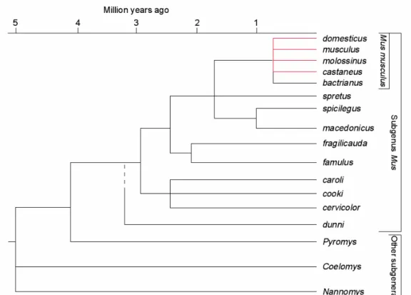
The mouse phylogeny and colonization history is well documented (e.g. Boursot et al. 1993). The genus Mus is thought to have emerged ~ 3 million years ago (Guénet and Bonhomme 2003) and the house mouse Mus musculus, a polytypic species, split up into the different subspecies less than one million years ago (Figure 1.2). It is thought that these subspecies originated on the Indian subcontinent from which they radiated outwards (Din et al. 1996) and have now spread across the world. Best known and described are the three subspecies M. m. musculus, the central house mouse, M. m. domesticus, the western house mouse and M. m. castaneus, the eastern house mouse, which can be distinguished morphologically (Boursot et al. 1993). They have been described as subspecies because they are only partially reproductively isolated from each other and hybrid zones exist in regions of secondary contact. M. m.
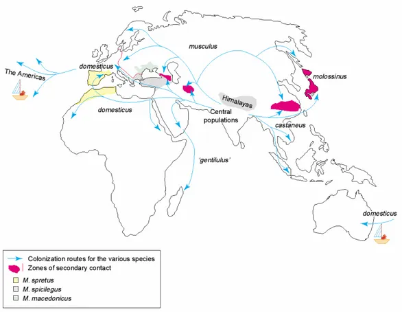
musculus is found in northern Asia as well as in Eastern Europe. M. m. domesticus is found in Western Europe and was introduced through human transport to Africa, America and Australia. The habitat of M. m. castaneus spans from Sri Lanka to South East Asia, including the Indian-Malayan archipelago (Figure 1.3). Due to competition in areas of sympatry with other rodents, house mice occur mainly in association with humans. Only in areas where other mouse species are absent they are able to exist in feral populations. M. m. castaneus is the only subspecies which lives exclusively commensal (Sage 1981, Boursot et al. 1993). Today’s distribution of the house mouse is clearly connected to the migration of the humans over the world. America and Australia as well as many islands lacked mice before humans started to enter those regions. Since Europeans have extensively roamed the oceans, the western house mouse, M. m. domesticus, is particular widespread over the world, though long- distance passive transport of mice is not restricted to M. m. domesticus (Boursot et al.
1993).
The genome of the sequenced laboratory strain C57/BL6J is a mixture of the three subspecies, M. m. domesticus, M. m. musculus and M. m. castaneus (Wade et al.
2002, Yang et al. 2007). According to the most recent analysis, M. m. domesticus provides up to 92% of the genome, musculus <7% and castaneus <1% (Yang et al.
2007).
Figure 1.2: Evolutionary tree of the genus Mus. The last node refers to the polytypic species Mus musculus (Figure from Guénet and Bonhomme 2003).
The fact that the house mouse subspecies are not completely reproductively isolated from each other makes them ideal for speciation studies. To study the origin and the evolution of reproductive barriers, it is intuitive to focus on study systems that are not completely isolated. Thereby it can be discriminated between causes and consequences of the genetic isolation (Macholán et al. 2007). Several areas of secondary contact – hybrid zones – occur between the subspecies (Figure 1.3).
M. m. musculus and M. m. domesticus meet in Europe, in the Caucasus, and in a
region southeast of the Caspian Sea. For M. m. musculus and M. m. castaneus a
contact zone in China and Japan has been described. In Japan, these two subspecies
have hybridized extensively, giving rise to a unique population often referred to as
M. m. molossinus (Yonekawa et al. 1988).
Figure 1.3: Geographical distribution of the different species of the genus Mus and routes of colonization. Mice of the American and Australian continents were imported by humans during colonization (Figure from Guénet and Bonhomme 2003).
The European hybrid zone between domesticus and musculus stretches from the Jutland peninsula to the Bulgarian coast of the Black sea (Boursot et al. 1993, Sage et al. 1993). It has formed as a consequence of the movement of M. m. domesticus into Western Europe within the last 3000 years (Cucchi et al. 2005). Several transects along the 2,400 km length of the hybrid zone have been studied (e.g. Hunt and Selander 1973, Sage et al. 1986, Tucker et al. 1992, Dod et al. 1993, 2005, Prager et al. 1993, Payseur et al. 2004, Raufaste et al. 2005, Macholán et al. 2007, Teeter et al.
2007), which has yielded insights into the genetic basis of reproductive isolation between the two subspecies. Heterogeneity in levels of introgression between genomic regions has been shown (Tucker et al. 1992, Boursot et al. 1993, Dod et al.
1993, Sage et al. 1993, Payseur et al. 2004); thus the genome of the house mouse seems to be “semipermable”, with some regions tolerant of gene flow between species and others not (Payseur et al. 2004). Particular for the X chromosomes limited introgression was found (Tucker et al. 1992, Payseur et al. 2004, Macholán et al.
2007). Changes in allele frequency at diagnostic markers occur very rapidly relative
to the geographic extent of the species ranges (Tucker et al. 1992, Dod et al. 1993, Payseur et al. 2004), which suggests that the hybrid zone is maintained by a balance between selection against hybrids and dispersal (Barton and Gale 1993). Secondary contact of the subspecies seems to have occurred recently (3000-10,000 years ago (Auffray et al. 1990, Cucchi et al. 2005)) in relation to the estimated divergence time, which is thought to be consistent with the accumulation of reproductive isolation in allopatry according to the Dobzhansky-Muller model (Payseur and Hoekstra 2005).
For hybrids between musculus and domesticus in the natural hybrid zone, elevated parasite loads, suggesting general reduced fitness, have been observed (Sage et al. 1986, Moulia et al. 1991, 1993) and reduced testis size has been documented (Britton-Davidian et al. 2005). Complementary to hybrid zone studies, several laboratory experiments with both inbred and wild outbred mice showed that M. m.
domesticus and M. m. musculus are isolated by hybrid male sterility (Forejt and Ivanyi 1975, Alibert et al. 1997, Storchová et al. 2004, Britton-Davidian et al. 2005, Vyskocilová et al. 2005, Good et al. 2007). In some cases, crosses between the subspecies produce fully fertile hybrid males (Vanlerberghe et al. 1986) or variation in the strength of sterility occurs (Alibert et al. 1997, Britton-Davidian et al. 2005).
Furthermore evidence exists that male F1 hybrid sterility is asymmetric, depending on the origin of the X chromosome (Britton-Davidian et al. 2005, Good et al. 2007). For some crosses there is also evidence for female sterility (Britton-Davidian et al. 2005).
Another contributor to reproductive isolation between musculus and domesticus may be assortative mating. Mate choice experiments hint to behavioral differences between the subspecies which may be important in preventing hybridization in some crosses (Smadja and Ganem 2002, Ganem et al. 2005). The genetic basis of male hybrid sterility seems to be fairly complex (Good et al. 2007). Studies which involve classical inbred domesticus strains identified two sets of Dobzhansky-Muller incompatibilities between the two subspecies. One set of dominant-autosomal interactions, which include one or more tightly linked loci on chromosome 17 (Hst1, Forejt and Ivanyi 1975, Vyskocilová et al. 2005). The other set involves the interaction of the X and autosomes (Storchová et al. 2004, Britton-Davidian et al.
2005, Oka et al. 2007), suggesting a crucial role of the X chromosome in reproductive isolation between M. m. domesticus and M. m. musculus. Only recently, Good et al.
(2007) discovered that at least a third set of Dobzhansky-Muller incompatibilities
occurs in wild derived strains of M. m. musculus and M. m. domesticus. Hybrid zone
analyses have identified multiple markers on the X-chromosome with reduced gene
flow across the zone (Tucker et al. 1992; Dod et al. 1993; Payseur et al. 2004,
Macholán et al. 2007). These regions probably harbor genes involved in reproductive
isolation. Only recently, in a detailed genetic survey, Teeter et al. (2007) identified
several regions in the genome which may be involved in reproductive isolation. They
found genes associated with a variety of biological processes, including such as
reproductive physiology, behavior and physiological and immune response. So far, no
specific gene has been identified that contributes to reproductive isolation between
house mouse subspecies. For hybrids between M. m. domesticus and M. spretus a
strong hybrid sterility candidate locus has been identified. Dnahc8, an axonemal
dynein protein expressed in the testis, has been mapped to the site of the hybrid
sterility locus 6, which has been shown to be involved in hybrid sterility between the
two species (Fossella et al. 2000).
1.4 Aim of the study
The study seeks to identify genes that are involved in postzygotic reproductive isolation in the house mouse Mus musculus with a genome wide expression screen.
The house mouse is particularly suited for studying the early stages of speciation,
since reproductive isolation is not complete yet and natural hybrids occur. I used a
combination of two classical approaches: a survey of laboratory F1 hybrids between
taxa and an analysis of animals from a natural hybrid zone. An Affymetrix
GeneChip ® expression analysis of three house mouse subspecies in comparison to
their reciprocal F1 hybrids for three different tissues was conducted to identify
candidate genes for regulatory hybrid incompatibilities. Transcripts with expression
differences between the parental subspecies, and between the hybrids and their
parents, respectively, are candidates which might be involved in reproductive
isolation. After validation of candidate genes with enlarged datasets, a hybrid zone
analysis was intended to infer if expression differences between parental subspecies
and their hybrids are connected to a reduction in fitness, i.e. to be of functional
consequence. Samples from a transect of the natural hybrid zone of M. m. musculus
and M. m. domesticus in Bavaria were used to make inferences about the fitness
effects of specific gene combinations in a hybrid genomic background by considering
the introgression of “expression phenotypes” across the hybrid zone. Genes with
negative fitness effects should not spread very far across the hybrid zone in
comparison to neutral genes. Additionally, sequence based population genetic
analyses for chosen candidate genes were applied to evaluate the genetic basis and
evolutionary forces that might contribute to differences in gene expression.
2 Genome wide expression patterns in house mouse subspecies and their F1 hybrids
2.1 Introduction
Microarray-based gene expression profiling has become widely applied as tool to uncover the genetic architecture of quantitative traits in the past several years.
Transcript levels are treated as phenotype and their intermediate position between DNA sequence polymorphism and organismal phenotype can be seen as “bridge”
between the two in mapping studies (Rockman and Kruglyak 2006). The genetic correlation between expression levels and an organism’s phenotype points to underlying molecular pathways. A co-localization of QTLs for expression and the organism’s phenotype facilitate the identification of causal mutations. “Expression quantitative trait locus” mapping (eQTL) is therefore highly suited to answer basic questions about the number of loci underlying variation in expression phenotypes, the proportion of heritable variance and the genetic complexity of traits (Brem et al. 2002, Schadt et al. 2003, Morley et al. 2004, Brem and Kruglyak 2005, West et al. 2007).
Despite of the usefulness in medical and agricultural genetics, uncovering general principles of the genetic architecture of expression traits will be helpful to reveal the basic forces responsible for phenotypic variation and evolution. In contrast to classical QTL mapping studies, eQTL mapping allows the assessment of thousands of traits simultaneously (Cui et al. 2006, Rockman and Kruglyak 2006). Therefore, a detailed description of possible architectures can be made and an unbiased set of traits in comparison to pre-selected single traits is provided. EQTL mapping studies can also be applied to assign variation in transcript abundance to differences in cis- respective trans-acting sites (Schadt et al. 2003, Yvert et al. 2003, Morley et al. 2004).
Significant levels of cis-polymorphism, controlling individual genes have been detected as well as evidence for clustered trans-eQTLs that simultaneously regulate a large fraction of the transcriptome has been revealed in yeast, mice and humans.
Recent studies indicate that expression levels are heritable and can be under multigenic control (Brem et al. 2002, Schadt et al. 2003, Brem and Kruglyak 2005).
Despite much effort, the relationship between transcript level-variation and
downstream organismal phenotypic trait variation is not well understood (Mackay
2001, Gibson and Weir 2005).
Two approaches are used to uncover the genetic basis of expression traits. One relates DNA polymorphism to differences in expression levels (Brem et al. 2002, Schadt et al. 2003, Doss et al. 2005, Storey et al. 2005). These studies revealed that expression traits are often affected by multiple underlying loci and interactions among them (Rockman and Kruglyak 2006). In yeast, the complex inheritance of transcript levels has been uncovered by detecting significant levels of nonadditive genetic variance, epistatic interactions and transgressive segregation (Brem and Kruglyak 2005). The second approach determines the within-locus mode of inheritance by comparing the transcript abundance of F1 hybrids to that of the parents and assesses if the expression is intermediate (additive) to that of the parents or not. A prevalence of nonadditively over additively expressed transcripts would suggest complex inheritance of expression traits. Recent studies treating this issue have come to inconclusive results concerning the prevalence of additivity and nonadditivity, respectively. Several studies found pervasive within-locus additivity of expression traits in F1 hybrids of laboratory mice, Drosophila and maize (Cui et al. 2006, Hughes et al. 2006, Stupar and Springer 2006, Swanson-Wagner et al. 2006). In contrast, two studies found most transcripts to be nonadditively expressed in Drosophila (Gibson et al. 2004) and the Pacific Oyster (Hedgecock et al. 2007), a third one reported similar numbers of additivity and non-additivity in Arabidopsis (Vulysteke et al. 2005), suggesting to consider epistasis as pervasive aspect of genetic architecture.
Comparative gene expression profiling of hybrids and their parents has also been
applied to identify genes which have the potential to cause hybrid dysfunctions via
regulatory incompatibilities. It is generally assumed that divergent evolution in two
lineages leads to coevolution of genes and that crosses between species can reveal
such coevolution (Landry et al. 2007). The formation of hybrids combines alleles that
have not been previously occurred together and the interaction may generate new
phenotypes. Uncovering the molecular basis of such nonadditive, epistatic
interactions is central to the understanding of the evolution of hybrid incompatibilities
(Dobzhansky-Muller incompatibilities (Dobzhansky 1937, Muller 1940). Theoretical
and empirical studies suggest the importance of gene regulation for hybrid
incompatibilities (Orr and Presgraves 2000, see Ortíz-Barrientos et al. 2007 for a
review). Regulatory incompatibilities are defined as interactions of the transcriptional networks that lead to novel expression phenotypes in interspecific hybrids (Landry et al. 2007). Several studies in Drosophila show that disruptions in transcriptional regulation may indeed be associated with hybrid incompatibilities such as hybrid sterility (Michalak and Noor 2003, 2004, Ranz et al. 2004, Haerty and Singh 2006, Moehring et al. 2007). Michalak and Noor (2003, 2004) for example found several genes severely underexpressed in hybrids between D. simulans and D. mauritiana, which are associated with spermatogenesis and other male-specific phenotypes. Still in the 5 th generation of backcross hybrids, sterility and misexpression of these transcripts was strongly correlated, which makes it possible that genes involved in spermatogenesis cause sterility in these hybrids. Interestingly, a large proportion of transcripts with disrupted expression in hybrids do not show expression level divergence between the parental species, suggesting that these genes are under stabilizing selection (Ranz et al. 2004, Haerty and Singh 2006). This finding underlines that species often accumulate divergent cryptic genetic changes in coding regions as well as regulatory regions that are revealed only if the two divergent regulatory architectures are brought together in a single individual (Moehring et al.
2007, Ortíz-Barrientos et al. 2007). If such disruptions in gene expression are the cause and not the consequence of hybrid incompatibilities, high-throughput “reverse genetics” approaches, like microarray analyses, have the potential to identify candidate genes or pathways that contribute to reproductive isolation via regulatory incompatibilities and thus to speciation (Noor and Feder 2006).
The present analysis was conducted to assess the within-locus mode of
inheritance of expression differences between three subspecies of the house mouse,
M. m. domesticus, M. m. musculus and M. m. castaneus. Differentially regulated
transcripts between the subspecies and between the F1 hybrids and their parents,
respectively, provide candidate genes, which can be involved in regulatory hybrid
incompatibilities. Transcripts with expression levels in the hybrids significantly over-
or under-expressed in comparison to the parents may be particular interesting with
regard to hybrid incompatibilities. It has to be stressed that a hybrid incompatibility
gene is any gene that contributes to reduced fitness in hybrids (Orr and Presgraves
2000).
2.2 Materials and Methods
2.2.1 Animals
Wild-derived strains of M. m. domesticus, M. m. musculus and M. m. castaneus were used for the microarray analysis. For M. m. musculus, strain JPC 2821 from the Czech Republic, for M. m. domesticus, strain JPC 2705 from Germany and for M. m. castaneus, strain CIM (from India), was used for the experiments. Animals from domesticus and musculus were collected in the wild and inbred by brother-sister matings for ~13 generations in the laboratory of J. Pialek in the Department of Population Biology in Studenec (Czech Republic). The castaneus strain was provided by A. Orth and F. Bonhomme and has been kept in the Laboratory of Genome, Populations, Interactions, Adaptation, Montpellier in France for more than 30 generations. Reciprocal crosses between the wild-derived-inbred subspecies were set up to obtain F1 hybrids (Table 2.1). M. m. musculus will be abbreviated as mus, M. m. domesticus as dom and M. m. castaneus as cas in figures and tables in the following.
Table 2.1: Breeding setup for F1 hybrids.
mother father F1 hybrid
M. m. musculus x M. m. domesticus mus-dom M. m. domesticus x M. m. musculus dom-mus M. m. castaneus x M. m. musculus cas-mus M. m. musculus x M. m. castaneus mus-cas
Three tissues each (brain, liver, testis) of two males each cross and direction and each of the parental pure subspecies were used for the experiment (see Supplement 1).
Altogether 42 microarray experiments were performed; 14 animals were analyzed for three tissues each. All mice were raised under identical standard laboratory conditions and were sacrificed at the age of 6-8 weeks.
2.2.2 Sample preparation
Animals were sacrificed using CO 2 . Organs were excised and immediately snap
frozen in liquid nitrogen. RNA was extracted using Trizol ® (Invitrogen, Carlsbad,
CA) following the manufacturer’s protocol. Quality and integrity of the total RNA
was controlled by using the Agilent Technologies 2100 Bioanalyzer and the RNA 6000 Nano LabChip ® Kit (Agilent Technologies Waldbronn, Germany). Only samples with RNA integrity numbers (RIN) >8.0 were used for analysis.
2.2.3 Microarray analysis
Expression profiles were determined for over 39,000 mouse transcripts using the Mouse Genome 430 2.0 Affymetrix GeneChip ® . The biotin-labeled target synthesis started from 1 µg of total RNA following the Ambion Message Amp II aRNA amplification Kit protocol. After hybridization the GeneChips ® were washed, using an Affymetrix GeneChip ® fluidic station 450, stained with SA-PE (Streptavidin R- phycoerythrin conjugate) and read using an Affymetrix GCS 300 G scanner.
2.2.4 Data analysis
Data processing and statistical analyses were performed using the statistical language R. Raw signal intensities were normalized and summarized according to the standard Affymetrix MA Suite 5.0 algorithm using the program Bioconductor (http://www.bioconductor.org/). Signal intensities were ln-transformed prior to statistical analyses. MA Suite 5.0 expression values have been submitted to the Gene Expression Omnibus (accession number GSE9338). All analyzed samples were defined as “groups”; a “group” consisting of either domesticus, musculus, castaneus, F1 hybrids from one direction of the cross, or F1 hybrids from the other direction of the cross. Transcripts are defined as “expressed” in a group if the average expression level among the replicate samples within a group is >500. Applying a threshold of
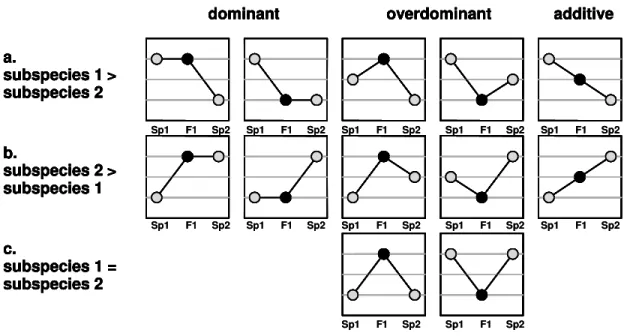
>500 yields about 11,000 expressed transcripts per group and tissue, a value that corresponds to previous studies (Su et al. 2004). The expression levels in the hybrids were determined as additive or nonadditive for two different groups:
1. transcripts that are differentially expressed between the parental subspecies (see Figure 2.1 a, b)
2. those transcripts, which do not differ in expression between the parental lines,
but which are expressed differently in the F1 hybrids relative to both parents
(Figure 1c):
a.
subspecies 1 >
subspecies 2
dominant overdominant additive
b.
subspecies 2 >
subspecies 1
c.
subspecies 1 = subspecies 2 a.
subspecies 1 >
subspecies 2
dominant overdominant additive
b.
subspecies 2 >
subspecies 1
c.
subspecies 1 = subspecies 2
Sp1 Sp1
Sp1
Sp1 Sp1 Sp1 Sp1
Sp1 Sp1
Sp1
Sp2 Sp2 Sp2 Sp2 Sp2
Sp2 Sp2
Sp2 Sp2
Sp2
Sp2 Sp2
Sp1 Sp1
F1
F1 F1
F1
F1 F1 F1 F1 F1
F1 F1 F1
a.
subspecies 1 >
subspecies 2
dominant overdominant additive
b.
subspecies 2 >
subspecies 1
c.
subspecies 1 = subspecies 2 a.
subspecies 1 >
subspecies 2
dominant overdominant additive
b.
subspecies 2 >
subspecies 1
c.
subspecies 1 = subspecies 2 a.
subspecies 1 >
subspecies 2
dominant overdominant additive
b.
subspecies 2 >
subspecies 1
c.
subspecies 1 = subspecies 2 a.
subspecies 1 >
subspecies 2
dominant overdominant additive
b.
subspecies 2 >
subspecies 1
c.
subspecies 1 = subspecies 2
Sp1 Sp1
Sp1
Sp1 Sp1 Sp1 Sp1
Sp1 Sp1
Sp1
Sp2 Sp2 Sp2 Sp2 Sp2
Sp2 Sp2
Sp2 Sp2
Sp2
Sp2 Sp2
Sp1 Sp1
F1
F1 F1
F1
F1 F1 F1 F1 F1
F1 F1 F1