Cyclase associated protein CAP in the regulation of the actin cytoskeleton and cell polarity in
Dictyostelium discoideum
INAUGURAL-DISSERTATION zur
Erlangung des Doktorgrades
der Mathematisch-Naturwissenschaftlichen Fakultät der Universität zu Köln
vorgelegt von Hameeda Sultana aus Bangalore, Indien
2004
Referees/Berichterstatter : Prof. Dr. Angelika A. Noegel Prof. Dr. Siegfried Roth
Date of oral examination : 06.07.2004 Tag der mündlichen Prüfung
The present research work was carried out under the supervision and the direction of Prof.
Dr. Angelika A. Noegel in the Institute of Biochemistry I, Medical Faculty, University of Cologne, Cologne, Germany, from June 2001 to July 2004.
Diese Arbeit wurde von Juni 2001 bis Juli 2004 am Biochemischen Institut I der Medizinischen Fakultät der Universität zu Köln unter der Leitung und der Betreuung von Prof. Dr. Angelika A. Noegel durchgeführt.
To my everloving Ammi-Abba
& family…
This thesis is not only the result of three years of bench work, data processing, reading and writing but it is also the result of the knowledge and the help of others. First and foremost I would like to express my sincere gratitude to my esteemed advisor, Prof. Dr. Angelika A. Noegel, Institute of Biochemistry I, Medical Faculty, University of Cologne, Germany. I am deeply grateful to her for the enormous freedom given to me to pursue my own interests while at the same time providing excellent guidance to ensure that my efforts contribute to the cytoskeletal research. Her creative suggestions, constructive criticism and steadfast encouragement is praiseworthy and unforgettable. Her analytical perusal of the manuscript is highly acknowledged. I would like to thank in perpetuity for all her help and superb guidance.
I acknowledge my special thanks to Dr. Francisco Rivero for his instant cooperation and help as and when required. His constructive suggestion during the course of my study is highly appreciable. I also thank him for providing RacA strains and GFP-VatB plasmid. I sincerely thank Dr. Ludwig Eichinger for providing me the lab facilities to carry out the microarray analysis and for his necessary suggestions. I owe special thanks to Patrick Farbrother for his help through out the microarray analysis. I also thank Dr. Akis Karakesisoglou for his friendly nature and laughs. A special note of thank to Dr. Christoph Clemen for his friendly gesture and support.
I owe a special gratitude to Bettina Lauss, who made all the necessary official things easy and fast.
Her encouragement and friendly support is admirable and unforgettable. My special thanks are due to Dr. Budi Tunggal for his valuable suggestions and wonderful company in our office. I thank Rolf for his necessary technical assistance, Berthold, Maria, Rosi, Bärbel and Roberto for their necessary help. I thank all my colleagues for providing a friendly atmosphere and a special one to Thorsten Olski. I record my special thanks to Inge Götz-Krichi for her friendly support and constant help.
I sincerely thank Dr. Salvatore Bozzaro (University of Torino), Dr. Kees Weijer (University of Dundee), Dr. Edward Cox (Princeton University) and Dr. Richard A. Firtel (University of California) for providing the mutant strains.
My everloving and wonderful parents Ammi-Abba were the source of inspiration and motivation throughout my life. I am deeply indebted for their love and affection, which stood by me as a strong support and without their blessings it would have been a difficult task to complete this work.
I owe a huge indebtedness to my everloving and affectionated Pops-deed for their constant encouragement and incredible love which bolstered my days to reach my goals and I thank them for their wonderful everlasting support throughout my studies. My everloving little sister lubu was my biggest inspiration and emotional support during all my studies. I thank her for her loving and caring nature. I express my heartiest gratitude to my everloving brother lulu for all his inspiration and constant support. I thank all my family members and beloved children abrar-pinky, shazu-nazu, wasimu-saleemu, faizu, kola and little adiyan for their affection.
I am greatly indebted to my wonderful husband and a word of appreciation is not at all sufficient to express my gratitude for all his great inspiration, understanding and heartfelt cooperation throughout this study.
Finally, the financial assistance received by me from the DFG is highly acknowledged.
Cologne Hameeda Sultana
03/05/2004
Table of Contents
Chapter Description Page(s)
ABBREVIATIONS
I. INTRODUCTION 1-10
1.0 Dictyostelium discoideum: a model system in motion 1 2.0 CAP is a highly conserved protein 2
3.0 Organization of CAP structure 3
4.0 Model of CAP function 5
5.0 The interaction of CAP with actin is PIP2 regulated 6
6.0 CAP is a multifunctional protein
7
7.0 Cell polarity and cAMP signalling in Dictyostelium 7
8.0 Overview 10
II. MATERIALS AND METHODS 11-51
1.0 Materials 11
1.1. Laboratory materials 11
1.1.1 Instruments and equipments 11
1.2. Kits 13
1.2.1 Enzymes, antibodies, substrates, inhibitors and antibiotics 13
1.3. Chemicals and reagents 14
1.4. Media and buffers 15
1.4.1. Media and buffers for Dictyostelium culture 15
1.4.2. Media for E. coli culture 16
1.4.3. Media and buffers for yeast culture 17
1.4.4. Buffers and other solutions 18
1.5. Biological materials 19
1.6. Plasmids 20
1.7. Synthetic Oligonucleotides 21
2.0 Cell biological methods 21
2.1 Growth of Dictyostelium 21
2.1.1 Growth in liquid nutrient medium 21
2.1.2 Growth on SM agar plates 22
2.2 Development of Dictyostelium 22
2.3 Preservation of Dictyostelium cells 22 2.4 Transformation of Dictyostelium cells 23 2.4.1 Transformation of Dictyostelium cells by CaCl2 23 2.4.2 Transformation of Dictyostelium cells by electroporation 24
Chapter Description Page(s)
2.5 Analysis of agglutination 24
2.6 Endocytosis assay 25 3.0 Molecular biological methods 25 3.1 Purification of plasmid DNA 25 3.2 Digestion with restriction enzymes 26 3.3 Generation of blunt ends in linearised plasmid DNA 26 3.4 Dephosphorylation of DNA fragments 27 3.5 Setting up of ligation reaction 27 3.6 Isolation of Dictyostelium genomic DNA 27 3.7 DNA agarose gel electrophoresis 28 3.8 Recovery of DNA fragments from agarose gel 28 3.9 Isolation of total RNA from Dictyostelium cells 29 3.10 RNA formaldehyde-agarose gel electrophoresis 29 3.11 Northern blotting 30 3.12 Radiolabelling of DNA 30 3.12.1 Chromatography through Sephadex G-50 spin column 31 3.12.2 Hybridisation of Southern- or northern-blot with radiolabelled DNA probe 31 3.13 Transformation of E. coli 32
3.13.1 Transformation of E. coli cells by the CaCl2 method 32 3.13.2 Transformation of E. coli cells by electroporation 32 3.14 Glycerol stock of bacterial culture 33 3.15 DNA colony blot for screening of E. coli transformants 33 3.16 Construction of vectors 34
3.16.1 Vectors for expression of GFP-fusion proteins of CAP and its domains (Noegel et al., 1999) 34 3.16.2 Vectors for expression of binding partner studies of CAP 35 3.16.3 Vectors for expression of GST fusions of sGC, ARPE and Vacuolar ATPases d subunit 35 3.17 DNA sequencing 35 3.18 Computer analyses 36
4.0 Methods for the Yeast Two Hybrid System 36
4.1 Transformation of Yeast by Lithium Acetate Method 36
4.2 DNA isolation from Yeast 36
4.3 β-galactosidase colony lift assay 37
4.4 Yeast strain maintenance 38
5.0 Biochemical methods 38
5.1 Preparation of total protein from Dictyostelium 38
5.2 Isolation of phagosomes 38
Chapter Description Page(s)
5.3 Cell fractionation studies to determine the subcellular
localization of CAP 39
5.4 Immunoprecipitation from Dictyostelium cell lysate 39
5.5 SDS-polyacrylamide gel electrophoresis 40 5.5.1 Coomassie blue staining of SDS-polyacrylamide gels 41 5.5.2 Silver staining of polyacrylamide gels 41
5.5.3 Drying of SDS-polyacrylamide gels 41
5.6 Western blotting using the semi-dry method 42 5.6.1 Ponceau S staining of western blots 42 5.7 Immunodetection of membrane-bound proteins 42 5.7.1 Enzymatic chemi-luminescence (ECL) detection system 43 5.8 Expression and purification of GST fusions of sGC, ARPE and VD 43 5.8.1 Small-scale protein expression 43 6.0 Immunological methods 44 6.1 Indirect immunofluorescence of Dictyostelium cells 44 6.1.1 Preparation of Dictyostelium cells 44 6.1.2 Methanol fixation 44 6.1.3 Picric acid-paraformaldehyde fixation 45 6.2 Immunolabelling of fixed cells 45 6.2.1 Mounting of coverslips 46 6.3 Phalloidin staining of fixed cells 46 6.4 Immunolabelling of Dictyostelium cells expressing GFP-CAP and its domains fixing during phagocytosis and pinocytosis 46 7.0 Microscopy 47 7.1 Live cell imaging of Dictyostelium cells expressing GFP-CAP 48 7.2 Imaging distribution and live dynamics of GFP-CAP during pinocytosis 48 7.3 Imaging the distribution of GFP-CAP during phagocytosis 48 7.4 Imaging the distribution and dynamics of GFP-CAP in aggregation competent cells 49 7.5 Microscopy of agar plates 49
8.0
Microarray analysis 498.1 RNA preparation 49
8.2 Spiking of internal mRNA controls 50
8.3 Quantitation, normalization and data analysis 51
8.4 Signal Quantification 51
Chapter Description Page(s)
III. RESULTS 52-117
1.0 Localization and dynamics of CAP in the signalling mutants 52 1.1 Expression levels of the endogenous CAP in the aca-, pia-, pia-.2ac-, 52 gα2- and gβ-, car1-/3-, pik 1-/2- and pka- cells
1.1.1 Localization of CAP in the signal transduction mutants 53 1.1.2 Localization of CAP in pka- cells 54
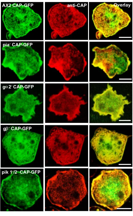
1.2 Expression of GFP-CAP fusion protein in the aca-, pia-, gα2- and gβ- and pik 1-/2- cells 56
1.2.1 Localization of GFP-CAP in signalling mutants 56 1.3 Re-localization of CAP in aca-, pia-, HSB101, gα2-, gβ-, cAR1-/3-,
pik1-/2-, and PKA- during phagocytosis 57 1.3.1 Determination of the presence of CAP in phagosomes 59 1.3.2 Association of CAP with membrane fractions 59 1.4 Analysis of CAP and CAP-GFP in the pia-, gα2-, gβ-, and
pik1-/2- cells during phagocytosis 60 1.4.1 Redistribution of GFP fusion proteins of CAP and its domains
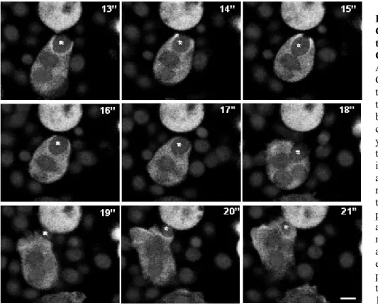
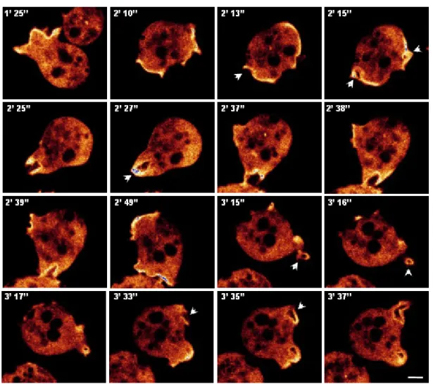
in aca- cells during phagocytosis 61 1.4.2 Live imaging visualizing the dynamics of GFP-CAP in aca-,
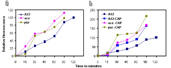
pia-, gα2-, gβ-, and pik 1-/2- cells 62 1.4.3 Quantitative analysis of phagocytosis in the aca-, pia-, gα2- and gβ- and pik 1-/2- cells 65 1.5 Re-localization of CAP during pinocytosis in the aca-, pia-, gα2-,
gβ- and pik 1-/2- cells 68 1.5.1 Live dynamics of GFP-CAP in the aca-, pia-, gα2- and gβ-
cells during pinocytosis 69 1.6 Dynamics of CAP-GFP in the pik 1-/2- cells during pinocytosis 71 1.6.1 Quantitative analysis of pinocytosis in pik 1-/2- cells and cells
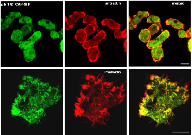
expressing GFP-CAP 72 1.6.2 Distribution of F-actin in the pik 1-/2- cells expressing GFP-CAP 73 1.7 Role of CAP in the aggregation of pik 1-/2- cells expressing GFP-CAP 75 1.8 Role of CAP during aggregation competence in the pik 1-/2- cells
expressing GFP-CAP 75 1.9 Role of CAP in the growth and development of pik 1-/2- cells
expressing GFP-CAP 77 2.0 Role of CAP in the aggregation and development of aggregation
deficient aca null cells 78 2.1 Expression of GFP fusions of CAP and its domains in aca- cells 78
Chapter Description Page(s)
2.2 Localization of GFP fusions of CAP and its domains in aca- cells 79
2.3 Moderate overexpression of CAP rescues the aggregation defect of aca- cells 80
2.3.1 Role of CAP-GFP fusions in the aca- cells during aggregation 82
2.4 Agglutination of the aca- cells expressing CAP-GFP and N-CAP-Pro-GFP 82
2.5 Developmental phenotypes of aca- cells expressing CAP-GFP 84
2.6 Cell adhesion mechanism in the aca- cells expressing CAP-GFP and N-CAP-Pro-GFP 85
2.7 Time course and quantification of cell-cell adhesion in the aca- cells expressing CAP-GFP and N-CAP-Pro-GFP 86
2.8 Expression of cAR1 receptor in the agglutinating aca- cells expressing CAP-GFP and N-CAP-Pro-GFP 88
2.9 Expression of cell adhesion molecules in the aca- cells expressing CAP-GFP and N-CAP-Pro-GFP 88
2.10 Distribution of polarity markers in the aca- cells and aca- cells expressing CAP-GFP and the N-CAP-Pro-GFP 91
3.0 Role of CAP in LimD- and AX2 cells expressing constitutively active and dominant negative RacA 94
3.1 Localization of CAP in AX2 cells expressing constitutively active and dominant negative RacA 94
3.2 Localization of CAP in LimD- cells 95
3.3 Complementation analysis of CAP bsr by GFP-LimD 96
3.4 Dynamics of GFP-LimD in CAP bsr during endocytosis 96
3.5 Expression of GFP-LimD restores pinocytosis of CAP bsr cells 98
3.4 Role of CAP in cAMP signalling 98
4.0 Identification of binding partners for Dictyostelium CAP 100
4.1 Does CAP physically interact with aggregation specific adenylyl cyclase (ACA)? 100
4.2 Does CAP interact with guanylyl cyclase? 102
4.3 Interaction of CAP with ARP2/3 complex subunits 103
4.4 Interaction of CAP with Vacuolar H+-ATPases (V-ATPases) complex 104
4.5 The vacuolar network is disturbed in the absence of CAP 106
4.6 Expression of CAP GFP fusion protein restores the disturbed vacuolar system in CAP bsr 108
5.0 Transcriptional analysis of CAP bsr using DNA microarrays 109
Chapter Description Page(s)
IV. DISCUSSION 114-131
1. Localization and dynamics of CAP in signalling mutants 114
2.0 CAP is a general regulator of phagocytosis 116
3.0 Dynamics of CAP during pinocytosis 117
3.1 CAP rescues the impaired pinocytosis of pik 1-/2- cells 117
3.2 CAP restores the altered distribution of actin and filamentous actin in pik1-/2- cells 118
4.0 Developmental regulation of CAP in pik 1-/2- cells 119
5.0 Role of CAP in aggregation and early development of aca- cells 119
5.1 Moderate CAP overexpression improves the development of aca- cells but does not allow complete restoration 120
6.0 An overview of the significance of CAP in the signalling mutants 121
7.0 Localization of CAP depends on LimD and is modulated by RacA 121
8.0 LimD expression restores the defect in endocytosis of CAP bsr 122
9.0 CAP is required for cAMP signalling 122
10.0 Does CAP interact with cyclases? 123
11.0 Does CAP interact with the Arp2/3 complex? 124
12.0 Interaction of CAP with the Vacuolar H+ ATPases complex 124
13.0 Microarray analysis signifies CAP as a regulator of signal transduction, multi-cellular development and cytoskeleton 127
14.0 Outlook 130
V. SUMMARY/ZUSAMMENFASSUNG 132-133
BIBLIOGRAPHY
ErklärungCurriculum Vitae/Lebenslauf
Abbreviations
AP alkaline phosphatase
APS ammonium persulphate
ATP adenosine 5’-triphosphate
bp base pair(s)
BSA bovine serum albumin
Bsr blasticidin resistance cassette cAMP cyclic adenosine monophosphate CAP cyclase associated protein
CCD cooled charge-coupled device
cDNA complementary DNA
CIAP calf intestinal alkaline phosphatase dNTP deoxyribonucleotide triphosphate DABCO diazobicyclooctane
DEPC diethylpyrocarbonate
DMSO dimethylsulphoxide
DNA deoxyribonucleic acid
DNase deoxyribonuclease
DTT 1, 4-dithiothreitol
ECL enzymatic chemiluminescence EDTA ethylenediaminetetraacetic acid
EGTA ethyleneglycol-bis (2-amino-ethylene) N, N, N, N-tetraacetic acid
G418 geneticin
GFP green fluorescent protein GST glutathione S-transferase
HEPES N-[2-Hydroxyethyl] piperazine-N’-2-ethanesulphonic acid HRP horse radish peroxidase
IgG immunoglobulin G
IPTG isopropyl-β-D-thiogalactopyranoside
kb kilo base pairs
kDa kilodalton
β-ME beta-mercaptoethanol
MOPS Morpholinopropanesulphonic acid
Mw molecular weight
NP-40 nonylphenylpolyethyleneglycol
OD optical density
ORF open reading frame
PAGE polyacrylamide gel electrophoresis PCR polymerase chain reaction
PEG polyethylenglycol
PIPES piperazine-N, N’-bis [2-ethanesulphonic acid]
PMSF phenylmethylsulphonylfluoride
RNA ribonucleic acid
RNase ribonuclease
rpm rotations per minute
SDS sodium dodecyl sulphate
TEMED N,N, N’,N’-tetramethyl-ethylendiamine TRITC tetramethylrhodamine isothiocyanate
U unit
UV ultra violet
vol. volume
v/v volume by volume
w/v weight by volume
X-gal 5-bromo-4-chloro-3-indolyl-β-D-galactopyranoside
Units of Measure and Prefixes
Unit Name
Ci curie
°C degree Celsius
D Dalton
g gram h hour
L litre
m meter
min minute
s sec
V volt
Symbol Prefix (Factor)
k kilo (103)
c centi (10-2)
m milli (10-3)
µ micro (10-6)
n nano (10-9)
p pico (10-12)
α alpha
β beta γ gamma
Introduction
1.0 Dictyostelium discoideum: a model system in motion
Movement is a manifestation of mechanical work, which requires a fuel (ATP) and proteins that convert the energy stored in ATP into motion. Also, all movements involve the cytoskeleton, a cytoplasmic system of fibres. The machinery that powers these movements is built from the actin cytoskeleton. The actin cytoskeleton is huge, larger than any organelle, filling the cytosol with actin filaments. The actin cytoskeleton of a cell is required for cell shape changes, cell motility and chemotaxis, as well as for cytokinesis, intracellular transport processes, development and signal transduction. The multiplicity of these actin related processes require the existence of actin in a variety of complex, dynamic structures, which are regulated by actin-binding proteins. Actin- binding proteins are particularly complex in their relationship with actin in its various forms where they can stimulate polymerisation and depolymerisation depending on the conditions. They are characterized by their ability and mode of interactions with G- or F-actin. The large number of F- actin binding proteins identified in various cells contrasts with very few G-actin binding proteins (Kreis and Vale, 1993).
Dictyostelium discoideum is a eukaryote that is related to animals and fungi, a position it shares with Acanthamoebae and the acellular slime moulds (Baldauf et al., 2000). The Dictyostelium discoideum genome (34 Mb) shows a high degree of sequence similarity to homologs, orthologs, and paralogs in other eukaryotic organisms including protozoan parasites and vertebrates; among them are the numerous genes interacting with G- and F- actin. Dictyostelium amoebae have a complex cytoskeleton whose dynamics is actively studied using the genetic approaches. The Dictyostelium undergoes a development, which is relatively simple and can be easily investigated with all the experimental techniques that are commonly used in developmental biology.
Dictyostelium cells have adopted a strategy for multicellular development that differs from that of metazoa. In their vegetative stage, Dictyostelium cells are single-celled amoebae that feed on bacteria and multiply by binary fission. The striking feature of Dictyostelium is that, when their food source is depleted, they undergo a switch in behaviour and the individual cells come together and form a fruiting body (Figure 1). This is a highly differentiated multicellular structure composed of spore cells supported by stalk cells that are arranged as a stalk and a basal disc, which anchors to the substratum. Dictyostelium is therefore a valuable and convenient experimental system for studies of the role of the actin cytoskeleton, cell motility, phagocytosis, macropinocytosis, chemotaxis, signal transduction, development and differentiation.
Figure 1: Dictyostelium development. Amoebae proliferate as single cells during the growth phase. Upon starvation, amoebae undergo chemotaxis towards a pulsatile cAMP source. During aggregation, cells coalesce into adherent cell
‘streams’ that eventually come together to form the mound, the first stage of multicellular development. The mound compacts to form a tight aggregate and then develops a ‘tip’, which coordinates further development. After extension to form the first finger, the developing structure either immediately forms a fruiting body, the process of culmination or forms a motile slug that migrates to seek conditions favourable for culmination. Scale shows relative timing of development (taken from Coates and Harwood, 2001).
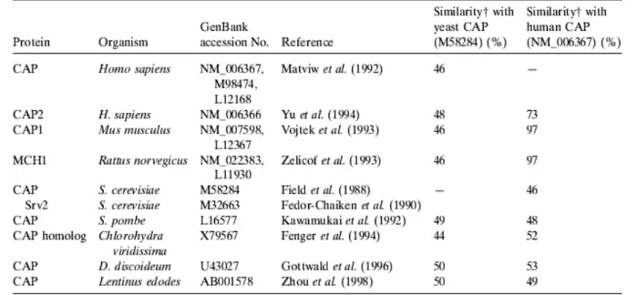
2.0 CAP is a highly conserved protein
Cyclase associated protein (CAP) is an evolutionarily conserved regulator of the G-actin/F-actin ratio and has been identified in a wide range of species (Figure 2). CAP, was first isolated as a 70 kDa component of the Saccharomyces cerevisiae adenylyl cyclase complex that serves as an effector of Ras during nutritional signalling, which is involved in Ras/cAMP-dependent signal transduction and serve as an adapter protein translocating the adenylyl cyclase complex to the actin cytoskeleton (Field et al., 1988). CAP and CAP-homologues have two domains, an N-terminal and a C-terminal domain separated by one (D. discoideum) or two (yeast, human) proline rich stretches, and distinct functions have been attributed to the domains as well as to the proline rich region.
Genetic and biochemical studies in yeast have demonstrated that a major target of Ras is adenylyl cyclase, and it has been shown that the N-domain of CAP is responsible for mediating the Ras sensitivity of adenylyl cyclase, whereas the C-domain is responsible for binding to monomeric actin and is required for regulating cellular morphology (Nishida et al., 1998, Gerst et al., 1991). Proline rich sequences often function as SH3 domain binding sites, which are thought to mediate formation of specific protein complexes, and the proline rich region 2 (P2) of yeast CAP was identified as an SH3 domain binding site and is known to interact with the SH3 domain of yeast actin binding protein Abp1p (Freeman et al., 1996). Furthermore, genetic studies in yeast have recently
implicated CAPs in vesicle trafficking and endocytosis (Hubberstey and Mottillo, 2002). In a screen to identify genes required for Drosophila oocyte polarity, the Drosophila homolog of yeast CAP was isolated. CAP preferentially accumulated in the oocyte where it inhibited actin polymerization resulting in a loss of asymmetric distribution of mRNA determinants within the oocyte (Baum et al., 2000). Benlali et al., 2000, identified CAP in a screen for mutations that disrupted eye development by increasing the F-actin levels and inducing premature photoreceptor differentiation. ASP56, the mammalian homologue of CAP, was isolated from pig platelets on the basis of its actin sequestering activity and shown to be a G-actin binding protein (Gieselmann et al., 1992). Taken together these studies suggest that CAP is a multifunctional protein with roles in signalling and regulation of the cytoskeleton that have been attributed to individual domains of the protein.
Figure 2: Comparison of CAP gene products. Similarity shows the degree of homology based on similar (not necessarily identical) amino acids (Taken from Hofmann et al., 2002).
3.0 Organization of CAP structure
In a search for novel actin binding proteins in Dictyostelium discoideum, a cDNA clone coding for a protein of approximately 50 kDa was isolated (Gottwald et al., 1996) that was found to be highly homologous to the class of adenylyl cyclase-associated proteins (CAP). Dictyostelium CAP shares approximately 40% identity and 60 % similarity to the other members of this protein family. It is composed of two domains separated by a proline rich stretch. Dictyostelium CAP homologue has a single proline rich region that can be aligned with the proline rich region P1 of the yeast protein.
The CAP homologue of Dictyostelium discoideum is a phosphatidylinositol 4, 5-bi phosphate (PIP2) regulated G-actin sequestering protein, which is present in the cytosol and shows enrichments at the
plasma membrane regions. The G-actin binding activity has been localized to the carboxyl-terminal domain of the protein residues (306-464), which is separated by a proline rich linker region of 39 residues from the N-terminal domain encompassing residues 1-226 (Figure 3).
Figure 3: Domain organisation of Dictyostelium CAP. Dictyostelium discoideum CAP has a highly conserved domain structure (Gottwald et al., 1996; Hubberstey and Mottillo, 2002; Paunola et al., 2002). An adenylate cyclase binding domain (AC) and a dimerization domain (Di) are located at the amino terminus and are followed by the proline- rich region (Pro) and the WH2 domain (which includes a highly conserved verprolin homology region (V)). At the carboxyl terminus is an actin binding domain (Act) and a second dimerization site (Di). The N-terminal domain consists of residues 51–226, the Proline rich region extends from 226-255 and the C-terminal domain constitutes 306-464 residues (taken from Ksiazek et al., 2003).
The N-terminal domain of CAP localizes the protein to the membrane (Noegel et al., 1999). A comparison of the amino-terminal domain of CAP proteins of 14 organisms revealed a conserved RLEXAXXRLE motif (Hubberstey and Mottillo, 2002). This highly conserved motif interacts with adenylyl cyclase in yeast and has been termed the ‘CAP signature’ motif. In the D. discoideum CAP this motif has been replaced by a RLD-RLE motif. It is yet unclear if this conservative change of one amino acid could affect the adenylyl cyclase binding activity of the D. discoideum CAP.
Figure 4: The overall folds of the structures solved by NMR and X-ray crystallography of CAP-N are very similar. Stereoview of the Cα-backbone of the X-ray structure (helices in red) superimposed on the minimized averaged NMR structure (the six helices are shown each in different colours) (taken from Mavoungou et al., 2004).
The structure of the N-terminal domain of CAP (residues 51-226) was solved by X-ray diffraction (Ksiazek et al., 2003). Recently the NMR characterization of the amino-terminal domain of CAP from D. discoideum CAP (1-226) and the determination of the 3D structure of the stable folded core of this domain has been solved. The three-dimensional structure of CAP-N indeed consists of six antiparallel helices, (Figure 4), each of them containing at least 10 to 20 amino acids. The helices are arranged into a six-helix bundle, which is connected in the complete protein to the C-terminal domain through a proline rich linker (Mavoungou et al., 2004). The structure of the C-terminal domain of S. cerevisiae CAP has been solved recently (Roswarski et al., to be published) (PDB ID:
1K4Z). In contrast to our N-terminal domain structure, the C-terminus of yeast CAP is built solely by parallel β-strands that form a right-handed β-helix of six turns. The β-helix itself forms a homodimer with two β-structures are arranged antiparallel to each other. It is interesting to note that the cyclase and actin binding sites are located in the whole protein on positions that are structurally independent from each other.
4.0 Model of CAP function
It is clear from the NMR, gel filtration, and structural data for CAP (1–226) and the C-terminal domain that CAP domains oligomerize and that this feature may be important for the biological activities of CAP. Oligomerization most probably is mediated by the first 50 N-terminal residues of CAP and/or by the β-helical C-terminal domain of CAP in its native form (Ksiazek et al., 2003).
Figure 5: Model for CAP multimerization: a schematic representation of CAP consisting of the amino-terminal domain (orange), poly-proline region (blue), and carboxyl-terminal actin binding domain (green). Several studies have demonstrated that CAP molecules strongly interact. Based on available evidence, several potential interaction models exist.
A) The N- and C-terminal domains interact, as revealed by two-hybrid and immunoprecipitation analysis. CAP molecules may simply interact through intramolecular associations between the N- and C- terminal domains. Since CAP has been found in large protein complexes in vivo, it potentially forms dimers or higher order intermolecular structures (B, C). That the N-terminal domain can interact with itself and the C terminus presents the possibility that either parallel (B) or antiparallel dimers (C) may form. These dimers may subsequently form intramolecular interactions (B) or create further intermolecular bonds between the N- and C-terminal domains (C) (taken from Hubberstey and Mottillo, 2002).
CAPproteins bind monomeric actin and can form oligomeric structures,probably dimers, although higher order structures have not been excluded. The amino terminus and carboxyl terminus can interactwith each other as well as with themselves suggesting that CAPmay form a parallel dimer in which the amino terminus interactswith the carboxyl terminus to potentially block actin binding.
Alternatively, antiparallel dimers that interact between theamino and carboxyl termini, which then fold over to interactwith themselves, may exist (Figure 5). Since the poly-prolinedomain resides essentially in the middle of the protein, bothmodels allow for the poly-proline SH3 interacting domain tobe free to bind target proteins (e.g., ABP1) and render properlocalization to the CAP molecule, though other domains may beinvolved (Hubberstey and Mottillo, 2002).
5.0 The interaction of CAP with actin is PIP
2regulated
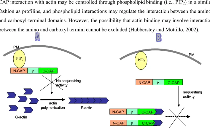
CAPinteraction with actin may be controlled through phospholipidbinding (i.e., PIP2) in a similar fashion as profilins, and phospholipid interactions may regulate the interactionbetween the amino- and carboxyl-terminal domains. However, the possibility that actin binding may involve interaction between the amino and carboxyl termini cannot be excluded (Hubberstey and Mottillo, 2002).
PIP2
N-CAP P C-CAP
actin polymerisation PM
No sequestring activity
sequestring activity
N-CAP P C-CAP
N-CAP P C-CAP
PIP2
N-CAP P C-CAP
PM
G-actin
F-actin PIP2
PIP2
N-CAP P C-CAP
actin polymerisation PM
No sequestring activity
sequestring activity
N-CAP P C-CAP
N-CAP P C-CAP
PIP2
PIP2
N-CAP P C-CAP
PM
G-actin
F-actin
Figure 6: PIP2 regulation and actin sequestering activity of CAP. Two possible scenaries could lead the PIP2
regulation of CAP. (A) Binding of the N-terminus of CAP to the PIP2 molecule prevents the actin sequestering activity of CAP by inhibiting the C-terminus of CAP to sequester the G-actin monomers. (B) The C-terminus of CAP allows the sequestering activity and inhibits the F-actin polymerization if there is no binding of the N-terminus of CAP to the PIP2
molecule.
It has been reported that PIP2 can regulate the binding of the Dictyostelium CAP to G-actin and it has been suggested that the PIP2 binding site is located to the N-terminal half of the protein. PIP2
may act in an inhibitory way, and increased PIP2 concentrations in the cell could transiently
stimulate actin polymerization predominantly near the plasma membrane by preventing the action of actin sequestering proteins (Figure 6). The regulation of CAP by PIP2 would fit into the cooperative mechanism of regulating the dynamics of the actin cytoskeleton (Gottwald et al., 1996).
6.0 CAP is a multifunctional protein
Different functions have been attributed to the different domains of CAP. The N-terminal domain seems to be required for ACA regulation, PIP2-regulation, cell polarity and development and for the correct localization of the CAP. The protein is present throughout the cytoplasm and shows enrichment near the plasma membrane especially at the rear and front ends. Furthermore, it undergoes rapid rearrangements in moving cells. The actin binding activity is assigned to the C- terminal domain and is involved in cytokinesis, cell polarity and development (Noegel et al., 2004).
Proline rich sequences often function as SH3 domain binding sites which are thought to mediate formation of specific protein complexes and possess the sequence APASSAPAAPV (position 228- 238) resembling the SH3 consensus binding sequence XPXXPPPYXPX (Y indicates a hydrophobic residue; prolines 2, 7 and 10 are essential) as analyzed for the SH3 domain of Ab1 (Freeman et al., 1996; Ren et al., 1993). Recently we also reported that the proline rich region is essential for phototaxis and cell size maintenance (Noegel et al., 2004). CAP bsr, a Dictyostelium mutant in which the CAP gene has beeninactivated by homologous recombination in such a way that the expression of the full-length protein was reduced to <5%of the protein concentration in wild-type AX2, revealed changesduring growth and development. Growing cells were heterogeneouswith regard to cell size and were often multinucleated. Themutant had an endocytosis and a chemotaxis defect. When chemotacticmotility was assayed by applying a cAMP gradient, the cells did not properly orientate in the direction of the chemotacticagent. Development was significantly delayed and developmentally regulated genes such as contact site A (csA) and cAMP receptor I were expressed significantly later than in wild type. However, the mutant was able to complete the developmental cycle and to form fruiting bodies containing viable spores (Noegel et al., 1999) implying that CAP a multifunctional protein.
7.0 Cell polarity and cAMP signalling in Dictyostelium
Cell polarity is defined as an asymmetry of cell shape andcellular functions that is stable for some time and requireslocalized assembly of signalling complexes, directed cytoskeletal rearrangements, and distinct recruitments of proteins (Nelson, 2003). During growth Dictyostelium cells do not
display a fixed polarity. They constantly change their shape and form new ends in response to environmental signals, which target them towards a food source. However, after the onset of starvation periodic signals of the chemoattractantcAMP lead to polarization of the cells and initiate the developmentinto a multicellular organism.
cAMP signalling is essential for the chemotactic aggregationof individual Dictyostelium cells into multicellular aggregatesand for progression through late development (Firtel and Meili,2000).
The aggregation centers produce cAMP pulses, which aredetected, amplified, and relayed to the surrounding cells. ThecAMP is sensed by a cAMP receptor on the cell surface, whichcouples to a heterotrimeric G protein. Dictyostelium harbours four cAR receptors whose expression levels are tightly regulated throughout development (Firtel and Chung, 2000). G-protein-mediated signal transduction pathways play an essential role in the developmental programme and are involved in regulating the actin cytoskeleton. Nine Gα subunits (Gα1-Gα9), and one Gβγ subunits have been identified (Devreotes, 1994). Gα2 is a key player, as the null cells of Gα2 do not aggregate and are unable to activate the adenylyl or guanylyl cyclases and Akt/PKB. They show no chemotaxis and do not respond to extracellular cAMP suggesting that Gα2 is the only Gα-subunit that interacts with any of the cAMP receptors (Wu et al., 1995). The Gα subunits are transiently expressed at specific stages while the single Gβγ subunits are expressed throughout growth and development.
Deletion of the gene encoding the Gβ subunit results in the inability of the cells to chemotax to chemoattractants and to activate the cyclases, and abrogates all signal transduction via G-proteins. It has been known that Gα2 mediates cAMP-induced activation of adenylyl or guanylyl cyclase and phospholipase C (Okaichi et al., 1992; Bominaar et al., 1994). The Gβγ complex is setfree and, together with CRAC, it activates the adenylyl cyclase (ACA) and leads to synthesis of cAMP (cAMP relay) (Firtel andChung, 2000).
The receptor mediated G-protein linked adenylyl cyclase systems are universal signal transducers and play a significant role in signalling leading to directed cell migration as well as in growth and development. Dictyostelium harbors three adenylyl cyclases: an aggregation specific adenylyl cyclase (ACA) is homologous to the G-protein regulated mammalian adenylyl cyclase, a germination-specific adenylyl cyclase (ACG) is expressed in prespore cells and spores and adenylyl cyclase B (ACB) is optimally expressed during culmination and is essential for the maturation of the spores and terminal differentiation (Pitt et al., 1992; Kim et al., 1998; Soderbom et al., 1999).
ACB (acrA) has recently been identified as functioning in a cell autonomous fashion during late development. ACA is expressed at high levels during aggregation and reduced levels during
multicellular development (Pitt et al., 1992), and acrA is expressed at low levels in growing cells and at more than 25 fold higher levels during development. Cells lacking ACA are capable of moving up the chemoattractant gradient, but are unable to stream. Although, ACA is not required for chemotaxis, it is essential for the cells to align in a head to tail fashion and stream into aggregates. ACA is highly enriched at the uropod of chemotaxing cells to align in a head to tail fashion and stream into aggregates and the asymmetric distribution of ACA-YFP is dependent on the actin cytoskeleton and on the acquisition of cellular polarity. Thus aca- cells have polarity defects and do not stream (Kriebel et al., 2003).
rearrangement of the actin cytoskeleton
cAMP
PM
CRAC
Gα2 Gβγ PIP2
PKA
?
PiaA
cAMP
PI3K
Akt/PKB
cAR Ras
Adenylyl cyclase
rearrangement of the actin cytoskeleton
cAMP
PM
CRAC
Gα2 Gβγ PIP2
PKA
?
PiaA
cAMP
PI3K PI3K
Akt/PKB Akt/PKB
cAR Ras
Adenylyl cyclase
Figure 7: Schematic representation of cAMP signalling pathways in Dictyostelium. Chemoattractant binding to a seven transmembrane receptor belonging to the group of G-protein-coupled receptors (GPCR) leads to the activation of Gα2, releasing the βγ subunit. The ACA, Gβγ subunits together with CRAC and PIA form a complex and activate adenylyl cyclase ACA. ACA activates further downstream effectors initiating a signalling cascade which activates pathways involving PI3-kinase and further downstream molecules, involved in the remodeling of the actin cytoskeleton.
cell polarity chemotaxis development cell polarity chemotaxis development
In a genetic analysis to identify novel genes involved in G protein-linked pathways controlling development, a new gene Pianissimo (piaA) has been identified which is a novel cytosolic regulator required for chemo-attractant receptor and G protein mediated activation of the 12 transmembrane domain adenylyl cyclase in Dictyostelium (Chen et al., 1997). PIA and CRAC, the previously identified cytosolic regulator of adenylyl cyclase have several common features and both are essential for activation of adenylyl cyclase and are involved in cAMP signalling. It is probable that the CRAC PH domain interacts with PIP3 and/or PIP2 producing the expected activation of PI3-
kinases in response to chemoattractants (Firtel and Chung 2000). Four PI3K genes have been identified in Dictyostelium: DdPIK5, which encodes a member of the PI3K-I subfamily and DdPIK1, 2 and 3, which encode members of the PI3K-2 subfamily, and DdPIK4 encodes a PI-4 kinase. The PI3K pathway has been implicated in the regulation of a multitude of intracellular events including mitogenesis (Carraway et al., 1995), membrane ruffling (Kotani et al., 1994), chemotaxis (Wennstrom et al., 1994), apoptosis (Yao et al., 1990), activation of neutrophils and oxygen radical formation (Ding et al., 1995; Traynor-Kaplan et al., 1989), receptor internalization (Joly et al., 1994; Kapeller et al., 1993), pinocytosis (Kotani et al., 1995) and neurite outgrowth (Kimura et al., 1994).
cAMP and cAMP-dependent protein kinase (PKA) are regulators of development in many organisms. All intracellular cAMP signalling is affected through PKA. PKA, the major downstream effector of the cAMP-ACA signalling pathway inside the cell, is activated when its regulatory subunit (PKA-R) binds cAMP and dissociates from the catalytic subunit (PKA-C). Therefore, PKA is essential in controlling aggregation and postaggregative development in Dictyostelium (Mann et al., 1997). cAMPalso initiates a network of signalling pathways such as cGMPsignalling, which is responsible for changes in the cytoskeleton(Liu and Newell, 1988).
8.0 Overview
The process of cAMP signalling involves the signalling molecules, the chemotactic machineryas well as components of the cytoskeleton such as CAP, whichin Dictyostelium is involved in actin cytoskeleton rearrangements. To decipher the multifunctional role of CAP in diverse cellular events will, undoubtedly, help in elucidating new and exciting biological mechanisms. As part of this approach we investigated the role of CAP in the mutants of cAMP signalling pathway, with the aim of understanding its cellular functions by investigating its localization and involvement in cytoskeleton-dependent processes using a green fluorescent protein (GFP)-tagged version of CAP, identifying its interacting partners, and its involvement in global regulation.
Materials and Methods
1.0 Materials
1.1 Laboratory materials
Cellophane sheet (Dry ease) Novex
Centrifuge tubes, 15 ml, 50 ml Greiner
Coverslips (glass), ∅ 12 mm, ∅ 18 mm, ∅ 55 mm Assistent
Corex tube, 15 ml, 50 ml Corex
Cryo tube, 1 ml Nunc
Electroporation cuvette, 2 mm electrode gap Bio-Rad
Gel-drying frames Novex
Hybridisation bag Life technologies
Microcentrifuge tube, 1.5 ml, 2.2 ml Sarstedt Micropipette, 1-20 µl, 10-200 µl, 100-1,000 µl Gilson
Micropipette tips Greiner
Needles (sterile), 18G–27G Terumo, Microlance
Nitrocellulose membrane, BA85 Schleicher and Schuell Nitrocellulose-round filter, BA85, ∅ 82 mm Schleicher and Schuell
Nylon membrane, Biodyne B Pall
Nucleopore® membrane filters Nucleopore
Parafilm American National Can
Pasteur pipette, 145 mm, 230 mm Volac
PCR softtubes, 0.2 ml Biozym
Petri dish (35 mm, 60 mm, 100 mm) Falcon
Petri dish (90 mm) Greiner
Plastic cuvette, semi-micro Greiner
Plastic pipettes (sterile), 1 ml, 2 ml, 5 ml, 10 ml, 25 ml Greiner
Quartz cuvette, Infrasil Hellma
Quartz cuvette, semi-micro Perkin Elmer
Saran wrap Dow
Scalpels (disposable), Nr. 10, 11, 15, 21 Feather
Slides, 76 x 26 mm Menzel
Syringes (sterile), 1 ml, 5 ml, 10 ml, 20 ml Amefa, Omnifix Syringe filters (Acrodisc), 0.2 µm, 0.45 µm Gelman Sciences Tissue culture flasks, 25 cm2, 75 cm2, 175 cm2 Nunc
Tissue culture dishes, 6 wells, 24 wells, 96 wells Nunc
Whatman 3MM filter paper Whatman
X-ray film, X-omat AR-5, 18 x 24 mm, 535 x 43 mm Kodak 1.1.1 Instruments and equipments
Centrifuges (microcentrifuges):
Centrifuge 5417 C Eppendorf
Centrifuge Sigma B. Braun Biotech Instruments Cold centrifuge Biofuge fresco Heraeus Instruments
Centrifuges (table-top, cooling, low speed):
Centrifuge CS-6R Beckman
Centrifuge RT7 Sorvall
Centrifuge Allegra 21R Beckman
Centrifuges (cooling, high speed):
Beckman Avanti J25 Beckman
Sorvall RC 5C plus Sorvall
Centrifuge-rotors:
JA-10 Beckman
JA-25.50 Beckman
SLA-1500 Sorvall
SLA-3000 Sorvall
SS-34 Sorvall
Electrophoresis power supply, Power-pac-200-300 Bio-Rad
Electroporation unit, Gene-Pulser Bio-Rad
Freezer (-80°C) Nunc
Freezer (-20°C) Siemens, Liebherr
Fluorimeter PTI
Gel-documentation unit MWG-Biotech
Heating block, DIGI-Block JR neoLab
Heating block, Dry-Block DB x 20 Techne
Hybridising oven Hybaid
Ice machine Ziegra
Incubators: Incubator, microbiological Heraeus
Incubator with shaker, Lab-Therm Kuehner
Laminar flow, Hera Safe (HS 12) Heraeus
Magnetic stirrer, MR 3001 K Heidolph
Microscopes:
Light microscope, CH30 Olympus
Light microscope, DMIL Leica
Light microscope, CK2 Olympus
Fluorescence microscope, DMR Leica Fluorescence microscope, 1X70 Olympus Confocal laser scan microscope, DM/IRBE Leica
Stereomicroscope, MZFLIII Leica
Stereomicroscope, SZ4045TR Olympus
Oven, conventional Heraeus
PCR machine, PCR-DNA Engine PTC-2000 MJ Research
pH-Meter Knick
Refrigerator Liebherr
Semi-dry blot apparatus, Trans-Blot SD Bio-Rad
Shakers GFL, Kuehner
Sonicator, Ultra turrax T25 basic IKA Labortechnik
Speed-vac concentrator, DNA 110 Savant
Spectrophotometer, Ultraspec 2000, UV/visible Pharmacia Biotech Ultracentrifuges:
Optima TLX Beckman
Optima L-70K Beckman
Ultracentrifuge-rotors:
TLA 45 Beckman
TLA 100.3 Beckman
SW 41 Beckman
UV-crosslinker, UVC 500 Hoefer
UV- transilluminator, TFS-35 M Faust
Vortex, REAX top Heidolph Video camaras
JAI CV-M10 CCD Camera Stemmer Imaging
SensiCam PCO Imaging
Waterbath GFL
X-ray-film developing machine, FPM-100A Fujifilm 1.2 Kits
FairplayTM Micrroarray labelling kit stratagene
Nucleobond AX Macherey-Nagel
NucleoSpin Extract 2 in 1 Macherey-Nagel
Nucleotrap Macherey-Nagel
Original TA Cloning Invitrogen
pGEM-T Easy Promega
Qiagen Midi- and Maxi-prep Qiagen
Qiagen RNAeasy Midi/Mini kit Qiagen
Stratagene PrimeIt II Stratagene
1.2.1 Enzymes, antibodies, substrates, inhibitors and antibiotics Enzymes used in the molecularbiology experiments:
Calf Intestinal Alkaline Phosphatase (CIAP) Boehringer
Deoxyribonuclease I (DNase I) Boehringer
Klenow fragment Boehringer
Lysozyme Sigma
Protein A Sepharose CL-4B Amersham
Restriction endonucleases Amersham, Life technologies, New England Biolabs
Reverse transcriptase, Superscript II Life technologies
Ribonuclease H (RNase H) Boehringer
Ribonuclease A (RNase A) Sigma
T4 DNA ligase Boehringer
Taq-polymerase Life technologies/Boehringer
Primary antibodies:
Mouse anti-CAP monoclonal antibody, 230-18-8 Gottwald et al., 1996 Mouse anti-CAP monoclonal antibody, 223-445-1 Gottwald et al., 1996 Mouse anti-actin monoclonal antibody, Act 1-7 Simpson et al., 1984 Mouse anti-GFP monoclonal antibody, K3-184-2 Gloss et al., 2003 Mouse anti-csA monoclonal antibody, 33-294-17 Bertholdt et al., 1985 Mouse anti-α-actinin monoclonal antibody, 47-62-17 Schleicher et al., 1984 Mouse anti-filamin- monoclonal antibody, 82-454-12 Brink et al., 1989 Mouse anti-myosin-monoclonal antibody, 56-395-2 Pagh and Gerisch, 1986 Mouse anti-comitin monoclonal antibody 190-340-2 Weiner et al., 1993
Mouse anti-vacuolin monoclonal antibody, 221-1-1 Rauchenberger et al., 1997 Mouse anti-V/H+ ATPase monoclonal antibody, 221-35-2 Jenne et al., 1998
Mouse anti-Rab5-monoclonal antibody Sigma
Goat anti-GST antibody Amersham
Mouse anti-profilin I -monoclonal antibody, 153-246-10 Haugwitz et al., 1991 Mouse anti-profilin II -monoclonal antibody, 174-336-12 Haugwitz et al., 1991
Secondary antibodies:
Goat anti-mouse IgG, peroxidase conjugated Sigma Goat anti-rabbit IgG, peroxidase conjugated Sigma Mouse anti-goat IgG, peroxidase conjugated Sigma
Sheep anti-mouse IgG, Cy3 conjugated Sigma
Substrates:
Hydrogen peroxide (H2O2) Sigma
Inhibitors:
Benzamidin Sigma
Complete Mini®, Protease inhibitor cocktail tablets Roche
Diethylpyrocarbonate (DEPC) Sigma
Leupeptin Sigma
Pepstatin Sigma
Phenylmethylsulphonylfluoride (PMSF) Sigma
Antibiotics:
Ampicillin Gruenenthal
Blasticidin S ICN Biomedicals
Chloramphenicol Sigma
Dihydrostreptomycinsulphate Sigma
Geneticin (G418) Life technologies
Kanamycin Sigma, Biochrom
Tetracyclin Sigma
1.3 Chemicals and reagents
Most of the chemicals and reagents were obtained either from Sigma, Fluka, Difco, Merck, Roche, Roth or Serva. Those chemicals or reagents that were obtained from companies other than those mentioned here are listed below:
Acetic acid (98-100%) Riedel-de-Haen
Acrylamide (Protogel: 30:0,8 AA/Bis-AA) National Diagnostics
Agar-Agar (BRC-RG) Biomatic
Agarose (Electrophoresis Grade) Life technologies
Amino acids sigma
Bacto agar Difco
Bacto-peptone Difco
Bacto-Trypton Difco
5-Bromo-4-chloro-3-indolyl-β-D-galactopyranosid (X-gal) Roth
Bromophenol blue (Na-Salt) serva
BSA (Bovine serum albumin) Roth
Chloroform Riedel-de-Haen
Calciumchlorid-Dihydrat Merck
Chloroform Riedel-de-Haen
Coomassie-Brilliant-Blue G 250 Roche
Coomassie-Brilliant-Blue R 250 Serva
p-Cumaric acid Fluka
Cyclohexamide Sigma
1,4-Dithiothreitol (DTT) Gerbu
Dimethylformamide (DMF) Riedel-de-Haen
Ethanol Riedel-de-Haen Ethylen diamine tetraaceticacid (EDTA) Merck
Glycerine Riedel-de-Haen
Glycine Riedel-de-Haen Isopropypl-β-D-thiogalactopyranoside (IPTG) Loewe Biochemica
Methanol Riedel-de-Haen
Morpholino propane sulphonic acid (MOPS) Gerbu N- [2-Hydroxyethyl] piperazine-N’-2-
-ethanesulfonic acid (HEPES) Biomol
Nonidet P40 Fluka
Peptone Oxoid
Sodium dodecyl sulphate (SDS) Serva
Sodium hydroxide Riedel-de-Haen
Triton X-100 Merck
TRIzol Gibco BRL
Tween 20 Roth
Yeast Nitrogen Base Difco
Yeast extract Oxoid
Radiolabelled nucleotide:
α-32P-deoxyadenosine triphosphate, (10 mCi/ml) Amersham 1.4 Media and buffers
All media and buffers were prepared with deionised water filtered through an ion-exchange unit (Membra Pure). The media and buffers were sterilized by autoclaving at 120ºC and antibiotics were added to the media after cooling to approx. 50ºC. For making agar plates, a semi-automatic plate- pouring machine (Technomat) was used.
1.4.1 Media and buffers for Dictyostelium culture AX2-medium (Claviez et al., 1982), pH 6.7:
7.15 g yeast extract
14.3 g peptone (proteose)
18.0 g maltose
0.486 g KH2PO4
0.616 g Na2HPO4.2H2O
add H2O to make 1 litre Soerensen phosphate buffer (Malchow, 1972), pH 6.0:
2 mM Na2HPO4
14.6 mM KH2PO4
Phosphate agar plates, pH 6.0:
9 g agar
add Soerensen phosphate buffer, pH 6.0 to make 1 litre
Salt solution (Bonner, 1947):
10 mM NaCl 10 mM KCl 2.7 mM CaCl2
Starvation buffer , (Shaulsky et al., 1998) pH 6.5:
10 mM MES, pH 6.5
10 mM NaCl 10 mM KCl 1 mM CaCl2
1 mM MgSO4
SM agar plates, (Sussman, 1951)pH 6.5:
9 g agar
10 g peptone
10 g glucose
1 g yeast extract 1 g MgSO4.7H2O
2.2 g KH2PO4
1 g K2HPO4
add H2O to make 1 litre 1.4.2 Media for E. coli culture
LB medium, pH 7.4:
(Sambrook et al., 1989) 10 g bacto-tryptone 5 g yeast extract 10 g NaCl
adjust to pH 7.4 with 1 N NaOH add H2O to make 1 litre
For LB agar plates, 0.9% (w/v) agar was added to the LB medium and the medium was then autoclaved. For antibiotic selection of E. coli transformants, 50 mg/l ampicillin, kanamycin or chloramphenicol was added to the autoclaved medium after cooling it to approx. 50ºC. For blue/white selection of E. coli transformants, 10 µl 0.1 M IPTG and 30 µl X-gal solution (2% in dimethylformamide) was plated per 90 mm plate and the plate was incubated at 37ºC for at least 30 min before using.
SOC medium (Sambrook et al., 1989), pH 7.0:
20 g bacto-tryptone
5 g yeast extract 10 mM NaCl 2.5 mM KCl
dissolve in 900 ml deionised H2O adjust to pH 7.0 with 1 N NaOH
The medium was autoclaved, cooled to approx. 50ºC and then the following solutions, which were
separately sterilized by filtration (glucose) or autoclaving, were added:
10 mM MgCl2.6H2O 10 mM MgSO4.7H2O 20 mM Glucose
add H2O to make 1 litre 1.4.3 Media and buffers for Yeast culture
YEPD-Medium: YEPD-Agar plates:
20 g/l Difco Pepton 20 g/l Difco Pepton 10 g/l Yeast extract 10 g/l Yeast extract
18 g/l Agar agar
100 x Adenine solution: 100 x Tyrosine solution:
200 mg (1,1 mmol) Adenine in 100 ml 300 mg (1, 7 mmol) Tyrosin in 100 ml water dissolve with addition of little dissolve with addition of NaOH solution and amounts of HCl and filter sterilize. filter sterilize.
100 x Histidine solution: 100 x Leucine solution:
200 mg (1 mmol) Histidine in 100 ml 1000 mg (7, 6 mmol) Leucin in 100 ml Water and filter sterilize. Water and filter sterilize.
100 x Tryptophan solution: 100 x Uracil solution:
200 mg (1 mmol) Tryptophan in 200 mg (1, 8 mmol) Uracil in 100 ml
100 ml Water filter sterilize. Water dissolve by warming in water bath and filter sterilize.
1 M 3-Amino-1,2,4-triazol solution: 100 x Cycloheximide solution:
8,4 g 3-Amino-1, 2, 4-triazol in 100 ml 1 mg/ml Cycloheximid in Water solution Water, filter once and filter sterilize. filter sterilize.
The composition of the selection media and agar plates is indicated in the following table. Agar agar and Yeast extract without nitrogen base was dissolved in water and autoclaved. The glucose solution was prepared in water and filter sterilised. The remaining stock solutions were added after cooling to 55 °C.
Selection plates
Reagents SD/-Trp SD/-Leu SD/-Leu/-Trp SD/-Leu/-His/- Trp/+3-AT
SD/-Leu/- His/-Trp/-Ade
Yeast Nitrogen Base [g] 6,7 6,7 6,7 6,7 6,7
Agar agar [g] 20 20 20 20 20
Water [ml] 750 750 770 745 780
20% Glucose [ml] 100 100 100 100 100
10X Amino acid solution [ml] 100 100 100 100 100
100X Tyrosine [ml] 10 10 10 10 10
100X Uracile [ml] 10 10 10 10 10
100X Histidine [ml] 10 10 10 - -
100X Leucine [ml] 10 - - - -
100X Tryptophane [ml] - 10 - - -
100X Adenine [ml] 10 10 10 10 -
3-AT-solution [ml] - - - 25 -
- indicates omission of the reagent
10 x Amino acid solutions:
300 mg (2, 3 mmol) Isoleucine 1500 mg (1, 1 mmol) Valine 200 mg (0, 9 mmol) Arginine 300 mg (1, 6 mmol) Lysine 200 mg (1, 34 mmol) Methionine 500 mg (3 mmol) Phenylalanine 2000 mg (16, 8 mmol) Threonine,
adjust final volume to 1l and filter sterilize.
1.4.4 Buffers and other solutions
The buffers and solutions that were commonly used during the course of this study are mentioned below.
10x MOPS (pH 7.0/ pH 8.0): 41.9 g MOPS
16.7 ml 3 M sodium acetate
20 ml 0.5 M EDTA
add H2O to make 1 litre
20x SSC (pH 7.0): 3 M NaCl
0.3 M sodium citrate
Hybridisation solution (50 µl): Hybridisation buffer 48 µl Fish sperm DNA [10 mg/ml] 1µl Oligo dA (18 mer, 100 µM) 1µl mix well
Hybridisation buffer: 1.2M Phosphate buffer, pH 6.8 2mM EDTA
50 % Formamide
1% Na-Laurylsarcosinate 0.2 % SDS
4 xDenhardt’s Reagent 100 x Denhardt’s reagent: 2 % Ficoll 400
2 % Polyvinylpyrolidone 2 % bovine serum albumin
Blocking Solution (282 ml): 270 ml 1-Methy-2-pyrrolidinone (solution should be colourless)
0.4 g Succinic anhydride (store desiccated) 12.1 ml 1M Sodium borate, pH 8.0 (42mM) 10x NCP-Puffer (pH 8.0): 12.1 g Tris/HCl
87.0 g NaCl
5.0 ml Tween 20
2.0 g sodium azide
add H2O to make 1 litre
PBG (pH 7.4): 0.5 % bovine serum albumin
0.1 % gelatin (cold-water fish skin)
in 1x PBS, pH 7.4
1x PBS (pH 7.4): 8.0 g NaCl
0.2 g KH2PO4 1.15 g Na2HPO4
0.2 g KCl
dissolve in 900 ml deionised H2O adjust to pH 7.4
add H2O to make 1 litre, autoclave Homogenisation buffer, pH 7.4: 30 mM Tris/HCl, pH 7,4
2 mM DTT 2 mM EDTA 4 mM EGTA 5 mM Benzamidin 0,5 mM PMSF
1 Tablet complete® mini Protease Inhibitor Mix (Roche) per 10 ml buffer
add 30 % sucrose, prepared freshly before use.
5X Immunoprecipitation Buffer: 0.5 m Potassium phosphate buffer 0.375 M NaCl
25 mM EDTA 5 mM Benzamidine 2.5 mM PMSF
Adjust the pH to 7.9, prepare fresh.
1.2 M Phosphate buffer (pH 6.8): 1.2 M Na2HPO4, pH 9.1 was mixed with 1.2 M NaH2PO4, pH 4.02 in the ratio of 2:1.
TE buffer (pH 8.0): 10 mM Tris/HCl, pH 8.0 1 mM EDTA
10X TAE buffer (pH 8.3): 27.22 g Tris
13.6 g sodium acetate
3.72 g EDTA
add H2O to make 1 litre 1.5 Biological materials
Bacterial strains:
Strain name Reference
E. coli DH5α Hanahan, 1983 E. coli XL1 blue Bullock et al., 1987 E. coli JM 38 Vieira and Messing, 1982 Klebsiella aeorgenes Williams and Newell, 1976