Cloning and characterization of Enaptin, a novel giant actin-binding protein connecting the
nucleus to the actin cytoskeleton
INAUGURAL-DISSERTATION zur
Erlangung des Doktorgrades
der Mathematisch-Naturwissenschaftlichen Fakultät der Universität zü Köln
vorgelegt von Sabu Abraham
aus
Thiruvaniyoor, Kerala, Indien
Köln, 2004
Referees/Berichterstatter: Prof. Dr. Angelika A. Noegel Prof. Dr. Jens Brüning Date of oral examination: 08.07.2004
Tag der mündlichen Prüfung
The present research work was carried out under the supervision of Prof. Dr. Angelika A.
Noegel, in the Institute of Biochemistry I, Medical Faculty, University of Cologne, Cologne, Germany from August 2001 to July 2004.
Diese Arbeit wurde von August 2001 bis Juli 2004 am Biochemischen Institut I der Medizinischen Fakultät der Universität zu Köln unter der Leitung von Prof. Dr. Angelika A.
Noegel durchgeführt.
To my beloved Pappa and Mummy
Acknowledgements
First of all, I would like to thank Prof. Dr. Angelika A Noegel for giving me an opportunity to work in her department and to do my PhD studies under her guidance. Her valuable guidance, creative suggestions, constructive criticism and constant encouragement during the course of work inspired and enabled me to complete this project. I would like to express here my deep sense of gratitude to her.
I cannot find words to say thanks to our group leader Dr. Iakowos Karakesisoglou for his constant encouragement and discussions during the course of this study. His ‘story for a lesson’ approach always lifted my spirit up. It was wonderful to work with him. Thank you Akis.
I would also like to thank Dr. Elena Korenbaum for guiding me in the first year of my study. Her valuable guidance enabled me to initiate the project. Thank you Elena.
I also owe my thanks to Dr. Francisco Rivero, Dr. Budi Tunggal, Dr. Ludwig Eichinger, and Dr.
Andreas Hasse for their cooperation and help as and when required.
Its hard to find words to say thanks to Kumar, my colleague and ‘partner’ for the Enaptin project.
I would also like to thank Thorsten Libotte, my ‘bench mate’ for his valuable friendship. Thanks also to Hafi, Thorsten Olski and Yen for their friendship and discussions during the life in Lab10.
I pay my sincere thanks to Martina for her technical assistance as and when required and helping me for the initial paper works.
I am thankful to Bettina Lauss, our secretary, for all the excellent assistance in the paper works.
I would also like to thank Berthold for teaching me the cell culture techniques, Maria for providing me the paraffin sections, and Rolf and Rosi for technical advices.
Now its the turn to say thanks to all my other colleagues and friends in the lab and in Köln for all the parties and laughter. Your friendships made life more meaningful. Thank you very much guys.
Last but not the least, I would like to thank my loving parents and brother for being there for me.
Sabu Abraham
Table of Contents
1 Introduction 1-12
1.1 The Cytoskeleton 1
1.2 The Actin Cytoskeleton 2
1.3 Actin binding proteins 3
1.4 Actin-binding proteins of a-actinin superfamily 4
1.5 Diseases related to actin-binding proteins 7
1.6 The Nuclear Lamina 8
1.7 Nuclear envelope 8
1.8 The nuclear lamina and inherited disease 9
1.9 Role of Actin-binding proteins in nuclear positioning 11
1.10 Aim of the study 12
2 Materials and methods 13-42 2.1 Materials 13
2.2 Instruments 14
2.3 Enzymes, inhibitors and antibodies 15
2.3.1 Enzymes for molecular biology 15
2.3.2 Antibodies 16
2.3.3 Inhibitors 16
2.3.4 Antibiotics 17
2.4 Reagents 17
2.5 Kits 18
2.6 Bacterial host strains 19
2.6.1 Media for E. coli culture 19
2.7 Eukaryotic cells 19
2.7.1 Media for cell culture 20
2.8 Vectors 20
2.9 Oligonucleotides 20
2.9.1 Oligonucleotides used for amplifying the full length cDNA of Enaptin 21 2.9.2 Knockout target vector primers 21
2.9.3 Primers for making GFP-dominant negative constructs for Enaptin 22
2.9.4 Primers for GST-specII fusion protein 22
2.9.5 Primers for probes for northern blotting 22 2.9.6 Primers for C-terminal syne-1 probe for multiple tissue expression array 22
2.10 Construction of vectors 22
2.10.1 Cloning of cDNA fragments 22
2.10.2 Generation of C-terminal Enaptin-GFP fusions 22
2.10.3 Cloning of GST-SpecII 23
2.10.4 Probes for northern blot analysis 23
2.11 Buffers and other solutions 24
2.12 Computer programs 24
2.13 Molecular biological methods 24
2.13.1 Plasmid-DNA isolation from E. coli by alkaline lysis miniprep 24 2.13.2 Plasmid-DNA isolation with a kit from Macherey-Nagel 25
2.13.3 DNA agarose gel electrophoresis 25
2.13.4 Isolation of total RNA from mouse tissue with TRIzol reagent 25 2.13.5 RNA agarose gel electrophoresis and northern blotting 26
2.13.6 Labeling of DNA probes 26
2.13.7 Generation of Riboprobes 27
2.13.8 Elution of DNA fragments from agarose gels 27 2.13.9 Measurement of DNA and RNA concentrations 27
2.13.10 Restriction digestion of DNA 28
2.13.11 Dephosphorylation of 5´-ends of linearized vectors 28
2.13.12 Creation of blund ends 28
2.13.13 Ligation of vector- and DNA-fragments 28
2.13.14 Polymerase chain reaction (PCR) 28
2.13.15 Transformation of E. coli cells with plasmid DNA 29
2.14 Protein methods and immunofluorescence 29
2.14.1 Extraction of protein homogenate from mouse tissues and cell cultures 29 2.14.2 SDS-polyacrylamide-gel electrophoresis (SDS-PAGE) 30
2.14.3 Gradient gel electrophoresis 31
2.14.4 Western blotting 31
2.14.5 Immunofluorescence 33
2.14.6 Small-scale GST fusion protein expression 33
2.14.7 Purification of GST-fusion proteins 34
2.14.8 Affinity purification of polyclonal antibodies by blot method 34
2.14.9 Affinity purification by the CNBr method 34 2.14.10 Immunohistochemical staining of formalin-fixed paraffin-embedded sections 35
2.14.11 Microscopy 36
2.15 Disruption of the cytoskeleton using various drugs 36
2.16 Digitonin experiment 36
2.17 Gene targeting protocols 36
2.17.1 Target vector construction 36
2.17.2 Probe generation 37
2.17.3 Embryonic stem cell culture 37
2.17.4 MEF cell culture and Mitomycin treatment 38
2.17.8 ES cell culture 38
2.17.9 ES cell transfection 39
2.17.10 Antibiotic selection and picking of ES cell clones 39
2.17.11 Splitting and freezing ES cells 39
2.17.12 Genomic DNA isolation 40
2.17.13 Southern blotting 40
2.17.14 Labeling of DNA probes and hybridisation 41
3 Results 43-80
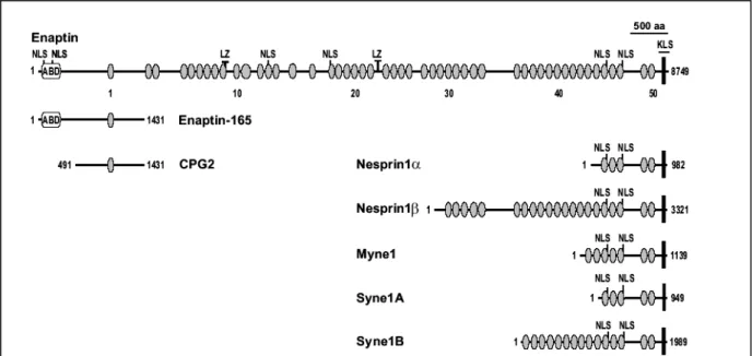
3.1 Sequence analysis of Enaptin 43
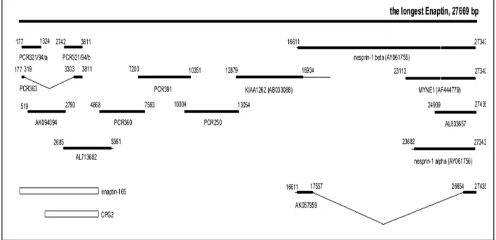
3.1.1 cDNA cloning of the longest isoform of Enaptin 43
3.1.2 Organisation of the Enaptin gene 45
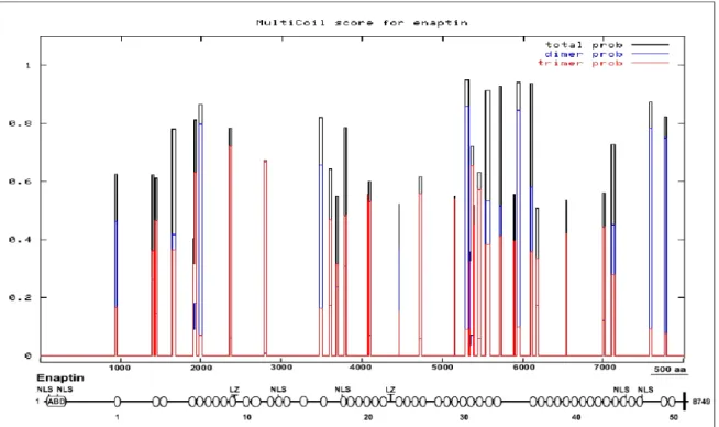
3.1.3 Domain analysis of Enaptin 45
3.1.4 Isoforms of Enaptin 51
3.2 Examination of Enaptin’s tissue distribution by northern blot analysis 52
3.2.1 Tissue distribution of Enaptin 52
3.2.2 Northern blot analysis of Enaptin 54
3.3 Western blot analysis of Enaptin 55
3.3.1 Western blots with an ABD Enaptin antibody of different tissue lysates 55
3.3.1 Expression and purification of GST-SpecII 56
3.3.2 Generation and purification of the Enaptin polyclonal antibody 57
3.4 Subcellular localization of Enaptin using the SpecII antibody 59
3.5 Enaptin is a component of the outer nuclear membrane 60
3.6 Nuclear membrane localization of Enaptin is not affected by drugs disrupting
microfilament and microtubule cytoskeleton 61
3.7 Tissue expression of Enaptin 63
3.7.1 Expression of Enaptin in muscle 63
3.7.2 Expression of Enaptin in cerebrum, cerebellum and hippocampus 65
3.7.3 Expression of Enaptin in the skin 66
3.7.4 Expression of Enaptin in a 16-day-old mouse embryo 67 3.8 Generation of GFP-fusion proteins containing the C-terminus of Enaptin 68 3.8.1 C-terminal Enaptin GFP fusions localize to the nuclear membrane 69
3. 9 Lamin dependent localization of Enaptin 72
3.9.1 Localization of Enaptin in Lamin A/C knockout cells 72 3.9.2 Dominant negative interference of Enaptin using a Xenopus lamin B construct 72 3.9.3 Nuclear localisation of Enaptin in fibroblasts from laminopathy patients 73 3.10 Distribution of Enaptin during myoblast differentiation 74
3.11 Generation of an Enaptin mouse mutant 76
3.11.1 Analysis of the structure of the mouse Enaptin gene 76 3.11.2 Construction of the targeting vector (Enaptin KO) 78
3.11.3 ES cell transfection 79
3.11.4 Screening of recombinant clones 80
4 Discussion 81-90
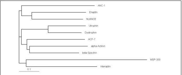
4.1 Enaptin is a giant protein of the α-actinin superfamily 81
4.2 Enaptin is a nuclear membrane protein 82
4.3 Expression and tissue distribution of Enaptin and its isoforms 85 4.4 Lamin dependent localization of Enaptin 86
4.5 Enaptin and Laminopathies 88
4.6 Possible functions of Enaptin 88
5 Summary 91-92
6 Bibliography 93-102
7 Abbreviations 103
Erklärung -
Curriculum Vitae / Lebenslauf -
I I n n t t r r o o d d u u c c t t i i o o n n
1 Introduction
1.1 The Cytoskeleton
Cells have to organise themselves in space and interact mechanically with their environment. They have to be correctly shaped, physically robust, properly structured internally and should retain their ability to move or change their shape. For all these cellular processes, cells depend on a remarkable system of filaments called the cytoskeleton. Three major cytoskeletal filamentous networks are present in eukaryotic cells, the actin microfilaments, intermediate filaments and microtubules. Microfilaments are fibers typically 7-9 nm of diameter whereas microtubules have a diameter of 24 nm. Intermediate filaments have a diameter of 10 nm which is in between the other two filaments and hence the name.
Together all these filamentous networks provide, not only a dynamic skeleton for the cells, but they are also involved in organelle transport, organelle and cell-fate-determinant positioning, cell polarity development, mitosis, cytokinesis, secretion and the formation of cell extensions, and the maintenance of cell integrity.
Actin filaments are two-stranded helical polymers of the protein actin. These flexible structures of about 8 nm in diameter are arranged in bundles and gel-like networks throughout the cell. The highest density of the actin filaments is found at the cell cortex, underneath the plasma membrane.
Microtubules are cylindrical tubes, composed of the monomer tubulin. They have a diameter of 25 nm and are stiffer than the actin filaments. Mostly these structures have its origin at the centrosome.
Intermediate filaments are rope like structures with a diameter of 10 nm. They are composed of various polypeptides. One of the filaments is present in the nuclear lamina. Others are localized throughout the cytoplasm, and contribute to the mechanical resistance of thecell.
Figure 1.1: The three different filament types of the cytoskeleton, each shown in an elelectron micrograph, a schematic drawing and the distribution in an epithelial cell. Figure is taken from Alberts et al., 1994.
1.2 The Actin Cytoskeleton
The major constituent of the microfilament is actin, and together with several actin- binding and associated proteins, it constitutes the actin cytoskeleton (Stossel, 1993). Some single cell eukaryotes like yeasts have a single gene for actin (Winsor and Schiebel, 1997), whereas many multicellular organisms contain multiple actin genes. Humans for example have six actin genes encoding various isoforms of the protein (Kedes et al., 1985), and plants such us Arabidopsis has 10 (Meagher et al., 1999). The six known actin isoforms in mammalian cells are two sarcomeric muscle actins (alpha-skeletal and alpha-cardiac), two smooth muscle actins (alpha and gamma), and 2 nonmuscle, cytoskeletal actins (beta and gamma). Actin is highly conserved throughout the evolution and is the most abundant intracellular protein in a eukaryotic cell. The recent discovery of MreB protein as a homologue of actin in bacteria changed the conventional theory that prokaryotes do not have actin (van den Ent et al., 2001). Actin is a protein consisting of 375 amino acids with a molecular mass of about 43 kDa. Actin exists either in a globular monomeric (G-actin) or in a filamentous polymeric form (F-actin). Each actin molecule can bind to ATP, which is hydrolyzed to ADP after incorporation of the actin molecule into the polymer. Polymers assemble spontaneously via noncovalent interactions between the monomeric subunits and are highly dynamic structures with subunit turnover at both ends. Energy is not required but contributes to the polymerization, as shown by the observation that ATP-bound actin polymerizes faster than ADP-bound actin (Engel et al., 1977). The rate-limiting step in actin polymerization is nucleation, the assembly of the first subunits to generate a new filament.
Actin filaments are structurally polarized, and the kinetics of polymerization at each end is different. The plus (barbed) end grows more quickly than does the minus (pointed) end. Actin is predominantly present in the cytoplasm and recently confirmed to be present also in the nucleus (Olave et al., 2002).
The actin cytoskeleton is directly involved in cell locomotion (Welch et al., 1997),
cytokinesis (Fishkind and Wang, 1995), cell-cell and cell-substratum interactions (Wehrle-
Haller and Imhof, 2002; Yamada and Geiger, 1997), vesicular and organelle transport (Kubler
and Riezman, 1993; Langford, 1995), establishment and maintenance of cell morphology
(Matsudaira, 1994) and the localization of signaling particles and mRNA (Bassell and Singer,
1997). All these functions of actin filaments are modulated and assisted by actin-binding
proteins.
1.3 Actin binding proteins
The association of the actin–binding proteins with actin is necessary for modulating the behavior and organization of the actin cytoskeleton. More than 162 distinct and separate actin-binding proteins have been identified excluding the synonyms and isoforms (dos Remedios et al., 2003) and they can be grouped into 48 classes (Kreis and Vale, 1999; Pollard and Cooper, 1986). These include monomer binding proteins [profilin (Ampe et al., 1988), cofilin (Abe et al., 1990)], barbed end capping proteins [capZ (Barron-Casella et al., 1995)], barbed end capping/severing proteins [gelsolin (Kwiatkowski et al., 1986),villin (Pringault et al., 1986)], lateral binding proteins [calponin (Strasser et al., 1993), tropomyosin (Lees-Miller and Helfman, 1991)], Cross linking proteins [α-actinin (Youssoufian et al., 1990), Spectrin (Winkelmann and Forget, 1993), dystrophin (Koenig et al., 1988),] membrane associated actin binding proteins [Synapsins or protein 4.1; (Sudhof, 1990)] and motor proteins such as myosins.
Profilin is a small actin-binding protein (12-19 kDa) originally identified as an actin sequestering protein that can inhibit actin filament growing.
Profilins appear to be multifunctional. They regulate actin polymerization, act as adaptor proteins, and possibly link transmembrane signaling to the actin cytoskeleton (Theriot and Mitchison, 1993). Cofilin is a widely distributed intracellular actin-modulating protein that binds and depolymerises filamentous actin and inhibits the polymerisation of monomeric G-actin in a pH-dependent manner (Kuhn et al., 2000).
Capping protein (capZ) is a heterodimeric actin-binding protein found in all eukaryotic cells and it binds to the barbed ends of actin filaments and nucleates polymerisation of actin (Schafer and Cooper, 1995). Gelsolin is best known for its involvement in dynamic changes in the actin cytoskeleton during a variety of forms of cell motility. Gelsolin severs assembled actin filaments in two, and caps the fast-growing plus end of a free or newly severed filament (Kwiatkowski, 1999). Calponin is a 33 kDa protein binding to actin and calmodulin and found
Figure 1.2: Actin binding proteins. Specific functions of actin-binding proteins are shown with a diagram of how each protein may interact with F-actin.
(Ayscough, 1998)
predominantly in smooth muscle and thought to be involved in the regulation or modulation of contraction (Strasser et al., 1993). Tropomyosin, in connection with the troponin complex regulates the interaction of F-actin and myosin (Farah and Reinach, 1995). Synapsin I is a neuronal phosphoprotein associated with the membranes of small synaptic vesicles and thought to regulate synaptogenesis and neurotransmitter release from adult nerve terminals (Ryan et al., 1996).
The organization of the actin cytoskeleton must be tightly regulated both temporally and spatially to perform all its biological functions. Rho-like GTPases are key regulators in signaling pathways that link extracellular growth signals or intracellular stimuli to the assembly and organization of the actin cytoskeleton (Hall, 1994).
1.4 Actin-binding proteins of α-actinin superfamily
At the leading margins of moving or spreading cells, actin filaments are organized into
two principal structures, bundles and meshworks. Actin bundles are parallel arrays of closely
packed actin filaments that stiffen membrane projections like filopodia, microvilli and
stereocilia. An actin meshwork is a criss-crossed array of actin filaments forming the
lamellipodia. When laminated to the cytoplasmic face of the plasma membrane, an actin
meshwork forms a two-dimensional elastic sheet that stiffens the cell membrane, anchors
integral membrane proteins and supports the shape of the cells. Actin cross-linking proteins
characterized by a pair of actin-binding sites form actin bundles and meshworks. The α-
actinin superfamily is the largest of the F-actin cross-linking protein families. Proteins in this
family share an actin-binding domain homologous to the ABD of α-actinin (Matsudaira,
1994). The family includes spectrins, fimbrin/plastins, dystrophins, gelation factor from
Dictyostelium and filamin subfamilies (Hartwig, 1995). The globular actin-binding domain is
contained in the first 250 amino acids at the N-terminus of all the members of the family and
may be composed of more than one subdomain (Puius et al., 1998). The actin-binding domain
(ABD) found close to the N-terminus in all members of the α-actinin superfamily consists of
two calponin-homology domains (Korenbaum and Rivero, 2002). The calponin homology
(CH) domain is a protein module of about 100 residues that was first identified at the N-
terminus of calponin, an actin-binding protein playing a major regulatory role in muscle
contraction (Strasser et al., 1993). Proteins of the α-actinin superfamily utilize a double
calponin homology domain to arrange the actin filaments in bundles and meshworks and link
them to the plasma membrane (Matsudaira, 1994). Fimbrins are the simplest of the family and
are modular proteins consisting of a calmodulin like calcium binding domain at the N-
terminus followed by a pair of α-actinin like actin binding domains (Lin et al., 1994). Filamin or actin-binding protein-280 (ABP-280) is a 280 kDa protein that cross-links actin filaments into orthogonal networks in the cortical cytoplasm and participates in the anchoring of membrane proteins to the actin cytoskeleton (Gorlin et al., 1990). Filamin and its homologues have an ABD at its N-terminus separated from a C-terminal dimerization domain by several immunoglobulin fold containing repetitive elements (Fucini et al., 1999). The third sub- family in the α-actinin superfamily consists of proteins like α-actinin, spectrin and dystrophin. The distinguishing feature of the subfamily is a repeated triple stranded α−helical motif (Yan et al., 1993) called the spectrin repeat. The presence of spectrin repeats in the rod domain classifies these proteins into the spectrin family. The structure of the ABD in dystrophin and utrophin has been solved, revealing a bundle of α-helices arranged in an extended head-to-tail dimer (Keep et al., 1999; Norwood et al., 2000). α-Actinin dimerises to cross-link actin filaments and spectrin forms a tetramer of (αβ)2 type (Viel, 1999). Even though the ABD of dystrophin also has been proposed to form a dimer by x-ray crystallographic studies (Norwood et al., 2000), the spectrin repeats in dystrophin do not form a dimer at least in-vitro (Chan and Kunkel, 1997).
In myofibrillar cells, α-actinin constitutes a major component of Z-disks in striated muscle and of the functionally analogous dense bodies and dense plaques in smooth muscle.
In nonmuscle cells, it is distributed along microfilament bundles and is thought to mediate their attachment to the membrane at adherens-type junctions (Blanchard et al., 1989).
Spectrin, first identified as supporting the plasma membrane of erythrocytes, is now found to be in association with other intracellular membranes (De Matteis and Morrow, 2000).
Dystrophin and utrophin are identified as proteins involved in Duchenne/Becker muscular dystrophies and associate with the plasma membrane of muscle and neuronal tissues.
Dystrophin is a 427 kDa protein which binds to cytoskeletal F-actin and to dystroglycan, a transmembrane protein associated with plasma membrane multimolecular complex.
Dystroglycan in turn binds to laminin-2 in the overlaying lamina. Thus dystrophin is a part of a complex that links the cytoskeleton to the extracellular matrix (Sunada and Campbell, 1995). Although utrophin is very similar in sequence to dystrophin and possesses many of the protein-binding properties ascribed to dystrophin, it is confined to the subsynaptic membrane at the neuromuscular junction (Blake et al., 1996).
Another family of large proteins exists with an α-actinin like ABD domain at the N-
terminal end and followed by several repeating domains, which are called the plakins. Plakins
are cytolinker proteins that associate with cytoskeletal elements and junctional complexes.
The plakin family consists of desmoplakin (Smith and Fuchs, 1998), plectin (Rezniczek et al., 1998), bullous pemphigoid antigen 1 [BPAG1 (Leung et al., 1999)], and microtubule–actin crosslinking factor [MACF/ACF7 (Karakesisoglou et al., 2000)]. This family of proteins is defined by the presence of a plakin domain and/or a plakin repeat domain. In addition to these two domains, plakins also harbor other domains that are common in some but not all members: the actin-binding domain (ABD), coiled-coil rod, spectrin-repeat-containing rod and microtubule-binding domain. Many plakins are expressed in tissues that experience mechanical stress, such as epithelia and muscle, where they play a vital role in maintaining tissue integrity by crosslinking cytoskeletal filaments and anchoring them to membrane complexes. The proteins in the spectrin family and plakin family can be classified together as spectraplakin family (Roper et al., 2002) composed of both the spectrin and plakin superfamilies. These superfamilies consist of proteins that contribute to the linkage between the plasma membrane and the cytoskeleton. Spectrin superfamily members bind and cross- link actin filaments and attach them to membrane receptors. Members of the plakin superfamily were first identified as components of desmosomes and hemidesmosomes, connecting the adhesion receptors to intermediate filaments, but they also can cross-link different cytoskeletal elements.
Figure 1.3: The spectrin protein superfamily. The figure depicted is a selection of members of the spectrin family of proteins, comparing human and fly orthologues for each member, and in the case of spectraplakins showing both mammalian genes (MACF1 and BPAG1). Taken from Roper et al., 2002.
1.5 Diseases related to actin-binding proteins
Members of the α-actinin superfamily are also involved in the pathogenesis of diseases
.The most famous disease associated with actin-binding proteins may be Duchenne and Becker muscular dystrophy (DMD & BMD). It is a X-linked degenerative disorder of muscle affecting 1 in 3500 live born males (Gospe et al., 1989). Patients with DMD have mutations in the gene encoding dystrophin. Dystrophin is thought to serve as a link from the actin-based cytoskeleton of the muscle cell through the plasma membrane to the extracellular matrix (Sunada and Campbell, 1995). In dystrophic muscle, where this linkage is disrupted, muscle fibers develop normally, but get easily damaged and degenerate (Menke and Jockusch, 1995).
Regeneration is insufficient and successive rounds of degeneration lead to a gradual replacement of muscle by connective tissue.
Spectrin, the predominant component of the membrane skeleton of the red blood cell, is essential in determining the properties of the membrane including its shape and deformability.
Mutations in the spectrin gene cause elliptocytosis and hereditary pyropoikilocytosis resulting in diminished elasticity or destabilization of the erythocyte skeleton (Delaunay, 2002;
Goodman et al., 1982)
Plectin is a widely expressed high molecular weight protein that is involved in cytoskeleton-membrane attachment in epithelial cells, muscle, and other tissues. Mutations in the gene encoding plectin (PLEC1) have been implicated in the pathogenesis of an autosomal recessive variant of epidermolysis bullosa simplex and is associated with cutaneous blistering starting in the neonatal period and muscular dystrophy in later life (McLean et al., 1996).
BPAG1 is a component of the basement membrane of the skin. BPAG1 is the major
autoantigenic determinant of autoimmune sera of patients with blistering disease bullous
pemphigoid (Stanley et al., 1988). Targeted removal of the BPAG1 gene in mice results in
severe dystonia and sensory nerve degeneration (Guo et al., 1995). Dystonin, is a neural
isoform of BPGA1 and deletion of dystonin causes a hereditary neurodegenerative disorder
called dystonia musculorum (dt) leading to a sensory ataxia in mice (Brown et al., 1995)
.In
the dt mice degenerating sensory neurons show abnormal accumulations of IFs and
disorganized MTs in the axons. Animals that survive longer develop less motor neuron
degeneration resulting in sensory neuropathy (Dalpe et al., 1998).
1.6 The Nuclear Lamina
The nuclear lamina is a mesh of intermediate filaments that mainly underlies the inner aspect of the inner nuclear membrane and also extends into the nuclear interior (Stuurman et al., 1998). The nuclear lamin filaments are made up of monomeric subunits, which each comprise a central coiled-coil, helical rod domain flanked by globular domains on the C- terminus and N-terminus. They can form parallel dimers and subsequently undergo polymerization to form the filamentous network of the nuclear lamina. In humans, there are three genes encoding the lamins, LMNA, LMNB1 and LMNB2. There are two major A-type lamin proteins (lamin A and C) and two major B-type lamins. All vertebrate cells express at least one B-type lamin, whereas the A-type lamins are developmentally regulated and expressed primarily in differentiated cells (Goldman et al., 2002). The locality of the nuclear lamins have implicated them in a wide range of nuclear functions such as nuclear growth, maintenance of nuclear shape,
DNA replication, chromatin organisation, RNA splicing, cell differentiation, apoptosis and cell- cycle-dependent control of nuclear architecture (Moir et al., 2000;
Zastrow et al., 2004).
1.7 Nuclear envelope
The most prominent feature of the nuclear envelope is a pair of inner and outer nuclear membranes (INM and ONM resp.) with a perinuclear space in between them.
These two membranes are periodically interrupted by nuclear pore complexes (NPCs), which are large macromolecular assemblies that form aqueous gated channels across the nuclear envelope and
mediate the transport of macromolecules. The ONM is continuous with the peripheral rough endoplasmic reticulum. The INM is enriched with a distinctive set of membrane proteins and
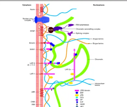
Figure 1.4: A schematic view of inner nuclear membrane proteins and their binding interactions with the nuclear lamina and nucleoplasmic components.
The outer and inner nuclear membranes (ONM and INM, respectively) are shown in cross-section, with a nuclear pore complex spanning the two membranes.
Twelve inner nuclear membrane proteins have been characterized in the mammalian nuclear envelope. These include: the multi-spanning membrane proteins nurim, lamin B receptor (LBR), ring-finger-binding protein (RFBP); the double-spanning membrane protein MAN1; and the single-spanning membrane proteins emerin, lamina-associated protein 2 (LAP-2) isoforms (a, b, g) and LAP- 1 isoforms.Interactions occur between the inner nuclear membrane proteins and the A-type lamins (shown in blue) and B-type lamins (shown in orange), which are helical filamentous proteins of the nuclear lamina and nucleoplasm.
Transcriptional regulators, cross-link inner nuclear membrane proteins and chromatin. These include: the retinoblastoma protein pRB; the ‘germ-cell-less’
protein GCL; the transcription factor E2F; and RNA polymerase, RNA splicing complex and DP protein. Taken from Maidment and Ellis, 2002
maintains close associations with the underlying lamina (Burke and Stewart, 2002). So far at least 20 inner nuclear membrane proteins were described and most of them with the exception of nurim interact with nuclear lamins (Rolls et al., 1999). The inner nuclear membrane proteins include two related families: the LAP-1 family, comprising LAP-1 A, B and C isoforms; and the LAP-2 family, comprising LAP-2 b, g, d and e isoforms (Dechat et al., 2000). LAP-2 has a fifth family member, LAP-2a, which is a soluble nucleoplasmic protein.
In addition, there are two related, but distinct, nuclear membrane proteins: emerin (Bione et al., 1994) and MAN1 (Lin et al., 2000). The LAP families and emerin both contain single transmembrane domains at their C-terminus, whereas MAN1 possesses two such domains.
These proteins are orientated in the inner nuclear membrane with their N-termini projecting into the nucleoplasm (known as type II orientation). All the LAP-2 isoforms, emerin and MAN1 share a homologous N-terminal domain, referred to as the ‘LEM domain’ (for LAP–
emerin–MAN) (Lin et al., 2000) which confers the ability to bind to ‘barrier to auto- integration factor’ (BAF), a DNA-bridging protein of unknown function (Haraguchi et al., 2001). Three unrelated multi-membrane-spanning proteins have also been identified: nurim, lamin B receptor (LBR) and a hormonally regulated atypical P-type ATPase termed ring- finger binding protein (RFBP) (Mansharamani et al., 2001; Rolls et al., 1999; Worman et al., 1990).
1.8 The nuclear lamina and inherited disease
The inherited human diseases associated with nuclear lamina components are called laminopathies. Laminopathies are a group of inherited diseases that arise through mutations in genes that code for A type lamins and lamina-associated proteins. The first report of such an association between the NE and disease was Emery–Dreifuss muscular dystrophy (EDMD) which arises with mutations in the X-linked gene STA (Bione et al., 1994). EDMD is the third most common X-linked form of muscular dystrophy (after Duchenne and Becker) and is usually characterized by contractions in the Achilles and elbow tendons, a rigid spine, with muscle weakness during early childhood. Abnormal heart rhythms, heart block and cardiomyopathy leading to cardiac arrest also arise (Emery, 1989). The STA gene encodes the nuclear protein emerin, a 29 kDa type-2 integral membrane protein that traverses the INM through its carboxy-terminal domain.
In the last few years a number of mutations has been identified in the LMNA gene, the
gene coding for lamin A/C, resulting in several diseases affecting different tissues (reviewed
in (Mounkes et al., 2003a). Figure 1.5 shows the identified mutations and the locations in the
lamin A/C gene. Three of these diseases affect striated muscle: autosomal dominant and recessive forms of Emery–Dreifuss muscular dystrophy (AR and AD-EDMD) (Bonne et al., 1999), autosomal dominant limb-girdle muscular dystrophy with a cardiac conduction disturbances (LGMD1B) (Muchir et al., 2000) and an autosomal dominant form of dilated cardiomyopathy with conduction defect (DCM-CD) (Fatkin et al., 1999). The fourth specifically involves the adipose tissue: autosomal dominant familial partial lipodystrophy (FPLD) (Cao and Hegele, 2000). The fifth, an autosomal recessive form of axonal neuropathy (AR-CMT2) (De Sandre-Giovannoli et al., 2002), specifically affects the peripheral nervous tissue. The sixth is an autosomal recessive disorder affecting adipose and bone tissues:
mandibuloacral dysplasia (MAD) (Novelli et al., 2002). The autosomal dominant form of Hutchinson–Gilford progeria syndrome (HGPS) is the seventh (De Sandre-Giovannoli et al., 2003). This list is not exhaustive and novel mutations in lamin A/C gene are being identified.
Abnormalities in nuclear structure in the cells and tissues of individuals with mutations in lamin A or C have been reported but the pathophysiological mechanism is unclear. Two mouse models are existing for laminopathies, one is a null mice for The LMNA gene showing muscular dystrophies similar to EDMD in humans (Sullivan et al., 1999) and another Hutchinson–Gilford progeria syndrome (Mounkes et al., 2003b). Analysis of the two animal models prompted researchers to form two theories about the pathogenesis of these diseases, one is the gene expression theory (Nikolova et al., 2004) and the other is the mechanical stress hypothesis (Lammerding et al., 2004). The “gene expression” hypothesis proposes that the nuclear envelope plays a role in tissue specific gene expression that can be altered by mutations in lamins and is based primarily on observed interactions between the nuclear envelope and chromatin components. The “mechanical stress” hypothesis states that abnormalities in nuclear structure, which result from lamin mutations, lead to increased susceptibility to cellular damage by physical stress. This hypothesis is supported by
Figure 1.5: Amino acid substitutions in the LMNA gene (top) and the resulting diseases (bottom) are shown, by color code.
This is not an exhaustive illustration of all the mutations but illustrates that mutations associated with the skeletal laminopathies are distributed throughout the gene. Those associated with FPLD and MAD tend to be clustered in the carboxyl globular domain. Abbreviations see text.
Taken from (Mounkes et al., 2003a)