Challenging the plant cell cycle Analysis of key cell cycle regulators
in Arabidopsis thaliana
Inaugural-Dissertation
zur
Erlangung des Doktorgrades
der Mathematisch-Naturwissenschaftlichen Fakultät der Universität zu Köln
vorgelegt von
Christina Weinl
aus Reutlingen
Köln 2005
Berichterstatter: Prof. Dr. Martin Hülskamp Prof. Dr. Wolfgang Werr
Prüfungsvorsitzender: Prof. Dr. Siegfried Roth
Tag der mündlichen Prüfung: 06. Juli 2005
Danke
Mein besonderer Dank gilt Dr. Arp Schnittger für die Überlassung des Themas, die intensive wissenschaftliche Betreuung und die tolle Offenheit gegenüber Fragen und Diskussionen. Bei Prof. Dr. Martin Hülskamp möchte ich mich für die freundliche Aufnahme in seinem Lehrstuhl bedanken und den guten Start in Köln. Für seine Übernahme des Zweitgutachtens möchte ich mich bei Prof. Dr. Wolfgang Werr bedanken.
Großer Dank gebührt dem Team Gärtnerei des MPIZ, v.a. Frank, Andreas und Thomas und den fleißigen Händen von Britta, Heidi und Parisa ohne deren große Hilfe hätte ich niemals die ganze Pflanzenflut bewältigen können. Für die großartige Hilfe bei den diversen Mikroskopen möchte ich mich bei Elmon Schmelzer, Rhiaz Bhat, Marcel Düggelin und Rolf-Dieter Hirtz bedanken. Auch möchte ich mich bei den freundlichen Damen in der Spülküche bedanken die einem den Laboralltag um einiges erleichtert haben.
Herzlichen Dank an das gesamte Team des Lehrstuhls Botanik III für eine tolle Atmosphäre und viel Unterstützung, v. a. an Birgit, Britta, Daniel, Elena, Katja, Martina, Rainer, Steffi, Uli und Viktor.
Was wäre ein Laborleben ohne die lieben Kollegen: von einem anfänglich recht überschaubaren Kreis - ein großer Dank an Martina für die besinnlich heiteren Stunden (unsere Beutezüge und die Öffnung des Ethanol-Kanisters werden mir unvergessen bleiben) hat sich doch ein kleines illustres Grüppchen gebildet. Auch wenn man unterschiedlicher nicht sein kann zeigte sich beim angeregten Mittagsgespräch über die abstrusen Dinge des Lebens eine große Gemeinsamkeit. Bedanken möchte ich mich für das tolle Raumklima bei Arp, Doris, Farshad, Marc, Moritz, Nico, Oliver, Regina, Sebastian, Stefan, Suzanne und Xiaoguo.
Zum Schluß noch ein ganz dickes DANKE SCHÖN für die geneigten Leser dieses Werkes Arp, Daniel, Moritz und Suzanne.
I
CONTENTS
Contents………I Zusammenfassung……….……….…………III Abstract……….………….IV Publications……….………V Abbreviations and gene names………..VI The who is who of the plant cell cycle genes ……….VIII Figure index……….………..IX Table index………IX INTRODUCTION
General features of cell cycle control………..1
Regulation of cyclin dependent kinases………..2
CDK inhibitors………6
Controlling the abundance of cell cycle regulators by protein degradation………8
Targets of CDK action: regulation of G1/S transition via the RB-E2F pathway…………9
Model systems to study the function of cell cycle regulators……….……..….12
Aim of this work………16
RESULTS 1 Studying KRP function: loss of function approach………...17
1.1. Isolation of a krp1 mutant………...………..………..17
1.2. The krp1 mutant…..……….………...18
1.3. RNAi approach………...…………20
2. Studying KRP function: gain of function approach……….22
2.1. Misexpression of Arabidopsis KRP1 and KRP4 in trichomes………22
2.2. Domain analysis of the KRP1 protein………..…………..……24
2.3. Trichome-neighboring cells in ProGL2:KRP1 misexpressing plants are enlarged and have an increased DNA content………..……….25
2.4. Intercellular localization of KRP1….………..…….………..29
2.5. Intracellular localization of KRP1……...……….…..……38
2.6. Premature endoreplication does not interfere with the adaptation of cell specific marker gene expression………...………...40
2.7. The induction of endocycles by KRP1 depends on the cell-cycle mode and the developmental state.……….………..……43
2.8. Misexpression of KRP1 in dividing epidermal cells of rosette leaves…………...….46
2.9. Mode of KRP1-induced endoreplication………....52
2.10. Expression of KRP1 in the siamese mutant………..53
2.11. Endoreplicated trichome socket cells re-enter mitosis………..56
3. Interactors of KRP1 and KRP1109………..60
3.1. A-type cyclin dependent kinase CDKA;1………..…….……60
3.2. B-type cyclin dependent kinase CDKB1;1……….61
3.3. D-type cyclin CYCLIN D3;1………..………….…...64
3.4. CDC KINASE SUBUNIT CKS1………..….……65
3.5. Conclusion………...………...…67
4. Analysis of RBX1a and CSN5A, proteins involved in protein degradation……...68
4.1. RBX1 the central component of the SCF complex………..…….…..68
4.2. CSN5 a component of the COP9 signalosome ………..……….……71
II
5. The RBR1-E2F pathway in Arabidopsis………...73
5.1. Retinoblastoma related RBR1………..….…..73
5.2. E2Fs and DPs……….……….75
5.3. Rescue of the glabra2 mutant ……….77
DISCUSSION The RBR-E2F pathway and the regulation of endoreplication………...79
CKIs as multiple cell-cycle switches……….81
Throwing the switch……….…….83
Non-cell-autonomous action of CKIs...86
Regulation of CKIs by their intracellular localization ………..87
Regulation of CKIs by protein degradation………...88
Endocycles and terminal differentiation………90
MATERIAL AND METHODS 1. Material...92
1.1. Chemicals and antibiotics……….………..92
1.2. Enzymes, primers and kits……….……….92
1.3. Cloning vectors and constructs……….………..92
1.4. Bacterial strains……….………..92
1.5. Plant lines ………93
2. Methods……….………93
2.1. Plant work……….………..93
2.1.1. Plant growth conditions……….………..93
2.1.2. Crossing of plants………93
2.1.3. Plant transformation……….93
2.1.4. Seed surface sterilization……….94
2.1.5. Selection of transformants………...…94
2.2. Microscopy and cytological methods ……….94
2.2.1. Microscopy………..94
2.2.2. GUS staining………95
2.2.3. Propidium iodide staining………95
2.2.4. DAPI staining………..95
2.2.5. Measurement of DNA content and YFP Intensity………..96
2.2.6. Fluorescent-Activated Cell Sorting Analysis………..96
2.3. Molecular-biological methods ………96
2.3.1. RNA isolation, reverse transcription and semiquantitative RT-PCR………..96
2.3.2. Genomic DNA preparation………..97
2.3.3. Plasmid DNA preparation from bacteria……….98
2.3.4. DNA-manipulation………..98
2.3.5. Isolation of T-DNA insertion lines………..98
REFERENCES APPENDIX Constructs………110
Plant lines……….122 Erklärung
Lebenslauf
Zusammenfassung
In der Entwicklung der Pflanze sind Zell-Differenzierung und Zell-Zyklus Kontrolle eng miteinander verknüpft. Eine Klasse von Serin/Threonin Kinasen, die Zyklin-abhängigen Kinasen (CDKs), kontrolliert den Ablauf des Zell-Zyklus. Ein wichtiger Mechanismus um die CDK Aktivität zu regulieren ist die Bindung von CDK-Inhibitoren. Auch in Pflanzen wurden vor kurzem CDK-Inhibitoren entdeckt. Missexpression von CDK- Inhibitoren in Arabidopsis führt zu verminderter Endoreplikation und einer Abnahme der Zell-Zahl. Diese Beobachtung ist konsistent mit der postulierten Funktion von CDK- Inhibitoren, den Zell-Zyklus während dem Übergang von der G1- zur S-Phase blockieren zu können. In dieser Arbeit konnte gezeigt werden, dass zumindest der CDK-Inhibitor KRP1 den Eintritt in die Mitose verhindern kann. Der Eintritt in die S-Phase wird nicht blockiert und Endoreplikation findet statt. Die Daten dieser Arbeit weisen darauf hin, dass KRP1 konzentrations-abhängig wirkt. KRP1 spielt eine wichtige Rolle während der Zell-Proliferation, dem Austritt aus dem Zell-Zyklus und dem Umschalten von einem mitotischen- in einen endoreplizierenden Zell-Zyklus-Modus. Endoreplikation wird meist mit einer terminalen Differenzierung assoziiert, interessanterweise wurden endoreplizierte Zellen entdeckt, die wieder in einen mitotischen Zell-Zyklus eintreten konnten. Diese Beobachtung betont die große Flexibilität pflanzlicher Zellen während ihrer Entwicklung. Darüber hinaus konnte in dieser Arbeit gezeigt werden, dass im Gegensatz zu CDK-Inhibitoren aus dem tierischen System, KRP1 sich von Zelle zu Zelle bewegen kann.
CDKs regulieren im tierischen System den Eintritt in die S-Phase durch Aktivierung des E2F-DP Transkriptionsfaktors. Dies geschieht indem CDKs das E2F-DP inhibierende RETINOBLASTOMA PROTEIN phosphorylieren. Mittlerweile sind orthologe Gene für Rb, E2F und DP in Arabidopsis isoliert worden. In dieser Arbeit wurde das RETINOBLASTOMA RELATED1 (RBR1) Gen und drei E2F Gene (E2Fa, E2Fb und E2Fc) in endoreplizierenden Trichomen missexprimiert. Die Ergebnisse weisen darauf hin, dass RBR1 ein negativer Regulator der Endoreplikation ist, wohingegen es sich bei E2Fa, E2Fb und E2Fc um positive Regulatoren handelt. Dieses Ergebnis läßt darauf schliessen, dass der RBR-E2F Regulations-Mechanismus in höheren Eukaryoten konserviert ist.
IV
Abstract
Throughout plant development cell differentiation is closely linked with cell cycle control. A class of highly conserved Serine/Threonine kinases, CYCLIN DEPENDENT KINASEs (CDKs) controls progression through the cell cycle. One important mechanism to regulate CDK activity is the binding of CDK inhibitors (CKIs). Recently, CKIs were also identified in plants and in previous studies, Arabidopsis plants misexpressing CKIs were found to have reduced endoreplication levels and decreased numbers of cells consistent with a function of CKIs in blocking the G1/S cell-cycle transition. I found that at least one inhibitor from Arabidopsis, KRP1, can also block entry into mitosis but allows S-phase progression causing endoreplication. The data presented in this work suggest that KRP1 acts in a concentration-dependent manner and ha s an important function in cell proliferation as well as in cell-cycle exit and in turning from a mitotic to an endoreplicating cell-cycle mode. Endoreplication is usually associated with terminal differentiation. Strikingly, endoreplicated cells were found to be able to re-enter mitosis emphasizing the high degree of flexibility of plant cells during development. Moreover, it could be shown that in contrast to animal CKIs KRP1 can move between cells.
In animals CDKs regulate entry into S-phase via activation of the E2F-DP transcription factor, by phosphorylating the E2F-DP inhibiting RETINOBLASTOMA protein.
Orthologs of Rb, E2F and DP have been identified in the Arabidopsis genome. In this work I misexpressed the RETINOBLASTOMA RELATED1 (RBR1) and three genes encoding for ADENOVIRUS E2 PROMOTOR BINDING FACTORs (E2Fa, E2Fb and E2Fc) in endoreplicating trichomes. The obtained data suggest that RBR1 negatively regulates endoreplication, whereas E2Fa, E2Fb and E2Fc act as positive regulators, indicating that the RBR-E2F regulatory pathway is conserved in higher eukaryotes.
Publications
Ectopic D-type cyclin expression induces not only DNA replication but also cell division in Arabidopsis trichomes
Schnittger A, Schöbinger U, Bouyer D, Weinl C, Stierhof YD, Hülskamp M Proc Natl Acad Sci U S A. 2001, 99: 6410-6415
For this publication I did some of the in situ hybridization experiments and RT-PCR experiments.
Misexpression of the cyclin-dependent kinase inhibitor ICK1/KRP1 in single-celled Arabidopsis trichomes reduces endoreduplication and cell size and induces cell
death.
Schnittger A, Weinl C, Bouyer D, Schöbinger U, Hülskamp M Plant Cell 2003, 15: 303-315
In this work I analyzed the crosses of the MAP:GFP and Talin:GFP reporter constructs with ProGL2:KRP1109 and with ProGL2:KRP1 and I did all RT-PCR experiments.
Novel functions of plant cyclin-dependent kinase inhibitors - ICK1/KRP1 can act non-cell-autonomously and inhibit entry into mitosis
Weinl C, Marquardt S, Kuijt SJH, Nowack MK, Jakoby MJ, Hülskamp M, Schnittger A Plant Cell 2005, 17:1704-1722
Besides Western-Blot analysis, images of DAPI stained mitotic nuclei in wild-type and ProGL2:KRP1109 and the generation of ProGL2:GUS:YFP:KRP1109 transgenic plants all data were made by myself.
Abbreviations and gene names
°C degree Celsius
35S 35S promotor from the Cauliflower Mosaic virus aa amino acid
AJH1 ARABIDOPSIS JAB1 HOMOLOG 1 APC/C anaphase-promoting complex/cyclosome ATP adenosine triphosphate
bp base pair
C DNA content of a haploid genome CAK CDK ACTIVATING KINASE CaMV Cauliflower Mosaic Virus CCS52 CELL-CYCLE SWITCH 52
CDC6 CELL DIVISION CYCLE DEFECTIVE 6 CDC25 CELL DIVISION CYCLE DEFECTIVE 25 CDK CYCLIN DEPENDENT KINASE
cDNA complementary DNA CDS coding sequence
CDT1 cdc10-DEPENDENT TRANSCRIPT 1 CFP cyan fluorescent protein
CKI cyclin dependent kinase inhibitor CKS1 CDC KINASE SUBUNIT 1
CLSM confocal laser scanning microscopy CPC CAPRICE
Col Columbia
COP9 CONSTITUTIVELY PHOTOMORPHOGENIC 9 CPR5 CONSTITUTIVE PATHOGEN RESPONSE 5 CSN COP9 Signalosome
CSN5 COP9 SIGNALOSOME SUBUNIT 5 CUL1 CULLIN 1
CYC CYCLIN
DAPI 4’,6’-Diamidino-2-phenylindole DEL DP-E2F LIKE
DNA desoxyribonucleic acid
DP DIMERIZATION PARTNER
EF1 ELONGATION FACTOR 1
E2F ADENOVIRUS E2 PROMOTOR BINDING FACTOR ER endoplasmatic reticulum
et al. et alterni [Lat.] and others Fig Figure
FZR FIZZY-RELATED
FZY FIZZY
GFP green fluorescent protein
GL2 GLABRA2
GL3 GLABRA3
GUS GLUCURONIDASE
ICK INTERACTOR/INHIBITOR OF CDKs kb kilo bp
kD kilo Dalton Ler Landsberg erecta
KRP KIP RELATED PROTEIN mRNA messenger RNA
n number
N/NLS nuclear localization signal/sequence PCR polymerase chain reaction
PD plasmodesmata PI propidium iodide
PTGS post transcriptional gene silencing
Rb RETINOBLASTOMA
RBR1 RETINOBLASTOMA RELATED1 RBX1 RING BOX PROTEIN1
RFP red fluorescent protein RNA ribonucleic acid RNAi RNA-interference rpm rounds per minute
RUB RELATED TO UBIQUITIN
RUX ROUGHEX
RT room temperature
RT-PCR reverse transcription PCR
SCF Skp1; Cdc53 (cullin); F-box protein SD standard deviation
SEL size exclusion limit
SEM scanning electron microscopy SIM SIAMESE
SKP1 S-PHASE KINASE-ASSOCIATED PROTEIN 1 T-DNA transferred DNA
TIS trichome initiation site TMM TOO MANY MOUTH TRY TRIPTYCHON
UTR untranslated region WT wild type
Y-2-H yeast two hybrid assay YFP yellow fluorescent protein WS-O Wassilewskaja
All gene and mutant names are written in italics. WT-genes are written in capital letters.
Proteins are written in non-italic letters.
The who is who of the plant cell cycle genes
CDKs
CDKA;1 = cdc2a (At3g48750) CDKB1;1 = cdc2b (At3g54180)
inhibitors of CDKs
KRP1 = ICK1 (At2g23430) KRP2 = ICK2 (At3g50630) KRP3 = ICK6 (At5g48820) KRP4 = ICK7 (At2g32710) KRP5 (At3g24810)
KRP6 = ICK4 = ACK1 (At3g19150) KRP7 = ICK5 (At1g49620)
E2Fs
E2Fa = E2F3 (At2g36010) E2Fb = E2F1 (At5g22220) E2Fc = E2F2 (At1g47870)
DP-E2F-like
DEL1 = E2Fe = E2L3 = ELP2 (At3g48160) DEL2 = E2Fd = E2L1 = ELP3 (At5g14960) DEL3 = E2Ff = E2L2 = ELP1 (At3g01330)
RING box
RBX1a = Rbx1;1 (At5g20570) RBX1b = Rbx1;2 (At3g42830)
COP9 signalosome subunits CSN5A = AJH1 (At1g22920) CSN5B = AJH2 (At1g71320)
The abbreviations used in this work are written in bold.
Figure index
Figure 1 Different cell cycle modes………2
Figure 2 CDK-regulation in Arabidopsis………5
Figure 3 The RBR-E2F pathway in Arabidopsis………..12
Figure 4 Model cells to study cell cycle regulation in Arabidopsis………..15
Figure 5 The krp1 mutant………..19
Figure 6 Misexpression of KRP1 and KRP4 in trichomes ………...23
Figure 7 The KRP1 domains ………25
Figure 8 Analysis of the DNA content of trichome neighboring cells……….28
Figure 9 Analysis of expression levels……….31
Figure 10 Localization of KRP1 in endoreplicating trichome cells………..36
Figure 11 Analysis of KRP1 movement………...37
Figure 12 Nuclear localization of YFP:KRP1 and YFP:KRP1108………39
Figure 13 Analysis of cell-division activity in trichome-neighboring cells………..42
Figure 14 Analysis of KRP1109 misexpression in embryonic epidermal cells…………..45
Figure 15 FACS-Analysis of KRP1109 misexpressed in leaf epidermal cells…………...48
Figure 16 Analysis of KRP1109 misexpression in TMM-positive cells……….49
Figure 17 Localization of KRP1 in dividing leaf epidermal cells………50
Figure 18 Misexpression of KRP1109 in siamese………..54
Figure 19 Analysis of late cell divisions in endoreplicated trichome-neighboring cells..59
Figure 20 Interactors of KRP1………..63
Figure 21 Analysis of ProGL2:RBX1a-RNAi misexpressing and csn5a mutant plants…..70
Figure 22 The glabra2 mutant………..77
Table index Table 1 KRP-RNAi constructs………..21
Table 2 Total surface area of trichome-neighboring cells……….27
Table 3 Trichome branch number……….34
Table 4 ProCYCB1;2:DB:GUS in socket cells of young trichomes………..53
Table 5 ProCYCB1;2:DB:GUS in socket cells of mature trichomes……….58
Table 6 Interactors of KRP1 and KRP1109………62
Table 7 E2F / DP / RBR1 misexpressing lines……….75
Table 8 RT-PCR primers……….……….97
Table 9 Screening and T-DNA Primers……..………..98
Introduction
INTRODUCTION
General features of cell cycle control
During development of higher eukaryotes many different cell types are produced all of which can substantially differ in their cell-cycle program, e. g. mitotic or endoreplication cycle. Also the presence and length of the distinct cell-cycle phases or the proliferation activity can vary between different cell types (Fig1) (Jakoby and Schnittger, 2004).
The prototype of a cell cycle is a mitotic cell cycle consisting of four phases, the synthesis-phase (S-phase) during which DNA is replicated, the mitosis-phase (M- phase), in which sister chromatids are separated and two gap phases, G1 and G2, which separate S- and M-phase. The transition from G1 to S-phase and the transition from G2 to M-phase are controlled by check points, wich are tightly regulated (Fig1).
At the G1/S transition multiple extrinsic and intrinsic signals are integrated, e.g. in animals the nutrition status of a cell. Also hormones can regulate the cell cycle, as shown for the plant hormone cytokinin, which activates cell division in Arabidopsis (Wang et al., 1998; Riou-Khamlichi et al., 1999). At the G2/M check point it is necessary to ensure that the complete genome has been replicated during S-phase in order to avoid chromosomal aberrations.
Common cell-cycle variants in both animals and plants are endocycles, in which cells replicate their DNA without undergoing a subsequent mitosis leading to polyploid cells (Fig1) (Edgar and Orr-Weaver, 2001). Endoreplication has been implicated in cell differentiation and cell growth, for instance in the development of Drosophila melanogaster nurse cells, Medicago truncatula nodule cells, or Arabidopsis thaliana leaf hairs (trichomes) (Kondorosi et al., 2000; Edgar and Orr-
Introduction
Weaver, 2001; Schnittger and Hulskamp, 2002; Sugimoto-Shirasu and Roberts, 2003;
Kondorosi and Kondorosi, 2004). The cellular need for endoreplication is still not fully understood. It has been suggested that endoreplication might be essential for an enhanced metabolic capacity, e.g. observed in plant endosperm tissue, or that higher ploidy levels might buffer mutations (Kowles and Phillips, 1985). Not much is known about how plant cells switch form a mitotic to an endoreplication cycle during their differentiation and how they manage to regulate starting another round of DNA replication while at the same time inhibiting mitosis. Also nothing is known about how cells enter, progress and terminate an endoreplication cycle in plants.
Figure 1 Different cell cycle modes
Simplified model of different cell cycle modes. The length of the individual phases (S, G2, M and G1) and the entry into an endoreplication cycle can vary.
Regulation of cyclin dependent kinases
Intrinsic and extrinsic cues are integrated at a central convergence point of eukaryotic cell-cycle control, which is represented by a group of Serine/Threonine kinases, CYCLIN DEPENDENT KINASEs (CDKs). To ensure a correct progression through
S G2
M
G2/M check point G1/S check point
G1
E
EN ND DO OR RE E PL P LI IC CA AT TI IO ON N
MI M IT TO OT TI IC C C C YC Y CL LE E
Introduction
the cell cycle these CDKs need to be tightly regulated. CDKs of higher eukaryotes are regulated at a transcriptional but most importantly at a post-translational level, i.e.
phosphorylation and dephosphorylation, subcellular localization and the binding of positive, e.g. cyclins, and negative, e.g. CDK inhibitors, regulators.
Four classes of CDKs have been described in Arabidopsis. The most prominent member is the A-type CDKA;1, that contains the PSTAIRE sequence which is conserved throughout eukaryotes. CDKA;1 has been shown to be constitutively expressed throughout the cell cycle, whereas expression of the plant- specific B-type CDKB1;1, which contains the variant PPTALRE motif, is upregulated at the G2/M transition (Menges and Murray, 2002). In maize overexpression of dominant-negative CDKA;1 inhibited endoreplication (Leiva-Neto et al., 2004) and completely abolished cell cycle progression in tobacco protoplasts arresting cells in G1 and G2 (Hemerly et al., 1995). Whereas cells were blocked in G2, in Arabidopsis plants misexpressing a dominant-negative CDKB1;1 (Boudolf et al., 2004). Taken together these data suggest that CDKA;1 is involved in the regulation of G1/S and G2/M transition, whereas B-type CDKs play only a role at G2/M transition.
In yeast and animals it has been shown that phosphorylation and dephosphorylation of specific CDK residues are essential for a fully active CDK/cyclin complex. WEE1 kinase phosphorylates CDKs at residues Thr14 and Tyr15, thereby inhibiting ATP fixation and substrate binding of the CDK (Fig2). In order to activate the CDK/cyclin complex the phosphogroups at position 14 and 15 have to be removed by the CDC25 phosphatase (Fig2). Additionally, CDKs need to be phosphorylated at Thr160 by CDK activating kinases. In the Arabidopsis genome orthologs have been identified for most of the components involved in the phosphorylation and dephosphorylation of CDKs (Vandepoele et al., 2002). Recently
Introduction
a CDC25-like gene has been identified in Arabidopsis .The protein has been shown to stimulate kinase activity of Arabidopsis CDKs in vitro (Landrieu et al., 2004b;
Landrieu et al., 2004a). The in vivo role of this CDC25-like protein, however, remains to be determined.
Also the spatial and temporal localization of the CDKs is important. In the study of Weingartner et al. the CDKA;2 from Medicago sativa was fused to GFP and its subcellular localization was followed in tobacco suspension culture (2001). The authors showed that during interphase CDKA;2 is localized in the nucleus and the cytoplasm. During mitosis CDKA;2 associates with mitotic structures like preprophase band, metaphase spindles and phragmoplast.
A prerequisite for an active CDK is the binding of a cyclin partner. A principal control mechanism is the abundance of cyclins, which involves transcriptional and post-translational regulation. To date, 49 putative cyclins have been identified in the Arabidopsis genome and are grouped into ten classes (Wang et al., 2004). The class of A-type cyclins is important for the G1/S and G2/M control; B-type cyclins play a key role at the G2/M transition and during mitosis; D-type cyclins are involved in the regulation of G1/S and G2/M transition (Riou-Khamlichi et al., 1999; Riou-Khamlichi et al., 2000; Schnittger et al., 2002b). The recently isolated H-type cyclin is part of the CDK-activating kinase (CDKD) (Fig2) (Shimotohno et al., 2004).
Introduction
Figure 2 CDK-regulation in Arabidopsis
Simplified model of the different regulatory steps during CDK activation
Moreover, the CDC KINASE SUBUNIT (CKS) which has been identified in fission yeast by its ability to rescue certain temperature sensitive CDK mutants, has shown to bind to the CDK/cyclin complex (Hayles et al., 1986). In Xenopus, binding of CKS to the CDK/cyclin complex stimulates the ability of this complex to be dephosphorylated or phosphorylated by cdc25 or WEE1, respectively (Patra et al., 1999). Only little information is available about the function of plant CKSs. Two genes encoding for CKS1 and CKS2 have been identified in Arabidopsis and overexpression of CKS1 has shown to inhibit cell cycle progression, but did not affect endoreplication (De Veylder et al., 2001a).
Another important regulatory mechanism of CDK activity is the binding of CDK inhibitors, which stochiometrically bind to cyclins and CDKs and inhibit the kinase activity (Fig2).
CYCA/B/D CDKA/B
P T160
CYCA/B/D CDKA/B
CDKA/B CYCA/B/D
CDK/cyclin complex formation
CYCA/B/D CDKA/B
P T160 T14 Y15
P P
inactive CDK/cyclin complex CYCH
CDKD WEE1
cdc25
active CDK/cyclin complex
KRP
Introduction
CDK inhibitors
In animals, two classes of CDK inhibitors (CKIs) have been identified, the INK4 class and the CIP/KIP family. The ankyrin containing INK4 class comprises p15, p16, p18, and p19, which inhibit CDK4 but can also bind to CDK6. Members of the CIP/KIP family (p21Cip1, p27Kip1 and p57Kip2) block cyclin D-, E-, and A-dependent kinases, but predominantly inhibit CDK2 activity (Pavletich, 1999; Sherr and Roberts, 1999).
Besides a negative role in CDK regulation, CKIs have also been found to help assemble and stabilize a CDK4-cyclin D complex (Sherr and Roberts, 1999). It is not clear, however, whether these CDK/cyclinD-CKI complexes are active or not (Olashaw et al., 2004).
Several mechanisms control the abundance of CKIs either on a transcriptional or a post-translational level. Recently, it has been reported in mouse that E2F1 binds to the p27Kip1 promotor thereby activating its expression and that depletion of E2F1 causes a reduction of the p27Kip1 expression level (Wang et al., 2005). Activated CDK2/cyclinE phosphorylates p27Kip1 on Threonin residue 187 (Sheaff et al., 1997;
Vlach et al., 1997; Montagnoli et al., 1999). This phosphorylated form of p27Kip1 is recognized by the nuclear localized E3 ligase SCFSkp2, and subsequently becomes ubiquitinated and degraded by the 26S proteasome during S- and G2-phase (Pagano et al., 1995; Carrano et al., 1999; Sutterluty et al., 1999; Tsvetkov et al., 1999). In addition, Kamura and colleagues have reported the existence of a Skp2 independent pathway for p27Kip1 degradation at G1-phase by the cytoplasmic Kip1 ubiquitination- promoting complex (KPC) (Kamura et al., 2004).
The subcellular localization of the CDK inhibitor p27Kip1 has been shown to play an important role for its action and regulation. p27Kip1 exerts its inhibitory function in the nucleus whereas p27Kip1 becomes degraded in the cytoplasm (Tomoda
Introduction
et al., 1999; Connor et al., 2003). Upon phosphorylation at the Serine residue (S10) by the nuclear human kinase interacting stathmin (hKIS) p27Kip1 is translocated from the nucleus to the cytoplasm (Boehm et al., 2002). To retain p27Kip1 in the cytoplasm Akt- mediated phosphorylation at Threonine 157 is necessary during G1, thereby the association of p27 with importin α is inhibited preventing re-entry into the nucleus (Shin et al., 2005). The mammalian COP9 signalosome subunit 5 (CSN5) but not p27Kip1 contains a nuclear export signal (NES). CSN5 can bind to p27Kip1 and functions as an adaptor between p27Kip1 and the exportin CRM1 to induce p27Kip1 nuclear export and its subsequent degradation (Tomoda et al., 1999; Tomoda et al., 2002).
Putative CKIs have also been found in plants (Wang et al., 1998; De Veylder et al., 2001b; Jasinski et al., 2002). In Arabidopsis, seven genes were identified, which display homologies to the animal p27Kip1, and thus were named KIP RELATED PROTEINS (KRPs) or INHIBITORs/INTERACTORs OF CDK (ICKs) (Wang et al., 1998; De Veylder et al., 2001b). The homology to p27Kip1 protein, however, is restricted to about 30 amino acids in the C-terminus. Information about plant CKIs is still very limited. In yeast two hybrid interaction assays it has been shown that KRP1 could bind to CDKA;1 and CYCLIN D3;1. Moreover, it has been demonstrated that KRP1 can inhibit the histone phosphorylation activity of CDKA;1 in vitro (Wang et al., 1997; Wang et al., 1998). In several misexpression studies it has been found that KRPs can block endoreplication and reduce cell numbers leading to dwarf plants, when ubiquitously expressed (Wang et al., 2000; De Veylder et al., 2001b; Zhou et al., 2002; Schnittger et al., 2003). All these results are consistent with the presumed function of KRPs as inhibitors of CDKs at the G1/S transition. However, analysis of the transcript profile of KRP1 in synchronized cell cultures suggested an additional
Introduction
role for KRP1 during G2/M transition, as expression levels are elevated during late G2-phase (Menges and Murray, 2002). To date not much is known about the regulation of plant CKIs, neither on the transcriptional level nor the post-translational level, such as localization and degradation.
Controlling the abundance of cell cycle regulators by protein degradation
Regulated protein degradation plays a crucial role in cell cycle progression. One mechanism for proteolysis in eukaryotes is the ubiquitin-proteasome pathway. First, a thiolester bond is formed between ubiquitin and an ubiquitin-activating enzyme (E1).
Second, ubiquitin is transferred to a Cystein residue within an ubiquitin-conjugating enzyme (E2). Third, the E2 interacts with an ubiquitin-protein ligase (E3) and transfers ubiquitin to E3-bound substrates. Finally, proteins with polyubiquitin chains are recognized and degraded by the 26S proteasome, a complex consisting of a 20S core and two 19S regulatory particles (Ciechanover, 1998).
The most important E3 enzymes involved in cell cycle regulation are the Anaphase Promoting Complex/Cyclosome (APC/C) and the Skp1-cullin F-box (SCF) complex; both complexes contain a RING-finger protein as the catalytical core. In animals, the most prominent targets of the APC/C are the B-type cyclins, which become rapidly degraded at the onset of anaphase. The SCF consists of four subunits:
a cullin, a S-phase kinase-associated protein1 (Skp1), a RING finger protein (RBX1) and a F-box protein. The F-box protein confers the substrate specificity for the SCF targets. One well-known example is the SCFSkp2 which is required for p27Kip1 ubiquitination (Carrano et al., 1999; Sutterluty et al., 1999; Tsvetkov et al., 1999). The APC/C is conserved in plants, but at present little is known about its substrates and regulation. Several SCF E3 enzymes have been described in Arabidopsis and more
Introduction
than 700 genes encoding for F-box proteins have been identified (Gagne et al., 2002;
Hellmann and Estelle, 2002).
Another component involved in protein degradation is the COP9 signalosome (CSN). The CSN is a multi-protein complex, which was first discovered through loss- of-function mutations that repressed photomorphogenesis in Arabidopsis (Wei et al., 1994; Chamovitz et al., 1996). It consists of eight subunits (CSN1-8), all of which are related to proteins of the 19S regulatory particle of the proteasome. Mutations in six of the eight CSN subunits destabilize the entire complex. Moreover, it has been shown that the turnover of LONG HYPOCOTYL 5 (HY5) is inhibited in csn mutants and that in these mutants elevated amounts of ubiquitinated proteins accumulate (Osterlund et al., 2000; Peng et al., 2001a, b; Holm et al., 2002). Moreover the mammalian COP9 signalosome subunit 5 (CSN5) has shown to be involved in the nuclear export of p27Kip1 (Tomoda et al., 1999; Tomoda et al., 2002).
The CSN interacts with the cullin and the RBX1 subunits of SCF E3s, suggesting a role of CSN in mediating SCF function (Schwechheimer and Deng, 2001). Rubylation (i.e. attachment of RELATED TO UBIQUITIN (RUB) to certain proteins) of the SCF subunit cullin, has shown to be an important regulatory step for of the SCF activation, by facilitating substrate polyubiquitination and E2 recruitment (Wu et al., 2000; Kawakami et al., 2001). The Arabidopsis CSN5A has shown to derubylate CUL1, thereby providing evidence for a positive role of the CSN in the regulation of Arabidopsis SCF through RUB deconjugation (Gusmaroli et al., 2004).
Targets of CDK action: regulation of G1/S transition via the RB-E2F pathway In mammals activated CDK/cyclin complexes phosphorylate the retinoblastoma (RB) tumor suppressor protein (Weinberg, 1995). In its non-phosphorylated form RB binds
Introduction
to the heterodimeric E2F-DP transcription factor (adenovirus E2 promotor binding factor; dimerization partner), thereby masking its transcriptional activation domain.
Upon phosphorylation the RB protein dissociates from the E2F-DP heterodimer thereby allowing the transcription factor to activate genes required for S-phase entry.
The mechanism that regulates G1/S transition appears to be conserved between animals and plants since close homologs exist in both systems.
In the Arabidopsis genome three genes encoding for E2F transcription factors (E2Fa, E2Fb and E2Fc) have been identified. E2Fs have also been isolated from carrot, rice, tobacco and wheat (Ramirez-Parra et al., 1999; Sekine et al., 1999);
(Albani et al., 2000; de Jager et al., 2001; Kosugi and Ohashi, 2002b). Plant E2Fs share common domains and motifs similar to their animal homologs, such as a DNA binding motif, a hetero-dimerization domain, a retinoblastoma binding motif and a transcriptional activation domain, this tranactivation domain is lacking in E2Fc.
Together with their DP dimerization partners E2Fs regulate the transcription of multiple genes via binding to specific E2F consensus sites in their promotor region.
5765 Arabidopsis genes have been found that contain potential E2F-sites in their promotors. E2F regulated genes include genes required for DNA replication such as CDC6 and DNA polymerase α (Ramirez-Parra et al., 2003).
The family of Arabidopsis E2F transcription factors can be divided into two classes. E2Fa and E2Fb act together with the appropriate dimerization partner as transcriptional activators whereas E2Fc, which lacks the transcriptional activation domain, might act as a repressor competing for the same E2F-sites (Fig3). This has been reported at least for the transcriptional regulation of CDC6, a subunit of the origin recognition complex (ORC) which has been shown to be upregulated in plants overepxressing E2Fa together with DPa whereas overexpression of E2Fc results in a
Introduction
decrease of CDC6 expression (De Veylder et al., 2002; del Pozo et al., 2002).
Moreover, Arabidopsis and tobacco plants misexpressing E2Fa and DPa together show ectopic cell divisions and excessive endoreplication (De Veylder et al., 2002;
Kosugi and Ohashi, 2003).
In the Arabidopsis genome, two genes have been identified encoding for DP proteins (DPa and DPb) (Magyar et al., 2000). Not much is known about DPs function in planta. So far no mutants have been described. The only insights into DP function came from the misexpression of DPa, that only led to morphological changes if overexpressed together with E2Fa (De Veylder et al., 2002).
The three Arabidopsis DP-E2F-like genes (DELs) might also act as repressors.
In contrast to the heterodimeric E2F-DP transcription factor which can only bind to DNA as a dimer, DELs can bind to the same promotor-E2F sites as monomers, because they contain two DNA binding domains. Like for E2Fc, DEL proteins lack the transcriptional activation domain suggesting that DELs act as competitors of E2Fa/b-DPa/b (Fig3) (Kosugi and Ohashi, 2002a). DEL proteins appear to be involved in the regulation of endoreplication since enhanced ploidy levels have been reported for the del1 mutant whereas overexpression results in a down-regulation of the expression of E2F target genes and a reduction of endoreplication (Vlieghe et al., 2005).
Recently, a gametophytic lethal rbr1 mutant has been isolated. Loss of function of RBR1 results in an overproliferation of gametophytic and endosperm nuclei (Ebel et al., 2004). Ectopic expression of RBR1 under control of promotors active in the shoot- or root-meristem results in cell cycle arrest, whereas the misexpression of RBR1-RNAi constructs under control of these promotors leads to ectopic cell divisions (Wilhelm Gruissem, personal communication). Similar
Introduction
observations were made by suppression of RBR1 from Nicotiana benthamiana via virus induced gene silencing (Park et al., 2005).
Figure 3 The RBR-E2F pathway in Arabidopsis
Simplified model about the regulation of the transcription of genes required for S- phase by the RBR-E2F pathway in Arabidopsis.
Model systems to study the function of cell cycle regulators
Since many mutants in cell cycle regulators are either embryonic or gametophytic lethal, e.g. rbr1, or display no alteration from wild type plants due to backup systems and redundancies, e.g. B-type cyclins (Farshad Roodbarkelari, personal communication) the analysis of plant cell cycle regulators has strongly relied on the use of misexpression experiments. For this purpose mostly the ubiquitously active 35S promotor (Pro35S) from the Cauliflower Mosaic Virus (CaMV) has been applied.
The positive aspect is that a wide range of different cell types can be analyzed for their reaction to the overexpression of the respective cell cycle regulator. However, ectopic expression of cell cycle regulators can cause severe effects on plant growth.
E2Fa/b
CYCD CDKA
P T160
DPa/b RBR
RBR
P P
P
Activation of genes required for S-phase
E2Fa/b DPa/b E2Fc DPa/b DEL
No activation of genes required for S-phase
Introduction
For examples plants misexpressing Pro35S:E2Fa together with Pro35S:DPa are tremendously retarded in growth (De Veylder et al., 2002) and overexpression of the N-terminally truncated KRP1109 under control of Pro35S was lethal (Zhou et al., 2003).
In these lines, it is difficult to distinguish whether the observed phenotype is caused by the misexpression of the cell cycle regulator directly, or whether this phenotype refelects the misregulation of multiple genes challenged by the misexpression, or whether it is an indirect effect, e.g. in Pro35S:KRP1 misexpressing plants also root development is severely affected.
Misexpression in specific cells, such as Arabidopsis leaf hairs (trichomes), have been proven to be suitable to study the function of cell cycle regulators in a developmental context, also largely avoiding general growth and fertility problems (Schnittger et al., 2002b; Schnittger et al., 2002a; Schnittger et al., 2003). Trichomes are single-celled leaf hairs, which are initiated with a controlled distance to each other in the basal part of young and developing leaves. Archetypical for many differentiating cells, incipient trichomes exit the mitotic program and switch to an endoreplication mode. Concurrent with outgrowth and initiation of branches, trichomes undergo approximately four rounds of endoreplication leading mature three-branched trichomes with a DNA content of approximately 32C (Marks, 1997;
Hulskamp et al., 1999).
To specifically study the role of cell cycle regulators in an endoreplicating context various promotors can be used, such as CAPRICE, GLABRA2 or TRIPTYCHON promotor. These three genes play important roles in trichome development and are expressed from very early stages until late stages of trichome development (Fig4C,D,E; Fig10A,F) (Szymanski et al., 1998; Schellmann et al., 2002). Besides its expression in trichomes GLABRA2 is expressed in alternating
Introduction
epidermal files of the hypocotyls of developing embryos, from late-heart stage until bent-cotyledon stage (Fig4A,B) (Costa and Dolan, 2003). Thus expression of cell cycle regulators under control of the GLABRA2 promotor provided a tool to analyze their function in a mitotic and an endoreplicating context.
To analyze the function of cell cycle regulators in dividing epidermal cells during post-embryonic development, the promotor of the TOO MANY MOUTH gene (TMM) has been used. TMM is involved in the control of stomata distribution and has been found to randomize the plane and alter the number of asymmetric divisions in stomata neighboring cells (Geisler et al., 2000). TMM is expressed during early leaf development in cells of the stomatal lineage. Expression could be detected in meristemoids, guard mother cells and some of their neighboring cells, but also in guard cells (Fig4F,G;Fig17A,B) (Nadeau and Sack, 2002a).
E C
ProCPC:GUS
D
ProGL2:GUS ProTRY:GUS A
G F
B
Figure 4 Model cells to study cell cycle regulation in Arabidopsis
(A) and (B) Expression of GLABRA2 (GL2) during embryo development is shown in (A) by in situ hybridization experiments; picture taken from (Costa and Dolan, 2003) and in (B) by laser scanning microscopy of a bent cotyledon stage embryo expressing ProGL2:nls:GFP:GUS.
(C) Expression pattern of CAPRICE (CPC) in rosette leaves revealed by Promotor:GUS analysis (D) Expression pattern of GLABRA2 in rosette leaves revealed by Promotor:GUS analysis
(E) Expression pattern of TRIPTYCHON (TRY) in rosette leaves revealed by Promotor:GUS analysis (F) Schematic drawing of guard cell development; picture taken from (Nadeau and Sack, 2002b) (G) Confocal scanning micrograph of leaf epidermal cell from plants expressing ProTMM:TMM:GFP; to visualize cell walls the leaf was stained with propidium iodide; picture taken from (Nadeau and Sack, 2002a). GMC: guard mother cell, SM: satellite merisetemoid
Introduction
Aim of this work
In this work I wanted to study the regulation of endoreplication in the context of cell differentiation in Arabidopsis thaliana. The analysis focused on two groups of key- regulators of the cell cycle. First, the CDK inhibitors (KRPs), which block the activity of CYCLIN DEPENDENT KINASEs. Second, the components of the RBR-E2F pathway, which are downstream targets of CDKs, involved in the regulation of entry into S-phase. To analyze their function cell type specific misexpression experiments in dividing or endoreplicating cells were performed.
Results
RESULTS
1. Studying KRP function: loss of function approach
1.1. Isolation of a krp1 mutant
One approach to learn more about the function of KRPs is to isolate mutants and analyze their phenotypes. Therefore I performed a PCR-based screen for T-DNA insertions in the KRP1 (At2g23430) and the KRP4 (At2g32710) gene in the Koncz T- DNA line collection, which contains more than 80000 individual Arabidopsis insertion-lines (Rios et al., 2002).
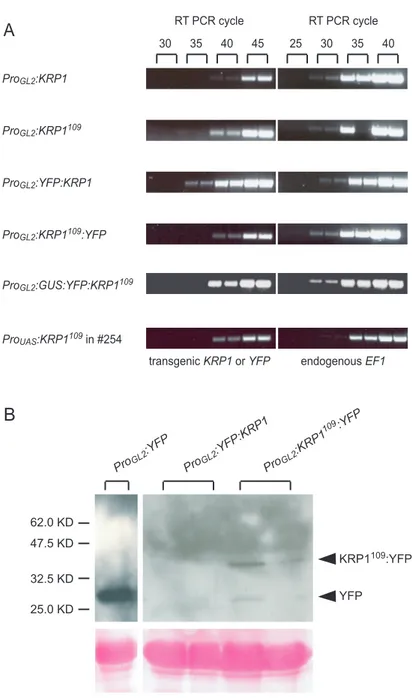
Whereas for KRP4 no insertion line could be found, for KRP1 one insertion line was found in Pool #36537. Sequencing of the PCR product obtained with the screening primer S1 and the left border primer T1 revealed that the T-DNA is inserted in the second intron, 387 bp downstream from the start codon (Fig5A). So far all PCR attempts, using the primer combinations S2+T2, S2+T4 and S2+T6, to proof that the complete 7 kb T-DNA was inserted in the KRP1 gene failed to reveal the insertion of the right border. However, plants were resistant to hygromycin and the HYGROMYCIN PHOSPHOTRANSFERASE (HPH) which confers resistance is located approximately 2 kb from the right border. Also no PCR products could be amplified with the S2 primer and any left border primer (T1, T3 and T5). To test whether the insertion resulted in a knock-out, a knock-down or knock-in of KRP1- function semiquantitative RT-PCR analyses were performed. No transcript could be detected in the homozygous mutant with a primer combination spanning the complete coding sequence of KRP1 (R1+R2) (Fig5B upper panel). However, using the primers R3 and R2, which anneal downstream of the T-DNA insertion, transcript could be
Results
obtained (Fig5B lower panel). This could be because the T-DNA contains promotor- like elements, which then result in a transcription of the KRP1 C-terminal domain.
Even though the transcript level is reduced in the mutant compared to wild type it cannot be ruled out that this mRNA becomes translated and that this peptide interferes for example with the CDK/cyclin complex, especially because it contains the cyclin- and CDK-interacting domains (see Fig7).
1.2. The krp1 mutant
Analysis of the phenotype of the homozygous krp1 T-DNA insertion plants revealed no obvious morphological alterations in comparison to wild type. Promotor-reporter analysis (Lieven de Veylder personal communication) and in situ hybridization of KRP1 mRNA suggested that KRP1 is expressed in endoreplicating trichome cells (Ormenese et al., 2004). Therefore I measured the trichome DNA content, which revealed a subtle enhancement of endoreplication in the homozygous krp1 mutant.
The median of the relative fluorescence of DAPI stained wild-type trichome nuclei was set as 32C (Fig5C). Three independent measurements of trichome DNA levels in the homozygous krp1 mutant revealed an elevated DNA content, 37.2C, 40.1C and 44.1C respectively, in comparison to wild type (Fig5C). This finding suggests that KRP1 might be involved in the termination of endocycles in trichomes.
Figure 5 The krp1 mutant
(A) Schematic drawing of the KRP1 gene showing the T-DNA insertion in the second intron.
Grey boxes represent the four exons, S1, S2 and T1 are the screening primers used for the identification of the insertion line. Also the primers used for the RT-PCR are shown (R1, R2 and R3).
(B) Semi-quantitative RT-PCR showing the relative expression strength of wild-type and the krp1 mutant. The used KRP1 primers are indicated on the left side. For the control, primers which amplify the ELONGATION FACTOR 1 (EF1) were used. Samples were taken after 30 or 40 cycles as indicated at the top of the figure.
(C) Distribution of trichome cell DNA contents are given in relative fluorescence units (RFUs). The median RFU of wild-type was set as 32C so that 2 RFUs represent approximately 2C. The sample size (n), the mean (m) +/- standard deviation and the median (md) are given.
A
5'UTR 3'UTR
T-DNA
Hygro
LB RB
S1
R2 R3
S2 T1
R1
388
1 902
T5 T3
T2 T4 T6
percentage of trichome nuclei
16 24 32 40 48 56 64 72 80
8
2 88
5 10
15 c) krp1
n=58 m=44.2 +/-16.0 md=44.1
C B
30 40 30 40 30 40
30 40
KRP1 EF1 KRP1 EF1
WT krp1
R1+R2 R3+R2
Figure 5 The krp1 mutant
relative fluorescence units b) krp1
n=51 m=41.0 +/-13.6 md=40.1
a) krp1
n=52 m=39.8 +/-16.3 md=37.2
Coln=58 m=34.2 +/-15.4 md=32.0
5 10 15
5 10 15
5 10 15
Results
1.3. RNAi approach
At the time the mutant was characterized no further insertion lines for KRP1 were available from other T-DNA collections to support the observed trichome phenotype.
Therefore I tried to knock-out KRP-function using a RNA interference approach by which introduction of double-stranded RNA should lead to a post-transcriptional silencing of the respective gene. In several attempts I tried to knock out KRP1 in trichomes. For that purpose I expressed double-stranded RNA of either the full-length KRP1 gene or the N-terminal domain of KRP1, which shows only low homology with the other members of the KRP family, by using the GLABRA2 promotor (ProGL2).
However analysis of seedlings in the T1 generation revealed a wild-type phenotype with respect to trichome morphology, leaf size and all over plant morphology (Tab1).
Additionally, I expressed double-stranded RNA of full-length KRP4, its N-terminal domain and a 141 bp fragment, which shows a high homology to KRP1, in trichomes.
Primary transformants did not display any morphological changes. Also the expression of double-stranded RNA of a short fragment of exon 3 from KRP1 or of two fragments of exon 4 from KRP7, which has shown to be expressed in endoreplicating and dividing cells (Ormenese et al., 2004), under control of the ubiquitously active CaMV35S promotor (Pro35S) did not result in a detectable phenotype in seedlings (Tab1).
In summary these results indicate that either the RNAi approach did not sufficiently reduce transcript levels of KRPs, or that the individual members of the KRP family act in a highly redundant manner, so that only in plants with a loss of function for more than one KRP gene a phenotype can be detected. The latter scenario is supported by the observation that even double and triple mutant combinations of
Results
krp2 with other krp mutants did not display any morphological alterations in comparison to wild type (Lieven de Veylder, personal communication).
TABLE 1
RNAI CONSTRUCTS TO KNOCK OUT KRPS
line template position
sense primer
position antisense
primer
Trichome or seedling*
phenotype
ProGL2:fl-KRP1-RNAi KRP1 Exon 1 Exon 4 WT ProGL2:N-KRP1-RNAi KRP1 Exon 1 Exon 3 WT Pro35S:Exon3-KRP1-RNAi KRP1 Exon 3 Exon 3 WT ProGL2:fl-KRP4-RNAi KRP4 Exon 1 Exon 3 WT ProGL2:N-KRP4-RNAi KRP4 Exon 1 Exon 1 WT ProGL2:cons-KRP4-RNAi KRP4 Exon 2 Exon 3 WT Pro35S:Exon4a-KRP7-RNAi KRP7 Exon 4 Exon 4 WT*
Pro35S:Exon4b-KRP7-RNAi KRP7 Exon 4 Exon 4 WT*
Results
2. Studying KRP function: gain of function approach
2.1. Misexpression of Arabidopsis KRP1 and KRP4 in trichomes
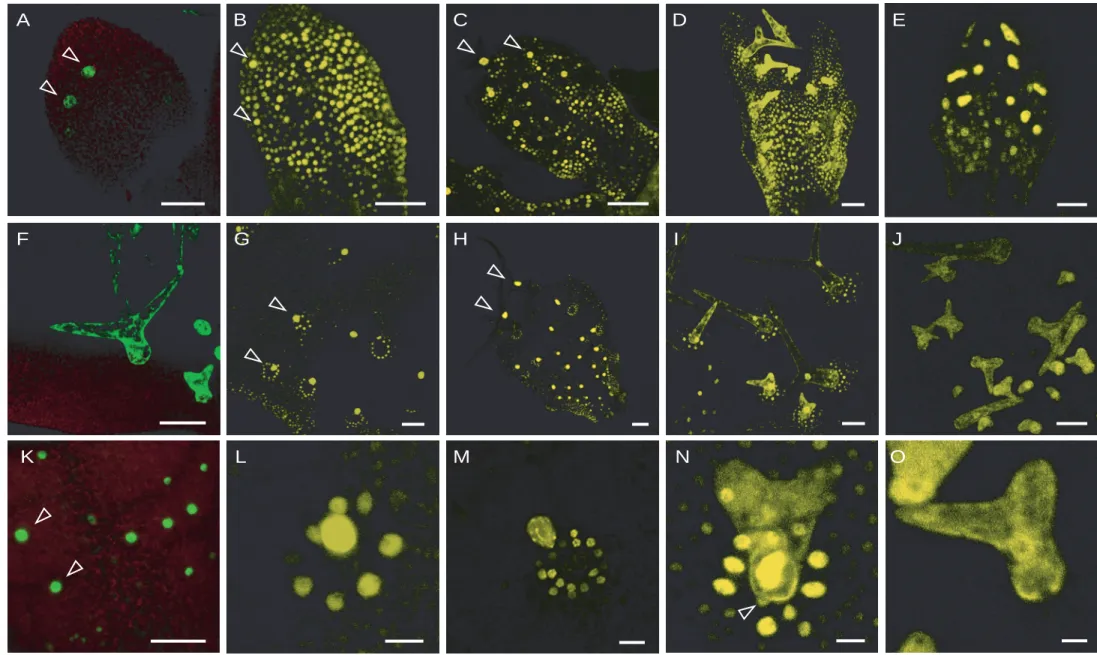
As described previously by Schnittger et al. the misexpression of KRP1 or the N- terminal truncated KRP1109 in trichomes under control of the GLABRA2 promotor results in smaller trichomes with reduced number of branches in comparison to wild type (Fig6A;B;E) (2003). In addition trichomes misexpressing KRP1 underwent cell death (Fig6G). DAPI stainings (see Fig6C,F for DAPI stained trichome nuclei) and DNA measurements revealed that endoreplication levels were reduced.
To test whether KRPs display similar functions in endorpelicating cells, I misexpressed another member of the KRP family, KRP4, which has not been characterized so far. The trichomes of the ProGL2:KRP4 transgenic plants also had fewer branches, the cell size was reduced and they showed the cell death phenotype as seen for ProGL2:KRP1 expressing plants (Fig6D). Taken together these data indicate that both KRP1 and KRP4 have similar effects, when misexpressed in trichomes.
Figure 6 Misexpression of KRP1 and KRP4 in trichomes
(A) to (C) Landsberg erecta wildtype In (A) an overview of a two week old seedling with mostly three-branched trichomes is given. (B) Scanning electron micrograph and (C) light micrograph of DAPI-stained mature trichomes with its neighboring cells, arrowheads point at trichome and trichome-neighboring cell nuclei.
(D) Overview of a two week old ProGL2:KRP4 misexpressing seedling with two- and unbranched trichomes
(E) to (G) ProGL2:KRP1109 misexpressing line. (E) and (G) Scanning electron micrographs showing in (E) a small and two-branched and in (G) a dead trichome. Note the enormously increased trichome-neighboring cells. (F) Light micrograph of DAPI-stained trichome with its neighboring cells, arrowheads point at trichome and the large trichome-neighboring cell nuclei.
(H) and (I) Scanning electron micrograph of (H) glabra3 and (I) cpr5 mutant trichomes, which have fewer branches, but normal sized trichome-neighboring cells
(J) and (K) Confocal laser scanning micrographs of enhancer trap line #254. (J) Showing the youngest state when GFP is detectable in the trichome-neighboring cells (indicated by arrowheads) and (K) a close up of line #254 showing GFP fluorescence in a mature trichome and its neighboring cells.
(L) Confocal laser scanning micrograph of ProGL2:KRP1109 crossed in enhancer trap line
#254, showing GFP expression in the enlarged trichome-neighboring cells.
Scale bar in all panels 100µm.
Figure 6 Misexpression of KRP1 and KRP4 in trichomes F C
E B
J
H I
D A
G
L K