Analysis of trichome differentiation in Arabidopsis thaliana:
From cell fate initiation to cell death
Inaugural-Dissertation
zur
Erlangung des Doktorgrades
der Mathematisch-Naturwissenschaftlichen Fakultät der Universität zu Köln
vorgelegt von
Daniel Bouyer aus Villingen
2004
Berichterstatter: Prof. Dr. Martin Hülskamp Prof. Dr. Klaus Harter
Prüfungsvorsitzender: Prof. Dr. Siegfried Roth
Tag der mündlichen Prüfung: 11.11.2004
DANK
schulde ich vielen. Zunächst möchte ich mich bei Martin Hülskamp bedanken für die Betreuung und die Freiheit, die er mir in der Auswahl und der Durchführung meiner Arbeit gegeben hat.
Deren Anfänge liegen im Tübinger Institut für Entwicklungsgenetik von Gerd Jürgens; daher auch ihm ein Dankeschön für die Aufnahme in den Kreis der Entwicklungsbiologen (wovon ich während des Studiums immer geträumt habe!).
Einiges in dieser Arbeit wurde von anderen begonnen oder in Zusammenarbeit mit anderen zu Wege gebracht. STICHEL wurde von Hilmar Ilgenfritz kloniert, die Analyse und Klonierung von CPR5 war ein Gemeinschaftsprojekt mit Viktor Kirik, die Herstellung von TTG-Promoter- Konstrukten wurde in unermüdlicher Arbeit von Martina Pesch bewältigt und die Idee der Nicht- Zell-Autonomie von TTG1 wurde von Arp Schnittger schon tatkräftig bearbeitet, bevor ich mich daran zu schaffen machte. Ihnen gilt daher ein dickes Danke!
Susanne Breiding war mir in Köln bei vielen Projekten behilflich und ich hoffe, es waren nicht zu viele! Danken möchte ich auch Herrn Weidner, der alle Verwaltungsangelegenheiten fest im Griff hat und bei dem ich immer gerne Prüfungsbeisitzer war. Uli Herrmann war immer zur Stelle um die Computer am Leben zu erhalten, auch wenn STICHEL mal wieder rumzickte. Dass Uschi Claßen eine tolle Arbeit in der Spülküche macht, merkt man natürlich erst, wenn sie nicht da ist, daher an dieser Stelle mal ein großes Dankeschön! Auch allen anderen, die mit ihren Jobs dafür sorgen, dass man im Labor überhaupt arbeiten kann und damit das Labor zum Labor machen, sei hier gedankt.
Die familiäre Atmosphäre hatte ich im Tübinger Labor Arp, Hilmar, Paul, Swen, Ulrike Folkers (UFO), Ulrike Schöbinger und Viktor zu verdanken. Dank Viktor, Susanne, Steffi, Martina, Katja, Britta und Birgit kann man sich auch in Köln ganz wohl fühlen.
Ganz besonders war ich auf die Hilfe bei der Durchsicht meines Manuskripts durch Christina Weinl, Viktor Kirik, Arp Schnittger, Swen Schellmann, Martina Pesch und Steffi Falk angewiesen! Ohne Euch würde ich wahrscheinlich selbst nichts mehr von dem verstehen, was ich da anfänglich so zusammengeschrieben habe ...
Dank einer glücklichen Fügung verschlug es Klaus Harter aus meiner alten Studienstätte Freiburg an das Kölner Nachbarinstitut. Für seine Übernahme des Zweitgutachtens möchte ich ihm danken und hoffe, dass ihm die Zugfahrt durch die Lektüre nicht zu lange wird.
Contents
CONTENTS
Zusammenfassung ... III Abstract... V Publications ... VII Abbreviations and Gene names ...VIII Figure index ... IX
A TRICHOME MOPRHOGENESIS... 1
A 1. Introduction ... 2
A 1.1. Different steps in the formation of cell shape... 2
A 1.2. Steps in trichome development... 2
A 1.3. Endoreplication-dependent trichome morphogenesis ... 3
A 1.4. Endoreplication-independent branch mutants... 4
A 2. Results ... 6
A 2.1. Analysis of STI ... 6
A 2.1.1. STI belongs to a group of novel genes ... 6
A 2.1.2. STI acts in a dosage-dependent manner... 10
A 2.1.3. STI is not involved in endoreplication... 11
A 2.1.4. Localisation of GFP-STI ... 12
A 2.2. Analysis of CPR5... 13
A 2.2.1. Trichome differentiation in the cpr5 mutant... 13
A 2.2.2. Cell proliferation defect in the cpr5 mutant... 14
A 2.2.3. Cell death in the cpr5 mutant... 16
A 2.2.4. Complementation of the cpr5 mutant... 16
A 3. Discussion... 17
A 3.1. Analysis of STI ... 17
A 3.1.1. The role of STI in cell morphogenesis... 17
A 3.1.2. Potential molecular function of STI... 18
A 3.2. Analysis of CPR5... 20
B TRICHOME PATTERN FORMATION... 23
B 1. Introduction... 24
B 1.1. Trichome initiation in Arabidopsis thaliana... 24
B 1.2. Elements of the trichome pattern system... 24
B 1.3. Molecular nature of the trichome patterning genes ... 25
B 1.4. Functions beside trichome patterning... 26
B 1.5. Special role of TTG1 and interactions between the patterning components... 26
B 1.6. A model to explain two-dimensional pattern formation... 27
B 1.7. Application of the activator-inhibitor model to the trichome patterning system ... 28
B 1.8. Intercellular protein movement in plants ... 30
B 1.9. Connection between intra- and intercellular mobility?... 31
B 1.10. Outlook and aim of the work ... 31
B 2. Results ... 33
B 2.1. TTG1-dependent and -independent trichome development ... 33
B 2.2. Expression analysis of TTG1... 36
B 2.3. TTG1-YFP localises to the nucleus... 39
B 2.4. TTG1 localisation depends on differentiation status ... 41
Contents
B 2.5. Non-cell-autonomous action of TTG1... 44
B 2.6. TTG1-YFP-fusion is able to move... 46
B 2.7. TTG1-YFP constructs show pattern defects ... 49
B 2.8. Blocking TTG1 mobility leads to strong pattern defects... 50
B 2.8.1. Exchanging endogenous TTG1 with an immobile TTG1... 50
B 2.8.2. Dependency of TTGim-phenotype on endogenous TTG1 function ... 52
B 2.8.4. Immobilised TTG1 induces ectopic trichome-specific reporters ... 55
B 2.8.5. Dominant negative effect of trichome-specific expression of the immobilised TTG1... 56
B 2.8.6. The dominant negative effect of GL2::TTGim is dependent on TRY/CPC ... 56
B 2.8.7. TTGim interacts with GL3 but not with GL1... 58
B 3. Discussion... 59
B 3.1. Non-cell-autonomous action of TTG1... 59
B 3.2. Developmental relevance of TTG1 mobility ... 60
B 3.3. Is TTG1 actively transported?... 62
B 3.4. Intracellular localisation of TTG1... 64
B 3.5. Interaction of TTG1 with other patterning genes... 65
B 3.6. Is the role of TTG1 in agreement with the activator-inhibitor system?... 67
B 3.7. Another view on the Meinhardt-model... 69
B 3.8. Outlook ... 73
Material and Methods ... 74
Materials ... 74
Chemicals, antibiotics... 74
Enzymes and molecularbiological materials... 74
Cloning vectors ... 74
Bacterial strains... 75
Plant lines ... 75
Methods ... 75
RNA isolation ... 75
Reverse transcription ... 76
Semiquantitative RT-PCR ... 76
Genomic DNA preparation... 76
Plasmid DNA preparation from bacteria ... 77
DNA-manipulation... 77
Cloning of the STI cDNA... 78
Constructs ... 78
Plant growth conditions ... 81
Crossing of plants... 81
Plant transformation ... 82
Seed sterilisation ... 82
Heat-shock treatment... 82
Histochemistry ... 82
Fluoresceine diacetate- & propidium iodid staining... 83
Microscopy ... 83
DAPI-staining and DNA-measurement ... 83
References... 85
Erklärung... 93
Lebenslauf... 94
Zusammenfassung
Zusammenfassung
In der folgenden Dissertationsarbeit habe ich mich mit Zelldifferenzierungsprozessen anhand der Blatthaarentwicklung der Modellpflanze Arabidopsis thaliana beschäftigt. Der Zelltyp ‚Trichom’
eignet sich besonders um entwicklungsbiologische Fragestellungen wie beispielsweise die Initiation eines bestimmten Zellschicksals, die Generierung eines geordneten Abstandsmusters oder auch die Prozesse hinsichtlich der Ausbildung einer streng festgelegten dreidimensionalen Zellform zu untersuchen.
In meiner Arbeit habe ich einige dieser Aspekte untersucht. Daher unterteile ich meine Ausführungen in die beiden Abschnitte Morphogenese (Untersuchung der Ausbildung einer bestimmten Zellform) und Trichom-Musterbilding (‚pattern formation’; Analyse der Prozesse, die für die Zellschicksalsfestlegung der Blatthaare aus anfänglich nicht unterscheidbaren Zellen notwendig sind).
Im Kapitel Morphogenese habe ich mich mit zwei Mutanten beschäftigt, die Störungen der Ausbildung der Zellform aufweisen. In normalen, sogenannten wildtypischen (WT) Blatthaaren wachsen die Zellen aus der Blattoberfläche aus und bilden anschließend ein stereotypisches Verzweigungsmuster. In der stichel-Mutante (sti) ist die Entstehung der Verzweigungen vollständig gestört, d.h., es entstehen keine Verzweigungen mehr. Es konnte in der Arbeit gezeigt werden, dass das STI-Gen diesen Prozess in einer Dosis-abhängigen Art reguliert. Eine Reduzierung der STI-Aktivität führt demnach zu einer Reduktion der Verzweigungen und eine Erhöhung zu einer vermehrten Verzweigung. Daneben legte die Klonierung des STI-Gens durch Hilmar Ilgenfritz eine Verbindung der Morphogenese mit bestimmten Zell-Zyklus-Prozessen nahe. Frühere Untersuchungen ergaben, dass Trichom-Zellen einen bestimmten Typ von Zellzyklus durchlaufen. Der DNA-Gehalt wird dabei wie bei einer normalen Zellteilung verdoppelt, die Zelle teilt sich jedoch nicht. Dieser Prozess wird als Endoreduplikation oder auch Endoreplikation bezeichnet. Tatsächlich kodiert STI für ein Protein, das Ähnlichkeiten mit einer DNA-Polymerase-Untereinheit besitzt, also mit einem Enzym, das maßgeblich an der Synthese von DNA während des Zellzyklus beteiligt ist. Allerdings konnte weder eine Verminderung des DNA-Gehalts in sti-Mutanten noch eine Erhöhung in Pflanzen, die STI vermehrt produzieren, beobachtet werden. Die weitere Analyse zeigte jedoch, dass STI zu einer phylogenetisch separierten Gruppe von Proteinen gehört, die bislang nur in Pflanzen gefunden wurde und wahrscheinlich nicht in direktem Zusammenhang mit den DNA-Polymerase-Untereinheiten steht.
Zusammenfassung
Diese Vermutung wurde weiter untermauert durch die Beobachtung, dass das STI-Protein nicht im Kern, also dem Ort der DNA-Synthese, sondern an den zukünftigen Verzweigungspunkten der Trichome gefunden wurde. STI scheint also direkt an der Ausbildung der Verzweigungen beteiligt zu sein.
Die zweite Morphogenese-Mutante (cpr5), die von mir in dieser Arbeit untersucht wurde, ähnelt sti insofern, als auch hier eine starke Reduktion der Trichom-Verzweigung zu beobachten ist.
Darüber hinaus konnte ich zeigen, dass es hierbei, im Gegensatz zu sti, zu einer Reduktion des DNA-Gehalts in den Trichomen der cpr5-Mutante kommt. Die weitere Analyse ergab, dass es in cpr5 zu einem Absterben der Trichome kommt, was auch in anderen Teilen der Pflanze beobachtet wurde. Daneben weist cpr5 ein vermindertes Wachstum auf und scheint in mehreren Prozessen gestört zu sein. Gemeinsam mit Viktor Kirik wurde das CPR5-Gen kloniert und es zeigte sich, dass es für ein Protein unbekannter Funktion kodiert. Die Proteinstruktur lässt keine genaueren Vermutungen über die molekulare Funktion von CPR5 zu.
Im zweiten Teil dieser Arbeit konzentriere ich mich auf die Prozesse der Ausbildung des regelmäßigen Abstandsmusters der Trichome zueinander. Dabei war vor allem eine Komponente dieses Musterbildungssystems, das TTG1-Gen, noch nicht genauer charakterisiert. Ich konnte die Aktivität des TTG1-Gens in zellulärer und zeitlicher Auflösung anhand von Promoter-GUS- Analysen aufzeigen und die Lokalisation des TTG1-Proteins innerhalb der Zelle klären. Dabei zeigte sich, dass TTG1 überall in der Zone der Trichom-Musterbildung exprimiert wird und dass das Protein anfänglich überwiegend in den Kernen und mit zunehmender Entwicklung des Blattes überwiegend im Cytoplasma der Zellen zu finden ist. Allerdings behalten die Trichome die Lokalisation von TTG1 in den Kernen über ihre gesamte Entwicklung hinweg bei. Daneben konnte ich zeigen, dass TTG1 nicht-zellautonom wirkt und das Protein zwischen Zellen mobil ist.
Diese Mobilität scheint auch für das Zustandekommen eines regelmäßigen Musters wichtig zu sein. Dies wird offensichtlich wenn die Mobilität des Proteins gestört oder gar verhindert wird, wobei es zu schwerwiegenden Störungen des Musters kommt.
Abstract
Abstract
In the following PhD thesis I studied cell differentiation processes of leaf hairs, trichomes, in Arabidopsis thaliana. This cell type is very well suited for the analyses of the initiation of a certain cell fate, the generation of a regular spacing pattern or the processes that are required for the formation of a three-dimensional cell form.
In my thesis I have investigated several of these aspects. Therefore this work is subdivided into the sections trichome morphogenesis (analysis of the formation of a certain cell form) and trichome pattern formation (analysis of the processes that are important for the commitment and the generation of a spacing pattern of a certain cell type that is derived from initially equal cells).
In the chapter morphogenesis I studied two mutants that show defects in the generation of the trichome-cell form. A typical trichome in Arabidopsis grows out of the leaf surface and forms three branches in a highly stereotypical manner. In the stichel (sti) mutant the development of these branches is completely abolished.
Previous genetic analysis and my studies suggest that STI acts in this process in a dosage- dependent manner. A reduction of STI activity leads to a reduction in branch number and a elevated STI activity leads to an increase in branch number. The cloning of the gene suggested a connection between morphogenesis and a certain kind of the cell cycle, called endoreduplication or endoreplication. This process leads to DNA-synthesis as observed in the usual cell cycle, however without division, which results in a higher DNA content in the cell. In fact STI encodes for a protein with sequence similarities to a DNA-Polymerase subunit, an enzyme that is involved in DNA-synthesis during the cell cycle. However neither sti mutants nor plants that ectopically express STI show changes in the DNA content.
The further analysis showed that STI belongs to a group of proteins that is separated from the conventional DNA-polymerase subunits. This assumption was supported by the observation that the STI protein is not found in the nucleus, the place of DNA-synthesis, but at the future branch initiation point in trichomes. Therefore STI seems to play a direct role in the formation of the trichome branches.
The second morphogenesis mutant (cpr5) that was subject of my work, resembles sti with respect to the reduction of trichome branches. However in contrast to sti, the DNA-content is reduced in the cpr5 mutant. Moreover, during further development the trichomes in cpr5 die, a process that
Abstract
is also observed in other parts of the mutant plant. Beside this, cpr5 is also impaired in the proliferation and growth and seems to be defective in several aspects. Together with Viktor Kirik the CPR5 gene was cloned and shown to encode a novel protein with unknown function.
In the second part of my thesis I examined the processes that control the formation of a regular trichome spacing pattern on the leaf surface. An important component of this patterning system, TTG1, was investigated in more detail. I could reveal the temporal and spatial activity of the gene in promoter-GUS analyses and the localisation of the protein in the cell. This revealed that TTG1 is expressed throughout the entire trichome-patterning zone and that the protein changes its localisation from predominantly nuclear in the early leaf-development to more cytoplasmatic during further development of the epidermal cells. However trichomes keep the nuclear localisation throughout their development. Moreover it was shown that TTG1 acts non-cell- autonomous and that the TTG1-protein is able to move between cells. This transport seems to be important for the generation of the spacing pattern, which is reflected by the patterning defects if the protein-mobility is impaired or even completely blocked. In the latter case the pattern is strongly impaired. These observations are summarised into a model to explain the early patterning events during trichome differentiation.
Publications
Publications:
CPR5 is involved in cell proliferation and cell death control and encodes a novel trans- membrane protein.
Kirik V, Bouyer D, Schöbinger U, Bechtold N, Herzog M, Bonneville JM, Hülskamp M Curr Biol 2001, 11: 1891–1895
Shared first authorship; I generated the rescue construct and did most of the mutant analysis, including trichome phenotype analysis, trichome DNA content measurements and cell-death analysis.
Ectopic D-type cyclin expression induces not only DNA replication but also cell division in Arabidopsis trichomes.
Schnittger A, Schöbinger U, Bouyer D, Weinl C, Stierhof YD, Hülskamp M Proc Natl Acad Sci U S A. 2001, 99: 6410-6415
For this work I did some of the endoreplication measurements and trichome morpho- genesis analysis.
The Arabidopsis STICHEL gene is a regulator of trichome branch number and encodes a novel protein.
Ilgenfritz H, Bouyer D, Schnittger A, Mathur J, Kirik V, Schwab B, Chua NH, Jürgens G, Hülskamp M
Plant Physiol 2003, 131: 643-655
Shared first authorship; I generated the STI cDNA together with Hilmar Ilgenritz and created all transformation constructs and transgenic plants. The analysis of the overexpression phenotype of 35S::STI was performed and the alignments with the STI- homologues. Birger Marin helped me in the generation of the phylogenetic tree.
Misexpression of the cyclin-dependent kinase inhibitor ICK1/KRP1 in single-celled Arabidopsis trichomes reduces endoreduplication and cell size and induces cell death.
Schnittger A, Weinl C, Bouyer D, Schöbinger U, Hülskamp M Plant Cell 15, 303-315
In this work I designed and generated the ICK deletion constructs and produced the corresponding transgenic plants. Furthermore I did most of the phenotypical characterisation and the endoreplication analysis of the trichomes in these plants.
Cell polarity in Arabidopsis trichomes.
Bouyer D, Kirik V, Hülskamp M Semin Cell Dev Biol 2001, 12: 353-356 Branching of single cells in Arabidopsis.
Bouyer D, Hülskamp M
In: Davies JA (Editor): Branching Morphogenesis 2003
Abbreviations
Abbreviations and Gene names
°C degree Celsius µ micro
35S 35S promoter from Cauliflower Mosaic virus AN ANGUSTIFIOLIA AN11 ANTHOCYANINLESS11 ATP Adenosine triphosphate
bp base pair
bHLH basic helix-loop-helix C DNA-content of a haploid genome CaMV Cauliflower Mosaic virus
CDK cyclin dependent kinase
cDNA complementary DNA
CDS coding sequence
CLSM confocal laser scanning microscopy
CPC CAPRICE Col Columbia
CPR5 constitutive pathogen response5
cv. cultivar/ecotype D Dalton
DAPI 4',6-Diamidino-2-phenylindole DIG Digoxygenin
DNA Desoxyribonucleic acid e.g. exempli gratia [Lat.] for example
ER endoplasmatic reticulum
ERH3 ELONGATED ROOT HAIR3 et al. et alterni [Lat.] and others ETC1/2 ENHANCER OF TRY CPC1/2 FDA fluorescein diacetate Fig. Figure
FRA FRAGILE FIBRE
FRC FURCA FS FASS
GA gibberelic acid
GFP green fluorescent protein GL1 GLABRA1 GL2 GLABRA2 GL3 GLABRA3 GUS glucuronidase ICK inhibitor of CDK k kilo KAK2 KAKTUS2
kb kilo bp
kD kilo Dalton
Ler Landsberg erecta
KRP kip related protein (=ICK)
MP movement protein
mRNA messenger RNA
MT microtubules n number
NLS nuclear localisation signal/sequence
NOK NOEK
PCR polymerase chain reaction p promoter PD plasmodesmata RFC replication factor C
RNA ribonucleic acid
rpm rounds per minute
RT-PCR reverse transcription PCR
SD standard deviation
SEL size exclusion limit SIM SIAMESE SPK SPIKE STI STICHEL T-DNA transferred DNA TIS trichome initiation site TRY TRIPTYCHON
TTG1 TRANSPARENT TESTA
GLABRA1
TON TONEAU
UTR untranslated region
WT wild type
YFP yellow fluorescent protein ZWI ZWICHEL WS-O Wassilewskaja
All gene- and mutant names are written in italics. WT-genes are written in capital letters. Proteins are written in non-italic letters.
Figure index
Figure index
Fig. 1: Steps in trichome development...3
Fig. 2: Sequence alignment of STI with prokaryotic DNA polymerase III γ-subunits and the small subunits of eukaryotic and archaebacterial replication factor Cs...7
Fig. 3: Sequence comparison between STI with its closest homologues...8
Fig. 4: Phylogenetic tree of the STI, the DNA polymerase III γ-subunit, and the RFC small subunit family...9
Fig. 5: Trichome-phenotypes of sti and 35S::STI...10
Fig. 6: Analysis of expression levels in 35S:STI lines by semiquantitative RT-PCR...11
Fig. 7: Confocal laser scanning micrographs of GFP-STI...12
Fig. 8: Endoreplication in cpr5...13
Fig. 9: Cell proliferation in cpr5...14
Fig. 10: Trichome and lesion phenotype in cpr5 mutants...15
Fig. 11: Trichome differentiation and mathematical modelling...28
Fig. 12: Trichome patterning model...29
Fig. 13: Mutant analysis of negative regulators in combination with ttg1 and gl1...35
Fig. 14: Expression analysis of TTG1...37
Fig. 15: Analysis of TTG1-YFP localisation...40
Fig. 16: TTG1-YFP localisation is dynamic...42
Fig. 17: Non-cell-autonomous action of TTG1...45
Fig. 18: TTG1-YFP fusion moves between cells...47
Fig. 19: Trichome phenotype of immobilised TTG1 (TTG1im)...51
Fig. 20: Effect of pTTG1::TTG1im in different ttg1 and other mutant backgrounds...53
Fig. 21: Dominant negative effect of pGL2::TTG1im...57
Fig. 22: Model to explain the early steps in pattern formation...71
Morphogenesis
Trichome
Morphogenesis A
Morphogenesis Introduction
A 1. INTRODUCTION
A 1.1. Different steps in the formation of cell shape
Formally, cell shape can be considered to be established in three steps (Hülskamp et al. 1998). In a first step, spatial information, e.g. cell polarity, is established by intracellular mechanisms or provided by outer cues. In a second step, this information is used to reorganize the cell, e.g.
change the cytoskeletal arrangement. Finally, actual growth takes place, which includes the incorporation of membrane and cell wall material at defined areas of the cell periphery.
Although some of the biochemistry of the last two steps of the cytoskeletal function and cell wall synthesis is known, the mechanisms underlying the spatial control of cell morphogenesis are largely elusive. Single-cell model systems such as pollen tubes, root hairs, and leaf hairs (trichomes) that are accessible to genetic approaches provide the means to study spatial control mechanisms (Aeschbacher et al. 1994, Marks 1997, Oppenheimer 1998, Hülskamp et al. 1999, Kost et al. 1999, Wilhelmi and Preuss 1999). Among these model cell types, trichomes in Arabidopsis are particularly well suited because they consistently develop a complex three- dimensional form, thus providing excellent criteria to isolate mutants affecting discrete aspects of morphogenesis (Oppenheimer 1998, Hülskamp et al. 1999).
A 1.2. Steps in trichome development
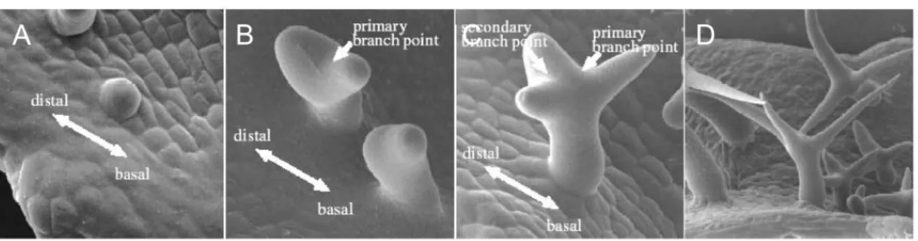
Leaf trichomes in Arabidopsis are large single cells that originate from the epidermis and are up to 500 µm tall. After trichome fate commitment the cells stop dividing but continue DNA synthesis (endoreduplication; Hülskamp et al. 1994). Fig. 1 shows the development of a trichome. The incipient trichome extends out of the leaf surface and undergoes two successive branching events (Hülskamp et al. 1994). The orientation of the first branching is co-aligned with the proximal-distal leaf axis. The primary branch, which points toward the leaf tip (main stem), undergoes a second branching in a plane perpendicular to the primary branching plane (Folkers et al. 1997). Subsequently, the trichome extensively elongates concomitant with an increase in vacuolization. The mature trichome has, on average, a DNA content of 32 C, suggesting that trichomes proceed through four endore-duplication cycles (Hülskamp et al. 1994). Trichome
Morphogenesis Introduction
branching requires the coordinated action of at least 18 genes (Marks 1997, Oppenheimer 1998, Hülskamp et al. 1999).
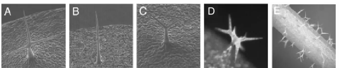
Figure 1: Steps in trichome development. Scanning electron micrographs of developing wild-type trichomes. (A) Incipient unbranched trichome. (B) Trichome with primary branch point. Note orientation of the branches with respect to the basal-distal leaf axis. (C) Trichome with primary and secondary branch.
(D) Mature trichome. [Figure modified from Schwab et al. 2000].
A 1.3. Endoreplication-dependent trichome morphogenesis
One group of genes appears to affect primarily the number of endoreduplication cycles and probably, as a consequence, also branch number (Hülskamp et al. 1994, Perazza et al. 1999, Kirik et al. 2001, Schnittger et al. 2003). The gl3 mutant is reduced in branching and shows a reduction of endoreplication cycles in the trichome from approximately 32C to 16C, whereas trichome-specific overexpression lead to enhanced branching and a DNA-content of approximately 128C (Hülskamp et al. 1994, Kirik et al. 2004c). Beside trichome morphogenesis and endoreplication, GL3 is also involved in trichome-pattern formation and encodes for a bHLH transcription factor (Payne et al. 2000). Moreover the try mutant that has been isolated as a patterning mutant, also shows trichomes with up to five branch points and has a DNA content of approximately 64C on average (Hülskamp et al. 1994). TRY encodes for a MYB-like transcription factor (Schellmann et al. 2002). Another group of mutants, the so-called kaktus group, show overbranching that is coupled with enhanced DNA content (Perazza et al. 1998).
Those studies revealed a link between gibberelic acid (GA) signalling and trichome morphogenesis, because the spindly mutant that shows a GA-oversensing phenotype also results in trichomes with enhanced branching (Perazza et al. 1998). The KAK2 gene has been cloned and shown to be an E3-ubiquitin ligase, thereby indicating that the control of protein degradation processes is crucial for the correct trichome morphogenesis (Downes et al. 2003, El Refi et al.
A B C D
Morphogenesis Introduction
2003).
Direct interfering with the cell-cycle machinery in Arabidopsis trichomes has been shown to result in altered morphogenesis (Schnittger et al. 2001a, Schnittger et al. 2001b, Schnittger et al.
2003). The trichome specific expression of an inhibitor of the cell cycle dependent kinase (CDK), the KRP1 (Kip related protein1)/ICK1 (Inhibitor of Cyclin dependent kinase1) gene results in less branched trichomes and a strong reduction in DNA content (Schnittger et al. 2003).
Surprisingly those cells eventually collapsed and died, which gave a hint to the connection of cell-cycle regulation and cell death control (Schnittger et al. 2003). The study of the cpr5 mutant, that shows reduced endoreduplication and trichome-branching, revealed a similar relationship (Kirik et al. 2001), and will be part of this work.
The sequence analysis of the STI gene, which was cloned by Hilmar Ilgenfritz, revealed a homology with prokaryotic DNA-replication factor C and DNA-polymerase γ-subunits. This suggested that DNA replication might be regulated by STI (Ilgenfritz et al. 2003). However the further analysis, which is given in this work, revealed no such relationship with STI.
A 1.4. Endoreplication-independent branch mutants
A second group of branching mutants affects branch number without affecting endoreduplication (Folkers et al. 1997, Luo and Oppenheimer 1999, Qiu et al. 2002). The genetic analysis of branching mutants suggests several redundant pathways control branch formation (Luo and Oppenheimer 1999). To date, five branching genes have been cloned and all appear to be involved in the regulation of the microtubule cytoskeleton at different levels. The ZWI (ZWICHEL) gene encodes a kinesin motor protein with a calmodulin-binding domain, indicating that microtubule-based transport is important for branch formation (Oppenheimer et al. 1997).
That the spatial organization of microtubules is important for trichome branching is suggested by the finding that in an (angustifolia) mutants, reduced trichome branching is correlated with the failure to establish a higher microtubule density at the tip of the developing trichome (Folkers et al. 2002). The underlying biochemical mechanism, however, remains unclear because AN encodes a novel protein with sequence similarity to C-terminal binding protein/BrefeldinA ribosylated substrates that are known to be involved in transcriptional regulation or in vesicle
Morphogenesis Introduction
budding but not in microtubule function (Folkers et al. 2002, Kim et al. 2002). The FRA2 (FRAGILE FIBER2)/ERH3 (ECTOPIC ROOT HAIR3) gene appears to be involved in the regulation microtubule assembly and disassembly. In fra2/erh3 mutants trichomes are underbranched; also, other cell types show morphogenesis defects (Burk et al. 2001, Webb et al.
2002). FRA2/ERH3 encodes for a katanin-p60 protein, suggesting that it functions as a microtubule-severing protein (Burk et al. 2001, Webb et al. 2002). In fs (fass)/ton2 (toneau2) mutants, shape changes of various cell types have been correlated with distortions of the microtubule cytoskeleton (Traas et al. 1995, McClinton and Sung 1997). Therefore, it is likely that the unbranched trichome phenotype in fs mutants (Torres-Ruiz and Jürgens 1994) is also linked to the microtubule phenotype. TON2 encodes a novel protein phosphatase 2A regulatory subunit, suggesting that microtubule organization in plants is controlled by the phosphorylation and dephosphorylation of proteins (Camilleri et al. 2002). Mutation in the SPIKE1 gene results in underbranched trichomes along with morphogenesis defects in various cell types. Microtubule organization is misregulated in spk1 mutants and the recent cloning of SPK1 revealed that it encodes an adapter protein involved in the integration of extracellular signals with the cytoskeletal organization (Qiu et al. 2002). These observations are supported by drug inhibitor studies that revealed distinct roles of actin and tubulin during trichome cell morphogenesis (Mathur et al. 1999, Szymanski et al. 1999, Mathur and Chua 2000). Although the inhibition of the actin cytoskeleton causes irregularities in the directionality of cell expansion, experiments with drugs disturbing the microtubule cytoskeleton result in reduced trichome branching.
To further elucidate the molecular mechanisms underlying branch formation, I have studied the STI (STICHEL) gene. sti mutants exhibit the strongest branch phenotype: All trichomes are unbranched. Positional cloning revealed that STI encodes a novel protein containing a domain with sequence similarity to eubacterial DNA-polymerase III γ-subunits (Ilgenfritz et al. 2003).
The cpr5 mutant is similar to the sti mutant because it also shows a trichome-branch defect.
However the further analysis revealed a link to cell-proliferation and cell-death control in a more pleiotropic manner and therefore the analysis of cpr5 will be separated from the analysis of sti.
Morphogenesis Results
A 2. RESULTS
A 2.1. Analysis of STI
A 2.1.1. STI belongs to a group of novel genes
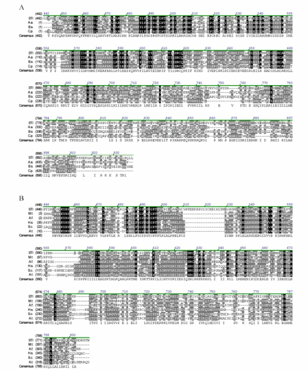
The sequence analysis of STI revealed that it encodes for a protein of 1,218 amino acid residues with a predicted molecular mass of 135.3 kD. Sequence comparison with other known proteins and motifs identified three putative functional domains. A large domain between amino acids 454 and 799 shows sequence similarity to eubacterial DNA polymerase III γ-subunits (Fig. 2A, Ilgenfritz et al. 2002). The prokaryotic DNA Polymerase γ III -subunit is the main component of the γ-complex, which is important for the formation of the replication initiation complex with the dimeric β-subunit. In principle, the DNA polymerase III is able to perform DNA replication without the γ-subunit, but the processivity is lower by several orders of magnitude. Upon ATP binding, the γ-complex loads the β-subunit onto a primer DNA template. Dissociation of the γ- complex from the β-subunit to allow the polymerase to bind the β-subunit requires ATP hydrolysis (Bertram et al., 1998). The similarity is 49% to 55% (identity 29%– 35%) within the homology region. Similarity to the family of 36- to 40-kD ATP-binding subunits of replication factor C (RFC, also known as activator 1), the archaebacterial and eukaryotic functional counterparts of the bacterial γ subunit (Chen et al., 1992a, 1992b), is less pronounced (42%–44%
similarity, 23%–27% identity). Fig. 2B shows the alignment of STI with this group of proteins, including the putative RFC from Arabidopsis. The latter is that member of a class of four putative RFCs in Arabidopsis that shows highest sequence similarity with the eukaryotic RFCs (Fig. 2B).
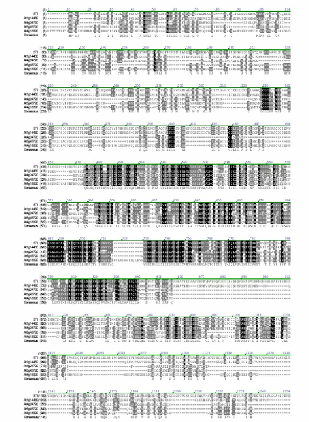
Figure 4 illustrates that STI and four STI homologues from Arabidopsis represent a phylogenetically separate branch, thereby defining a new, potentially plant-specific, subfamily among the γ-subunit homologues. The five proteins of this subfamily are markedly larger than the relatively small RFC proteins and the prokaryotic DNAPol III γ subunits and show sequence similarity outside the RFC/γ subunit domain (Fig. 3).
Morphogenesis Results
Figure 2: Sequence alignment of STI with prokaryotic DNA polymerase III γ-subunits and the small subunits of eukaryotic and archaebacterial replication factor Cs.
(A): Alignment of the STICHEL sequence with related protein sequences of prokaryotic DNA polymerase III γ-subunits. Black-shaded amino acids are identical, dark grey-shaded amino acids are conserved, and light grey indicates weak similarity. The numbering at the top corresponds to amino acid positions in STI.
A.a., Aquifex aeolius (aq 1855); B.s., Bacillus subtilis (Bsu0019); C.p., Chlamydias pneumoniae (CPn0040).
(B): Alignment of STI with related protein sequences of the small subunit of replication factor C. A.t., Arabidopsis (At1g2169); A.f., Archaeoglobus fulgidus (AF20608); M.t., Methanobacterium thermoautotrophicum (MTH241); S.c., yeast (YSCRFC2); H.s., human (Homo sapiens).
[Figure adopted from Ilgenfritz et al. 2003]
Morphogenesis Results
Figure 3: Sequence comparison between STI with its closest homologs. STI is a member of a class of five homologs that share sequence similarity outside the DNA polymerase III γ-subunit/RFC domain.
Black-shaded amino acids are identical, dark grey-shaded amino acids are conserved, and light grey indicates weak similarity. [Figure from Ilgenfritz et al. 2003]
Morphogenesis Results
Two regions, one between amino acids 273 and 304 and a second between amino acids 425 and 449, show similarity to PEST domains known to mediate rapid protein degradation. According to the score calculated based on the PEST hypothesis by Rodgers et al. (1986), the two PEST domains found in STI have a very high score of 9.24 and 9.58. Three nuclear localization signals (NLSs) suggest that STI is targeted to the nucleus with a probability of 87%. One NLS is located in the N-terminal part, and the two others are located tandemly at the very C terminus.
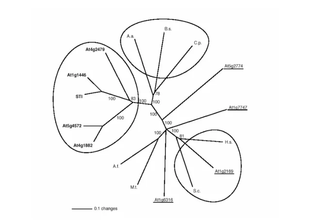
Figure 4: Phylogenetic tree of the STI, the DNA polymerase III γ-subunit, and the RFC small subunit family. The phylogenetic tree of STI, the DNA polymerase III γ subunit and the RFC small subunit family were calculated using only the core homology region corresponding to amino acids 449 to 799 in STI. The phylogenetic distance is shown as an unrooted dendrogram. The scale bar indicates 10% changes of amino acids. The bootstrap values are indicated for each branch. The five closest related STI homologs are bold and the four putative RFC-like proteins in Arabidopsis are underlined. The five closest STI homologs fall in a class that is separate from both, the prokaryotic DNA polymerase III γ-subunits and the eukaryotic RFC small subunits (the three groups are marked by circles). Note that only one of the Arabidopsis RFCs, At1g2169, is in the same group as the known eukaryotic RFC small subunits. A.a. A.
aeolius (aq 1855); B.s., B. subtilis (Bsu0019); C.p., C. pneumoniae (CPn0040); A.f., A. fulgidus (AF20608); M.t., M. thermo-autotrophicum (MTH241); S.c., yeast (YSCRFC2); H.s., human.
[Figure adopted from Ilgenfritz et al. 2003]
Morphogenesis Results
D E
A 2.1.2. STI acts in a dosage-dependent manner
Previous experiments using different sti alleles and the genetic interactions of representative strong and weak alleles with other mutants affecting branching and/or endoreduplication revealed that STI acts in a dose-dependant manner (Fig. 5, Ilgenfritz et al. 2003). This analysis showed that strong sti alleles invariably exhibit almost only unbranched trichomes, two weaker alleles have an increased frequency of two-branched trichomes (Ilgenfritz et al. 2003). The sti-40 allele has been shown to contain a mutation in the splice-donor site, thereby leading most likely to premature STOP if the transcript is spliced in the wrong way, however stronger alleles have more C-terminally mutations leading to premature STOP codons (Ilgenfritz et al. 2003). The mutation in the weak sti-47 allele leads to the most N-terminal premature STOP of all mutant alleles. This effect was explained by a reinitiation of translation at the next ATG bearing a minimal Kozak sequence (Ilgenfritz et al. 2003). In addition double and triple mutant combinations of strong and weak sti alleles suggest that STI acts in a dose-dependent manner (Folkers et al. 1997, Ilgenfritz et al. 2003).
Therefore the question was if an ectopic expression of STI would have an effect on branching.
Therefore the cDNA of STI was fused to the 35S Cauliflower Mosaic virus promoter and this construct was introduced into the sti mutant. Sixty-seven transgenic lines were studied for the rescue of the mutant phenotype. Seventeen lines showed a weak rescue, and 34 showed complete rescue with up to three branches. In addition, we found in 11 lines trichomes with more than two branch points, occasionally up to six branch points (Fig. 5D). Semiquantitative RT-PCR analysis of the 35S:STI specific expression levels did not reveal clear differences in the RNA levels between lines exhibiting weaker or stronger rescue (Table 1; Fig. 6). Thus, in summary, the overexpression of STI does not only rescue the sti phenotype, but also leads to extra branch formation.
Figure 5: Trichome-phenotypes of sti and 35S::STI. (A) – (C) scanning electron micrographs of trichomes, (A) WT three-branched trichome, (B) sti-146 mutant unbranched trichome, (C) sti-40 mutant two-branched trichome, (D) 35S::STI leaf trichome with six branch points, (E) 35S::STI stem trichomes with branches. [Figure adopted from Ilgenfritz et al. 2003].
Morphogenesis Results
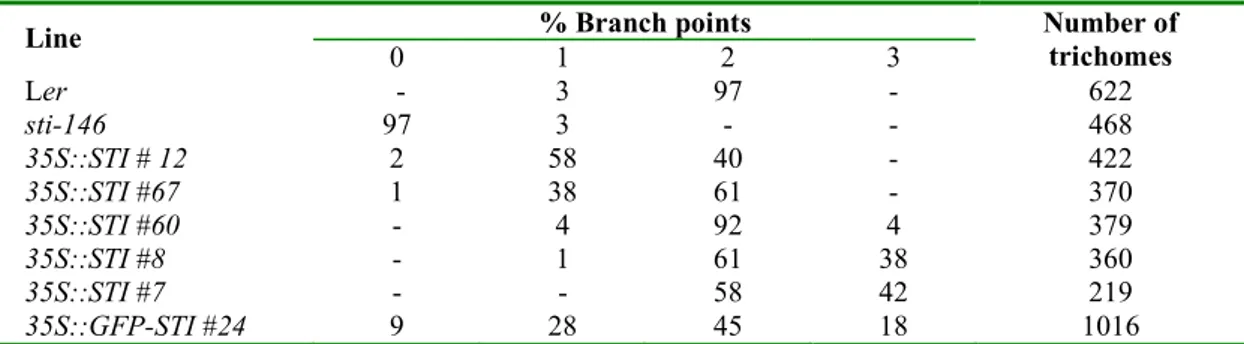
% Branch points Line
0 1 2 3
Number of trichomes
Ler - 3 97 - 622
sti-146 97 3 - - 468
35S::STI # 12 2 58 40 - 422
35S::STI #67 1 38 61 - 370
35S::STI #60 - 4 92 4 379
35S::STI #8 - 1 61 38 360
35S::STI #7 - - 58 42 219
35S::GFP-STI #24 9 28 45 18 1016
Table 1: Effect of 35S::STI on trichome branching. Trichomes were counted on the third and fourth leaf of 10 or more plants.
Also, in 35S:STI lines, stem trichomes that are normally unbranched exhibited up to two branch points (Fig. 5E), suggesting that organ-specific differences in trichome branching are controlled by STI.
A 2.1.3. STI is not involved in endoreplication
Previous analysis of the DNA content in sti trichomes revealed no effect in endoreplication (Ilgenfritz et al. 2003).
Because of the homology of STI to DNA-polymerase III γ subunits, the possible role of STI in endoreplication was further analysed by measuring the DNA-content of the 35S::STI lines that showed an overbranched phenotype (Table 1). This was studied in two independent lines. In both lines, trichomes had a DNA content indistinguishable from wild type (38 ± 15 C, n = 149; 37 ± 16 C, n = 60; WT: 38 ± 17 C, n = 49).
Figure 6: Analysis of expression levels in 35S:STI lines by semiquantitative RT-PCR.
The numbers of the lines correspond o those in Table 1. The elongation factor1 EF1 was used as a control. [Figure adopted from Ilgenfritz et al. 2003].
Morphogenesis Results
A 2.1.4. Localisation of GFP-STI
The homology to the DNAPolIII γ /RFC subunit and the existence of three nuclear localisation signals (NLS) point towards a nuclear function of STI. However the nuclear DNA content was neither impaired in the mutant nor in the 35S::STI lines. Therefore it was important to reveal the intracellular localisation of STI.
The STI-CDS was fused N-terminally to a GFP and expressed under the CaMV 35S promoter in sti-146 plants. Rescued lines (see table 1) were selected and further analysed for GFP- fluorescence using confocal laser scanning microscope (CLSM).
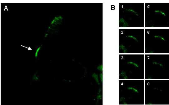
Interestingly fluorescence could not be detected in nuclei, but quite specific in young trichomes that started to branch (Fig. 7A). This fluorescence was visible at the just initiated side branch and ceased during the outgrowth of the branch (Fig. 7A). Single optical sections of the trichome branch sites show a speckled fluorescence of the GFP-signal (Fig. 7B). Ulrich Herrmann could confirm this localisation with an epifluorescence microscope, however the signal is barely visible (Ulrich Herrmann unpublished observation). Although STI is expressed in all organs of the plant, it was not possible to detect a specific fluorescence in other cells beside trichomes (data not shown).
Figure 7: Confocal laser scanning micrographs of GFP-STI.
(A) Above-view on a young trichome. GFP-signal appears at trichome branch initiation (arrow) (B) Series through a branch point showing speckle-like structures.
Morphogenesis Results
A 2.2. Analysis of CPR5
A 2.2.1. Trichome differentiation in the cpr5 mutant
In a screen of T-DNA mutagenised plants for trichome mutants, Viktor Kirik found two recessive mutants exhibiting reduced trichome branching, the ctz8 mutant and the 5758-1 mutant (Fig.
10B) (Kirik et al. 2001). Both mutants exhibited the same phenotypes and resulted from a T- DNA insertion in the CPR5 gene (accession number AY033229). The ctz8 mutant and the 5758-1 mutant were renamed cpr5-T1 and cpr5-T2, respectively.
In the cpr5-T1 mutant, the number of trichome branches is drastically reduced (58% unbranched, 38% two branches, 3% three branches, n = 661) compared to the corresponding wild-type WS (0% unbranched, 10% two branches, 90% three branches, n = 1029) (Fig. 10b). Because trichome cells appeared to be reduced in size, I determined whether the ploidy level is also reduced (Fig.
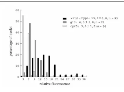
8). The comparison of the relative fluorescence of DAPI-stained nuclei in cpr5-T1 trichomes with that of wild-type (32C) and the glabra3 (16C) mutant revealed that the DNA content in cpr5-T1 trichomes corresponds to approximately 8C, suggesting that endoreduplication cycles stop after the second cycle is completed (Fig. 8). It was also found that the cell size and the nuclear size of epidermal pavement cells was greatly reduced (Figure 9B), such that, in 3-week-old plants, cell size and nuclear size of 98% of all cells in cpr5 mutants corresponded to about 50% of the smallest cells in wild-type. This suggests that endoreduplication levels are also reduced in epidermal cells.
Figure 8: Endoreplication in cpr5.
Relative fluorescence of trichome nuclei is blotted against the per- centage of nuclei that were grouped into classes of three fluorescence units. The distribution of nuclei in cpr5-T1, gl3, and wild-type is shown in the diagram, and the mean values and standard deviations are shown at the side.
[Figure from Kirik et al. 2001]
Morphogenesis Results
A 2.2.2. Cell proliferation defect in the cpr5 mutant
As cpr5 mutant plants are much smaller than wild-type plants (Fig. 10A), it was tested whether cell divisions are generally affected. The number of pavement cells on rosette leaves of 3-week- old plants along the length and the width axis was compared. Along the length axis, cpr5 mutant leaves had approximately 70% fewer cells than wild-type (wild-type: 471 ± 65; cpr5: 149 ± 35, n
= 20). Along the width axis, the cell number was reduced by about 60% (wild-type: 269 ± 23;
cpr5: 109 ± 35, n = 20). In order to determine whether these phenotypes are caused by growth retardation or by a premature growth arrest, the leaf elongation between cpr5 mutants and wild- type was compared (Fig. 9A). Initial growth rates were indistinguishable. After 5 days, cpr5 mutant leaves stopped growing, while wild-type leaves continued to grow.
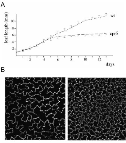
Figure 9: Cell proliferation in cpr5.
(A)Leaf length measurements of leaf number 5 at daily intervals. The standard deviation is shown for wild- type toward the top and for the cpr5 mutant toward the bottom.
(B) Comparison of wild-type (left) and cpr5 mutant (right) pavement cells at the same magnification.
[Figure adopted from Kirik et al.
2001]
B A
Morphogenesis Results
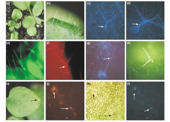
Figure 10: Trichome and lesion phenotype in cpr5 mutants.
(a) A mature wild-type (right) and cpr5 mutant plant (left).
(b) cpr5 mutant trichomes on rosette leaves.
(c) DAPI-stained cpr5 mutant trichome; the nucleus is marked with an arrow.
(d) DAPI-stained mature wild-type trichome cell; the nucleus is marked with an arrow.
(e) Mature FDA-stained cpr5 mutant trichomes; note that the cytoplasmic region around the nucleus and the thin lining of the cytoplasm is stained, indicating that the cell is alive.
(f) Propidium iodide-stained cpr5 mutant cell; staining indicates that membranes are not intact anymore and that the cell is dead. Note that the nucleus still has a normal morphology (arrow).
(g) DAPI-stained cpr5 mutant trichome cell, the nucleus is drastically reduced in size and is condensed (arrow).
(h) Collapsed trichome cell.
(i) Lesions on rosette leaves of cpr5 mutants (arrow).
(j) Propidium iodide-stained leaf tissue; note that single cells have started to die (arrow).
(k) Light micrograph of the same leaf area as in (j); note the single brownish dead cells (arrow).
(l) Same leaf area as in (j) and (k), using the UV filter set. Note that autofluorescent cells are different from the brown cells shown in (k) (arrow).
[Figure adopted from Kirik et al. 2001].
Morphogenesis Results
A 2.2.3. Cell death in the cpr5 mutant
During the course of experiments, I noticed that cpr5- T1 mutant trichomes on mature leaves eventually died and collapsed (Fig. 10h). Fluorescein diacetate (FDA), which labels only living cells, was found to stain mature cpr5-T1 mutant trichomes (Fig. 10e), indicating that these trichomes had completed their normal differentiation program before cell death occurred. On 4- week-old plants, trichomes began to die, as indicated by propidium iodide staining (Fig. 10f).
Initially, the nucleus appeared to be normal. However, eventually trichomes were found containing extremely small and condensed nuclei, indicating that the nucleus disintegrated (Fig.
10g). On 6- to 7-week-old plants, all trichome cells were collapsed, leaving the cell wall remnants behind (Fig. 10h).
Cell death was also observed in other cell types. On 6- to 7-week-old plants, single cells (Fig.
10k) or small leaf areas (Fig. 11i) were found to form lesions containing brownish cells (Figure 10i & 10k). In propidium iodide-stained leaves, single cells or cell groups were stained, indicating that cell death had occurred. Frequently, cells displaying a high autofluorescence were found (Fig. 10j, 10l), which is indicative of the production of high amounts of phenolic compounds and often correlates with cell death.
A 2.2.4. Complementation of the cpr5 mutant
The cpr5-T1 and cpr5-T2 mutants were isolated from screens of two different Arabidopsis T- DNA insertion mutant populations by Viktor Kirik. The genomic DNA sequences flanking the T- DNA insertions mapped to the P1 clone MXK3 on chromosome V (Kirik et al. 2001). According to the annotated sequence information, both T-DNA insertions are located in the first intron of the CPR5 gene and most likely result in a complete loss of gene function (Kirik et al. 2001). In order to prove the correct identity of the gene, I used a 6340-bp BamHI fragment containing 3.407 bp 5’ and 500 bp 3’ of the annotated gene for rescue. A total of 16 transgenic lines were recovered, which all showed complete rescue of the cpr5-T1 mutant phenotype.
Sequence analysis revealed that the CPR5 gene encodes a novel putative transmembrane protein containing five putative transmembrane helices at the C terminus. CPR5 is predicted to be a Type IIIa membrane protein, with the N terminus being cytoplasmatic (PSORT). In addition, a bipartite NLS is found at the N terminus at position 40–56 (PSORT) (Kirik et al. 2001).
Morphogenesis Discussion
A 3. DISCUSSION
Although the genetic and cell biological analysis of trichome branching provides a well-defined framework for the formal logic of the system, little is known about the underlying molecular mechanisms. The molecular analysis of several branching mutants revealed links to the control of the microtubule function at different regulation levels, including the microtubule-based transport processes (Oppenheimer et al. 1997), the regulation of microtubule assembly and disassembly (Burk et al. 2001, Webb et al. 2002), and the control of microtubule organization (Traas et al.
1995, Camilleri et al. 2002, Folkers et al. 2002, Kim et al. 2002, Qiu et al. 2002). Although these findings provide an excellent entry point into the understanding of the final steps of cell morphogenesis, earlier steps such as the control of branch initiation and its spatial control remain misunderstood.
The trichome phenotype of the sti and the cpr5 mutants are both similar in the reduction of branches. The elucidation of the protein structure of STI suggested a function of the gene in the context of DNA replication that is also affected in the cpr5 mutant. However further analysis of the 35S::STI and the sti mutant lines revealed no direct function of STI in this context. In addition the localisation of GFP-STI does not hint towards a nuclear function of STI although the protein contains three nuclear localisation signals.
On the other side the cpr5 mutant was shown to be pleiotropic and has initially been identified as a mutant showing constitutive pathogen response (and was named accordingly). Therefore both mutants do not provide evidence for acting at a common pathway regulating trichome morphogenesis and will therefore be discussed separately.
A 3.1. Analysis of STI
A 3.1.1. The role of STI in cell morphogenesis
Three lines of evidence suggest that STI regulates branching in a dosage-dependent manner. First, mutations in the STI gene do not simply eliminate its function, but depending on the severity of
Morphogenesis Discussion
the defect, intermediate phenotypic defects are also observed. This suggests that less STI activity results in fewer branches. Second, conversely, lines overexpressing STI can trigger extra branch formation. A third line of evidence supporting a regulatory role of STI in branch formation comes from the genetic analysis of double mutants. In a previous study, the finding that sti mutants can be rescued in double mutants with nok but not with try has led to the assumption that STI and NOK might specifically counteract each other (Folkers et al. 1997). However, the findings that weak sti alleles can also be rescued by try and that the additional removal of TRY in a sti nok background results in an even better rescue suggests that mutations in the sti gene can be bypassed in several ways (Ilgenfritz et al. 2003). This suggests that STI is not required to make branches, but involved in the regulation of their number.
The cell biological analysis of sti mutants revealed no deviation from wild type at the subcellular level (Ilgenfritz et al. 2003). One criterion to monitor cell differentiation is the timing and extent of cell vacuolization. Reduced vacuolization was found to be associated with severe growth abnormalities of root hairs in rhd3 mutants (Galway et al. 1997). Vacuolization in sti mutant trichomes, however, was normal (Birgit Schwab & Martin Hülskamp, unpublished result). A second important aspect is the organization and function of the actin and microtubule cytoskeleton. The general organization of both cytoskeletal elements was normal, suggesting that STI is not involved in the control of the microtubule or actin organization (Ilgenfritz et al. 2003).
A 3.1.2. Potential molecular function of STI
The sequence similarities of STI to other proteins provide few clues about its molecular function.
The presence of NLS domains and a DNA-polymerase III γ-subunit/RFC domain suggest that STI might be involved in the regulation of DNA replication. This, however, seems to be unlikely because the ploidy level in trichomes is normal in sti mutants and in lines overexpressing STI, indicating that replication is not affected in both situations. Consistent with this interpretation is the finding that STI belongs to a group of five genes that is clearly distinct from the putative Arabidopsis RFC genes. These five genes also show sequence similarity outside the DNApolymerase III γ-subunit/RFC domain, suggesting that they may have adopted a new plant- specific role.
The analysis of the GFP-STI fusion leads towards such a potential role of STI during trichome