1
The role of PATJ in the development of Drosophila melanogaster
DISSERTATION ZUR ERLANGUNG DES DOKTORGRADES DER NATURWISSENSCHAFTEN (DR. RER. NAT.) DER
FAKULTÄT FÜR BIOLOGIE UND VORKLINISCHE MEDIZIN DER UNIVERSITÄT REGENSBURG
vorgelegt von Arnab Sen
aus
Kolkata, India
im Jahr
2014
2 Das Promotionsgesuch wurde eingereicht am:
27.05.2014
Die Arbeit wurde angeleitet von:
Junior Prof. Dr. Dr. Michael Krahn
Unterschrift:
3
Acknowledgements
I would like to thank first of all Prof. Michael Krahn for giving me this wonderful opportunity to do my research work in his laboratory and to mention further all the help that he have offered during the last three years which not only helps me to finish on a successful note but also helps me to gain a lot of experience in the process. My sincere thanks also go to Prof.
Frank Sprenger and Prof. Eugen Kerkhof for their mentoring and providing important inputs in the research work.
I thank all the members and colleagues of Prof. Andreas Wodarz in Göttingen for a successful and wonderful start to my doctoral studies. Without their help it would have been tougher to have a great start and on the later days I would like to extend my thanks to all colleagues of Prof. Ralph Witzgall in Regensburg for providing nice facilities and environment in the department.
My gratitude also goes towards my own laboratory colleagues namely Florian, Gudrun, Christian, Laura, Giada, Sabine, Thomas, Maria and Olga for their continuous support in all aspects of work in the laboratory. I like to thank them for all the nice times spent together in academics and also non-academic times outside the lab in small gatherings. It was absolute fun to have you all around all throughout these years.
Lastly but not least my sincere thanks are bestowed towards my parents without whom nothing would have been possible. They have given a constant courage and inspiration for all the hard times and supported me through thick and thin.
4
Table of contents
1. SUMMARY 5
2. INTRODUCTION 2.1 Cell Polarity 6
2.2 Epithelial cell polarity in vertebrates and Drosophila 8
2.3 The Crumbs complex 10
2.4 PATJ 11
2.5 Actin-Myosin Cytoskeleton 14
2.6 Myosin-II 15
2.7 The PAR-complex 17
2.8 PAR-6 17
2.9 Research objectives 19
3. RESULTS 21
3.1 PATJ localization and function in Drosophila is regulated by two distinct apical complexes 22
3.2 Drosophila PATJ supports adherens junction stability by modulating Myosin Light Chain activity 48
3.3 PAR-6 regulates apical-basal polarity in epithelia by preventing degradation of Sdt/Pals1 95
4. DISCUSSION 107
4.1 Upstream regulation mechanisms of PATJ 107
4.2 The role of PAR-6 in PATJ localization and stabilization of the Crb complex 111
4.3 PATJ and its role in cell polarity and beyond 112
5. REFERENCES 116
6. APPENDIX 125
6.1 Abbreviations 125
5
Summary
In the due course of the formation of apical-basal polarity the transmembrane protein Crumbs (Crb) and its intracellular adaptor protein Pals1 (Protein associated with Lin seven 1, Stardust, Sdt in Drosophila) have been found to play key roles in the establishment and maintenance of cell polarity in various types of tissues. Research in Drosophila revealed that PATJ (Pals1 associated tight junction protein) which have been reported to be a part of the trimeric complex with Crb-Sdt localizes at the apical cell-cell contacts and plays roles in the formation of the tight junction and cell migration in mammalian cells. However it is not yet fully understood how PATJ has been localized to the apical cell junctions and its role in the regulation and maintenance of cell polarity.
In this vivid study in elucidating functional significance of PATJ, a systematic structural- functional analysis have been carried out with deletion constructs tagged with green fluorescent protein (GFP) in transgenic flies to elucidate the roles of each conserved domain of the protein. In our study we found that the N-terminally located L27 domain along with a redundancy of the PDZ domains is required for proper functionality of the protein. Further we also found that PATJ attaches to both Baz-Sdt and Crb-Sdt complexes for its proper functionality. On our way to decipher how PATJ shuffles between these two complexes, we reveal the role of PAR6 in stabilizing Crb-Sdt complex via selective inhibition of Sdt degradation by ubiquitin mediated proteosomal pathway.
Additionally we have found that PATJ is not per se crucial for the establishment or maintenance of apical-basal polarity, but rather regulates Myosin dynamics. PATJ directly binds to the Myosin Binding Subunit of Myosin Phosphatase and decreases Myosin dephosphorylation, resulting in activated Myosin dynamics. Thereby PATJ supports the stability of the Zonula Adherens. Notably, weakening of Adherens Junction (AJ) in a PATJ- mutant epithelium leads first to a loss of Myosin from the AJ, subsequently to a disassembly of the AJ and finally to a loss of apical-basal polarity and disruption of the tissue.
6
2. Introduction 2.1. Cell Polarity
Cell polarity mainly arises from the asymmetric division of cells in respect of cell shape, protein distributions and cell functions in different tissues. Cell polarity spans its evolutionary diversity from single cell to multi-cellular organisms. It has been found to function in important biological aspects like the establishment of cell barriers, directed growth, migration of cells and so forth.
To date various forms of cell polarity have been described, such as planar cell polarity, anterio-posterior polarity, apical-basal polarity (Fig.1) and radial cell polarity. Extensive research in the past has shed light on many polarity landmarks which play an active role in the establishment of apical-basal polarity. Fortunately most of the cues are conserved to a greater extent throughout evolution in various organisms from invertebrates like the nematode Caenorhabditis elegans or the fruitfly Drosophila) to vertebrates (and mammals in particular) (Nelson, 2003).
Riding on the success of developmental biology which opens up a scope to study model organism like Drosophila melanogaster, it has been possible to study cell polarity with versatile tools enabling the opportunity to study ‘in vivo’ various mechanisms, candidate genes, proteins etc. Drosophila also provides the possibility to study several cell types which are polarized and serves as a perfect model for investigation.
Several cell types of Drosophila melanogaster have been established as model systems for in vivo studies on different aspects of cell polarity:
1. The oocyte: Unlike the C. elegans oocyte, which lacks polarity before fertilization the Drosophila oocyte is a highly polarized cell that contains a large number of localized messenger RNAs and proteins along an anterior-posterior (and dorsal-ventral) polarity
7
axis. The oocyte is surrounded by the mesodermal derived folliclular cell epithelium.
Unlike other epithelial cells the apical domain of these follicle cells is not directed towards the lumen or the outside surface; instead it forms cell contacts with the germline cells (oocyte and nurse cells).
2. The ectodermal epithelia: Ectodermal epithelia eg. the epidermis, fore- and hindgut and tracheal system emerge from primary epithelia which originate directly (without any non-epithelial intermediates) from the blastoderm.
3. The neural stem cells (neuroblasts): Drosophila neuroblasts offer a nice model for studying asymmetric cell division. Neural development which starts during stage 9 of embryogenesis also provides a distinct model for studying apical-basal polarity.
Figure 1: Various polarized cell types used as a model in Drosophila melanogaster (edited from Suzuki and Ohno 2006)
8
2.2 Epithelial cell polarity in vertebrates and Drosophila
The ectodermal epidermis serves as a good model for studying fundamental mechanisms of apical-basal polarity. The first epithelia to form in the Drosophila embryo is the blastoderm which develops from a syncytium by multiple invaginations of the plasma membrane causing the formation of the cleavage furrows, a process known as cellularization. With an increase in the surface area and orderly segregation of around 5000 nuclei, establishment of cell polarity takes place concomitantly with the growth of the polarized plasma membrane (Lecuit. et al., 2002). Previous studies on epithelia of various species have revealed many highly conserved genes which are responsible for cell polarity (Knust and Bossinger, 2002).
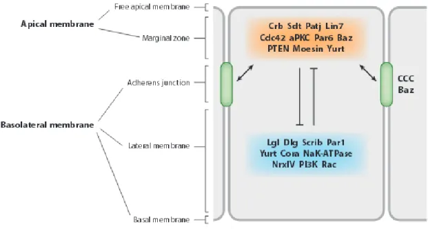
Apical-basal polarity has been mainly formed by mutual segregation of certain proteins and lipids distributed between certain distinct domains, an apical membrane domain, lateral cell contacts and a basal zone. Often the last two domains are annotated as basolateral domain.
Cell-cell and cell-matrix interactions (Wang et al., 1990; O’Brien et al., 2002) and in particular the assembly of apical and basolateral junctional complexes are prerequisites for the proper development of cell polarity: First epithelial cells have an adhesive belt encircling the cell just below the apical domain known as zonula adherens (ZA).
In Drosophila and vertebrate epithelial cells the transmembrane protein E-Cadherin (and other proteins belonging to the same family) binds directly to β-catenin which in turn recruits α- catenin which forms the pre-requisite for the linkage to the actin cytoskeleton, partly directly and partly via actin binding proteins like vinculin or α-actinin (Nelson, 2008; Perez-Moreno et al., 2003). However further studies have shown that linkage of actin cytoskeleton to the cadherin-catenin complex is more complicated than a simple interaction of the proteins (Weis et al., 2006).
Second the boundary between the apical and lateral domains is marked by the tight junctions (TJ), which contain a number of homophilic adhesion molecules, such as Occludin, Junctional
9
Adhesion Molecules (JAMs), and the Claudins, which create the paracellular barrier and an intramembranous diffusion barrier between apical and basolateral transmembrane proteins (Johnston et al., 2010) (Fig.2). Apart from the junctional proteins some other transmembrane and cytoplasmic proteins also accumulate near the TJ, namely the PAR/aPKC complex (PAR- 3, aPKC, PAR-6) and the Crumbs complex Crumbs (Crb) / PALS1 (protein associated with Lin7) / PATJ (PALS1-associated TJ protein). These apical protein complexes are mutually excluded and controlled by a basolateral Discs Large (Dlg) / Scribble (Scrb) / Lethal (2) giant larvae (Lgl) complex.
Figure 2: Intercellular Junctions in Epithelial Cells of Drosophila and vertebrates (from Daniel St Johnstonand Julie Ahringer 2010)
In Drosophila epithelia components of the AJ are highly conserved with respect to vertebrate system but differ in the arrangement of lateral junctions. As Drosophila epithelia do not express Occludins they do not form real TJ, instead they develop a distinct region apical to the ZA, known as sub-apical region (SAR), that localize at a region homologues to the TJ in vertebrate cells (Knust and Bossinger, 2002). The mentioned PAR complex is localized to the ZA and SAR while the Crb complex is located slightly apical to the PAR complex in the SAR. The basolateral proteins like Dlg, Scrib and Lgl are on the other hand localized to a special junction called Septate Junction (SJ). SJs are specific to the invertebrate system but
10
they do posses analogy to the vertebrate TJs as many TJ specific protein homologues like Claudins [Sinuous (Sinu), Megatrachea (Mega) and Kune-kune (Kune)], the Na+/K+-ATPase, Nrg (Neuroglian), Neurexin-IV (Nrx-IV), Contactin (Cont), Lachesin (Lac), Gliotactin (Gli) and two intracellular components,Coracle (Cora) and Varicose (Vari) are localized to SJ. TJ and SJ also share similar functions in restriction of paracellular molecular transport across epithelial sheets and helping to polarize plasma membrane and the underlying cytoskeletal elements along apical-basal axis (Tepass et al., 2001; Furuse and Tsukita, 2006; Cereijido et al., 2008; Miyoshi and Takai, 2008) (Fig.3).
Figure 3. Localization of protein complexes in the Drosophila epithelium (from Tepass 2012)
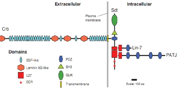
2.3 The Crumbs complex
The Crumbs complex is comprised of four core proteins: Crb, its intracellular adaptor protein Pals1, PATJ and Lin7 (Fig.4). Crb and Sdt are highly conserved from fly to men and are known to play key roles in the maintenance of apical-basal polarity and integrity in Drosophila epithelia and photoreceptor cells as well as in vertebrate epithelia and polarized
11
cells of the retina (Tepass and Knust, 1993; Tepass et al., 1990; Bulgakova and Knust 2009).
The formation of the Crb complex is achieved via physical interaction of the PDZ (Psd95, Disc large, ZO-1) domain of Sdt and the C-terminal ERLI motif of Crb. (Bachmann et al., 2001; Hong et al., 2001). The two L27 domains of Sdt bind to the L27 domains of PATJ and Lin-7 (Bachmann et al., 2004; Bulgakova et al., 2008; Roh et al., 2002). This complex interestingly has an asymmetric localization in the apical cortex in the SAR, just above the ZA, irrespective of species or cell type (Berger et al., 2007; Johnson et al., 2002; Pellikka et al., 2002; Richard et al., 2006a; Tepass, 1996).
Figure 4. Schematic diagram of the core proteins of the Drosophila Crumbs complex (from Bulgakova and Knust 2009)
2.4 PATJ
The PATJ gene has been first identified by Bhaat et al. in a yeast two-hybrid screen for binding partner of Nrx-IV, a component of the SJs in the Drosophila embryo. They mistakenly thought to have a PATJ mutant and annotated it as disc lost (Dlt) (Bhaat et al,.
1999). Later on it has become evident that Dlt is a neighbouring gene which is unrelated to
12
PATJ (Pielage et al., 2003). Drosophila PATJ contains four PDZ domains and a single L27 (Lin-2, Lin-7) domain at the N-terminus (Pielage et al., 2003) (Fig.5). Studies have reported that PATJ forms the third member of the Crumbs complex by binding to the N-terminal L27 domain of Sdt via its own L27 domain in Drosophila embryos (Klebes and Knust, 2000; Roh et al., 2002b) and also in adult flies (Pellikka et al., 2002). In contrast, its mammalian homologue consists of 10 PDZ domains. Another multiple PDZ domain containing protein MUPP1 (13 PDZ domains) is also referred to have close similarity with mammalian PATJ with partly overlapping functions of regulating TJs (Adachi et al., 2009), including binding to Claudins and JAM (Hamazaki et al. 2002, Poliak et al. 2002) (Fig.5).
Figure 5. Alignment of different PATJ homologues in vertebrate and invertebrate system (edited from Roh et al., 2002)
Of mammalian PATJ’s ten PDZ domains, binding partners have been identified for only two.
PATJ interacts with ZO-3 via its sixth PDZ domain and with Claudin-1 via its eighth PDZ domain. (Lemmers et al. 2002, Roh et al. 2002a,b). PATJ also have been reported to play a role in development of mammalian cell polarity in MDCKII (Madine-Darby canine kidney) cells (Shin et al., 2005). Suppression of PATJ expression in Caco- 2 (human epithelial colorectal adenocarcinoma) cells resulted in decreased stability of the CRB3 complex and localization of CRB3 to the intracellular compartment (Michel et al. 2005). Furthermore, PATJ is required for the formation of TJ (Latorre et al. 2005). Similar effects were observed overexpressing a dominant-negative version of PATJ in MDCK cells (Hurd et al., 2003).
Recent research sheds light on the role of PATJ in apical constriction of epithelial cells via AJ
13
associated Acto-Myosin belt by modulating direct or indirect recruitment of small GTPase RhoGEFp114 to the apical junction (Nakajima and Tanoue, 2011). In turn Lulu2 (the mammalian homologue of Drosophila Yurt) interacts with and activates RhoGEFp114 in regulation of the circumferential Acto-Myosin belt. RNAi knockdown of either PATJ or Lulu2 results in loss of the Acto-Myosin belt and consequently apical constriction (Nakajima and Tanoue, 2012). It has been also shown that PATJ plays significant role in cell migration by wound healing assays (Shin et al., 2007). This observation was supported by the findings of PATJ acting as a scaffold for Angiomotin and the RhoGEF Syx in migrating endothelial cells (Ernkvist et al., 2009).
In Drosophila the role of PATJ has been obscurely described in various studies. In follicular epithelium PATJ forms a complex with Crb and have been postulated to regulate the formation of follicle cell epithelium by stabilizing Crb in this cell type (Tanentzapf et al., 2000). In adult Drosophila eyes PATJ has been shown to be necessary to stabilize the Crumbs complex at the stalk membrane of photoreceptor cells (Richard et al., 2006). PATJ also have been shown to affect planar cell polarity (PCP) through interaction of one of the key players Frizzled (Djiane et al., 2005). During cellularization in early embryonic stage PATJ is associated with the leading edge of the invaginating membrane, although a role for this localization has not yet been established.
Due to a lack of a clean PATJ mutant, further investigation of the role of this gene in cell polarity and development of Drosophila was not possible. Discrepancies in the functions described for PATJ before could have risen from the use of artificial construct, like the N- terminal of PATJ to rescue deletion mutants (Nam and Choi, 2006; Pielage et al., 2003). In other reports RNAi mediated down regulation of PATJ might have resulted in off targets and dose dependent effects (Nam and Choi, 2006). Although PATJ have been studied to certain extent in Drosophila photoreceptor cell epidermis in the eye, the role of Crb seems to differ than in other polarized tissues like embryonic epidermis or follicle cell epithelium (reviewed
14
by Bulgakova and Knust, 2009). However it is likely that PATJ may function differently in different tissue types, a matter that needs to be elucidated.
2.5 Actin-myosin cytoskeleton
The cell cytoskeleton consists of a scaffold embedded in the external environment of cell cytoplasm. Actin polymers mainly comprises of the cytoskeleton along with other molecular motors like myosin and accessory proteins which initiate actin polymerization, control growth of actin filaments and protein turnover. Actin and myosin first discovered in the muscles makes up for half of the total protein content of the cytoskeleton. Under physiological conditions actin monomers polymerize in a spontaneous manner into polar long stable filaments where one end of the filament grows faster than the other (Pollard 2007). Through a cascade of hydrolysis of Adenosine triphosphate (ATP) to Adenosine diphosphate (ADP) the mobility of the actin filaments have been maintained. Polymerization of actin filaments give rise to certain physiological cell activities like establishing and maintaining cell morphology, cell motility, cell division and intracellular transport (Pollard and Cooper, 2009).
Interaction of actin filaments with myosin motors results in production of a force which helps the actin filaments to contract forming a cleavage producing subsequent cell division and formation of tissue architecture. On the other hand myosin motors also helps to move cargos like macromolecular complexes of RNA and proteins along actin filaments. Different types of myosin motors have been reported so far. Among them the most described ones are Myosin I, Myosin II and Myosin V. Myosin I binds to the Arp2/3 complex which helps in nucleation of actin filaments and subsequently helps in the process of endocytosis, while Myosin II (also known as non-muscle myosin) polymerizes into bipolar filaments, which can produce a contraction by pulling actin filaments together influenced by RhoGTPase (Miller at al., 2009).
15
2.6 Myosin II
Myosin II is one of the several identified motor proteins which binds to Actin filaments and control mechanistic regulation of cell migration and movement. Myosin structurally consists of six different parts: two heavy chains known as Myosin Heavy Chain (MHC) each of which contains a head domain and long coiled-coiled domains; two regulatory light chains (MRLC);
two light chains known as Myosin Light Chain (MLC) which separates the head and the coiled-coiled domains (Mooseker MS et al., 1995, Foth BJ 2007). Stabilization of this hexameric structure comes through dimerization of the coiled-coiled domains. The globular head domain of MHC has an ATPase activity, whereupon ATP hydrolysis catalysed by the enzyme induces conformational change and contractility (Spudich JA 2001). Generation of contractile forces for Actin filament crosslinking requires activation of Myosin II which is achieved via phosphorylation of two conserved amino acid residues at Threonine 18 and Serine 19 of the regulatory light chain. Although Myosin Regulatory Light Chain Kinase (MRLCK) has been the principle kinase phosphorylating MRLC, other kinases like ROCK-I, ROCK-II, MRCK, PAK kinases, and citron kinase, also phosphorylate it (Aguilar-Cuenca R et al., 2014). On the other hand a trimeric complex consisting of Myosin phosphatase, a class 1 protein phosphatase (PP1cδ), a protein of unknown function and the Myosin-Binding Subunit (MBS), dephosphorylates and thereby inactivates Myosin (Matsumura and Hartshorne, 2008). Vice versa Myosin phosphatase is inactivated via phosphorylation of MBS by ROCK-I (Kawano et al., 1999).
In Drosophila the hexameric Myosin II complex is highly conserved. The MHC protein is encoded by the zipper (zip) gene while the MRLC is encoded by spaghetti squash (sqh).
Previous studies have reported that zip deficient embryos have morphogenetic defects like impaired dorsal closure, head involution, and axon patterning (Young et al., 1993), while
16
expression of a tagged version of the zip protein in amneoserosa cells can restore the cortical localization (Franke et al., 2005). The process could possible mediated by cell-cell adhesion to reorganize actin along with morphogenetic forces.
Drosophila development has been associated with the activity of actin-myosin dynamics.
Actin plays an important role in co-coordinating several events in reorganization of the cytoskeleton as the embryo starts its developmental cycle. Actin filaments condense above the nuclei at early stages but with start of the process of cellularization they are remodeled more towards the invaginating furrow where myosin II interacts with Actin filaments to form a contractile apparatus, inducing the polarized blastoderm epithelium (Warn et al., 1980 and Miller KG, Kiehart DP, 1995).
The role of Myosin II has been further implicated in the Germ Band Extension (GBE), one of the morphogenetic movements in the embryonic development. Zip and Sqh are reported to co- localize with the β-catenin (Armadilo in Drosophila)/E-cadherin complex during GBE in intercalating cells where the contractile acto-myosin force might regulate remodeling E- Cadherin based cell-cell contacts (Lecuit et al., 2002; Bertet et al,. 2004; Zallen and Wieschaus, 2004). On another occasion Myosin II, reported to be localized at the leading edge of the lateral epidermis during the onset of dorsal closure in late embryogenesis (Young et al., 1993).
Along with cell polarity regulators Myosin-II have been also known to regulate AJ. Rho- dependent activation of Myosin via Rok is crucial for the accumulation of E-Cadherin at cell- cell contacts thereby stabilize the AJ (Ivanov et al., 2007; Shewan et al., 2005; Yamada and Nelson, 2007). In mammalian epithelial cells as well as in the Drosophila epidermis, Myosin- II accumulates at the AJ (Ivanov et al., 2007; Krendel and Bonder, 1999; Shewan et al., 2005;
Yamada and Nelson, 2007; Sen et al., 2012), however, activated phosphomyosin-II (measured by its phosphorylation) might not occur at all AJ but mainly at newly established ones (Yamada and Nelson, 2007).
17
2.7 The PAR complex
One of the important regulators of apical-basal polarity is the PAR-aPKC (partitioning defective– atypical protein kinase) complex. Along with the Crb complex it forms the apical domain in establishing polarity in different polarized cell types. It is highly conserved throughout evolution from worm to man (Suzuki and Ohno, 2006). The PAR complex consists of three core proteins namely the scaffolding protein PAR-3 (Bazooka (Baz) in Drosophila), PAR-6 and the serine-threonine kinase aPKC. Except the core components of the PAR complex, a small GTPase Cdc42 also has been reported to indirectly bind to the PAR complex. On the onset of epithelial polarization, PAR-3 associates itself with the PAR- 6/aPKC hetero-dimer via PDZ domain interactions (Lin et al., 2000; Suzuki et al., 2001;
Joberty et al., 2000). PAR-6 interacts with aPKC via the PB1 domains of both the proteins while the semi-CRIB domain of PAR-6 associates with Cdc42. Upon binding of Cdc42, aPKC is activated and phosphorylates PAR-3 which leads to the release of the PAR-6/aPKC complex from PAR-3 (Horikoshi et al., 2009). In Drosophila, the PDZ domain of PAR-6 was shown to bind to Crb resulting in the release of Baz from the trimeric complex (Lemmers et al., 2004).
2.8 PAR-6
PAR-6, a core member of the PAR/aPKC complex is known to bind to aPKC and to PAR-3 as PAR-6/aPKC heterodimer. PAR-6 consists of three distinct domains: the PDZ binding domain by which it interacts with PAR-3/Baz, the PB1 binding domain interacting with aPKC and semi CRIB Drosophila domain which associates with Cdc42. The binding of PAR-6 to aPKC modulates its kinase activity and thereby regulates cell polarity in various tissues
18
(Suzuki and Ohno, 2006). PAR-6 has been found to interact with both the known apical complexes: the PAR/aPKC and the Crb complex. The PDZ domain of PAR-6 binds to the N- terminal region of Sdt/Pals1 or the C-terminus of Crb/CRB3 ( Hurd et al., 2003; Lemmers et al., 2004; Wang et al., 2004). On the other hand the N-terminus of PAR-6 interacts with the third PDZ domain of Drosophila PATJ (Nam and Choi, 2003). Thus PAR-6 mediates interplay between the two known protein complexes localized to the apical domain of a polarized cell. In Drosophila epithelial cells Crb is required for the apical localization of PAR-6 (Kempkens et al., 2006), while in case of mammalian epithelial cells the dominant homologue of Crb, CRB3 is able to recruit PAR-6 in unpolarized cells (Hurd et al., 2003).
New study reported of a WD40 protein Morg1 (mitogen-activated protein kinase organizer 1) to be a potential interaction partner of PAR-6 and also CRB3 simultaneously thereby regulating the translocation of PAR-6/aPKC to the apical junctions in MDCK (Madin-Darby- canine-kidney) cells (Hayase et al., 2013). In a separate study PAR-6 phosphorylation by aPKC induces epithelial to mesenchymal transition (EMT), a canonical pathway to tumorigenesis. (Gunaratne A, Guglielmo GM, 2013).
19
2.9 Research Objectives
Over the last decade research in cell polarity have spanned in various domains of cell function. Among them apical-basal polarity is worth mentioning as different apical and basal cues have been explored regulating the establishment and maintenance of the polarity in polarized epithelial cells. In the apical domain Crb complex forms an important cluster.
Although many studies have been reported on two members of the Crb complex, Crb and Sdt, little was known with contradictory results about the third member of the complex, PATJ.
Hence it is necessary to clarify the roles of PATJ in context of cell polarity and probable other functions.
In order to achieve this aim, a null mutant of PATJ has been created in Drosophila where the whole open reading frame of the gene has been deleted. Immuno-localization studies on embryonic epithelium and follicle cell epithelium in ovaries have shown that PATJ is not crucial for the establishment or stability of apical-basal polarity. Instead it plays a role in modulating Myosin-II dynamics by regulating Myosin-II phosphorylation and by direct binding to its regulatory light chain.
Secondly we analyzed the function of different conserved domains of PATJ. Here we found that the L27 domain is most important to the protein’s function and the PDZ domains act in redundancy along with the L27 domain. Further studies also reveals that PATJ, as known before a part of the Crb complex can also form a complex with Baz and Sdt by which it gets translocated to the apical junctions. Additionally through expression of chimeric proteins we show that binding of PATJ to both Baz-Sdt and Crb-Sdt complex is necessary in embryonic epithelium.
Finally on deciphering the mechanism of how PATJ shuffles between the two complexes, PAR-6 has been found to play a central role. In oppose to the known facts that PAR-6 binds directly to Crb and Sdt ( Hurd et al., 2003; Lemmers et al., 2004; Wang et al., 2004), we
20
found that PAR-6 stabilizes the Crb-Sdt complex through the selective inhibition of degradation of Sdt via proteosomal linkage. PAR-6 has been found before to interact with the proteosomal receptor Rpn13 and we found that downregulation of Rpn13 or core components of the proteasomal pathway in PAR-6-mutant cells rescues Sdt degradation and localization..
21
3. Results
Every chapter of the results with a short description of:
the main aim of the particular manuscript in context of the complete thesis
the authors and their contribution to the work
the status of the manuscript
22
3.1 PATJ localization and function in Drosophila is regulated by two distinct apical complexes
This project aims mainly at the structural-functional analysis of the multiple PDZ containing protein PATJ. With the use of ubiquitin promoter to express proteins close to the endogenous levels, various deletion constructs of the protein have been studied in the context of the localization pattern and functionality of the truncated proteins in rescuing the PATJ-null mutant allele. Further to elucidate the upstream regulators which are responsible for proper localization of PATJ, rescue experiments are performed with chimeric PATJ, able to bind Baz and Crb at the same time. The interaction of PATJ with Baz and Crb has been shown through biochemical assays.
Arnab Sen and Michael P. Krahn Author contribution to work:
Arnab Sen: All experiments and partly writing of the manuscript Michael P. Krahn: Editing of the manuscript
Status: In revision at Molecular Biology of Cell (MBoC)
.
23
PATJ localization and function in Drosophila is regulated by two distinct apical complexes
Arnab Sen* and Michael P. Krahn*†
*Molecular and Cellular Anatomy, University of Regensburg, Universitätsstr. 31, 93053 Regensburg, Germany
†author for correspondence: Michael.Krahn@vkl.Uni-Regensburg.de, phone: +49-941- 9432879, fax: +49-941-9432868
Running title: Upstream regulators of PATJ
Abbreviations List
AJ, Adherens Junctions; aPKC, atypical protein kinase C; Baz, Bazooka; CR, conserved region; Crb, Crumbs; DE-Cad, Drosophila E-cadherin; Dlg, Discs Large; PATJ, Pals1- associated tight junction protein; Sdt, Stardust; TJ, Tight Junctions; Yrt, Yurt.
24 Abstract
The transmembrane protein Crumbs (Crb) and its intracellular adaptor protein Pals1 (Stardust, Sdt in Drosophila) play a crucial role in the establishment and maintenance of apical-basal polarity in epithelial cells in various organisms. In contrast the multiple-PDZ-domain containing protein PATJ, which has been described to form a complex with Crb/Sdt, is not essential for apical basal polarity or for the stability of the Crb/Sdt complex in the Drosophila epidermis. Here we show that Sdt is essential for the correct subcellular localization of PATJ in maturing epithelial cells but not during cellularization. Consistently the L27-domain of PATJ is crucial for the correct localization and function of the protein. We further demonstrate that the four PDZ domains of PATJ function to a far extent in redundancy regulating the protein’s function.
Interestingly the PATJ-Sdt heterodimer is not recruited to the apical cell-cell contacts by binding to Crb but depends on functional Bazooka (Baz). Using chimeric proteins we demonstrate that the association of PATJ with both complexes, the Baz-Sdt and the Crb-Sdt complex, is crucial for PATJ’s function during development of Drosophila.
Highlight summary
The conserved multiple PDZ-domain containing protein PATJ is recruited to the apical cell- cell contacts by the cell polarity regulators Crumbs and Bazooka. Indirect binding to both proteins via the adaptor molecule Stardust is necessary to accomplish PATJ’s function during development of Drosophila.
25 Introduction
Apical-basal polarization of epithelia is regulated by conserved complexes determining the apical versus the basolateral domain (Tepass, 2012; Roignot et al., 2013): At the apical tip of the lateral plasmamembrane, the PAR(partitioning-defective)-aPKC(atypical protein kinase C)-complex regulates assembly of the Crumbs(Crb)-complex, whereas the activity of these two complexes is counterbalanced by Scribble-Lethal(2) Giant Larvae-Discs Large(Dlg) which localize to the basolateral domain. Recently, various studies have demonstrated that both apical complexes are rather dynamic and that their composition might be tissue- dependent and temporally and/or developmentally regulated (Hurd et al., 2003; Nam and Choi, 2003; Penkert et al., 2004; Sotillos et al., 2004; Wang et al., 2004; Kempkens et al., 2006; Krahn et al., 2010a).
In Drosophila, the multiple PDZ-domain containing protein PATJ has been described to function in a complex with Crb and Stardust (Sdt, the Drosophila homologue of Partner of Lin-7 one, Pals1) to regulate apical-basal polarity in follicle epithelial cells and photoreceptor cells (Tanentzapf et al., 2000; Nam and Choi, 2006; Richard et al., 2006). Recently, we and others reported that loss of PATJ in Drosophila epithelia does not affect apical-basal polarity in the embryonic epidermis or in follicle epithelial cells but rather modulates Myosin activity to support Adherens Junction (AJ) stability (Penalva and Mirouse, 2012; Sen et al., 2012;
Zhou and Hong, 2012). Only in photoreceptor cells and to some extent in the follicular epithelium, PATJ seems to be essential for the correct subcellular localization of the Crb-Sdt complex, either by directly stabilizing this complex or indirectly by regulating photoreceptor morphology/development (Sen et al., 2012; Zhou and Hong, 2012).
Two mammalian orthologues of PATJ are expressed in epithelia: mammalian PATJ (mPATJ, encoded by INADL in mice) and Multiple PDZ-domain protein 1 (MUPP1). Both proteins are very similar to DmPATJ: Beside an N-terminal L27 domain they exhibit several PDZ domains (DmPATJ four, mPATJ ten, MUPP1 thirteen) and localize to the Tight Junctions (TJ) in mammalian epithelial cells (Adachi et al., 2009). However, Abachi et al. showed that
26
despite its domain similarity, mPATJ but not MUPP1 regulates TJ stability (Adachi et al., 2009). These data are in line with previous findings describing TJ-formation delay or defects upon loss of mPATJ in cultured epithelial cells (Michel et al., 2005; Shin et al., 2005). Other studies describe a role of mPATJ in Myosin-driven processes like apical constriction and cell migration (Shin et al., 2007; Ernkvist et al., 2009; Nakajima and Tanoue, 2011).
In this study we report that in the embryonic epidermis of Drosophila PATJ can be found in the described Crb-Sdt complex but additionally associates with the Baz-Sdt-complex we described previously (Krahn et al., 2010a). Notably deletion of Baz and Sdt but not of Crb leads to mislocalization of junctional PATJ during gastrulation and in mature epithelia of the embryonic epidermis. In contrast, localization of PATJ at the tip of the invaginating plasmamembrane during cellularization is independent of Baz/Sdt. Consequently, deletion of the L27-domain of PATJ leads to an abolished junctional accumulation and impaired function of the protein. Studies with chimeric proteins further suggest that binding to the Baz-(Sdt) complex as well as to the Crb-(Sdt) complex are inevitable for PATJ’s function. Finally we investigated the functionality of PATJ’s four PDZ domains and demonstrate that under close to endogenous expression levels, these domains function partly in redundancy.
27 Results and Discussion
PATJ is recruited by Sdt to a complex with Baz at the apical junctions in the embryonic epidermis
Upon the formation of apical AJ in late cellularization/early gastrulation in Drosophila, PATJ is recruited to the apical cell-cell contact region whereas staining at the basal membrane ceases (Sen et al., 2012). Studies in Drosophila and cultured mammalian epithelial cells proposed that PATJ associates with Sdt/Pals1 which in turn binds to the transmembrane protein Crb which targets the complex at the TJ in vertebrates and in the corresponding
“subapical region” in Drosophila (Klebes and Knust, 2000; Roh et al., 2002).
We recently found that in the embryonic epidermis of Drosophila Sdt is initially localized to the apical junctions in early gastrulation before Crb is expressed and even remains at the junctional region of mature epithelial cells when Crb is absent (Krahn et al., 2010a). This is accomplished by a direct interaction of the PDZ-domain of Sdt with Baz. Upon phosphorylation of Baz by aPKC at Serine 980, Sdt is released from Baz and available to stabilize the Crb complex (Krahn et al., 2010a). We therefore tested whether the subcellular localization of PATJ is dependent on Crb or Baz or both. In crb-mutant embryos, PATJ shows a normal localization not only during cellularization (data not shown) but also after gastrulation as long as apical-basal polarity is still intact (stage 6-9, Fig. 1A, B). Only in later stages (from stage 10/11 on), apical-basal polarity is impaired upon loss of Crb, finally resulting in a multilayered epithelium. Here, PATJ is cytoplasmic or in aggregates (Fig. 1C).
Notably, loss of cortical PATJ in these embryos is accompanied by a loss of membrane- associated Baz (Fig. 1C).
In contrast, in maternal and zygotic baz mutant embryos (baz815-8 germ line clones), accumulation of PATJ at the tip of the furrow canal during plasma membrane invagination is not affected (Fig 1D) but targeting of the protein to the apical junctional region after cellularization is abolished (Fig. 1E).
28
Furthermore, we found endogenous PATJ and Sdt to coimmunoprecipitate with endogenous Baz in lysates from wild type embryos (Fig. 1G). Consequently, in embryos lacking Sdt, PATJ is correctly localized during cellularization (data not shown) but fails to relocalize to the apical AJ during gastrulation (Fig. 1F), indicating that PATJ is recruited by Sdt to the apical junctions. This is consistent with studies in cultured mammalian cells demonstrating that PATJ directly binds to Pals1 via hetero-dimerization or even hetero-oligomerization of its L27 domain with the (more N-terminal) L27 domain of Pals1 (Roh et al., 2002; Li et al., 2004; Feng et al., 2005). Beside its association with Baz-Sdt, PATJ can also be co- immunoprecipitated with Crb-GFP expressed from its endogenous promoter (Klose et al., 2013, Fig. 1H), pointing to a second complex, consisting of Crb-Sdt-PATJ, which might be formed later in development as soon as Sdt is released from Baz upon phosphorylation by aPKC (Krahn et al., 2010a). However the fact that PATJ remains correctly localized in the absence of Crb even in later stages (stage 8/9) indicates that Baz can complement Crb’s function regarding junctional targeting of Sdt/PATJ.
PATJ localization in the follicular epithelium depends on Sdt, Baz, and partly on Crb Similar to the embryonic epidermis loss of Sdt in the epithelial cells surrounding the oocyte (follicular epithelium) abolishes apical accumulation of PATJ (Fig 2A, mutant clones are marked by the absence of RFP). In baz-mutant clones, Sdt as well as PATJ are lost from the apical junctions (data not shown and Fig. 2B, mutant clones are marked by the absence of RFP and Baz staining, note that the follicular epithelium becomes partly multilayered (arrow)). Notably, in crb-defective follicle cells, apical Sdt and PATJ staining is drastically diminished but a minor fraction of the protein still accumulates apically (Fig. 2C, arrow), although it is unclear whether this is the primary consequence of loss of Crb or the result of impaired Baz localization, which is affected upon removal of Crb in the follicular epithelium, too (Fig. 2D). Thus, the follicular epithelium represents an intermediate phenotype between the epidermis (PATJ localization is only dependent on Baz-Sdt but not on Crb-Sdt) and pupal
29
photoreceptor cells, where PATJ localization depends on Crb (Richard et al., 2006). Vice versa in photoreceptor cells, PATJ seems to be crucial for the stabilization of the Crb-Sdt complex (Nam and Choi, 2006; Richard et al., 2006; Zhou and Hong, 2012), whereas this phenotype is much weaker in the follicular epithelium and not seen at all in the embryonic epidermis: In follicle epithelial cells, loss of PATJ results in decreased apical-junctional accumulation of Crb/Sdt but without subsequent disassembling of the complex and polarity defects (Penalva and Mirouse, 2012; Sen et al., 2012).
The L27 domain is essential and sufficient for apical junctional localization
To test which domains are crucial for PATJ’s correct subcellular localization and function, we generated deletion constructs of the N-terminal L27 domain and each of the PDZ domains as well as truncated versions of PATJ, all C-terminally tagged with GFP (Fig. 3A). To avoid artificially increased protein levels, we expressed the modified proteins under a ubiquitous promoter (Ubiquitin) and used the PhiC31-Integrase system (Groth et al., 2004) to generate transgenic lines with identical genomic background, ensuring comparable protein levels.
Indeed, wild-type PATJ-GFP expressed in this system is expressed at similar levels as endogenous PATJ (Fig. 3B), localizes indistinguishable from endogenous PATJ (Fig. 3C) and is capable to rescue the PATJ1 null allele (79% surviving flies, Fig. 3A).
In mammalian epithelial cells, mPATJ has been shown to be targeted by Pals1 to the TJ via a heterodimerization of their L27 domains (Roh et al., 2002; Li et al., 2004; Straight et al., 2004). Likewise, deletion of the L27 domain of Drosophila PATJ results in a cytoplasmic accumulation of the mutant protein in the embryonic epidermis as well as in follicle cells (Fig.
3D and data not shown). Consequently, the PATJL27-GFP is unable to rescue a PATJ-null allele, resulting in similar phenotypes as the null allele (PATJ1, pupal lethality).
In contrast to deletion of the L27-domain, removal of any of the four PDZ domains alone does not impair the subcellular localization of the modified protein at the apical junctions (data not shown). Furthermore, ubiquitous expression of all single deletion constructs can complement
30
for PATJ’s function and can be maintained as a stable stock with the homozygous PATJ1 allele. However, analysis of the hatching rates showed that deletion of the first PDZ-domain affects functionality of the protein far more than deletion of PDZ2, 3 or 4 (34% in comparison to 58, 55, 68%, respectively, Fig. 3A).
As a truncated version of PATJ has been reported to be capable to partly rescue a PATJ- mutant (Nam and Choi, 2006; Richard et al., 2006; Penalva and Mirouse, 2012), we determined which minimal region of PATJ is sufficient for the protein’s function: As expected, ubiquitous expression of the isolated L27 domain (PATJ1-151) shows a mostly junctional localization, although not as delimited as the wild-type protein (Fig. 3E). This protein, lacking all PDZ domains, shows no rescue capacity. Experiments with flies lacking zygotic PATJ expression and ubiquitously expressed PATJ1-240-GFP (L27 domain and the first PDZ domain) produced occasionally adult flies. However the majority of flies died during late pupal stages but in contrast to the null allele, pupae in the PATJ1-240 rescue undergo complete morphogenesis and die only shortly before hatching (or fail to hatch).
Hatched flies are sterile and died after a few days, indicating that the truncated version exhibits sufficient functionality to overcome the pupal lethality of PATJ1 but is not capable to fully replace the wild-type protein. Overexpression of the same construct using arm::GAL4 resulted in increased rescue capacity and the recued flies can be maintained as a stable stock.
Thus only artificially increased levels of the protein consisting of the L27 domain and the first PDZ domain can accomplish function of PATJ during development, which is in line with previous studies using overexpressed proteins (Nam and Choi, 2006; Penalva and Mirouse, 2012).
In contrast to PATJ1-449, a protein consisting of the first 449aa, including the L27 domain as well as the first two PDZ domains expressed close-to-endogenous levels can fully rescue the PATJ null allele and rescued flies can be kept as a stable stock. Deletion of the first PDZ domain in this construct (resulting in PATJ1-449 PDZ1-GFP) results in a loss of functionality as estimated in rescue experiments.
31
These results suggest that none of the PDZ domains is inevitable for the proteins function but that they function in redundancy and under overexpression conditions, the first PDZ-domain is sufficient for viability of the fly. This is further supported by the observation that upon deletion of the first two PDZ domains (PATJPDZ1+2) or the first and the fourth PDZ domain (PATJPDZ1+4) the mutated protein can still rescue PATJ1. However, survivor rates (Fig. 3A) indicate that deletion of more than one PDZ domain strongly reduces PATJ’s functionality.
Thus the multiple PDZ domains of PATJ might contribute to its physiological function and further enhance junctional recruitment of PATJ as under endogenous expression levels the isolated L27 domain shows a certain cytoplasmic mislocalization which is not observed in constructs compromised of several PDZ domains (data not shown).
Taken together, our data revealed a surprising redundancy of the PDZ domains during Drosophila development. This fact is even more unusual as all four PDZ-domains share only 50-60% identity and similar amino acids between each other.
Association with both Crb-Sdt and Baz-Sdt complexes rather than apical junctional localization is essential for PATJ’s function
In order to test whether the association of PATJ with junctional Baz/Crb is crucial for its function or whether an apical junctional accumulation is sufficient, we cloned the PDZ- domain of Sdt to PATJL27-GFP (PATJL27-PDZ(Sdt), Fig. 4A). Notably the localization of this chimeric protein is more or less cytosolic with only a minor fraction accumulating at the apical junctions (Fig. 4B). This might be due to the fact that Sdt-levels are restrictively controlled: Even moderately increased protein levels lead to an entirely cytosolic localization (data not shown). Nonetheless PATJL27-PDZ(Sdt) restores to some extent the rescue-capacity of the protein (19% hatching flies, Fig. 4A). The addition of the PDZ domain of PAR6, which is capable to directly bind to both, Baz (Joberty et al., 2000; Lin et al., 2000) and Crb (Lemmers et al., 2004; Kempkens et al., 2006) to PATJL27 results in a more junctional localization of the chimeric protein, although a substantial amount is still cytosolic (Fig. 4C). PATJL27-
32
PDZ(PAR6) rescues the PATJ null allele similar to PATJL27-PDZ(Sdt) (13% hatching flies, Fig.
4A). In contrast, a protein composed of the four PDZ-domains of PATJ and a fragment of Baz which accumulates at the apical junctions by direct binding to the plasmamembrane (Krahn et al., 2010b) is to a far extent correctly targeted to the apical junctions (PATJL27-LB(Baz), Fig.
4D) but does not rescue the PATJ-null allele (Fig. 4A).
These results suggest that an association with the apical junctional complexes is essential for PATJ’s function and that the targeting competence to these complexes is the most important (indispensible to life) feature of the L27 domain.
As outlined above, Baz is essential to initially recruit Sdt to apical junctions –in later stages, this complex is (in part) released by phosphorylation of Baz by aPKC, resulting in apically enriched Sdt which is capable to stabilize Crb. To dissect, whether PATJ exhibits its function through a Baz-Sdt or via a Crb-Sdt complex, we established chimeric PATJ proteins lacking the Sdt-binding domain and exhibiting either a Crb-binding domain (FERM domain of Yurt (Yrt), Laprise et al., 2006) or a Baz-binding domain (oligomerization domain CR1, Benton and Johnston, 2003; Desai et al., 2013).
Interestingly although PATJL27-CR1(Baz) and PATJL27-FERM(Yurt) localize to a great extent correctly at the apical junctions (Fig. 4E-F) none of these chimeric proteins is capable to rescue PATJ1 (Fig. 4A). Thus, association of PATJ with both complexes, Baz-Sdt and Crb- Sdt is essential for the proteins function. This might be explainable by the implication of PATJ in regulation of the cytoskeleton: By modulating Myosin-Phosphatase PATJ regulates Myosin activity which is essential for several morphological processes, including metamorphosis. Baz in turn associates with the AJ (Harris and Peifer, 2005; Bulgakova et al., 2013) which anchors Actin-Myosin filaments as well as Myosin-modulating enzymes (Shewan et al., 2005; Yamada and Nelson, 2007). On the other hand, Crb has been described to link the Actin-Cytoskeleton via Moesin and heavy-chain spectrin to the plasmamembrane (Medina et al., 2002). Therefore PATJ seem to be indispensible in both complexes to
33
modulate Myosin dynamics in different compartments of the apical junctional region during metamorphosis.
34 Materials and Methods
Drosophila genetics
The following mutant alleles were used: PATJ1 (Sen et al., 2012), baz815-8 (McKim et al., 1996; Krahn et al., 2010b), sdtK85 (Berger et al., 2007) and crb11A22 (Jürgens et al., 1984).
Germ line clones were generated with the mutant alleles recombined with FRT using dominant female sterile technique (Chou et al., 1993). Homozygous mutant embryos were identified using GFP- and RFP-marked Balancer chromosomes. Ubi::PATJ-GFP (mutant/chimeric) constructs were generated using phiC31-mediated germ line transformation using attP40.
DNA and constructs
The QuickChange Site-Directed Mutagenesis Kit (Stratagene) was used to generate domain deletions with full length PATJ cDNA in pENTR (Sen et al., 2012) as template. The following oligonucleotides were used for mutagenesis:
PATJL27: 5’-GCGGATATTTCCAGCTCCATGTTGCCCAAC-3’
PATJPDZ1: 5’-GCCATAGAGCTGGTCCGTCCCGTTGAGCAG-3’
PATJPDZ2: 5’-GAAACGGAGAAGCTTCGCTACCTGAGGGGC-3’
PATJPDZ3: 5’-GGCTCCGATGTGGAGTGCGGTCGCAACAGG-3’
PATJPDZ4: 5’-ATGTGGTCGTCCCAACGCATTGGTGTGGCC-3’
To generate truncated versions of PATJ, the following primers were used:
PATJ-F: 5‘-CACCATGCACCTCAGCGCGGA-3’
PATJ-151-R: 5‘-CTCTATGGCCTGGATCTGAGC-3‘
PATJ-256-R: 5’-CAGGGCGTACTGGGG-3’
PATJ-449-R: 5’-TGATGGTGTAGTTGTGGC-3‘
For PATJL27 PDZ(Sdt), the PDZ domain of Sdt was amplified with Sdt-PDZ-F: 5’- GCGGCCGCCCCCTTCACCATGCGTATCATCCAGATCGAG-3’ and Sdt-PDZ-R: 5’-
35
GCGGCCGCCGGTGGACTACCCGCTGG and inserted with NotI (underlined) into PATJL27 pEntry.
Similar the PDZ domain of PAR6, the FERM domains of Moe and Yrt and the CR1 domain of Baz were cloned into NotI of PATJ pEntry using the following oligonucleotides:
PAR6-PDZ-F: 5’-GCGGCCGCCCCCTTCACCATGAGAAGAGTGCGGCTACTG-3’,
PAR6-PDZ-R: 5’-GCGGCCGCCTTCACGGTGATTATCAGATTG-3’; Yrt-FERM-F: 5’-
GCGGCCGCCCCCTTCACCATGGTGCTCGGAAAGGATGGC-3’,Yrt-FERM-R: 5’-
GCGGCCGCTTTGACCGGCGCCCTAA-3’; Baz-CR1-F: 5’-
GCGGCCGCCCCCTTCACCATGAAGGTCACCGTCTGCTTCGGC-3’, Baz-CR1-R: 5’- GCGGCCGCATCTCCGCCTCCTTGC-3’. Baz733-1221 was cloned into an endogenous SacII site (aa 633) of PATJL27. All constructs were recloned into destination vectors (modified UWG, Murphy lab, DGRC) using the gateway technology (Life technologies).
Immunoprecipitation and Western blotting
For immunoprecipitations, w- embryos from an overnight collection were dechorionated and lysed in lysis buffer (1% Triton X-100, 150mM NaCl, 1mM CaCl2, 1mM MgCl2, 50mM TRIS-HCl pH 7.5) supplemented with protease inhibitors. After centrifugation, 2 l of rabbit anti Baz (Wodarz et al., 1999), 2 µl of guinea pig anti PATJ (Sen et al., 2012) or 2 µl of the corresponding preimmune sera were added to embryonic lysate corresponding to 500 µg total protein. Immune complexes were harvested using protein A-conjugated agarose (BioVision).
GFP-binder (Chromotek) was used to immunoprecipitate Crb-GFP. Wild-type flies served as control. Beads were washed five times in lysis buffer and boiled in 2x SDS sample buffer before SDS-PAGE and Western blot. Western blotting was done according to standard procedures. Primary antibodies used for Western blotting were as follows: Mouse anti Crb (Cq4, 1:50, DSHB), guinea pig anti PATJ (1:1000, Sen et al., 2012), mouse anti Sdt (1:20, Berger et al., 2007), rabbit anti Baz (1:2000, Wodarz et al., 1999).
36 Immunohistochemistry
Embryos were fixed in 4% formaldehyde, phosphate buffer pH 7.4 as described before (Krahn et al., 2009). Primary antibodies used for indirect immunofluorescence were as follows:
Guinea pig anti PATJ (1:500, Sen et al., 2012), mouse anti Sdt (1:20, Berger et al., 2007), rabbit anti Baz (1:1000, Wodarz et al., 1999), mouse anti Crb (Cq4, 1:50, DSHB), mouse anti Dlg (1:50, DSHB), rat anti DE-Cad (1:50, DSHB), rabbit anti GFP (#A11122, 1:1000, Life technologies). Secondary antibodies conjugated with Alexa 488, Alexa 568 and Alexa 647 (Life technologies) were used at 1:400.
Images were taken on a Zeiss LSM 710 Meta confocal microscope and processed using Adobe Photoshop.
Author contributions
Arnab Sen and Michael P. Krahn performed the experiments and wrote the manuscript.
Acknowledgements
We thank E. Knust, U. Tepass, A. Wodarz, the Bloomington Drosophila stock center at the University of Indiana and the Developmental Studies Hybridoma Bank at the University of Iowa for sending reagents. This work was supported by grants of the DFG to M. P. K.
(DFG3901/1-1, DFG3901/2-1) and by the SFB699.
37 References
Adachi, M., Hamazaki, Y., Kobayashi, Y., Itoh, M., Tsukita, S. and Furuse, M. (2009) 'Similar and distinct properties of MUPP1 and Patj, two homologous PDZ domain-containing tight-junction proteins', Mol Cell Biol 29(9): 2372-89.
Benton, R. and Johnston, D. S. (2003) 'A conserved oligomerization domain in drosophila Bazooka/PAR-3 is important for apical localization and epithelial polarity', Curr Biol 13(15):
1330-4.
Berger, S., Bulgakova, N. A., Grawe, F., Johnson, K. and Knust, E. (2007) 'Unraveling the genetic complexity of Drosophila stardust during photoreceptor morphogenesis and prevention of light-induced degeneration', Genetics 176(4): 2189-200.
Bulgakova, N. A., Grigoriev, I., Yap, A. S., Akhmanova, A. and Brown, N. H. (2013) 'Dynamic microtubules produce an asymmetric E-cadherin-Bazooka complex to maintain segment boundaries', J Cell Biol 201(6): 887-901.
Chou, T. B., Noll, E. and Perrimon, N. (1993) 'Autosomal P[ovoD1] dominant female-sterile insertions in Drosophila and their use in generating germ-line chimeras', Development 119(4):
1359-69.
Desai, R., Sarpal, R., Ishiyama, N., Pellikka, M., Ikura, M. and Tepass, U. (2013) 'Monomeric alpha-catenin links cadherin to the actin cytoskeleton', Nat Cell Biol 15(3): 261-73.
Ernkvist, M., Luna Persson, N., Audebert, S., Lecine, P., Sinha, I., Liu, M., Schlueter, M., Horowitz, A., Aase, K., Weide, T. et al. (2009) 'The Amot/Patj/Syx signaling complex spatially controls RhoA GTPase activity in migrating endothelial cells', Blood 113(1): 244-53.
Feng, W., Long, J. F. and Zhang, M. (2005) 'A unified assembly mode revealed by the structures of tetrameric L27 domain complexes formed by mLin-2/mLin-7 and Patj/Pals1 scaffold proteins', Proc Natl Acad Sci U S A 102(19): 6861-6.
38
Groth, A. C., Fish, M., Nusse, R. and Calos, M. P. (2004) 'Construction of transgenic Drosophila by using the site-specific integrase from phage phiC31', Genetics 166(4): 1775-82.
Harris, T. J. and Peifer, M. (2005) 'The positioning and segregation of apical cues during epithelial polarity establishment in Drosophila', J Cell Biol 170(5): 813-23.
Hurd, T. W., Gao, L., Roh, M. H., Macara, I. G. and Margolis, B. (2003) 'Direct interaction of two polarity complexes implicated in epithelial tight junction assembly', Nat Cell Biol 5: 137- 142.
Joberty, G., Petersen, C., Gao, L. and Macara, I. G. (2000) 'The cell-polarity protein Par6 links Par3 and atypical protein kinase C to Cdc42', Nat Cell Biol 2(8): 531-9.
Jürgens, G., Wieschaus, E., Nüsslein-Volhard, C. and Kluding, H. (1984) 'Mutations affecting the pattern of the larval cuticle of Drosophila melanogaster. II. Zygotic loci on the third chromosome', Wilhelm Roux's Arch 193: 283-295.
Kempkens, O., Medina, E., Fernandez-Ballester, G., Ozuyaman, S., Le Bivic, A., Serrano, L.
and Knust, E. (2006) 'Computer modelling in combination with in vitro studies reveals similar binding affinities of Drosophila Crumbs for the PDZ domains of Stardust and Dm', Eur J Cell Biol 85(8): 753-67.
Klebes, A. and Knust, E. (2000) 'A conserved motif in Crumbs is required for E-cadherin localisation and zonula adherens formation in Drosophila', Curr Biol 10(2): 76-85.
Klose, S., Flores-Benitez, D., Riedel, F. and Knust, E. (2013) 'Fosmid-based structure- function analysis reveals functionally distinct domains in the cytoplasmic domain of Drosophila crumbs', G3 (Bethesda) 3(2): 153-65.
39
Krahn, M. P., Buckers, J., Kastrup, L. and Wodarz, A. (2010a) 'Formation of a Bazooka- Stardust complex is essential for plasma membrane polarity in epithelia', J Cell Biol 190(5):
751-60.
Krahn, M. P., Egger-Adam, D. and Wodarz, A. (2009) 'PP2A antagonizes phosphorylation of Bazooka by PAR-1 to control apical-basal polarity in dividing embryonic neuroblasts', Dev Cell 16(6): 901-8.
Krahn, M. P., Klopfenstein, D. R., Fischer, N. and Wodarz, A. (2010b) 'Membrane targeting of Bazooka/PAR-3 is mediated by direct binding to phosphoinositide lipids', Curr Biol 20(7):
636-42.
Laprise, P., Beronja, S., Silva-Gagliardi, N. F., Pellikka, M., Jensen, A. M., McGlade, C. J.
and Tepass, U. (2006) 'The FERM protein Yurt is a negative regulatory component of the Crumbs complex that controls epithelial polarity and apical membrane size', Dev Cell 11(3):
363-74.
Lemmers, C., Michel, D., Lane-Guermonprez, L., Delgrossi, M. H., Medina, E., Arsanto, J. P.
and Le Bivic, A. (2004) 'CRB3 binds directly to Par6 and regulates the morphogenesis of the tight junctions in mammalian epithelial cells', Mol Biol Cell 15(3): 1324-33.
Li, Y., Karnak, D., Demeler, B., Margolis, B. and Lavie, A. (2004) 'Structural basis for L27 domain-mediated assembly of signaling and cell polarity complexes', EMBO J 23(14): 2723- 33.
Lin, D., Edwards, A. S., Fawcett, J. P., Mbamalu, G., Scott, J. D. and Pawson, T. (2000) 'A mammalian Par-3- complex implicated in CdC42/Rac1 and aPKC signalling and cell polarity', Nat. Cell Biol. 2: 540-547.
40
McKim, K. S., Dahmus, J. B. and Hawley, R. S. (1996) 'Cloning of the Drosophila melanogaster meiotic recombination gene mei-218: a genetic and molecular analysis of interval 15E', Genetics 144(1): 215-28.
Medina, E., Williams, J., Klipfell, E., Zarnescu, D., Thomas, G. and Le Bivic, A. (2002) 'Crumbs interacts with moesin and beta(Heavy)-spectrin in the apical membrane skeleton of Drosophila', J Cell Biol 158(5): 941-51.
Michel, D., Arsanto, J. P., Massey-Harroche, D., Beclin, C., Wijnholds, J. and Le Bivic, A.
(2005) 'PATJ connects and stabilizes apical and lateral components of tight junctions in human intestinal cells', J Cell Sci 118(Pt 17): 4049-57.
Nakajima, H. and Tanoue, T. (2011) 'Lulu2 regulates the circumferential actomyosin tensile system in epithelial cells through p114RhoGEF', J Cell Biol 195(2): 245-61.
Nam, S. C. and Choi, K. W. (2003) 'Interaction of and Crumbs complexes is essential for photoreceptor morphogenesis in Drosophila', Development 130(18): 4363-72.
Nam, S. C. and Choi, K. W. (2006) 'Domain-specific early and late function of Dpatj in Drosophila photoreceptor cells', Dev Dyn 235(6): 1501-7.
Penalva, C. and Mirouse, V. (2012) 'Tissue-specific function of Patj in regulating the Crumbs complex and epithelial polarity', Development 139(24): 4549-54.
Penkert, R. R., DiVittorio, H. M. and Prehoda, K. E. (2004) 'Internal recognition through PDZ domain plasticity in the -Pals1 complex', Nat Struct Mol Biol 11(11): 1122-7.
Richard, M., Grawe, F. and Knust, E. (2006) 'DPATJ plays a role in retinal morphogenesis and protects against light-dependent degeneration of photoreceptor cells in the Drosophila eye', Dev Dyn 235(4): 895-907.
41
Roh, M. H., Makarova, O., Liu, C. J., Shin, K., Lee, S., Laurinec, S., Goyal, M., Wiggins, R.
and Margolis, B. (2002) 'The Maguk protein, Pals1, functions as an adapter, linking mammalian homologues of Crumbs and Discs Lost', J Cell Biol 157(1): 161-72.
Roignot, J., Peng, X. and Mostov, K. (2013) 'Polarity in mammalian epithelial morphogenesis', Cold Spring Harb Perspect Biol 5(2).
Sen, A., Nagy-Zsver-Vadas, Z. and Krahn, M. P. (2012) 'Drosophila PATJ supports adherens junction stability by modulating Myosin light chain activity', J Cell Biol 199(4): 685-98.
Shewan, A. M., Maddugoda, M., Kraemer, A., Stehbens, S. J., Verma, S., Kovacs, E. M. and Yap, A. S. (2005) 'Myosin 2 is a key Rho kinase target necessary for the local concentration of E-cadherin at cell-cell contacts', Mol Biol Cell 16(10): 4531-42.
Shin, K., Straight, S. and Margolis, B. (2005) 'PATJ regulates tight junction formation and polarity in mammalian epithelial cells', J Cell Biol 168(5): 705-11.
Shin, K., Wang, Q. and Margolis, B. (2007) 'PATJ regulates directional migration of mammalian epithelial cells', EMBO Rep 8(2): 158-64.
Sotillos, S., Diaz-Meco, M. T., Caminero, E., Moscat, J. and Campuzano, S. (2004) 'DaPKC- dependent phosphorylation of Crumbs is required for epithelial cell polarity in Drosophila', J Cell Biol 166(4): 549-57.
Straight, S. W., Shin, K., Fogg, V. C., Fan, S., Liu, C. J., Roh, M. and Margolis, B. (2004) 'Loss of PALS1 expression leads to tight junction and polarity defects', Mol Biol Cell 15(4):
1981-90.
Tanentzapf, G., Smith, C., McGlade, J. and Tepass, U. (2000) 'Apical, lateral, and basal polarization cues contribute to the development of the follicular epithelium during Drosophila oogenesis', J Cell Biol 151(4): 891-904.
42
Tepass, U. (2012) 'The apical polarity protein network in Drosophila epithelial cells:
regulation of polarity, junctions, morphogenesis, cell growth, and survival', Annu Rev Cell Dev Biol 28: 655-85.
Wang, Q., Hurd, T. W. and Margolis, B. (2004) 'Tight junction protein Par6 interacts with an evolutionarily conserved region in the amino terminus of PALS1/stardust', J Biol Chem 279(29): 30715-21.
Wodarz, A., Ramrath, A., Kuchinke, U. and Knust, E. (1999) 'Bazooka provides an apical cue for Inscuteable localization in Drosophila neuroblasts', Nature 402(6761): 544-7.
Yamada, S. and Nelson, W. J. (2007) 'Localized zones of Rho and Rac activities drive initiation and expansion of epithelial cell-cell adhesion', J Cell Biol 178(3): 517-27.
Zhou, W. and Hong, Y. (2012) 'Drosophila Patj plays a supporting role in apical-basal polarity but is essential for viability', Development 139(16): 2891-6.