Analysis of structure and function of Chriz complex in Drosophila melanogaster
D i s s e r t a t i o n
zur Erlangung des akademischen Grades d o c t o r r e r u m n a t u r a l i u m ( Dr. rer. nat.)
im Fach Biologie
eingereicht an der Lebenswissenschaftlichen Fakultät der Humboldt-Universität zu Berlin
von
Diplom-Biologe Alexander Glotov
Präsident der Humboldt-Universität zu Berlin Prof. Dr. Jan-Hendrik Olbertz
Dekanin/Dekan der Lebenswissenschaftlichen Fakultät Prof. Dr. Richard Lucius
Gutachter/innen: 1.
Prof. Dr. Harald Saumweber
2.
Prof. Dr. Ann Ehrenhofer-Murray
3.
Prof. Dr. Gunter Reuter
Tag der mündlichen Prüfung: 04.12.2015
Table of Contents
Table of Contents……… 2
Abstract (German) ……… 4
Abstract (English) ……… 5
Acknowledgements……… 6
1. Introduction……… 7
1.1. Chromatin architecture……… 7
1.2. Histone modifications……… 10
1.3. Polytene chromosomes ………..……… 14
1.4. Chriz/Z4/Jil-1 complex………..……… 16
1.5. Boundary elements ……….………..……… 20
1.6. Aims of the work………..……… 25
2. Materials and Methods ……….……… 26
2.1. Materials ………..……… 26
2.1.1. Chemicals………..……….… 26
2.1.2. Bacterial strains……….……. 26
2.1.3. Cell culture………..…………..……… 26
2.1.4. Fly work………..……….…… 26
2.1.5. Crossing schemes ………..……….. 27
2.1.6. Primers: ………..……… 29
2.1.7. Antibodies: ……… 32
2.2. Methods……….……… 32
2.2.1. Cloning……… 32
2.2.2. SDS-PAGE and western blot………..………….… 33
2.2.3. Chromatin immunoprecipitation……….……… 33
2.2.4. RNA expression analysis……… 34
2.2.5. Real-time PCR analysis……… 34
2.2.6. RNAi in S2 cell culture……….… 34
2.2.7. Co-immunoprecipitation……… 35
2.2.8. IIF ………35
2.2.9. Microscopy……….. 35 2
2.2.10. Quantitative image analysis……… 35
2.2.11. Calculation of correlation coefficient……… 37
2.2.12. Establishing of 42patt61C_BEAF1+2 fly strain……… 38
3. Results……….… 39
3.1. Chriz complex contributes to chromatin structure and histone modifications 3.1.1. RNAi of Chriz complex in S2 cells……… 39
3.1.1.1. Chriz RNAi affects the amount of Z4 and the Jil-1 kinase..……… 39
3.1.1.2. Chriz RNAi affects interphase H3S10 phosphorylation state………. 40
3.1.2. Chriz RNAi knockdown in salivary glands……….…….… 42
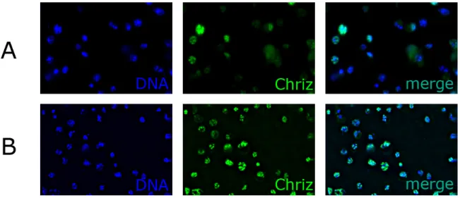
3.1.3. DeGradFP protein knockout of Chriz in salivary glands……… 43
3.2. Role of Chriz complex in gene expression... 48
3.2.1. Chriz and Z4 RNAi in S2 cells……… 48
3.2.1.1. Chriz and Z4 RNAi modulate the expression of many genes…….… 48
3.2.2. Correlation of Chriz binding with gene expression………..… 50
3.3. Insulator proteins BEAF-32 and CP190 interact with the Chriz complex……… 53
3.3.1. Co-immunoprecipitation………..………… 53
3.3.2. Pulldown assay……….……… 55
3.4. BEAF-32 contributes to recruitment of Chriz complex………..…… 57
3.4.1. RNAi in S2 cells……….………. 57
3.4.2. BEAF binding motif mutation………..…… 57
3.4.3. RNAi of BEAF-32 and CP190 in salivary glands……….………… 59
3.5. Role of Chriz complex in open chromatin domain formation. ………..………… 65
4. Discussion………..……… 69
4.1. Chriz complex contributes to chromatin structure and histone modifications … 69 4.2. Role of Chriz complex in gene expression……… 71
4.3. Insulator proteins BEAF-32 and CP190 interact with the Chriz complex…….……… 73
4.4. BEAF-32 contributes to recruitment of Chriz complex……….……… 75
4.5. Role of Chriz complex in open chromatin domain formation. .……….………… 77
5. Conclusion……… 79
Literature……… 80
Supplementary Figures ……… 94
3
Abstract (German)
Die Struktur des Chromatins spielt eine bedeutende Rolle bei der Stadien- und Gewebs- spezifischen Genexpression. Der epigenetische Status von Chromatindomänen wird mit Hilfe einer Anzahl von Proteinen und Histonmodifikationen etabliert und aufrechterhalten. Das Ziel der vorliegenden Arbeit ist die Untersuchung der Bedeutung und Funktion des Chriz Protein Komplexes bei Drosophila, der spezifisch in dekondensierten Regionen von Interphase Chromosomen gebunden ist. Mehrere Proteine waren als Bestandteile des Chriz Komplex bereits bekannt, darunter das Zink-Finger Protein Z4 und die H3S10 Kinase Jil1.
Meine RNAi Experimente an embryonalen S2 Zellen zeigten, dass die Rekrutierung von Z4 und der Kinase Jil-1 an das Chromatin sowie dessen H3S10 Phosphorylierung während der Interphase von der Gegenwart von Chriz abhängen. Diese Ergebnisse lieferten einen eine starken Hinweis auf eine Ähnlichkeit bei der Bildung des Komplexes in polytänen Zellen und diploiden S2 Zellen. Ich führte eine vergleichende Analyse der Bindungsprofile von Proteinen des Chriz Komplex zwischen Speicheldrüsen im 3.Larvenstadium und S2 Zellen im chromosomalen Intervall 61C7-8 durch. Dabei fand ich heraus, dass die Chriz Bindungsprofile im proximalen Teil der Domäne konserviert waren. Dagegen beobachtete ich eine veränderte Chriz Bindung im distalen Teil, die mit einem veränderten Transkriptionsprofil in dieser Region übereinstimmte. Verfügbare genomweite Daten zeigen eine Tendenz, dass Chriz in der Nähe von Transkriptionsstart-Stellen (TSS) und Promotoren aktiver Gene bindet.
Ich untersuchte daher die Korrelation zwischen der Chriz Bindung an Promoter Regionen von elf in S2 Zellen und Speicheldrüsen differentiell exprimierten Genen. Chriz und Z4 RNAi Experimente in S2 Zellen führten zu einer Veränderung der Expression vieler Chriz- und Z4- bindender Gene. Mittels Co-Immunpräzipitation und Protein „Pull-down“ konnte ich die Bindung der Insulator Proteine BEAF-32 und CP190 im Chriz-Komplex nachweisen und die für die Protein-Protein Interaktion notwendigen Proteinabschnitte grob kartieren. Schließlich überprüfte ich die Möglichkeit einer Rekrutierung des Chriz Komplexes durch BEAF-32 mit Hilfe von BEAF-32 RNAi bzw. nach Induktion von Punktmutationen in bekannten BEAF-32 Bindemotiven. Meine Ergebnisse wiesen eine Wechselwirkung zwischen Chriz und Insulator Proteinen nach und zeigten, dass der Chriz-Komplex eine wichtige Bedeutung bei der strukturellen Organisation von Chromatin Domänen besitzt.
4
Abstract (English)
Chromatin structure is important for the correct stage and tissue-specific expression of the genetic material. The epigenetic state of chromatin domains is established and maintained by a number of proteins and histone modifications. The aim of current thesis is to investigate the role and function of Chriz protein complex, specifically bound to decondensed regions of interphase chromosomes in Drosophila. Several proteins were known to compose Chriz complex - chromodomain protein Chriz, zinc finger protein Z4 and H3S10 kinase Jil-1. I performed RNAi experiments on S2 cells which demonstrated that recruitment of Z4 and Jil- 1 kinase to chromatin and interphase H3S10 phosphorylation are dependent on the presence of Chriz, and pointed to the high similarity of complex assembling between polytene cells and S2 diploid cells. I accomplished the comparative analysis of binding profiles of Chriz complex components between 3rd instar larvae salivary glands and S2 cell culture within 61C7-8 chromosomal interval. I found that Chriz binding profiles between two tissues are conserved at proximal part, however variant Chriz binding in distal part of 61C7-8 domain coincides with differences in transcription profile in the same region. Publicly available genome-wide data shows a tendency of Chriz to bind near Transcription Start Sites (TSS) and promoter regions of active genes. I investigated the correlation of Chriz binding to promoter regions of 11 differentially expressed genes with the expression of these genes in S2 cells and in salivary glands of 3rd instar larva. Chriz and Z4 RNAi experiments, performed in S2 cells resulted in expression changes of many Chriz- and Z4-binding genes. Using co- immunoprecipitation and pull-down assays, I identified insulator proteins BEAF-32 and CP190 to be present in the Chriz complex and performed rough mapping of interacting domains. Using BEAF-32 RNAi and introduced point mutations to BEAF-32 binding motifs I examined the possibility for recruitment of Chriz complex by BEAF-32. The obtained results revealed the interplay between Chriz and insulator proteins and point to important architectural role of Chriz complex in organizing chromatin domains.
5
Acknowledgements
I wish to express my sincere appreciation to my supervisor, Professor Harald Saumweber. I consider it a great opportunity to do my doctoral research under his guidance and to learn from his research expertise. I hope that I could be as lively, enthusiastic, and energetic as Harry and to someday be able to manage a research group as well as he can.
I thank all the present and former members of our Cytogenetic group whom I had a chance to work with for their friendly help, comments and suggestions. Thomas Zielke, Dandan Zhao, Ansgar Klebes, Dereje Negeri, Jenni Jammrath, Hussein Baaj, Shaza Dehne, Jorge Ferreira – you are the best colleagues I’ve ever known. We’ve got a really great atmosphere in the group, which I will definitely miss.
A big “Thank you!” also goes to Petra Binting and Irina Passow for their brilliant technical assistance.
I would also like to take this opportunity to thank all the students, to whose bachelor or master project I had a possibility to contribute to as a practical supervisor. Sorry for being strict with them sometimes.
I especially thank my Mom, Dad, and Sister. I love them so much and I would not make it this far without them.
Finally, I thank God for letting me through all the difficulties.
6
1. Introduction 1.1 Chromatin architecture
Large genomes of eukaryotic cells are densely packed into chromatin to fit inside nuclei that have diameter of few microns. One of fundamental questions in molecular biology is how such a compacted structure is established and maintained in a way to provide correct and virtuously regulated expression of encoded genetic material.
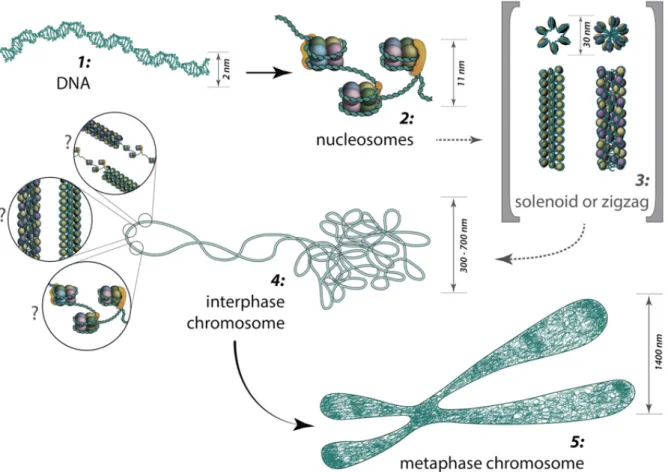
The fundamental unit of chromatin is a nucleosome comprising 147 base pairs (bp) of DNA wrapped around a histone octamer core in ≈1.67 left-handed superhelical turns as shown on the Fig. 1 (Cutter et al. 2015). The four core histones (H2A, H2B, H3 and H4) are relatively small (11–15kDa), very basic proteins that are highly conserved among eukaryotic species (White 2001). N-terminal “tails” of histones protruding out of nucleosomal core are strikingly prone to such post-translational modifications as acetylation, methylation or phosphorylation in a multitude of residues (Bannister et al. 2011; Feng et al. 2011).
Fig. 1. Structural details of a nucleosome core. (A) Model of a nucleosome core. A view down the superhelical axis, and a view rotated 90˚ about a horizontal axis. H2A, green, H2B, blue, H3, yellow, H4, red. Proteins in lower half of nucleosome are lighter in color (Cutter et al. 2015).
The core DNA is in tight association with the core histones and is protected from nuclease digestion whereas the linker DNA is rapidly digested. This fact determined historically the term ‘‘nucleosome core particle’’, which was originally defined as the product of extensive micrococcal nuclease digestion of native chromatin (Ausio et al. 1989).
The nucleosomal folding reflects the first ‘beads on a string’ fiber level of DNA compaction with a diameter of 10 nm. Linker histones (H1 and H5) bind to the DNA linker regions in close proximity to the sites of DNA entry, and organize the nucleosomal particles into a more
7
condensed 30-nm chromatin fiber, which represents the second level of DNA compaction (Thoma et al. 1979; Widom et al. 1985).
To describe three-dimensional organization of nucleosomes into 30-nm chromatin fibers, a number of models, including the solenoid (Finch et al. 1976), twisted-ribbon (Worcel et al.
1981), cross-linker (Staynov et al. 1983), and superbead (Zentgraf et al. 1984) models, had initially been proposed, based on the early studies of native chromatin in nuclei or isolated from nuclei by various biochemical and biophysical studies. Despite three decades of intense research, the precise structure of the 30-nm chromatin fiber remains elusive, however, recently Guohong and coworkers proposed 3D-cryo-EM structure which showed a left- handed twist of the repeating tetra-nucleosomal structural units with a two-start ‘‘Zig–Zag’’
configuration (Guohong et al. 2015) (Fig. 2).
Fig. 2. 3D structure of 30-nm chromatin fiber. (B) The three tetranucleosomal structural units of the 30-nm chromatin fibers reconstituted on 12 x 187 bp DNA arrays are highlighted in different colors. (C) A schematic representation of the cryo-EM structure of a 30-nm chromatin fiber as shown in B. (Guohong et al. 2015)
The 30 nm fibres could be further compacted or coiled, or just be arranged side to side, forming a hierarchy of folding levels, resulting in fibres of 60– 300 nm (Daban 2000).
Other microscopy-based studies suggested complex topologies co-existing within linear interphase chromosome structures (Bian et al. 2012). Based on cryo-EM and ESI contrast for conventional EM, the existence of an in vivo interphase chromosome structure which consists nearly entirely of 10 nm fibers, locally dispersed or concentrated in compact local domains was proposed (Bian et al. 2012).
During the interphase, chromatin is folded into domains 300-700 nm sizes, which comprise a chromosome territory. The structure and organization of chromatin loops inside a chromosome territory remain still unclear and was proposed to exist in the form of solenoid, or zigzag, or nucleosomes, or a hybrid of those. According to the chromonema model, chromosome structure arises from three helical folding levels of chromatin fibres.
Fibres of 60–80 nm in width are coiled into fibres of 100– 130 nm which are further coiled to the 200–300 nm structure of the metaphase chromatid (Heun et al. 2001) (See Fig. 3).
8
Figure 3. From DNA to metaphase chromosome: The major structures in DNA compaction.
(MBInfo).
During such processes as transcription or replication chromosomal organization is highly dynamic, varying both between different cell types and during the cell cycle. Recent studies revealed importance of spatial nuclear disposition of different chromatin regions and their relationships to the nuclear envelope for regulation of gene expression (Bickmore et al.
2013). However the exact logic of chromosome organization at the sub-megabase scale, which is the level where most gene regulatory landscapes and long range interactions are thought to occur (Kleinjan et al. 2009, Sanyal et al. 2012) had remained somewhat of a blackbox.
Chromosome conformation capture (3C) experiments have uncovered the presence of an additional level of compartmentalization of the genome (Dekker et al. 2002). Chromosomes of a wide range of species were found to be organized as a string of so called Topologically Associated Domains (TADs), which are characterized by preferential chromatin interactions within them, and spatial separation of loci located in different domains (Dekker et al. 2015).
In mammalian genomes these domains are several hundred kb in size, up to 1–2 Mb (Dixon et al. 2012), whereas they are smaller in bacteria (∼170 kb) (Le et al. 2013) and in flies (∼60 kb) (Hou et al. 2012). TADs (also named as “physical domains”) were found to correlate strongly with epigenetic features, including active gene density, association with the nuclear
9
lamina, replication timing, nucleotide and repetitive element composition (Sexton et al.
2012). Analyzing of expression patterns of genes located within the same TAD across ES cell differentiation, Nora and coworkers revealed that genes united in the same TAD show similar dynamics of expression during differentiation, whereas genes located in different TADs were less correlated (Nora et al. 2012).
Figure 4. Architectural proteins mark TAD borders. Cartoon schematics depicting regions of highly associating chromatin called TADs, which are separated by TAD borders. Interaction frequency is shown as a continuum from white to dark red. (A) The strength of a TAD border, defined as the ratio between intra- and inter-TAD interactions around border sequences, depends on architectural protein occupancy (occupancy shown by peak size and a continuum from light to dark blue) (Cubenas-Pottis et al. 2015).
Moreover, the calculated physical domains from the Hi-C contact map strongly correlated with numerous linear epigenetic profiles describing enrichment for histone modification or such DNA-binding factors as chromodomain protein Chriz, insulator proteins CP190, BEAF-32 and dCTCF (Sexton et al. 2012) (See Fig. 4). These findings support the statement that TADs are critical chromosome structural units of long-range gene regulation (Sexton et al. 2012, Cubenas-Pottis et al. 2015).
1.2 Histone modifications
Interphase chromatin is not static. Although nucleosomes themselves are stable and have limited mobility, a number of remodeling complexes can mobilize and/or eject the nucleosome to regulate access to DNA (Saha et al. 2006). Moreover, depending on the needs of the cell, the N-terminal and C-terminal tails of histones may undergo reversible post- translational modifications that change their interaction with DNA and convert them to
“docking stations” for different classes of nuclear proteins (Martin et al. 2005)
To date, a wide variety of histone post-translational modifications (PTMs) are known, including methylation, acetylation, phosphorylation, glycosylation, carbonylation, ubiquitylation, biotinylation, sumoylation, citrullination, ADP-ribosylation, N-formylation, crotonylation, propionylation, and butyrylation, as well as proline and aspartic acid isomerization (Sadakierska-Chudy et al. 2015). Known enzymes, which execute this huge variety of modifications are listed in Table 1.
10
Table 1. Histone modifying enzyme activity. 1 Histone or residue if known. (Modified from Swaminathan et al. 2012)
Phosphorylation, in general, represents one of the major forms of histone post-translational modifications. It can be found at, threonine (North et al. 2011), tyrosine and histidine
11
(Besant et al. 2012), but more often at serine residues (Sotero-Caio et al. 2011) in each or the four core histones. In particular, phosphorylation of histone H3 at serine 10 is observed frequently. This modification is observed both at interphase chromatin and during mitosis and is linked with different processes, such as transcription activation or mitotic chromatin condensation (Nowak et al. 2004). During interphase in Drosophila, the majority of H3S10 phosphorylation is performed by JIL-1, an essential, ubiquitously expressed, nuclear tandem kinase (Jin et al. 1999) (will be discussed later). At mitosis, genome-wide H3S10ph (and also H3S28ph) marks are established by Aurora B kinase and erased by PP1 phosphatase (Giet et al. 2001, Goto et al. 2002).
Histone acetylation in Drosophila is known a while ago to be associated with transcription activation (Allfrey et al. 1964). To date, several histone acetyltransferases have been identified and grouped into three main families - MOF (KAT8), chameau (KAT7) and Tip60 (KAT5) belong to the MYST HAT family. dGCN5 (KAT2) and Elp3 (HAT9) represent members of the GNAT HAT family, while nejire (KAT3) is the single Drosophila CBP/p300 homologue (Lee et al. 2010, Boros 2012).
Methylation of histones may occur at lysine and arginine residues and is associated both with transcription activation and repression (Martin et al. 2005). Lysine methylation, which is more characterized, was found at five positions of histone H3 and at single position of histone H4. Methylation of H3K4, H3K36 and H3K79 was identified in association with active transcription, whereas H3K9, H3K27 and H4K20 methylation associated with repression (Sims et al. 2003, Martin et al. 2005).
In Drosophila methylation of H3K4 is executed by several enzymes. To date, Trx, absent, small and homeotic discs 1 (Ash1), and Trithorax-related (Trr) have been identified as H3K4 methyltransferases. Little imaginal discs (LID) and Su(var)3-3, the Drosophila homolog of LSD1, are H3K4 specific demethylases (Swaminathan et al. 2012)
Histone H3 lysine 27 trimethylation, was specifically found to play an important role in polycomb-mediated gene silencing. It is known to be found at repressed genes containing PREs – Polycomb Response Elements, which recruit PcG proteins. Trimethylation of H3 K27 spreads out from this sites by enzymatic activity of E(z), the SET domain-containing subunit of PRC2 (Muller et al. 2002).
As can be seen, the majority of histone modifications can be associated with two opposite processes, namely transcriptional activation and repression (Cohen, 2011). However, regulation of these processes is tightly connected with another important task – determination of chromatin state (Brower-Toland et al. 2009).
Heterochromatin is generally repressive and deprived of such histone modifications as acetylation. In metazoan, two types of heterochromatin can be defined – facultative heterochromatin which contains developmentally or tissue-specifically silenced genes (for instance, homeotic genes, Antennapedia or Bithorax complexes) sometimes marked by
12
H3K27me3, and H3K9 methylated constitutive heterochromatin, which contains permanently silenced genes in genomic regions such as the centromeres and telomeres.
Active genes and their regulatory regions are located in more open, decondensed euchromatin, containing a number of typical histone modifications, such as H3S10 phosphorylation or H3K9 acetylation. However, high degree of overlap between different histone modifications is observed, proposing, that there are no simple rules governing their localization and function (Bannister et al. 2011).
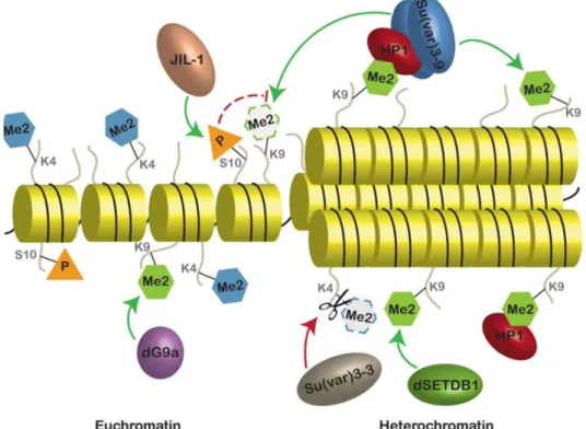
A simplified scheme of euchromatic and heterochromatic regions, established by action of distinct histone modifying enzymes is shown on Fig. 5.
Figure 5. Chromatin states with typical histone modifications. The major enzymes and their location of action are indicated. Green arrows indicate the addition of the mark and red arrows with scissors indicate the removal of the specified modification. Dashed arc indicates an indirect effect. Dashed lines and margins indicate disappearance of the mark (Swaminathan et al. 2012, modified).
Genome-wide profiles identifying the location of the histone marks, as well as analysis of phenotypes resulting from altered levels of the modifying enzymes indicate the existence of
“cross talk” between certain modifications where, for instance, one mark facilitates another or is even required for the second mark to occur or when one modification prevents the modification of a second residue (Lee et al. 2010).
Such comparative analysis required systematic collection of a rapidly increasing amount of genome-wide data. In 2007 over 2 000 datasets containing the information about positions 13
of modified histones and other chromatin marks, origins of DNA replication, RNA transcripts and the transcription factor binding sites of Drosophila were combined to a single database by modENCODE Consortium (2007-2012) aimed to discover and provide biologically informative characterizations of as many genomic elements as possible (Brown et al. 2015).
The modENCODE platform provided unique possibilities for epigenetic studies purposed to reveal the principles of underlying chromatin regulation (Schubeler et al. 2010).
The analysis of genome-wide distribution of numerous epigenetic factors leaded to a statement that functional state of chromatin may be accompanied by a certain local combinations of bound proteins and/or histone modifications (Filion et al. 2010). The development of such ideas resulted in establishing of several “maps of chromatin state”
(Filion et al. 2010; Kharchenko et al. 2011; Zhimulev et al. 2014), which were attempted to identify the necessary epigenetic features for chromatin structure determination. Active chromatin types in their models were accompanied with local enrichment of Pol II, active transcription and presence of such modifications as H3K9Ac and H3K4me3. Repressive chromatin types featured Polycomb group protein binding, lamin association, H3K27me3, etc. Using the available database Zhimulev and coworkers reported correlations found for a number of regions between the cytogenetic structure of polytene chromosomes and selected features of inactive/active chromatin of S2 cells (Vatolina et al. 2011; Demakov et al. 2011).
1.3 Polytene chromosomes
Polytene chromosomes of Drosophila have been used for epigenetic studies since 1934 (Turner et al. 1992). Due to chromatid amplification, polytene chromosomes of larval salivary glands provide a precious possibility of direct visualization of chromosomal architecture during interphase, location of proteins bound and modified histones involved in the regulation of chromatin structure and expression of genetic material.
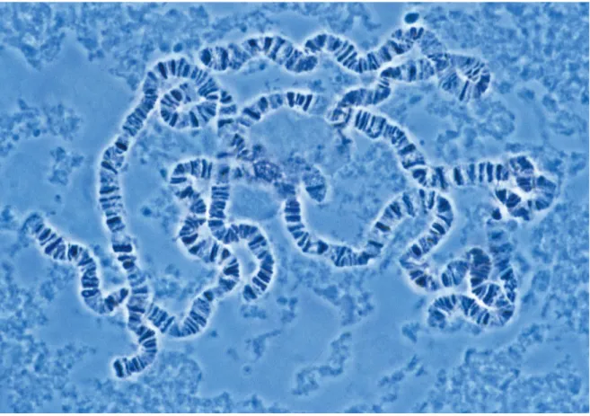
On microscopic squash preparations of polytene chromosomes a reproducible pattern of clearly distinguishable compacted bands interrupted by less compacted interbands can be observed (See figure 6) (Painter et al. 1934; Beermann et al. 1972).
Heat-shock or steroid hormone treatment leads to local loosening of polytene chromatin structure known as “puffing”, which is associated with the induction of extremely high levels of transcription of steroid response or stress-activated genes located at relevant polytene chromosome regions (Ashburner 1967, 1970).
14
Fig. 6. Polytene chromosomes from Drosophila salivary glands. (The Cell, 4th Edition, Fig.
5.26, 2006)
RNA-polymerase enrichment as well as detected ongoing transcription process in interbands pointed to localization of active genes there (Alcover et al. 1982). Till recent time, the hypothesis which postulated interbands as the sites of location of 5' parts of genes, while the 3' gene ends were assigned to the adjacent bands was poplular among cytologists (Zhimulev et al. 2013). However latest findings purposed to merged the cytological observations with molecular biological datasets consider interbands as distinct open chromatin domains, containing active genes (See Fig. 7) (Sexton et al. 2012; Zhimulev et al.
2014; Zielke et al. 2015, in press). Therefore, experimental evidences, strengthening the statement that the boundaries of chromatin marks coincide with borders of the physical domains, are provided (White 2012).
15
Figure 7: The domain organization of the genome. In the domain model, the different chromatin states are represented by domains of different chromatin folding, separated by boundaries (open dots). Blue, green and black lines corresponds to different types of condensed chromatin, red and yellow – to active chromatin states (according to Filion et al.
2010). Physical domains can be visualized at polytene chromosomes as band/interband pattern (Modified from White 2012).
Continuing technical and methodological advances together with the wealth of existing studies on the Drosophila genome make the fly a major model for the analysis of the many remaining questions concerning how genome packaging relates to genome function (White 2012).
1.4 Chriz/Z4/Jil-1 complex
The proper organization of eukaryotic chromosomes determines the manner in which the DNA sequence is interpreted in a large number of cellular processes, such as DNA replication, repair and transcription (Sexton, 2009). Therefore, for keeping the dynamic structure of chromatin, a huge number of proteins and protein complexes are employed.
Current study is focused on Chriz complex, discovered in our group in 2004.
16
Several proteins are known to be part of this complex, however, the “core” consists of zinc- finger protein Z4 and “the chromodomain protein interacting with Z4” - Chriz (also called Chromator by some authors (Rath et al. 2004).
Chriz has a calculated molecular weight of 100 kDa (apparent 130-140 kDa), and can be divided into two main domains, an amino-terminal domain containing the chromodomain and a carboxy-terminal domain containing a nuclear localization signal (Fig. 8)(Rath et al.
2004, Gortchakov et al. 2005). Chriz protein is ubiquitous, essential and is known to fulfil several functions during fly development (Gortchakov et al. 2005; Rath et al. 2006; Wasser et al. 2007; Ding et al. 2009). During the interphase Chriz is localized at interband regions of polytene chromosomes, but not in puffs. However, during cell division Chriz redistributes to form a macro molecular spindle matrix complex together with at least three other nuclear- derived proteins Skeletor, Megator, and EAST (Walker et al. 2000; Rath et al. 2004; Qi et al.
2004, 2005). The studies of Gan with colleagues showed that N-terminal part of Chriz is responsible for correct targeting to chromatin (Gan et al. 2011). Same domain later was found to interact with histone H1 (Yao et al. 2012). C-terminal domain was reported to be sufficient for localization to mitotic spindle (Ding et al. 2009).
Figure 8. Schematic picture of the full-length chromodomain protein Chriz (926 amino acid residues). Chomodomain is displayed as yellow box. The extent of the domains is given by aa numbers.
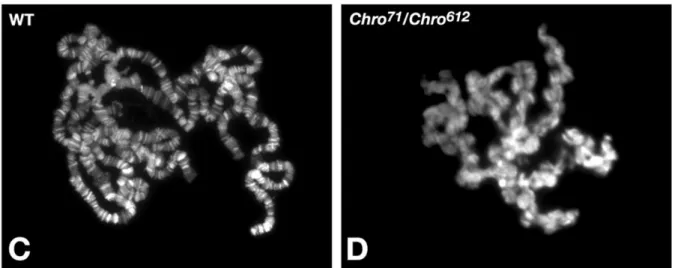
Chriz is required for maintenance of chromatin structure – polytene chromosomes of flies with a combination of hypomorphic mutant alleles showed loss of band/interband structure together with numerous ectopic contacts connecting non-homologous chromosomal regions (Rath et al. 2006)(Fig. 9, 10).
17
Fig. 9 Localization of Chriz in mutant polytene chromosomes. Polytene chromosome preparations from third-instar larvae were labeled with Hoechst to visualize the chromatin.
Preparations are shown from a wild-type female larvae (C) and from a female Chro71/Chro612 mutant larvae (D). Reduced levels of wild-type Chromator protein have a severe effect on the structure and organization of larval polytene chromosomes. Note the disruption and misalignment of interband and banded regions and the extensive coiling and folding of the chromosome arms in Chro71/Chro612 mutant chromosomes (D). (Modified from Rath et al 2006)
Fig. 10. Ultrastructure of Chro71/Chro612 mutant polytene chromosomes. (A) TEM micrograph of a wild-type polytene chromosome. Note the clear segregation into bands and interbands and the orderly alignment of euchromatic chromatid fibrils. (B,C) Chromosomes from Chro71/Chro612 polytene salivary gland nuclei. The micrograph in B shows the disorganization and misalignment of band/interband polytene chromosome regions (arrows). The micrograph in C shows the folding and coiling of the chromosomes with numerous ectopic contacts connecting non-homologous regions (arrows). (Modified from Rath et al 2006).
Chriz significantly co-localizes and interacts with the tandem kinase Jil-1, which phosphorylates histone H3 at serine 10 residue during the interphase (Rath et al. 2006). Jil-1 was shown to play a significant role in maintaining polytene chromosome structure and is enriched about two-fold on the male X-chromosome (Jin et al. 1999; Wang et al., 2001; Deng 18
et al., 2005; Zhang et al., 2006). In the absence of JIL-1 non-orderly intermixing of euchromatin and the compacted chromatin banded regions was observed (Deng et al., 2005) along with is a striking redistribution of the heterochromatin markers dimethyl H3K9 and HP1 to ectopic chromosome sites (Zhang et al., 2006). Deng and colleagues demonstrated that ectopic recruitment of JIL-1 to a cluster of Lac operator repeats, mediated by fusion with a lacI DNA-binding domain leaded to local decondensation of polytene chromatin. Their results also provided evidence that observed reorganization of chromatin structure was dependent on the kinase activity of JIL-1 (Deng et al. 2008). Corces and coworkers have proposed that histone H3S10 phosphorylation executed by Jil-1 is required for active transcription by the RNA polymerase II machinery (Ivaldi et al. 2007; Kellner et al. 2012).
However, later it was demonstrated that RNA polymerase II mediated transcription occurs at robust levels in the absence of H3S10 phosphorylation (Regnard et al. 2011). Recently, Cai and coworkers provided experimental evidence for the functional role of JIL-1-mediated H3S10 phosphorylation in maintenance of active gene expression by serving as a protective epigenetic mark counteracting H3K9 dimethylation and gene silencing (Cai et al. 2014). It has been shown that the C-terminal domains of Jil-1 kinase and Chriz directly interact with each other (Rath et al. 2006). Consequently, Gan and colleagues demonstrated that Jil-1 requires Chriz for targeting to polytene chromosomes (Gan et al. 2011).
Another component of Chriz complex – seven zinc-finger protein Z4 with a molecular weight of about 160 kDa is essential for fly development and acts in a dose-dependent manner on the development of several tissues (Fig. 11)(Eggert et al. 2004).
Figure 11. Schematic picture of the full-length zinc-finger protein Z4 (996 amino acid residues). Z4 contains 7 zinc-finger (ZnF) domains of the classical C2H2 type (green box). The extent of the domains is given by aa numbers.
Z4 is localized to open interband domains of polytene chromosomes (not in puffs) and is involved in maintenance of chromatin structure; chromosomes from 3rd instar larvae of hypomorphic Z4 mutants loose the organization into bands and interbands and altogether appear as a less compact mass of chromatin (Eggert et al. 2004). Z4 has been shown to be an important cofactor in at least three different pathways related with chromatin remodeling:
the NURF and the TRF2/DREF remodeling complexes, where it acts as an activator (Kugler et al. 2007; Kugler et al. 2010); and in the JAK/STAT pathway, where Z4 acts as a co-repressor (Kugler et al. 2011). In the work of Silva-Sousa the role of Z4 in maintenance of telomere stability is discovered (Silva-Sousa et al. 2012).
19
Chriz and Z4 precisely co-localize in interbands along the whole length of polytene chromosomes (Gortchakov et al. 2005) (see Figure 12) and interact by their central and N- terminal domain, respectively (Gan et al. 2011).
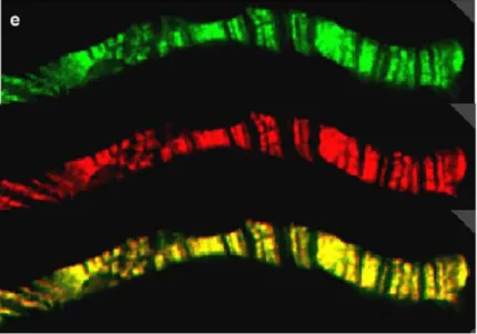
Fig. 12. Z4 and Chriz colocalize on polytene chromosomes; top: Z4 (green); middle: Chriz (red); bottom: colocalization is demonstrated by the yellow color resulting from the overlay of both wavelengths (Gortchakov et al. 2005)
Interestingly, loss of function of Chriz complex components also influences the coherence and organization of bands although neither protein is present in these regions. Rath and coworkers suggested that their function may affect the distribution and/or activity of other molecules important for influencing chromatin structure such as boundary elements. (Rath et al. 2006, Van Bortle et al. 2014)
1.5 Boundary elements
Chromatin insulators play a key role in determining the structural organization of eukaryotic chromatin.
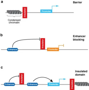
Generally, insulator elements possess two key properties indicative of the capacity to define a chromatin domain, characteristic for each insulator family. The first is termed enhancer blocking, the ability to interfere with enhancer– promoter communication only when placed between the two elements (Reitman et al. 1990; Cai et al. 1995). The second feature is termed barrier activity, the ability to protect a flanked transgene from position-dependent silencing (Kellum et al. 1992). Numerous research work in chromatin studies provided strong support for both types of models, and, therefore defined two major classes of insulators:
‘enhancer blocking’ insulators which block communications between adjacent regulatory elements in a position dependent manner and ‘barrier insulators’ which prevent the
20
silencing of euchromatic genes by blocking the spreading to nearby heterochromatin. Both types are schematically visualize on Fig. 13
Figure 13. Insulators block enhancer and silencer elements in a position-dependent manner. (a) Barrier elements block the linear spread of silenced chromatin protecting the reporter gene from silencing. (b) Enhancer-blocking elements interfere with enhanced transcription when placed between an enhancer element and the promoter. (c) Flanking a transgene with insulator elements generates a functionally independent domain protected from position effects. Regulatory interactions can occur within the domain whereas the insulators block external signals (Valenzuela et al. 2006).
Pioneer studies on boundary elements were focused on the 87A7 locus in Drosophila. This locus contains two divergently transcribed heat shock genes (hsp70) actively transcribed at increased temperature and reflected by puffing in salivary gland polytene chromosomes.
DNAse sensitivity analysis of DNA regions flanking the 87A7 locus identified two zones with an unusual chromatin structure, called scs and scs′ (specialized chromatin structures) (Udvardy et al. 1985). These regions were shown to be located very close to the border between the decondensed 87A7 locus and the flanking condensed chromatin. Further research on these elements identified them as insulators (Kellum et al. 1991). Kellum and coworkers hypothesized that if insulator elements established borders between chromatin domains, they should protect a reporter gene from position effects when integrated into the genome. Random P-element mediated insertion of white minigene with a minimal promoter resulted in flies with a range of eye color from white to red due to position effects. Such position effects were considered to be a consequence of interactions between the promoter of the reporter gene and enhancer and silencer elements present near the integration site.
When the white minigene was placed between the scs and scs′ elements, its level of 21
expression was uniformly low in the transformed lines, indicating that scs and scs′ protected the expression of the miniwhite gene from euchromatin (Kellum et al. 1991).
Several enhancer-blocking assays were designed to test whether these DNA sequences inserted between an enhancer element and a target promoter can prevent them from interacting, and both scs and scs′ were shown to insulate reporter genes from different enhancer elements as well (Kellum et al. 1992). Although they do not share any sequence homologies, both scs and scs′ elements were recently shown to be close to promoters of genes (Avramova et al. 1999). The scs insulator is found in a region that contains the promoter for CG31211 whereas the scs′ insulator maps to the promoter of two divergently transcribed genes, CG3281 and aurora (Glover et al. 1995). Later, two proteins bound to scs and scs’ insulators were characterized, the zinc-finger Zeste-white 5 (Zw5) protein and the
“boundary element-associated factor” - BEAF-32 (Gaszner et al. 1999; Zhao et al. 1995).
These studies gave rise to further characterization of function of identified insulator proteins and revealed additional sites throughout the genome as well as protein complexes associated (Hirose et al. 1996, Xu et al. 2004). Distinct families of insulators have been described to date, defined by insulator binding protein that is essential for their activity (Maeda et al. 2007). In Drosophila five insulator families were identified: Suppressor of Hairy-wing [Su(Hw)], boundary element-associated factor (BEAF), Zeste-white 5 (Zw5), the GAGA factor (GAF) (Maeda, 2007), and dCTCF, a homologue of mammalian CTCF (Moon, 2005). Each category of insulator complexes also contains the common centrosomal protein 190 (CP190) (Matzat et al. 2014).Genome wide analysis identified thousands of binding sites for most of these proteins (Bushey et al. 2009; Schwartz et al. 2012; Van Bortle et al. 2014).
Recent observations proposed a model for insulator function in which direct DNA-binders (BEAF-32, dCTCF, Su(Hw)) provide DNA specificity and serve as “first layer proteins”, whereas such proteins as CP190, Mod(mdg4) or Chriz may interact with them and form a “second layer” being responsible for the physical interactions required for long-range contacts (Vogelmann et al. 2014).
In the current study we will focus on two insulator proteins – BEAF-32 insulator and CP190.
The BEAF-32 insulator protein is known to have two isoforms, A and B, which differ in their N-terminal part, where two slightly different atypical C2H2 zinc-fingers, termed BED fingers are located (see Fig 14) (Aravind et al. 2000). C-terminus of BEAF-32 contains BESS domain and coiled-coil CC-domain, involved in protein-protein interactions (Hart et al. 1997). Gilbert and colleagues showed that this domain is necessary for BEAF-32 self-interaction and, demonstrated the capacity of BEAF-32 to form trimers (Gilbert et al. 2006).
22
Figure 14. Schematic picture of the full-length boundary element – associated factor BEAF- 32 (282 amino acid residues). BEAF-32 contains BED domain (blue box) at the N-terminus , coiled-coil domain (brown box) and BESS domain (orange box) at the C-terminus. The extent of the domains is given by aa numbers.
According to ChIP studies both isoforms bind specifically to scs′ as well as many other sites in the genome, however the more abundant BEAF-32B protein apparently is associated with more sites than BEAF-32A (Jiang et al. 2009). It was shown experimentally that a number of BEAF-32 binding sites possess enhancer blocking activity (Hart et al. 1999; Schwartz et al.
2012), supporting the statement that BEAF-32 is a general insulator factor. BEAF-32 was found to bind DNA directly and is preferentially associated with a CGATA motif (Hart et al.
1997). Same DNA motif is also bound by the DREF transcription factor proposed to compete with BEAF-32 for chromatin binding (Hart et al. 1999). Comparative analysis of the genome- wide binding profile of BEAF-32 with the distribution of its binding motif suggests that BEAF- 32 prefers to bind to dual clusters containing two or three individual binding motifs, totaling five or six sites, spaced by a 200 bp central AT-rich region (Emberly et al. 2008). These “dual cores” as well as the majority of BEAF-32 binding sites correspond to transcription start sites (Jiang et al. 2009). Furthermore, BEAF-32 has a clear tendency to bind at promoter regions of head-to-head oriented genes (Jiang et al.2009). Yang and colleagues hypothesized that BEAF-32 may play an evolutionarily conserved role in preventing undesired transcriptional regulatory crosstalk between the individual genes forming the pair (Yang et al. 2012).
Knockdown studies of BEAF-32 in cell culture have provided insight into its role in regulation of gene expression. RNAi of BEAF-32 in cells results in few changes in gene expression with no apparent growth arrest (Schwartz et al. 2012; Van Bortle et al. 2012). However, after longer time of BEAF-32 depletion, growth arrest is observed, as well as increase in cellular DNA content and chromosome segregation defects (Emberly et al. 2008). These effects may partially result from relief of competitive binding with DREF, which activates a subset of cell cycle control genes (Matzat et al. 2014). Interestingly, global H3K9me3 rises in BEAF-32 knockdown cells, in contrast, to unchanged global H3K27me3 levels or gene expression within H3K27me3 islands bordered by CTCF sites (Van Bortle et al. 2012). In addition to DREF, BEAF-32 also overlaps considerably with other insulator proteins, particularly centrosomal protein CP190 (Bushey et al. 2009).
CP190 is a protein of 1,096 amino acids with a predicted molecular weight of 121 kDa and an apparent molecular weight of about 190 kDa. The protein contains an N-terminal BTB/POZ (Broad-complex, Tramtrack and Bric-abrac/Poxvirus and Zinc Finger) domain; an aspartic- acid rich D-domain; three C2H2 zinc finger motifs; and a C-terminal E-rich domain (see Fig.
15) (Ahanger et al. 2013).
23
Figure 15. Schematic picture of the full-length Centrosomal Protein 190 (1,096 amino acid residues). CP190 contains BTB/POZ domain (light-orange box) at the N-terminus, D-rich (red box), CENT (violet box) and zinc-finger (ZnF) domains (green box) in the center and an E-rich domain (blue box) at the C-terminus. The extent of the domains is given by aa numbers.
CP190 contains a centrosomal targeting domain (CENT) for its localization to centrosomes during mitosis (Whitfield et al. 1995). The BTB/POZ, the aspartic-acid rich (D-rich) and the C- terminal glutamic-acid rich (E-rich) domains are essential for its association with insulator subclasses and insulator function. The E-rich region was shown to be important for the dissociation of CP190 from the chromosome after heat-shock, which may provide a mechanism for regulating insulator function (Oliver et al. 2010). CP190 was identified to be associated with centrosomes throughout the nuclear division cycle in syncytial Drosophila embryos (Frasch et al. 1986). However, after the cellularization of the embryo, CP190 is exclusively found in nucleus during the interphase (Callaini et al. 1990). Using indirect immunofluorescence staining of polytene chromosomes from salivary glands, CP190 was found at a number of sites along the entire length of the chromosomes localizing mainly to interbands and band/interband boundaries (Whitfield et al. 1995). Recent genome-wide studies revealed an extensive overlap of CP190 with the target sites of main insulator factors; dCTCF, Su(Hw), BEAF and GAF (Negre et al. 2010). It was demonstrated that 80%
robust CP190 binding sites overlap with dCTCF, Su(Hw) or BEAF-32 (Schwartz et al. 2012).
The number of sites in the genome where CP190 binds independently of Su(Hw), dCTCF and BEAF-32 were later identified as binding sides for recently discovered proteins IBF1, IBF2, Pita and ZIPIC (Maksimenko et al. 2015; Cuartero et al. 2014).
As can be seen, insulator proteins associate in clusters of overlapping binding sites more often than would be expected by chance, suggesting that these factors could bind as a complex to the same genetic locus (Vogelmann et al. 2014). Significant co-localization of insulator protein BEAF-32 with Chriz complex components on polytene chromosomes was revealed by Gan and colleagues (Gan et al. 2011). Later, this observation was confirmed by computational analysis of overlapped binding sites between insulator proteins BEAF-32, CP190 and Chriz (Vogelmann et al. 2014) (Fig. 16) More detailed systematic screen of chromatin profiles performed by Sexton and coworkers showed that such proteins as Chriz, CP190, CTCF, and BEAF-32 were identified at domain borders. At the same loci, active histone mark H3K4me3 and high DNase hypersensitivity showed a striking enrichment compared to background regions (Sexton et al. 2012). Interestingly, CP190 and Chriz were found to be most represented at the borders of physical domains. Boundary behavior was significant but somewhat weaker at peaks of BEAF-32, H3K4me3, and CTCF and was minimal at peaks of Su(Hw) (Sexton et al. 2012).
24
Fig. 16. Venn diagram showing the genome-wide overlap between BEAF-32, CP190 and Chromator (Chriz) in S2 cells. Binding sites were calculated from publicly available modENCODE ChIP-chip data (Vogelmann et al. 2014).
All these findings proposed a possibility of physical interaction between Chriz complex and insulator proteins. Co-IP experiments performed by Gan and colleagues revealed interaction between Chriz complex component Z4 and insulator BEAF-32 (Gan et al. 2011). Direct interaction between Chriz and Mod(mdg4), which is the component of Su(Hw) complex was reported (Golovnin et al. 2014).
The variety of identified connections between Chriz complex and other proteins, allows us to assume that the role, which Chriz complex plays in the cell may not be restricted to maintenance of chromatin structure. It is still unclear to which extent the knowledge about Chriz protein complex obtained on the polytene chromosomes can be extrapolated to diploid cell chromatin. Interactions between Chriz and insulator proteins BEAF-32 and CP190 are still weakly studied and mechanism how Chriz complex is recruited to chromatin remains unknown.
1.6 Therefore, current work has the following aims:
1. Determine the interactions between known and putative components of Chriz complex – Z4, Jil-1, BEAF-32, Chriz and CP190.
2. Identify the role of “core” Chriz complex components in gene expression.
3. Compare the composition and localization of Chriz complex in polytene and diploid cells.
4. Explore the possibility of Chriz complex recruitment by identified factors.
25
2. Materials and Methods:
2.1. Materials 2.1.1 Chemicals
If not specified, standard chemicals from Serva, Roth, Merck and Sigma were used.
2.1.2 Bacterial strains
For cloning and plasmid DNA production E. coli strain XL1-Blue (recA1 endA, gyrA96 thi-1 hsdR17 supE44 relA1, lac [F‘, proAB, lacIqZDM15, Tn10 (Tetr)]c) was used.
For protein expression E. coli strain BL-1-DE3 (B F– dcm ompT hsdS(rB– mB–) gal λ(DE3)) was used.
For E. coli cultivation the following media were used:
LB-Medium (Luria/Miller) LB-Agar (Luria/Miller)
10 g/l Trypton 10 g/l Trypton
5 g/l Yeast extract 5 g/l Yeast extract
10 g/l NaCl 10 g/l NaCl
15 g/l Agar-Agar 2.1.3 Cell culture
Drosophila S2 cells were kept in 10-50 ml tissue culture flasks in commercial Drosophila S2 medium (Gibco, Lot 1627148) with 10% FCS under conventional conditions at 28 ˚C. Cells were split 1-2 x weekly.
2.1.4 Fly work
Drosophila strains were kept in standard media at 18˚C (see receipt in the table below).
Crosses were performed at 25˚C.
Standard fly media (Bloomington)
39l Water
675g Yeast 390g Soy flour 2,85g Cornmeal 225g Agar-Agar 3l Sugar syrup 188ml Propionic acid
26
In current work the following Drosophila strains were used
2.1.5 Crossing schemes:
Double BEAF/CP190 double RNAi strain was obtained by the following crossing scheme:
PBEAF: BEAFRNAi BEAFRNAi;+
+ X CyO
If ;
TM6, Tb
MKRS, Sb PCP190 + + ;
CP190RNAi
CP190RNAi X CyO If ;
TM6, Tb MKRS, Sb
F1: BEAFRNAi
If ;MKRS, Sb
+ X CyO + ;
CP190RNAi TM6, Tb
F2: BEAFRNAi
CyO ;CP190RNAi MKRS, Sb X
BEAFRNAi
CyO ;TM6, Tb +
F3: BEAFRNAi
CyO ;CP190RNAi TM6, Tb X
BEAFRNAi
CyO ;CP190RNAi TM6, Tb OR
F3: BEAFRNAi
BEAFRNAi;CP190RNAi TM6, Tb X
BEAFRNAi
BEAFRNAi;CP190RNAi TM6, Tb
Final Stock: BEAFRNAi
BEAFRNAi;CP190RNAi CP190RNAi
Name Marker Chr. Notes
WT-Oregon Wild type
Tft/CyO 2 Balancer strain
TM3/TM6 3 Balancer strain
Tft/CyO; TM6/MKRS 2,3 Balancer strain
G231.1 Gal4 2 Gal4 salivary gland specific driver
(Dr. U. Hinz, Cologne)
G61 Gal4 X Gal4 salivary gland specific driver
(Dr. U. Hinz, Cologne)
ΔKG12 Chriz 3 Chriz deletion (Gortchakov et al.
2005)
42pattP 2 (Zielke, Saumweber 2014 )
BEAF RNAi 2 Bloomington, 35642
CP190 RNAi 3 Bloomington, 33903
Chriz RNAi 2 (Gortchakov et al. 2005)
UAS-NB 2 y1 w*; P{UAS-Nslmb-vhhGFP4}2 ,
Bloomington, 38422
27
Crosses were performed during a lab rotation project by M. El Genedy under my supervision.
The Chriz-GFP strain was obtained using the following scheme:
P: w−
w−; ChrizGFP, w+ ChrizGFP, w+;+
+ X w− CyO If ;
TM6, Tb
MKRS, Sb w−
w−;Chriz∆KG12
Chriz∆KG12 X w− CyO If ;
TM6, Tb MKRS, Sb
F1: w−
w−; ChrizGFP, w+
CyO ;TM6, Tb
+ X w− CyO + ;
Chriz∆KG12 MKRS, Sb
F2: w−
w−; ChrizGFP, w+
CyO ; TM6, Tb
Chriz∆KG12 X w− CyO
ChrizGFP, w+;Chriz∆KG12 TM6, Tb F3: w−
w−; ChrizGFP, w+
ChrizGFP, w+;Chriz∆KG12 Chriz∆KG12 DeGradFP strain 1 was obtained using the following scheme:
P: Gal4, w+ Gal4, w+; +
+ ; +
+ × w− CyO If ;
TM6, Tb
MKRS, Sb F1:Gal4, w+
w− ; + CyO ;
TM6, Tb
+ , Gal4, w+ w− ;+
If ; +
MKRS, Sb × w−; ChrizGFP, w+
ChrizGFP, w+;Chriz∆KG12 Chriz∆KG12 F2: Gal4, w+
Gal4, w+; + CyO ;
TM6, Tb
+ × Gal4, w+; If
ChrizGFP, w+; MKRS, Sb Chriz∆KG12 F3: Gal4, w+
Gal4, w+; ChrizGFP, w+
CyO ;Chriz∆KG12 TM6, Tb F4: Gal4, w+
Gal4, w+; ChrizGFP, w+
ChrizGFP, w+;Chriz∆KG12
Chriz∆KG12 × w−; UAS−NB, w+
UAS−NB, w+;Chriz∆KG12 TM6, Tb
F5: Gal4, w+; UAS−NB, w+
ChrizGFP, w+ ;Chriz∆KG12 Chriz∆KG12 ,
Gal4, w+
w− ; UAS−NB, w+
ChrizGFP, w+;Chriz∆KG12 Chriz∆KG12
DeGradFP strain 2 was obtained using the following scheme:
P: y10 y10;+
+ ;
Chriz∆KG12
TM6, Tb × w− CyO If ;
TM6, Tb
MKRS, Sb F1: y10; +
CyO ;
MKRS, Sb Chriz∆KG12
P: w−
w−; UAS−NB, w+ UAS−NB, w+;+
+ × w− CyO If ;
TM6, Tb MKRSSb
28
F1: w−
w−; UAS−NB, w+
CyO ; +
TM6, Tb × y10; + CyO ;
MKRS, Sb Chriz∆KG12
F2: w−; UAS−NB, w+
CyO ;Chriz∆KG12 TM6, Tb ×
w−
w−; UAS−NB, w+
CyO ;Chriz∆KG12 TM6, Tb
w−; UAS−NB, w+
UAS−NB, w+;Chriz∆KG12 TM6, Tb
Both DeGradFP strains were established by Y. Rehn under my supervision.
2.1.6 Primers:
Name Sequence 5’-3’ Name Sequence 5’-3’
Primer used for ChIP/qPCR:
cg3523ChrizF TTCCAGTTTTACTCGCGTGGT rev1ChrizF GCGAAAAGAGAGTTGCCACA cg3523ChrizR ATTTGCCCGCCATAGTCGGA rev1ChrizR TCGAAGTAGCCGCCCTG tctpChrizF GTTCGCCTTAATATTCCCT cg6405ChrizF ATAAACTTTATTCTTGCGCCAT tctpChrizR GACCTGCCACGCGCTCA cg6405ChrizR AAATCAAGTGAAAACCGTTACC galeChrizF TACCACCCCTATTCGCGTCTC spz4ChrizF ACAAAGTGCACAGTTTCGC galeChrizR CCACGCGACATGGCT spz4ChrizR CCCAAGTCCCACGGCAAA cg25cChrizF CCGTCGCCCATCCGTTC cg9040ChrizF TACTAGCAGGTCGCCTAACGC cg25cChrizR GCCCCATCTTCACTCGT cg9040ChrizR CGTCTTAATTTTCGGTGGCT baccChrizF ATCAACAGAAAGATTGGCACA sgs7ChrizF AACATTTTACATGCCCTT baccChrizR TGCAACCCCATAAGATTCGAAA sgs7ChrizR ATTTCAGTGAGCACATCCAA cg3402ChrizF TTGTCGCCCACAACAAACTCT sageChrizF ACTGCCGGACAACTGGAC cg3402ChrizR ACTAAGAAACATTGCCAAACAG sageChrizR GCGATGACGGATCAACTGCT med30ChrizF ATTACCCTGGTTTTGACCGTA
med30ChrizR TGGTAATTTAAAACCCGCGACT Primers used for qRT-PCR in correlation experiments:
q_cq6405-F GGAAATCAACGAGAGTCTCTGTC q_cg3523-R GCGTCAATAATAGCTTCATGGGT q_cq6405-R AATCTTGTGTTCCAATCGGTGT q_tctp-F TGATCTACGAGGTGTACGGAAA q_cg9040-F TATTGGTTGCCTTCGTAACGG q_tctp-R TCGGTGGTTAAGCACAACATC q_cg9040-R CCGAACTGGACTCTACATCAGA q_gale-F TCAATGCGGGCTACAACGTC q_sgs7-F TTCTCCGATCTAGCCCTGGG q_gale-R CCGGTGATTTCCTGCACCC q_sgs7-R AAAGTTGGGGCTTTTCGGGA q_cg25c-F AGGGCGAAATGGGTTTCCC q_sage-F GGGCTTGGAATGCAACAAACC q_cg25c-R CCCTTATCACCACGCTGTCC q_sage-R GTGCTATTGGCTATACTACCGC q_spz4_F CGGCGATGTAAGGCACATTTC q_cg3523-F TGACCAACAGTTCTTCGGTGT q_spz4_R GTCCGCCATCCTTGCTATATC q_ef1-F GCGTGGGTTTGTGATCAGTT q_bacc-F AATCCGCAGAATCAAAGAAGGC q_ef1-R GATCTTCTCCTTGCCCATCC q_bacc-R TCGCTCTCGATTTCACTGTCG q_med30-F AGCACGGCAATATGCAGCA q_cg2402-F ATGGCGAAAATGGCATTCCAG q_med30-R CAGGTCCTTGGGGATTCATCT q_cg2402-R GCCGCACTTGAGGATCTCG q_rev1-F ATgACCCgCgATgAggATAAT
q_rev1-R ggTCCgACTTgCgAAATggAT Primers used for ChIP/qPCR of 61C7-8 domain:
F61C7-8_10 ACCGTTCAATGACGAATTTTACAG F61C7-8_21 AAGCAAAAACTCAGCGCCAC R61C7-8_10 CAGCTCTTCTCGCGTTTTCC R61C7-8_21 CCACAGAGAAGCGAAGAACG F61C7-8_11 ACAACACATAGGAAAACGCGAG F61C7-8_22 TGCGTGAAAAACGCTCAGATG
29