Analysis of Chriz involved in Drosophila polytene chromosome structuring and binding
DISSERTATION
zur Erlangung des akademischen Grades doctor rerum naturalium
(Dr. rer. nat.) im Fach Biologie eingereicht an der
Mathematisch-Naturwissenschaftlichen Fakultät I
Humboldt-Universität zu Berlin
von
Frau Miao Gan
geboren am 23.06.1977 in Heilongjiang, P.R.China
Präsident der Humboldt-Universität zu Berlin:
Prof. Dr. Dr. h.c. Christoph Markschies
Dekan der Mathematisch-Naturwissenschaftlichen Fakultät I:
Prof. Dr. Lutz-Helmut Schön Gutachter:
1. Prof. Dr. Achim Leutz
2. Prof. Dr. Harald. Saumweber 3. Prof. Dr. Ansgar Klebes
eingereicht am: 27.01.2009
Tag der mündlichen Prüfung: 27.05.2009
Abstract
Drosophila polytene chromosomes are compacted into a series of bands and interbands. Z4 is a protein to keep this pattern of polytene chromosomes, since Z4 mutant larvae show a decompaction of chromosomes and a loss of banding pattern (Eggert et al., 2004). By coimmuno-precipitation, we iden- tified a chromodomain protein, which we named Chriz, for chromodomain protein interacting with Z4 (Gortchakov et al., 2005).
In my PhD thesis, I tested the interactions between the full length pro- teins and different fragments of Chriz and Z4 which showed that Chriz could directly interact with Z4 in vivo. The interaction domains were mapped and it was determined that the N terminus of Z4 and the C terminus of Chriz are sufficient for mutual interaction. GST pull down confirmed these data and more precisely localized the interaction domains. Chriz, like Z4, is present in many interbands of interphase polytene chromosomes. The overexpression of different domains of Chriz demonstrated that both the N and C termi- nus are sufficient for targeting of Chriz to interbands. The C terminus was shown to be sufficient for rescue of Chriz null mutations into larva stage.
Chriz full length proteins, with site directed mutations within the chromod- omain, could still partially rescue the null mutant. Chriz RNAi knock down resulted in a loss of structure of polytene chromosome. The similar chro- mosomal phenotype of Z4 and Chriz indicate that they cooperate in the formation of chromosomal structure. Using the Chriz RNAi, I showed that Z4 chromosomal binding is dependent on Chriz. However, by a similar assay I showed that Chriz binding did not depend on Z4. Finally, the decondensed interphase chromatin marker Jil-1, a H3S10 histone kinase, and H3pS10 are decreased in Chriz RNAi line.
From these data, I conclude that Chriz/Z4/Jil-1 form an interband bind- ing complex. Chriz is the fundamental factor for the chromosomal targeting and stabilitation of the complex that is required to maintain locally chro- matin structure.
Zusammenfassung
Polytäne Chromosomen von Drosophila sind in eine Abfolge von Ban- den und Interbanden unterschiedlichen Kompaktionsgrades gegliedert. Das Protein Z4 ist notwendig, um dieses Muster aufrecht zu erhalten, da Lar- ven, die für Z4 mutant sind, eine Dekompaktierung von Chromosomen und einen Verlust des Bandenmusters aufweisen (Eggert et al., 2004). Durch Koimmunpräzipitation mit Z4 wurde in unserer Arbeitsgruppe ein Chromod- omänen Protein identifiziert, das von uns als Chriz bezeichnet wurde, für:
“Chromodomain- Protein interacting with Z4” (Gortchakov et al., 2005).
In meiner Arbeit testete ich die Interaktion zwischen den vollständigen Proteinen Chriz und Z4, sowie verschiedenen Fragmenten beider Proteine.
Ich konnte dabei zeigen, dass beide Proteine in vivo direkt miteinander in- teragieren. Die kartierten Interaktionsdomänen am N-Terminus von Z4 und am C-Terminus von Chriz sind hinreichend für die wechselseitige Interak- tion beider Proteine. Die Ergebnisse wurden über GST-Pulldown Experi- mente abgesichert, wobei die Interaktionsdomänen weiter eingeengt werden konnten. Chriz ist wie Z4 in vielen Interbanden polytäner Interphasechro- mosomen gebunden. Die Überexpression verschiedener Domänen von Chriz zeigte, dass sowohl der N- als auch der C-Terminus von Chriz für die Interban- denbindung von Chriz ausreichend sind. Der Chriz C-Terminus ist darüber hinaus notwendig, um das Überleben von Tieren mit einer Chriz Null Muta- tion bis in das larvale Stadium zu gewährleisten. Chriz Proteine mit gezielten Mutationen innerhalb der Chromodomäne konnten ebenfalls Chriz Null Mu- tationen partiell komplementieren. Tiere mit induziertem Chriz RNAi knock down zeigten eine verringerte DNA Kondensation polytäner Chromosomen.
Die Ähnlichkeit des chromosomalen Phänotyps von Z4 und Chriz Mutationen legt nahe, dass beide Proteine in einem gemeinsamen Komplex in Interbanden vorkommen. Unter Ausnutzung von Chriz RNAi bzw. Z4 RNAi konnte ich zeigen, dass die chromosomale Bindung von Z4 von Chriz abhängt. Weiterhin sind die Proteinkinase Jil-1 und an Serin 10 phosphoryliertes H3 (H3pS10), beides Marker für dekondensiertes Chromatin, in Chriz RNAi Tieren ver- ringert.
Aus meinen Daten schliesse ich, dass Chriz/Z4/Jil-1 in einem gemein- samen Komplex an Interbanden gebunden sind. Chriz ist dabei fundamental wichtig für die zielgerichtete Bindung und Stabilität des Komplexes. Der Komplex selbst ist erforderlich, um die lokale Chromatinstruktur aufrecht zu erthalten.
Contents
1 Introduction 1
1.1 Chromatin . . . 3
1.2 DNA Methylation . . . 3
1.3 Covalent histone modifications . . . 6
1.4 Histone Acetylation . . . 6
1.5 Histone ubiquitination . . . 9
1.6 Histone phosphorylation . . . 9
1.7 Histone methylation . . . 12
1.8 Chromatin organization and histone modifications . . . 14
1.8.1 Drosophila polytene chromosome . . . 17
1.9 Previous work of my project . . . 19
1.10 The aim of my work . . . 22
2 Material and Methods 23 2.1 General used molecular biological applications . . . 23
2.1.1 Bacteria and Yeast strains . . . 23
2.1.2 Plasmids . . . 24
2.1.3 PCR and cloning of plasmid constructs . . . 24
2.1.4 Digestion of DNA with restriction enzyme . . . 28
2.1.5 DNA ligation . . . 29
2.1.6 Setting up competent cells . . . 29
2.1.7 Bacterial Transformation . . . 31
2.1.8 Mini and Midi DNA preparation . . . 32
2.2 Protein -protein interaction assays . . . 34
2.2.1 Yeast two hybrid assay . . . 34
2.2.2 GST pull down assay . . . 38
2.3 Antibodies . . . 41
2.4 Fly work . . . 41
2.4.1 Flies strains . . . 41
2.4.2 Fly food . . . 41
2.4.3 Fruit juice medium . . . 43
2.4.4 Microinjection of Drosophila embryos . . . 43
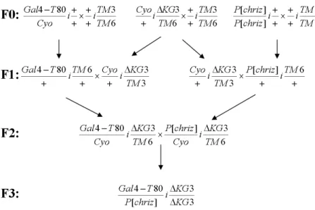
2.4.5 Crossing map of rescue assay . . . 43
2.4.6 Preparation of imaginal discs . . . 44
2.4.7 Immunostaining of imaginal discs . . . 45
2.4.8 Squash preparations of polytene chromosomes . . . 45
2.4.9 Preparation of gland extracts from third instar larvae . 47 3 Results 48 3.1 Molecular interaction between Chriz and Z4 . . . 48
3.1.1 Chriz directly interact with Z4 in yeast . . . 48
3.1.2 Chriz fragment (from 279-768aa) is sufficient for inter- action with Z4 in yeast . . . 49
3.1.3 Z4 N terminus interacts with Chriz . . . 51
3.1.4 Chriz fragment (500-768aa) can mediate Chriz selfin- teraction . . . 53
3.1.5 Test of direct Chriz-Z4 interaction by pull down exper- iments . . . 55
3.2 Genetic interactions between Chriz and Z4 alleles . . . 56
3.3 The activity of Chriz fragments are determined by comple- mentation . . . 58
3.3.1 overexpression of Chriz fragments . . . 58
3.3.2 Complementation and dominant negative effects by the Chriz fragments . . . 59
3.4 Identification of the domain for Chriz targeting to interband . 62 3.5 Effects of Chriz knockdown on chromatin protein binding . . . 63 3.5.1 Chromosome phenotype of Chriz knocked down flies . 63
3.5.2 Z4 protein binding following ChrizRNAi knockdown . . 66 3.5.3 Jil-1 and Histone 3 phosphorylated at S10 (H3pS10)
levels are decreased in ChrizRNAi lines . . . 70
4 Discussion 75
4.1 Chriz interactors . . . 75 4.1.1 The Chriz protein is a central element of a chromatin
complex located in interbands . . . 75 4.2 Chriz is responsible for targeting the complex to interbands . . 77 4.3 Chriz and the function of the complex . . . 79 4.3.1 The Chriz chromodomain . . . 79 4.3.2 Chriz is required for maintainance of the chromosome
structure . . . 80 4.3.3 Chriz is required for maintenance of Jil-1 activity in
interbands . . . 82 4.4 Targeting of Jil-1 by Chriz/Z4 is required for the interbands . 83
Bibliography 85
List of Figures 95
List of Tables 97
Chapter 1 Introduction
The genetic information, present on a linear DNA molecule of considerable length has to be condensed to fit inside a cell nucleus of usually several µm diameter only. Often over one metre of DNA is packaged into the nucleus in an orderly manner that allows for regulation nuclear activities, like transcrip- tion, replication and repair, to occur. This is accomplished by the wrapping of DNA around nucleosomes that fold into a structure known as chromatin.
This structure is subject to various modifications that have profound influ- ences on gene expression (Schones and Zhao, 2008). Previously, the control of transcription has been considered to be largely dependent on the genetic information provided by local DNA sequences. However, numerous biological phenomena cannot be explained by simple genetics, such as position effect variegation in the Drosophila in which the local chromatin environment of a gene determines its expression or paramutation in korn, that reflects an in- teraction between two alleles in which one allele causes heritable changes in the other allele (Goldberg et al., 2007). Apparently gene activity depends on the local chromatin structure provided by binding of proteins or RNAs, their modification as well as modification of the DNA molecule itself. Collectively for these processes the name epigenetics was created. Conrad Waddington (1905-1975), embryologist and professor of animal genetics, defined epige- netics as the branch of biology which studies the causal interactions between genes and their products, which bring the phenotype into being(Waddington,
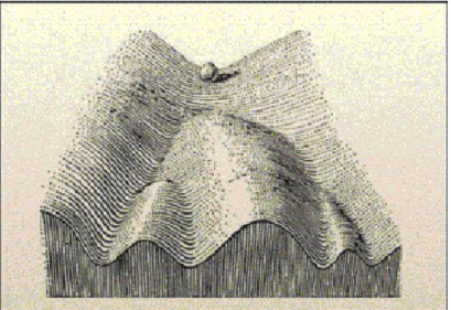
1942). Epigenetics, in a broad sense, is a bridge between genotype and pheno- type, a phenomenon that changes the final outcome of a locus or chromosome without changing the underlying DNA sequence (Goldberg et al., 2007). For example, even though the cells in a multicellular organism share an identi- cal genotype, organismal development produces a diversity of cell types with disparate, yet stable, profiles of gene expression and distinct cellular func- tions. Cellular differentiation may be considered as epigenetic phenomenon, largely governed by changes in what Waddington described as the epigenetic landscape provided by chromatin modification rather than by alterations in genetic inheritance (Goldberg et al., 2007) (Figure 1.1).
Figure 1.1: Waddingtons Classical Epigenetic Landscape. In 1957, Conrad Waddington proposed the concept of an epigenetic landscape to represent the process of cellular decision-making during development. At various points in this dynamic visual metaphor, the cell (represented by a ball) can take specific permitted trajectories, leading to different outcomes or cell fates (Goldberg et al., 2007).
Today’s epigenetic research is mainly focused on the study of covalent modification and noncovalent modifications of DNA and histone proteins and the mechanisms by which such modifications affect chromatin structure and cell fates. Noncovalent means chromatin structural remodelling and the incorporation of histone variants as a way to introduce variation into the
1.1. Chromatin
chromatin template. However, since my Phd work will not cover this field I will not introduce this in further detail.
1.1 Chromatin
Chromatin is composed of DNA, RNA and proteins, 50% of which are his- tones and the remaining proteins collectively called the nonhistone proteins.
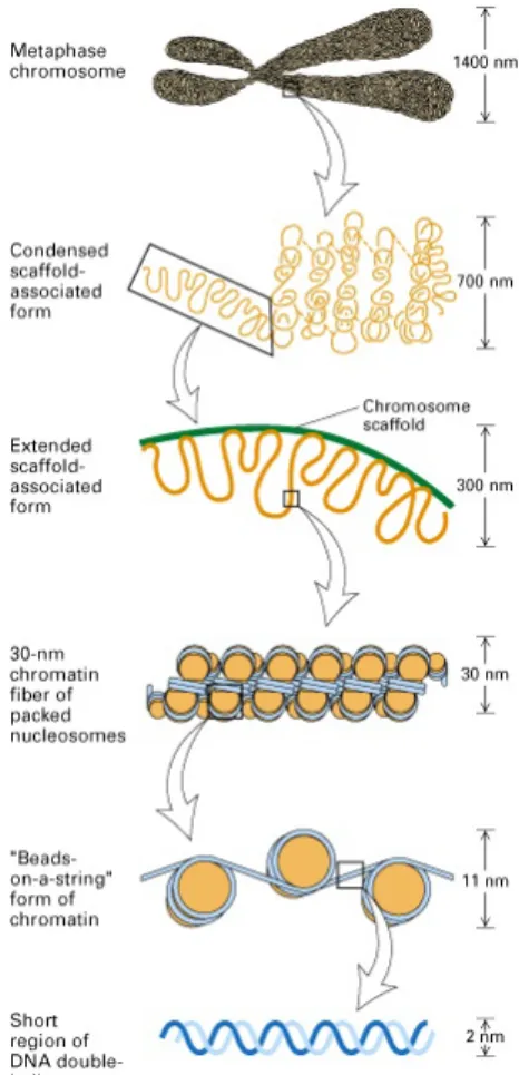
The basic structural unit of chromatin is the nucleosome. Nucleosomes com- prise 147 base pairs of DNA wrapped in a left-handed superhelix 1.7 times around a protein core, the histone octamer. The histone octamer core con- tains two molecules of each of the histones H2A, H2B, H3 and H4. The DNA connecting adjacent nucleosomes is called linker DNA. In addition to the core histone molecules, the histone H1 is also contained in chromatin and covers part of the linker DNA. DNA in the nucleosome is compacted into the 11nm fiber. This fiber folds into higher order structures, forming chromatin structures that locally differ in their degree of condensation (Figure 1.2).
1.2 DNA Methylation
Locally, the folding of the chromatin is dynamically regulated by several biochemical processes. Two important categories of epigenetic modifications are DNA methylation and histone modifications. DNA methylation is a rather well characterized chemical modification. In mammals, nearly all DNA methylation occurs on cytosine residues of CpG dinucleotides. Re- gions of the genome that have a high density of CpGs are referred to as CpG islands. Seventy to 80 percent of all CpG sites in human DNA are methylated. DNA methyltransferases are responsible for this modification, which adds activated methyl groups from a donor to cytosine residues at selected sites (Figure 1.3). Mammalian DNMTs have been identified (Bestor TH, 2000). DNMT3A and DNMT3B are thought to be involved in the es- tablishment of DNA methylation while DNMT1 is thought to be involved in the maintenance of this pattern through replication since their substrate is
1.2. DNA Methylation
Figure 1.2: DNA double helix coils are around the histone octomer to form a bead-like structure (11 nanometer diameter). histone proteins that form the octamer include H2a, H2b, H3, H4 (2 molecules each) that form a cylinder- like structure that binds DNA. A short sequence of DNA (10-70bp) connects the nucleosomal beads giving it a beads on a string-like appearance. The beads are packed together aided by histone H1 that binds to the beads, pulling them together to form the packed nucleosome or chromatin fiber that has a diameter of 30 nanometers. The chromatin fiber can be further looped or organized onto the nuclear matrix in interphase cells or condensed into chromosomes at metaphase (Rinehart, 2004).
1.2. DNA Methylation
hemimethylated DNA (Okano et al., 1999).
Figure 1.3: DNA methylation refers to the transfer of a methyl (CH3 group) to one of the bases that constitute DNA. The reaction is catalyzed by a DNA methyltransferase (Mtase), and uses S-Adenosyl Methionine (SAM) as a methyl donor. In humans, normal DNA methylation is limited to the Cytosine base (Anderson, 2008).
Alteration of DNA methylation patterns changes gene expression and genome stability by affecting chromatin structure and is therefore associated with a number of disorders (Jones and Baylin, 2002). Mechanistically, a methylated cytosine base can function to promote or preclude recruitment of regulatory proteins. In the former case, the methyl mark can be read through a family of methyl-CpG binding proteins thought to mediate transcriptional repression through interactions with histone deacetylases (Bird, 2002). Al- ternatively, the methyl mark can exclude DNA binding proteins from their target sites as was shown for CTCF binding at the Igf2/H19 region (Hark et al., 2000). Moreover, the formation of heterochromatin in many organisms is mediated in part by DNA methylation, binding of proteins to this mark in combination with RNA and histone modifications characteristic of silent chromatin (Zaratiegui et al., 2007).
For a long time the fruit fly Drosophila was considered to lack DNA methylation (Rae and Steele, 1979), but now there is evidence for a func- tional DNA-methylation system in Drosophila as well (Lyko et al., 2000).
Drosophila genomic methylation is restricted to the early stages of embryo development. However, the significance of this methylation is unclear until
1.3. Covalent histone modifications
of reduced methyltransferase expression (Lyko et al., 2000).
DNA methylation plays a role in many cellular processes including si- lencing of repetitive and centromeric sequences from fungi to mammals, X chromosome inactivation in female mammals and mammalian imprinting, all of which can be stably maintained by the hemimethylated DNA specific DNMT1 (Yang and Kuroda, 2007). Taken together, DNA methylation pro- vides a stable, heritable, and critical component of epigenetic regulation.
1.3 Covalent histone modifications
At the core of the nucleosomes are the highly conserved histone proteins (H3, H4, H2A, H2B). Once thought of as static, structural elements, it is now clear that histones are integral and dynamic components of the ma- chinery responsible for regulating gene transcription. An accumulating data shows post-translational modifications of histone proteins including acety- lation, phosphorylation, methylation, ubiquitination and ADP-ribosylation (Strahl and Allis, 2000) (Figure 1.4). Many of these modifications take place on the ’tail’ domains of histones. Indeed, such histone tail modifications can alter DNA-histone interactions within and between nucleosomes and, thus, influence nucleosomal and higher-order chromatin structures (Hansen et al., 1998; Wolffe and Hayes, 1999). Regarding this, histone covalent modifications alone or in combination influence a multitude of cellular processes, including transcription, replication, DNA repair and cell cycle progression.
1.4 Histone Acetylation
Histone acetylation neutralizes the positive charge of the target lysine and occurs at specific lysines on the four core histones (Figure 1.5). As a re- sult, histone acetylation can alter histone-DNA interactions, creating a more open chromatin architecture and it serves as a tag for protein binding (Shah- bazian and Grunstein, 2007). This modification is catalyzed by histone acetyltransferases (HATs) through the transfer of the acetyl moiety from
1.4. Histone Acetylation
Figure 1.4: The modifications include acetylation (ac), methylation (me), phosphorylation (ph) and ubiquitination (ub1). Most of the known histone modifications occur on the N-terminal tails of histones, with some exceptions including ubiquitination of the C-terminal tails of H2A and H2B and acety- lation and methylation of residues within the globular domain of H3 at K56 and K79, respectively. Globular domains of each core histone are represented as colored ovals (Bhaumik et al., 2007).
1.4. Histone Acetylation
acetyl-coenzyme A to theε-amino group of target lysine residues (Table 1.1).
Consistently, given that histone acetylation can create a more open chro- matin structure, many transcriptional coactivators, such as Gcn5/PCAF, CBP/p300 and SRC-1, have been shown to possess intrinsic HAT activity.
Histone acetylation can be reversed by the enzymatic action of the histone deacetylases (HDACs). The interplay between HAT and HDAC activities thus regulates cellular histone acetylation levels. Complementary to tran- scriptional coactivators possessing HAT activity, many transcriptional core- pressor complexes, such as mSin3a, NCoR/SMRT and NURD/Mi-2, contain subunits with HDAC activity (Denslow and Wade, 2007; Shahbazian and Grunstein, 2007).
Figure 1.5: Acetylation of lysine or phosphorylation of serine (MBMB, 2007).
1.5. Histone ubiquitination
1.5 Histone ubiquitination
Histone ubiquitination is catalyzed by the formation of an isopeptide bond between the carboxy-terminal glycine of ubiquitin and the amino-group of a lysine residue on histones. This bond is formed by the sequential catalytic actions of E1-activating and E2-conjugating enzymes and E3-ligases (Shilati- fard, 2006; Bhaumik et al., 2007). Whereas the same E1-activating enzyme is involved in the ubiquitination of all target proteins, different E2-conjugating enzymes are required for the ubiquitination of different substrates. E3-ligases provide protein target specificity (Shilatifard, 2006; Bhaumik et al., 2007).
Histone ubiquitination can be reversed by deubiquitinases. Often ubiquitina- tion is related to changes in protein conformation or degradation. However, there are also links to gene activation.Ubp8 associates with Gcn5-containing complexes (SAGA and SLIK) and its activity is needed for the full expression of SAGA- and SLIK-regulated genes (Daniel et al., 2004; Bhaumik et al., 2007). An activating role of ubiquitination is reported in transcriptional elongation by the histone chaperone FACT (Denslow and Wade, 2007). The mechanism of ubiquitination for providing this function is unclear. Maybe due to its huge size, ubiquitination physically keep chromatin open through wedging.
1.6 Histone phosphorylation
Histones are phosphorylated at specific sites during cell division (see Figure 1.4) (Barber et al., 2004; Bhaumik et al., 2007). Several distinct kinases are required for the phosphorylation of histones on different residues (Table 1). Phosphorylation of histone H2A is induced by a DNA-damage signaling pathway, and this modification is dependent on phosphatidylinositol-3-OH kinases, such as Mec1 in yeast (Foster and Downs, 2005; Bhaumik et al., 2007). Histone H2B phosphorylation is catalyzed by the sterile-20 kinase in yeast and Mst1 (mammalian sterile-20-like kinase) in mammals (Ahn et al., 2005). Phosphorylation at histone H3S10 and H3S28 during mitosis is regu- lated by the Aurora kinases, which are highly conserved from yeast to humans
1.6. Histone phosphorylation
Covalent modifications Enzymes
H3K4 methylation Set1 (Sc), SET7/SET9 (Hs), MLL (Hs), Smyd3 (Hs) H3K9 methylation
SUV39H1 & SUV39H2 (Mm, Hs), G9a (Mm, Hs) Eu-HMTase1 (Hs), ESET & SETDB1 (Mm, Hs), Clr4 (Sp), Dim5 (Nc), Kryptonite (At), Ash1 (Dm) H3K27 methylation E(z) (Dm), EZH2 (Hs, Mm)
H3K36 methylation SETD2/HYPB (Hs), NSD1 (Hs), Set2 (Sc) H3K79 methylation DOT1 (Sc), DOT1L (Hs)
H4K20 methylation Pr-SET7/SET8 (Hs, Dm), SUV4-20 (Hs),SET9 (Sp)
H3R2 methylation CARM1 (Mm, Hs)
H3R26 methylation CARM1 (Mm, Hs) H4R3 methylation PRMT1 (Hs), RMT1 (Sc) H3K9 acetylation Gcn5 (Sc), Src1 (Mm) H3K14 acetylation
Gcn5 (Tt, Sc), Src1 (Mm), TAF1 (Dm, Hs), CBP & p300 (Hs), Sas3 (Sc),MOZ & MORF (Hs), PCAF & hGcn5 (Hs)
H3K18 acetylation Gcn5 (Sc), CBP & p300 (Hs) H3K23 acetylation Gcn5 (Sc), CBP (Hs), Sas3 (Sc) H3K36 acetylation Gcn5 (Sc)
H3K56 acetylation Rtt109 (Sc)
H4K5 acetylation Esa1 (Sc), Hat1 (Tt, Dm, Hs), p300 (Hs), Tip60 (Mm), HBO1 (Hs)
H4K8 acetylation p300 (Hs), Esa1 (Sc), Tip60 (Mm), p300 (Hs), HBO1 (Hs)
H4K12 acetylation Hat1 (Sc), Esa1 (Sc), Tip60 (Mm) and CBP
& p300 (Hs), HBO1 (Hs)
H4K16 acetylation Mof (Dm), hMof (Hs), Sas2 (Sc), Tip60 (Mm), Esa1 (Sc)
H3S10 phosphorylation
Snf1 (Sc), Jil-1 (Dm), Rsk2 (Mm, Hs), Msk1 (Mm), lp11(Sc), Aurora B (Ce, Dm, Hs), NIMA (An)
H3S28 phosphorylation Aurora B (Mm, Hs) H4S1 phosphorylation Sps1 (Sc), CKII (Sc) H2BS phosphorylation
(S14 in human; S10 in yeast)
Mst1 (Hs), Ste20 (Sc) H2BK11 acetylation Gcn5 (Sc)
H2AS129 or H2AXS139
phosphorylation Tel1 & Mec1 (Sc), ATM & ATR & DNAPK (Hs) H2AK5 acetylation Tip60 (Hs, Dm)
H2AK119 ubiquitination Ring1B (Dm, Mm, Hs) H2BK ubiquitination
(K120 in human, K123 in yeast)
Rad6 (Sc), Bre1 (Sc), HR6A (Hs), HR6B (Hs)
H2AZK14 acetylation Esa1 (Sc), Gcn5 (Sc)
Table 1.1: The enzymes responsible for covalent histone modifications (Bhau- mik et al., 2007). An, Aspergillus nidulans; At, Arabidopsis thaliana; Ce, Caenorhabditis elegans; Dm, Drosophila melanogaster; Hs, Homo sapiens;
Mm, Mus musculus; Nc, Neurospora crassa; Sc, Saccharomyces cerevisiae;
Sp, Schizosaccharomyces pombe; Tt, Tetrahymena thermophila.
1.6. Histone phosphorylation
(Nowak and Corces, 2004). Other kinases of the MSK/RSK/Jil-1 family can mediate phosphorylation of histone H3 at Ser10 during interphase that is correlated to gene activity (Nowak and Corces, 2004; Bhaumik et al., 2007).
InDrosophila, H3S10 phosphorylation is also found in dividing cells (See (Adams et al., 2001)) and the gene responsible for phosphorylation of H3S10 at mitosis is known as Aurora B. In Aurora B RNAi cells, the phosphory- lation of H3S10 decreased during mitosis and resulted in a failure to recruit condensin to the chromosomes (Adams et al., 2001; Giet and Glover, 2001).
As a consequence many defects were observed including lack of sister kine- tochore separation, lagging chromatids, and extensive chromatin bridging at anaphase. These data support that the phosphorylation of histone H3S10 is essential for the regulation of mitotic processes. However, phosphorylation of H3S10 is also observed during interphase. The transcriptionally active heat- shock locus usually shows significant H3S10 phosphorylation. Intriguingly, Labrador (Labrador and Corces, 2003) observed that when transcription was driven from an hsp70 promoter, H3S10 was hyperphosphorylated, but when transcription was driven from a P-element transposase promoter, phospho- rylated H3S10 was not detected. Therefore, histone H3S10 phosphorylation may be only required in the context of specific promoters. Phosphoryla- tion of the histone H3S10 residue was also described to be associated with chromosomal condensation (Hendzel et al., 1997; Wei et al., 1999). How- ever, as already mentioned phosphorylation of H3S10 also has been found to occur at transcriptionally activated heat shock puffs (Nowak and Corces, 2000) implying a role for phosphorylation of histone H3S10 in establishment of the decondensed state in addition to its role in chromosome condensa- tion (Mizzen et al., 1998; Thomson et al., 1999). Furthermore, it has been demonstrated that MSK1/2 kinase activity and histone H3S10 phosphory- lation have roles in chromatin remodeling and gene transcription in mam- mals (Dunn et al., 2005). Thus, histone H3S10 phosphorylation is associated with two opposed chromatin states, highly condensed mitotic chromosomes and the relaxed chromatin of activated genes during interphase (Prigent and Dimitrov, 2003). This apparent contradiction has sparked the speculation that the effect of the H3S10 modification on chromatin structure might be
1.7. Histone methylation
context dependent and be influenced by adjacent epigenetic marks such as histone acetylation or H3S28 phosphorylation (Turner, 2000; Johansen and Johansen, 2006).
1.7 Histone methylation
Histones may be methylated on lysine residues, arginine residues or both.
Histone arginine methylation can occur in the mono- or dimethylated form at specific positions on histone H3 and histone H4 in higher eukaryotes (Figure 1.4). Histones also can be mono-, di-, or trimethylated on lysines 4, 9, 27, 36 and 79 of histone H3 and Lys20 of histone H4. The machinery and the sites of histone methylation are highly conserved from yeast to humans, in particular for methylation of H3K9, H3K27 and H4K20 (Kouzarides, 2007).
Unlike HATs, which can be promiscuous in their histone substrate specificity and can modify several residues within the same or different histones, histone methyltransferases are typically more specific for their targets. Almost all of the histone lysine methyltransferases characterized to date contain a SET domain, named after D. melanogaster Su(var)3-9, Enhancer of zeste (E(z)), and Trithorax (Trx) enzymes. SET domain containing enzymes can catalyze methylation of specific lysines on histones H3 and H4 (Table 1). Although some histone methyltransferases share the same substrate specificity, it is likely that each enzyme may regulate different genes or different cellular processes in vivo.
Set1 and Set2 have been shown to methylate Lys4 and Lys36 of histone H3, respectively (Shilatifard, 2006). Histone H3K4 methylation is a hallmark of actively transcribed genes. Therefore, the identification of complexes that mediate this modification has been the focus of many laboratories (Shilati- fard, 2006). The yeast Set1 protein associates with seven other polypeptides to form the COMPASS (complex of proteins associated with Set1). Several COMPASS components contain WD domains, also found in other trithorax- related complexes (Shilatifard, 2006). COMPASS was the first identified H3K4 methyltransferase and can catalyze the mono-, di- and trimethylation of this residue (Shilatifard, 2006; Bhaumik et al., 2007).
1.7. Histone methylation
More recently it was found that histone lysine methylation is also re- versible. Demethylation is carried out by two families of enzymes, amine oxidases such as LSD1 and hydroxylases of the JmjC family (Schneider and Shilatifard, 2006; Klose and Zhang, 2007; Shi, 2007). Because LSD1 requires a protonated methyl ammonium group, only mono- and dimethyl forms can be substrates. In contrast, JmjC family members can also demethy- late trimethylated lysines (Klose and Zhang, 2007; Shi, 2007). LSD1 can demethylate Lys4 or Lys9 of H3, depending on its associated proteins (Klose and Zhang, 2007; Shi, 2007). Many JmjC family members have unique sub- strate specificities, with demethylases for H3K4, H3K9, H3K27 and H3K36 being characterized recently (Shi, 2007). Despite the theoretical ability of JmjC members to demethylate mono-, di- or trimethylated lysine, it seems that some JmjC family members are more specialized. For exam- ple, D. melanogaster Lid only demethylates H3K4me3 (trimethylated H3K4) to H3K4me2 in vivo, although its mammalian homologs can also convert H3K4me2 to H3K4me1 (Bhaumik et al., 2007; Eissenberg et al., 2007; Iwase et al., 2007).
Histone methylation plays different roles in biological processes depend- ing on the site and type of the histone modified. Arginine methylation of histones H3 and H4 regulates transcriptional activation of steroid-responsive genes (Lee et al., 2005). Histone lysine methylation is also involved in the transcriptional process since Lys4 methylation on histone H3 in present at the 5 end of transcription units and is associated with the early elongating form of RNA polymerase II at actively transcribed genes (Shilatifard, 2006).
H3K36 methylation by Set2 is associated with the elongating form of RNA polymerase II and is found more towards the 3 end of transcribed genes (Shi- latifard, 2006). In contrast, H3K27, H4K20 and histone H3K9 methylation is linked to heterochromatin formation. The Suv39 protein methylates his- tone H3K9 and localizes to transcriptionally silent heterochromatin, where it recruits the transcriptional repressor protein HP1 (Richards and Elgin, 2002;
Ayyanathan et al., 2003; Grewal and Moazed, 2003). HP1 in turn recruits more Suv39 and both recruit H4K20 histonemethyltransferases which place further silence marks. However, the molecular mechanism of chromatin com-
1.8. Chromatin organization and histone modifications
paction by this protein complex is not known in detail. Like histone H3K9 methylation, histone H3K27 methylation is involved in gene silencing (Shi- latifard, 2006; Sims and Reinberg, 2006; Bhaumik et al., 2007). This often alters developmentally regulated genes that have to be permanently silenced in certain tissues.
1.8 Chromatin organization and histone mo- difications
Histone modifications main functions are: the establishment of global chro- matin environments and the local targeting of proteins or protein complexes for the orchestration of DNA-based biological processing. As to establish a global chromatin environment, modifications help to partition the genome into distinct domains such as euchromatin, where DNA is kept accessible, and heterochromatin, where chromatin is inaccessible for transcription. This may be a very local function, such as the modification of a nucleosome at the promoter or it may be more global like in the regulation of chromoso- mal domains or whole chromosomes. In any case, this requires the ordered recruitment of the machinery to unravel DNA, manipulate it and keep it an altered state or eventually to reset it to the initial chromatin state.
Text books describe two different types of chromatin environments in the genome, silent heterochromatin and active euchromatin. In reality however, both eu- and heterochromatin may be further subdivided in domains different in structure and function. Each of these domains is associated with a distinct set of modifications.
Heterochromatin that makes up to 30% of the total genome has a low gene density, a compacted appearance revealed by intense staining and is late replicating in S-phase. Heterochromatin is required for the protection of chromosome ends and functional centromeric heterochromatin is important for the separation of chromosomes in mitosis. Formation of facultative het- erochromatin is required for X inactivation and silencing of developmental regulators to avoid inappropriate expression. In mammals the silent hete-
1.8. Chromatin organization and histone modifications
rochromatic state is associated with low levels of acetylation and high levels of methylation of H3K9, H3K27 and H4K20. The recruitment of PRC-1 to H3K27me is required for developmental gene silencing and thought to be in- volved in the maintenance of the inactive X chromosome. The recruitment of HP1 to H3K9me is thought to play an important role in the maintenance of pericentric heterochromatin silencing.
Euchromatin is a large proportion of the genome. In this environment DNA has extended flexibility for biological output. Genes can be turned on or kept off, DNA can be unravelled for repair or replication. Euchromatin is less condensed reflected by faint staining of chromatin. It has a higher gene density and replicates early in S-phase. The histone modification pattern in euchromatin reflects this open chromatin state. In the transcriptionally inactive state, already significant levels of acetylation, methylation, and phos- phorylation can be detected on genes, but these are still insufficient to elicit transcription. Further enzymatic activities are necessary for transcription to take place and typically, actively transcribed euchromatin has high levels of acetylation and is trimethylated at H3K4, H3K36, and H3K79.
Impressive evidence for switching of opposing chromatin states (hete- rochromatin and euchromatin) emerged from (Ebert et al., 2004). Three copies of the Su(var)3-9 H3K9 methyltransferase result in a strong enhancer of PEV phenotype. This strain also shows increased H3K9 di and trimethy- lation throughout the genome, notably, also in many euchromatic regions.
Conversely, mutants were isolated showing a strong suppressors of PEV phe- notype. These mutants were special alleles of the Su(var)3-1 gene, which encodes the Jil-1 kinase that controls phosphorylation of H3S10 within eu- chromatin (Wang et al., 2001) and all alleles contained mutations in the C ter- minus of Jil-1, leaving the kinase activity unchanged. These striking results show that Su(var)3-1 can completely antagonize the function of Su(var)3-9 in heterochromatin assembly and spreading, and that it may act to main- tain a balance between euchromatin and heterochromatin. This antagonistic relationship is in support of the histone switch hypothesis (Fischle et al., 2003a) in which the functional readout of local chromatin may be modified by opposing histone modifications, such as methylation and phosphorylation.
1.8. Chromatin organization and histone modifications
Opposing chromatin modifications also can be responsible for creation of insulator elements required as boundaries between euchromatin and hete- rochromatin that are established or regulated by mutual exclusion or inhibi- tion of different histone modifications as shown for the globin domain (West et al., 2004). The insulator elements can be classified as enhancer-blocking or barrier elements depending on whether they interfere with enhancer- promoter interactions or act as barriers against the spreading of heterochro- matin. The latter class functions as described: in the globin domain the spreading of H3K9 trimethylated heterochromatin is blocked by USF de- pendent establishment of a local patch of H3K9 acetylated chromatin (West et al., 2004). The former class may exert its function by attaching the chro- matin fiber to a nuclear substrate such as the nuclear matrix, resulting in the formation of structural or functional chromatin loops. The best known examples of such looped domains are the globin loci in vertebrates, in par- ticular the chickenβ-globin locus. In chicken red blood cells the globin genes are coordinately regulated within a 33 kb chromosomal domain, which dif- fers by its chromatin structure from the flanking chromatin already early in development. Inherent to the concept gene activities within a given domain are kept insulated from the activity of adjacent domains. The globin domain is flanked by two constitutive hypersensitive regions, HS4 and 3’HS, whose function as boundary elements was shown by their ability to act as enhancer blockers and by conferring position-independent expression of transgenes in vertebrates and invertebrates. In addition, the CTCF protein, which also was identified as an essential boundary element factor in other species, is needed for their function. Upstream, the HS4 element blocks the spread- ing of a 16 kb heterochromatic chromatin region and inhibits the regulatory crosstalk with the strong folate receptor gene enhancer. The downstream element keeps the expression of the globin genes independent from that of the immediate flanking odorant receptor gene, which is normally expressed in olfactory epithelia and certain neurons only (Bell et al., 2001; Bulger et al., 2002; Eggert et al., 2004).
In Drosophila, the heat-shock locus in 87A7 is a well studied domain model. The two divergently transcribed hsp70 genes are flanked by strong
1.8. Chromatin organization and histone modifications
DNAseI hypersensitive sites which are part of the so called specialized chro- matin structure elements scs and scs’, respectively (Udvardy et al., 1985).
On heat-shock induction these elements mark the edges of a decondensed puff formed at this site. Both scs and scs’ elements function as bound- ary elements. They confer position-independent expression on Drosophila transgenes and are functional as enhancer blockers in transgene expression (Kellum and Schedl, 1991). The insulating activity of the scs element is me- diated by the Zeste-white 5 protein (Zw5) (Gaszner et al., 1999), whereas the scs’ activity is mediated by the boundary element associated factor 32 (BEAF-32) (Zhao et al., 1995). It was shown that Zw5 and BEAF-32 pro- teins interact. Results from ChIP experiments using Drosophila cell lines provided evidence that the scs and scs’ elements are close in space to each other in nuclei in vivo, although they are separated by 15 kb of intervening sequence on linear DNA (Blanton et al., 2003). This is consistent with a role of these elements in forming the base of a looped domain.
1.8.1 Drosophila polytene chromosome
Polytene chromosomes are a good model to study chromatin organisation since they have reproducible band-interband pattern that is visible in normal light microscopy. By cytophotometry and high resolution in situ hybridiza- tion data, the DNA packaging ratio in bands was determined as 60 to 100 fold and that of interbands to about 5 to 8 fold (Rykowski et al., 1988). Thus, interband chromatin structure is likely a 10-nm nucleosome fiber, whereas chromatin in bands is condensed at least into 30nm fibers (Rykowski et al., 1988).
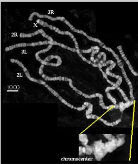
Polytene chromsomes are interphase chromosomes common to many Dip- tera, plants and Ciliates. Through repeated rounds of DNA replication with- out mitosis and cell division (called endoreplication), they become large bun- dles of chromatids with their chromomeres in register. As a consequence of polytenization, interphase chromosome organization becomes apparent where phase dark bands are separated by phase light interbands, resulting in a species-specific banding pattern. (see Figure 1.6). For unknown reasons,
1.8. Chromatin organization and histone modifications
the centromeric regions of the chromosomes are less replicated. As a result, the centromeres of all the chromosomes bundle together in a mass called the chromocenter.
Figure 1.6: Drosophila polytene chromosomes are viewed on light microscopy.
Chromosome arms and chromocenter are labelled (SEDAT, 2008).
Polytene chromosomes are usually found in the larvae, reflecting the mode of growth at that stage that occurs by hypertrophy of cells mainly. Because each cell now has many copies of each gene, it can express its genes more efficiently than with only two copies as in diploid cells. A similar pattern of bands is consistently observed in all polytene tissues down to the lowest level of polyteny amenable to cytological analysis. Thus, the band/interband pattern may reflect a common structural organization of interphase chromo- somes in general. Intuitively, the difference in compaction between bands and interbands suggests the existence of different mechanisms of formation of the two elements. This difference in chromatin organization may reflect important aspects of how gene expression is regulated (Semeshin et al., 2008).
1.9. Previous work of my project
1.9 Previous work of my project
However, little is known about the molecules and molecular mechanisms that are responsible for controlling the establishment and maintenance of these chromosomal subdivisions. In order to address this question interband spe- cific binding proteins are investigated in our group. We cloned the Z4 gene which encodes an 996 amino acid protein with a calculated molecular mass of 113KDa that is found in about 80% of all interbands. The protein has 7 zinc fingers of the classical C2H2-type, between amino acid 239-515 (Eg- gert et al., 2004). Z4 is an essential protein that is ubiquitously present in all tissues during embryonic and larval development and exerts its function within interphase, as it does not bind to the chromosomes during mitosis.
Z4 null (Z4-1.3) and hypomorphic (Z4-7.1) mutants were obtained by impre- cise excision. These mutations can be complemented by the 7.4 kb genomic region encoding the complete Z4 gene, resulting in viable adults. The ho- mozygous hypomophic mutation, Z4-7.1 shows a progressive disintegration of the chromosomal structure. Regions of the polytene chromosomes loose their band/interband organization and develop a decompacted and cloudy appearance. Z4 can directly bind to DNA, however so far no sequence spe- cific interaction has been found. It is still unclear, how Z4 is involved in structuring the chromosome. A possibility may be the modification of nucle- osomes. More recently a Z4 mutation was found independently that shows growth defects and therefore was called Putzig (Kugler and Nagel, 2007). As was shown in their paper, a consequence of down regulation of Z4 in the loss of H3K4 methylation in imaginal disc nuclei. H3K4-trimethylation is also a hallmark of interband chromatin. However, it still has to be demonstrated how loss of H3K4 methylation is connected to loss of Z4.
The Chriz protein was first found by coimmunoprecipitation with Z4- specific antibodies. Mass spectrometric analysis of gel purified protein iden- tified a chromodomain protein with an apparent molecular weight of about 140 kDa which was named Chriz, for Chromodomain protein interacting with Z4 (Eggert et al., 2004). Chriz is a single copy gene located in 79F on chromosome 3L close to the centromeric heterochromatin (Gortchakov
1.9. Previous work of my project
et al., 2005). The chromodomain, an evolutionary conserved protein module of about 50 amino acids, is located at the N terminal part of Chriz. Hete- rochromatin protein 1(HP1) and Polycomb (PC) also have chromodomains which mediate the specific recognition of methylated lysine residues K9 and K27 of histone H3, respectively (Nielsen et al., 2002; Fischle et al., 2003b).
Comparison of the Chriz chromodomain sequence with those of HP1 and PC reveals significant conservation in residues. Nevertheless, two out of the three aromatic residues in the chromodomain of HP1/PC that were identi- fied to be crucial for the recognition of histone H3 di- and trimethylysine are substituted in the chromodomain of Chriz for charged amino acids. Besides the chromodomain, no other strongly conserved domains were found in the protein database. Chriz perfectly colocalizes with Z4 and like Z4, Chriz is ab- sent from bands and from decondensed puffs. The latter observation rules out the possibility that selective binding to interbands would simply be explained by enhanced accessibility. Chriz is essentially required for development and also dosage sensitive since the ubiquitous overexpression of Chriz under the control of act-Gal4 and T80-Gal4 driver lines is early larval lethal. Using different driver lines with a more restricted GAL4 expression pattern, organ- and tissue- specific defects were observed. By imprecise excision, Chriz null and hypomorphic mutants were obtained which can be complemented with a 7.1kb genomic Chriz fragment (Gortchakov et al., 2005).
Chriz was independently identified as Chromator, as an interaction part- ner of the putative spindle matrix component Skeletor, that localizes to the spindle and to the centrosomes during mitosis (Rath et al., 2004). Further- more, functional assays using RNAi-mediated depletion in S2 cells suggest that Chromator directly affects spindle function and chromosome segrega- tion (Rath et al., 2004). However, localization of Chromator to polytene interbands suggested them that it also has a functional role in maintaining chromatin structure during interphase. They performed an EMS mutagen- esis screen that generated two new Chro hypomorphic alleles. The analysis of these alleles showed that impaired Chromator function leads to disorgani- zation and misalignment of band/interband regions resulting in coiling and folding of the polytene chromosomes (Rath et al., 2006). In addition, they
1.9. Previous work of my project
demonstrated that Chromator directly interacts with JIL-1 kinase and that the two proteins extensively co-localize at polytene interband regions(see below). These findings suggest that Chromator and JIL-1 interact in an interband-specific complex to establish or maintain polytene chromosome structure in Drosophila.
Drosophila Jil-1 is a tandem kinase, which localizes specifically to eu- chromatic interband regions of polytene chromosomes. Jil-1 is upregulated almost 2-fold on the hypertranscribed male X chromosome compared to au- tosomes. (Jin et al., 1999) and is the predominant kinase regulating histone H3S10 phosphorylation at interphase (Wang et al., 2001). Analysis of Jil- 1 null and hypomorphic alleles showed that Jil-1 is essential for viability and that reduced levels of JIL-1 protein lead to a misalignment of inter- band polytene chromatin fibrils that is further associated with coiling of the chromosomes and an increase of ectopic contacts between non-homologous regions (Zhang et al., 2003; Deng et al., 2005). This results in a shortening and folding of the chromosomes with a non-orderly intermixing of euchro- matin and the compacted chromatin characteristic of banded regions (Deng et al., 2005). The intermingling of non-homologous regions can be so exten- sive that these regions become fused and confluent, further shortening the chromosome arms. Based on these findings a model was proposed where JIL-1 functions to establish or maintain the parallel alignment of interband chromosome fibrils as well as to repress the formation of contacts and inter- mingling of non-homologous chromatid regions (Deng et al., 2005). Reducing the level of JIL-1 results in the spreading of the major heterochromatin mark- ers dimethyl H3K9 and HP1 to ectopic locations on the chromosome arms, with the most pronounced increase on the X chromosomes. Genetic interac- tion demonstrated that JIL-1 functions in a pathway that includes Su(var)3-9 (Zhang et al., 2006). By using a LacI-tethering, it was shown that Jil-1 me- diated ectopic histone H3S10 phosphorylation that is sufficient to induce a change in higher-order chromatin structure from a condensed state to a more open euchromatic state(Deng et al., 2008).
1.10. The aim of my work
1.10 The aim of my work
Studies of the relationships between nuclear architecture and chromatin bind- ing proteins and histone modifications reveal novel principles underlying nu- clear compartmentalization (Branco and Pombo, 2007). The chromatin as- sociated complex Z4/Chriz/Jil-1 was identified by us and others and accu- mulating evidence shows that it may play a role in the maintenance of the Drosophila polytene chromosome structure. The proteins colocolize in most interbands, they interact with each other and mutants result in loss of chro- matin structure. However, there are still many questions to be answered.
So the aim of my work is to address the mechanism of the complex func- tion in Drosophila chromatin structure. How do the proteins interact with each other? Z4 and Chriz were identified by our group by CoIp. It is clear that they are in the same complex, but whether they interact directly is not known. Both of the proteins have conserved domains, and I became inter- ested if these domains may mediate the observed interactions. The second question was how the proteins and the complex as a whole were targeted to interbands. Z4, Chriz and Jil-1 all are interband specific proteins whereby Z4 and Chriz have identical chromosomal distribution, colocalizing largely with Jil-1. Having identified the targeting region it would be interesting to unravel which protein is the fundamental factor for interband targeting of the complex. The third question relates to the function of the complex in interbands. Interbands are considered as less condensed compared to bands.
Intuitively, this is related to local chromatin modification. So I will use avail- able RNAi lines to knockdown the Chriz and Z4 protein level to elucidate consequences on protein binding, modification and chromatin structure.
Chapter 2
Material and Methods
2.1 General used molecular biological appli- cations
2.1.1 Bacteria and Yeast strains
The E.coli strains, XL-1, BL21 and the yeast strain SFY56 used for protein expression and for protein-protein interaction testes are summarized in Table 2.1.
Strains Genotype Reportergene
E. coli XLI
recA1 endA, gyrA96 thi-1 hsdR17 supE44 relA1, lac [F’, proAB, lacIqZDM15, Tn10 (Tetr)]c E. coli BL21
(DE3) pLysS
E. coli B, F-, dcm, ompT, hsdS(r−Bm−B), gall(DE3) [pLysS Camr].
S. cerevisiae SFY526
MATa, ura3-52, his3-200, ade2-101, lys2-801, trp1-901, leu2-3, canr, gal4- 542, gal80-538,URA3::GAL1U AS- GAL1T AT A-LacZ
LacZ
Table 2.1: Bacteria and yeast strains used in this thesis.
The E.coli strain XL-1 was used for the cloning and amplification of plasmids. The BL-21 was used for the expression of protein. The yeast
2.1. General used molecular biological applications
strain SFY was used for the protein-protein interaction in yeast two hybrid assay.
2.1.2 Plasmids
All plasmids that have been used in this work are listed in Table 2.2. The property of these plasmids, their application, resistence and their source sup- ply are as indicated. All of these plasmids have the ability to be replicated in E. coli cells and they allow the selection of cells which contain them since they give their host bacteria an antibiotic resistance. The plasmids pGEX- 2T, pGEX-6p-1 and pET−(Myc)3−(His)6 are used to express GST- and MH- recombinant proteins respectively. The pUAST are P-element based plasmids used to incorporate genes into the Drosophila genome by embry- onic injection. pπ25.7 wc plasmids are used to express transposase to help pUAST integrate into genome of fruit fly.
Additionally, two other yeast plasmids, pGBT9 and pGAD424 were used in the yeast two hybrid system for analysing protein-protein interaction. The selection marker for these two plasmids is the growth of the yeast cells of on medium lacking Tryptophan/Leucin (Trp/Leu). The pGAD424 and pGBT9 plasmids were applied to express Activation Domain (AD) fusion proteins and DNA Binding Domain (DBD) fusion proteins respectively.
2.1.3 PCR and cloning of plasmid constructs
Different plasmids which were used for the working with yeast, bacteria and fruit fly were constructed. The cloning of these plasmids occurred in the following procedures.
Polymerase chain reaction (PCR)
Polymerase chain reaction provide a basis to amplify DNA fragments and to create also a new enzyme restriction site in the amplified DNA fragment needed for the cloning. Specific enzyme restriction sites were introduced by
2.1. General used molecular biological applications
Name Application Resistence/
selection marker
Source of supply
PET3a- Myc-His
Myc-His fusion pro- tein expression in BL21 E.coli cell
Amp/- Novagen Rosen-
berg et al., 1987;
PGEX-6p- 1
GST fusion protein expression in BL21 E.coli cells
Amp/- Invitrogen
PGEX- 2TK
GST fusion protein expression in BL21 E.coli cells
Amp/- Pharmacia
Biotech Smith and Johnson, 1988
pUAST Expression in Dro- sophila (Gal4/UAS system)
Amp/ mini white gene
Brand & Perirri- mon 1993
Pπ25.7 wc Helper Plasmid Amp/- Lab stock pGBT9 DBD fusion protein
expression in yeast cells
Amp/ TRP Clontech
pGAD424 AD fusion protein ex- pression in yeast cells
Amp/LEU Clontech
Table 2.2: Lists of plasmids are used for cloning strategies and for expression of fusion proteins. Shown are also the application, the resistance and selection marker and the source of supply of each of these plasmids.
2.1. General used molecular biological applications
using the following primers (Table 2.3) in order to amplify and clone the Chriz and Z4 constructs.
PCR is performed using the following standard reaction mixture:
Template DNA 50-100 ng
10x amplification buffer 1:10 of the final volume
Mixture of dNTPs 4 µM
Primer1 10µM ('100pmol)
Primer2 10µM ('100pmol)
Taq DNA polymerase 5 Units H2O to a final volume of 50µl
Primer used in this work
Name Primer sequence Enzyme site
chrizR72 Ttagcgtcgactcgatcctaatggctatgc SalI chrizR92 Ttagcgtcgactacgttgggatgttgagcg SalI chrizF92 Tctagtcgacggcacgccaaaaacttact SalI chrizR82 Ttagcgtcgacttggtcttggaagttcgag SalI chrizF82 Tctagtcgacatcaccaaccggtaagctg SalI chrizF72 Tctagtcgacaatgttggcacaggagattt SalI chrizF73 Gtattgcggccgcttggcacaggagatttc NotI chrizF83 Gtattgcggccgcaccagaaaatcaccaac NotI chrizF93 Gtattgcggccgcgcacgccaaaaacttac NotI chrizF4g Gtcagatctttggcacaggagatttcacct BglII chrizF5g Gtcagatctcgcatagccattaggatcgat BglII chrizF6g Gtcagatctcaattggcacgccaaaaact BglII chrizR7 Ttagcggtacctcgatcctaatggctatgc KpnI chrizR8 Ttagcggtaccttggtcttggaagttcgag KpnI chrizR9 Ttagcggtacctacgttgggatgttgagcg KpnI chrizF4 tctggatccttggcacaggagatttcacct BamHI chrizF5 tctggatcccgcatagccattaggatcgat BamHI chrizF6 tctggatcccaattggcacgccaaaaact BamHI chrizR4 Gacgaattctcgatcctaatggctatgcg EcoRI
2.1. General used molecular biological applications
chrizR5 Gacgaattctccagacccttctccctagt EcoRI chrizR6 Gacgaattcttacgttgggatgttgagcg EcoRI chrizF7 Atcgaggtacctaatgttggcacaggagat KpnI chrizF8 Atcgaggtaccaatcaccaaccggtaagct KpnI chrizF9 Atcgaggtacctggcacgccaaaaacttac KpnI chrizF10 Gcggccgctgcagcagtcgctaagcgcttc NotI chrizR10 Tgctgcagcggccgcctcttcggtgcgctg NotI chrizF11 Gatgaagagaccgacgttgatcgctcgcat
chrizR11 Gtcggtctcttcatcctcgtgggtcttacg chrizF12 Gccgcgttcgatgacgtgatggccaatctc chrizR12 Gtcatcgaacgcggcatgatgcgagcgatc
chrizF1 Tacgaattcttggcacaggagatttcacct EcoRI chrizF2 Tacgaattccgcatagccattaggatcgat EcoRI chrizF3 Tacgaattccaattggcacgccaaaaact EcoRI chrizR1 Tgactgcagtcgatcctaatggctatgcg PstI chrizR2 Tgactgcagtccagacccttctccctagt PstI chrizR3 Tgactgcagttacgttgggatgttgagcg PstI chrizF10-1 Gccgcagtcgatgacgtcgctaagcgcttc
chrizR10-1 Gtcatcgactgcggcctcttcggtgcgctg
Z4R1 Tgacagatctcttggtggccttgcgttgga BglII chrizF2-1 Tgacgaattctcctcagttccctctgccgg EcoRI Z4F1 Tgacgaattcaacaaccaactgaatccggc EcoRI Z4R2 Tgacagatctagttgccttcttgtcacgtc BglII Z4F2 Tgacgaattcgtccaacgcaaggccaccaa EcoRI Z4F3 Tgacgaattcagacgtgacaagaaggcaac EcoRI Z4R3 Tgacagatctgtcggtctttgtctccgtaa BglII Z4R4 Tgacctcgagcttggtggccttgcgttgga XhoI Z4R5 Tgacctcgagagttgccttcttgtcacgtc XhoI Z4R6 Tgacctcgaggtcggtctttgtctccgtaa XhoI CF1 Gactgaattccttactagggagaagggtct EcoRI CR1 Gactctgcagcgaaacgggtgccggtgcct PstI
2.1. General used molecular biological applications
CF2 Gactgaattccgggttgaaaattccgaggc EcoRI CR2 Gactctgcagtacctggtaaacagtgccgt PstI CF3 Gactgaattccaagctcagttgtgtccaat EcoRI
Table 2.3: List of the primers used in this work. F: for- ward primer; R: reverse primer.
Overlap PCR
Overlap Extension PCR is used to create long DNA fragments from short ones or changing its genetic code through mutations. In this work, I used overlap PCR to create Chriz chromodomain mutant1 and mutant3, in which the mutant amino acids were introduced by PCR primers. First Chriz primer F73 and R10-1, F10-1 and R9 were used as two pair primers to amplify two fragments using Chriz gene as template. These two fragments have 15 bp overlap, in which were the mutated sequence. Gel extraction was used to clear up the 690bp and 2100bp correct size band. The new round PCR reaction was set up using cleaned up fragments as "template" without adding new primers into the reaction tube. Proofreading enzyme was used for extension.
Ten PCR cycles were run without end primers (Template extension step).
F73 and R9 primers were added, then continue PCR cycling for another 20 rounds. The corrected band Chriz mutant 1 (2770bp) product was extracted from Gel. Chriz mutant1 contruct were cloned into pUASTmychis vector with NotI and KpnI enzyme. Chriz mutant 3 were also constructed in this way by using F73-R11, F11-R9, F73-R12 and F12-R9 primers. See Figure 2.1 for procedure of overlap PCR.
2.1.4 Digestion of DNA with restriction enzyme
Enzymes were purchased from New England Biolabs. For each digestion, the following solution were added and mixed in a 1.5 ml microcentrifuge tube.
2.1. General used molecular biological applications
Figure 2.1: Procedure of overlap PCR (Heckman and Pease, 2007).
DNA up to 1 µg
10×buffer 2 µl
Enzyme 10 unit
H2O up to 20 µl
The mixture was incubated at 37◦C for 2-3 hours.
2.1.5 DNA ligation
The ligation of the insert (PCR fragment) to the specific plasmids (vectors) was prepared as following.
In a microcentrifuge tube the following reaction mixture were mixed and incubated for 16 hours at 22◦C (Table 2.4).
2.1.6 Setting up competent cells
The preparation of competent bacteria was done according to Hanahan pro- tocol (Hanahan, 1983) with changing.
2.1. General used molecular biological applications
Linear vector DNA 5-10 µl (50-400 ng)
Insert DNA use a 1:1 up to a 3:1 molar ratio of insert DNA to vector DNA 10× ligation buffer for T4
DNA Ligase
2 µl
Water to 20 µl
T4 DNA Ligase 0.2-0.4 µl (1-2 u) for sticky ends;
1µl (5 u) for blunt ends Table 2.4: DNA digestion procedure.
First day, frozen stock of XL1 bacteria were streaked on LB plate and incubated overnight at 37◦C. The second day, a single colony was inoculated into a 4ml LB medium, which grow overnight with shaking at 37◦C. The next day, one flask containing 400 ml LB medium were set up. Two ml overnight culture was given to the flask. The cell culture was incubated with shaking until it reaches an OD600 of ∼ 0.4-0.5. Bacteria are centrifuged at 4000 rpm at 4◦C for 10 minutes and resuspend the pellet on ice gently in 60 ml TFBI. After cooling on ice for 10 min, centrifuge bacteria at 4000 rpm, 4 ◦C for 10 minutes. Resuspend the pellet gently on ice in 8 ml of TFBII and immediately dispense 100 µl of cell suspension into each cold microcentrifuge tubes on ice. Once all 8 ml of competent cells are dispensed into microcentrifuge tubes, immediately freeze them in liquid nitrogen and stored at -80◦C.
• LB medium:
– Tryptone (10 g/l) – Yeast extract (5 g/l) – NaCl (5 g/l)
• TBFI solution:
– 100 mM Rubidium Chloride – 50 mM Manganese Chloride – 30 mM Potassium Acetate
2.1. General used molecular biological applications
– 10 mM Calcium Chloride – %15 w/v Glycerol
– Adjust pH to 5.8 with acetic acid 2M and Sterilize by filtration
• TFBII solution:
– 10 mM MOPS
– 10 mM Rubidium Chloride – 75 mM Calcium Chloride – 15% w/v Glycerol
– Sterilize by filtration
2.1.7 Bacterial Transformation
Frozen competent cells were taken out from -80◦C and place on ice, waiting for cells to thaw. Note: Keep cells chilled on ice to ensure high transformation efficiency.
Before adding DNA, it is better to mix cells by flicking the tube gently, then 100 µl per transformation was taken into a sterile pre-chilled (on ice) microcentrifuge tube. 1-50ng of DNA (in a volume no greater than 10µl) was added to 100µl cells. Quickly flick the tube several times to ensure the even distribution of DNA. Tubes were immediately placed on ice for at least 10 minutes. Heat shock the cells for 45-50 seconds in a water bath at exactly 42◦C without shaking. Then tubes were immediately placed on ice for 2 minutes. 800µ of room temp (or 37◦C) LB medium were added and incubated for 1 hour at 37◦C with shaking at ∼150 rpm. 100-800 µl of the transformation mix was plated onto antibiotic plates. The plating volume depends on the concentration of DNA. The cells may be pelleted by centrifugation at 1000 rpm for 1 minute, then the cells can be resuspended in 50-200 µl of LB medium and plated. (The maximum amount of solution that may be spread on a plate is ∼200 µl). For the positive control DNA, a 1:100 to 1:1000 dilution is recommended for plating on LB plates. Plates
2.1. General used molecular biological applications
the cell growth rate (XL1 usually grow slower than BL21; it’s better to keep an eye on the growth of the cells the next day).
For the positive control DNA, a 1:100 to 1:1000 dilution is recommended for plating on LB plates. Plates were put in the 37◦C incubator and grow overnight 14-18 hrs depending on the cell growth rate (XL1 usually grow slower than BL21; it’s better to keep an eye on the growth of the cells the next day).
2.1.8 Mini and Midi DNA preparation
Mini DNA preparation was done according to standard molecular cloning protocol. You may find the detail in molecular cloning book (Sambrook et al., 2001). The midi DNA preparation was done according to Hispeed plasmid midi kit protocol (Qiagen).
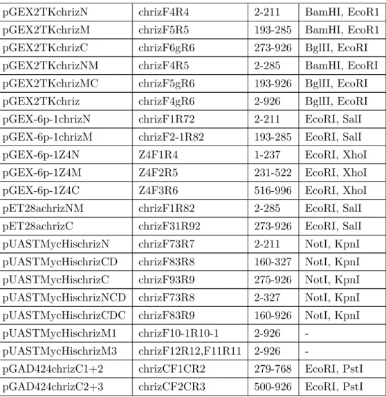
In this work, I have constructed 35 plasmids for bacterial expression, yeast two hybrid and fly work respectively. All the plasmids are listed as following. Their names, cloning enzyme sites and protein regions are also indicated (Table 2.5).
Name of plasmids Used primers Regions
(AA) Cloning site
pGAD424chriz chrizF1R3 2-926 EcoR1, PstI
pGAD424chrizN chrizF1R1 2-211 EcoR1, PstI pGAD424chrizM chrizF2-1R2 193-285 EcoR1, PstI pGAD424chrizC chrizF31R3 273-926 EcoR1, PstI pGAD424chrizN M chrizF1R2 2-285 EcoR1, PstI pGAD424chrizMC chrizF2-1R3 193-926 EcoR1, PstI
pGBT9Z4 Z4F10R10 1-996 EcoRI, BglII
pGBT9Z4N Z4F1R1 1-237 EcoRI, BglII
pGBT9Z4M Z4F2R2 231-522 EcoRI, BglII
pGBT9Z4C Z4F3R3 516-996 EcoRI, BglII
pGAD424chrizC1 chrizCF1CR1 279-509 EcoRI, PstI pGAD424chrizC2 chrizCF2CR2 500-768 EcoRI, PstI pGAD424chrizC3 chrizCF3CR3 700-926 EcoRI, PstI
2.1. General used molecular biological applications
pGEX2TKchrizN chrizF4R4 2-211 BamHI, EcoR1 pGEX2TKchrizM chrizF5R5 193-285 BamHI, EcoR1 pGEX2TKchrizC chrizF6gR6 273-926 BglII, EcoRI pGEX2TKchrizNM chrizF4R5 2-285 BamHI, EcoRI pGEX2TKchrizMC chrizF5gR6 193-926 BglII, EcoRI pGEX2TKchriz chrizF4gR6 2-926 BglII, EcoRI pGEX-6p-1chrizN chrizF1R72 2-211 EcoRI, SalI pGEX-6p-1chrizM chrizF2-1R82 193-285 EcoRI, SalI
pGEX-6p-1Z4N Z4F1R4 1-237 EcoRI, XhoI
pGEX-6p-1Z4M Z4F2R5 231-522 EcoRI, XhoI
pGEX-6p-1Z4C Z4F3R6 516-996 EcoRI, XhoI
pET28achrizNM chrizF1R82 2-285 EcoRI, SalI pET28achrizC chrizF31R92 273-926 EcoRI, SalI pUASTMycHischrizN chrizF73R7 2-211 NotI, KpnI pUASTMycHischrizCD chrizF83R8 160-327 NotI, KpnI pUASTMycHischrizC chrizF93R9 275-926 NotI, KpnI pUASTMycHischrizNCD chrizF73R8 2-327 NotI, KpnI pUASTMycHischrizCDC chrizF83R9 160-926 NotI, KpnI pUASTMycHischrizM1 chrizF10-1R10-1 2-926 -
pUASTMycHischrizM3 chrizF12R12,F11R11 2-926 -
pGAD424chrizC1+2 chrizCF1CR2 279-768 EcoRI, PstI pGAD424chrizC2+3 chrizCF2CR3 500-926 EcoRI, PstI
Table 2.5: list of constructs in this work.
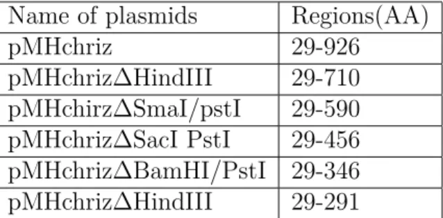
In addition to the above plasmids, the following plasmids were also used in this work. They were constructed by former Phd students Gortchakov AA(Gorchakov et al., 2004). Table 2.6.
2.2. Protein -protein interaction assays
Name of plasmids Regions(AA)
pMHchriz 29-926
pMHchriz∆HindIII 29-710 pMHchirz∆SmaI/pstI 29-590 pMHchriz∆SacI PstI 29-456 pMHchriz∆BamHI/PstI 29-346 pMHchriz∆HindIII 29-291
Table 2.6: List of constructs taken from Gortchakov AA.
2.2 Protein -protein interaction assays
2.2.1 Yeast two hybrid assay
In order to study Chriz-Z4 interaction, I performed yeast two hybrid assay and GST pull down. Yeast two hybrid system is working with SFY526 yeast cells. Two plasmids pGBT9 and pGAD424 are used to achieve Chriz-Z4 interaction test. The truncations of Chriz gene were cloned into pGBT9 and pGAD424, the truncations of Z4 gene were were cloned into pGBT9 as shown in Table 5. Yeast competent cells and LiAc yeast transformation procedure are as following.
One ml of YPD medium was inoculated with several colonies, 2-3 mm in diameter from the fresh prepared working stock plate. Vortex vigorously for 5 min to disperse clumps and were transferred into a flask containing 50 ml of YPD. Incubate at 30◦C for 16-18 hr with shaking at 250 rpm to stationary phase (OD6001.5). Next day, transfer 30 ml of overnight culture to a flask containing 300 ml of YPD. The OD600 of the diluted culture were measured up to 0.2-0.3. Incubate at 30◦C for 3 hr with shaking at 230 rpm until an OD600 of 0.4-0.6. Cells were placed in 50 ml tubes and centrifuge for 5 min at 1000 rpm at room temperature.
The supernatants were discarded and thoroughly resuspend the cell pel- lets in 25-50 ml sterile TE or distilled H2O. Centrifuge at 1000 rpm for 5 min at room temperature. The supernatants were discarded. The cell pellets were resuspended in 1.5 ml of freshly prepared, sterile 1X TE/1X LiAc. Add 0.1 µg pGBT9Z4, 0.1 µg pGAD424Chriz plasmids DNA and 0.1 mg herring
2.2. Protein -protein interaction assays
testes carrier DNA to a fresh 1.5 ml tube and mix. Since I wanted to test different fragments of Chriz and Z4 interaction, so different combination of pGBT9Z4 fragments with pGAD424Chriz fragments were added to individ- ual tubes respectively. 0.1 ml of yeast competent cells were added to each tube and mixed well by vortexing. 0.6 ml of sterile PEG/LiAc solution was added to each tube and vortexed at a high speed for 10 sec to mix. Incubate all the tubes at 30◦C for 30 min with shaking at 200 rpm. Later added 70 µl of DMSO to each tube and mixed well by gentle inversion. In a 42◦C water bath heat shocked them for 15 min. Chill cells for 2 min on ice. Cells were centrifuged for 5 sec 14,000 rpm at room temperature. Then I removed the supernatant. Cells were resuspended in 0.5 ml of sterile 1XTE buffer.
Finally, the cells were plated on SD agar plates lacking Trp and Leu nutrient that will select for the desired transformants.
Plates were incubated at 30◦C for 2-4 days until colonies appear. The growing clones indicated that both pGBT9Z4 and pGAD424Chriz plasmids were successfully transformed into the yeast cells.
• YPD medium:
– 20 g/l Difco peptone – 10 g/l yeast extract – 20 g/l agar (for plate)
• PEG/LiAc solution (polyethylene glycol/lithium acetate) per 10 ml solution:
– PEG 4000: 8 ml of 50% PEG – TE buffer: 1ml of 10X TE – LiAc: 1ml of 10X LiAc
• 50% PEG:
– 50g PEG4000 in 100 ml sterile deionized H2O
• 10× TE buffer: