Functional characterisation of the Sterile 20 like kinase Slik in tracheal morphogenesis in Drosophila melanogaster
Inaugural-Dissertation
zur
Erlangung des Doktorgrades
der Mathematisch-Naturwissenschaftlichen Fakultät
der Universität zu Köln
vorgelegt von
Fiona Paul Ukken
aus Cochin, Indien
Köln, Februar 2011
1. Berichterstatter: Prof. Dr. Maria Leptin
2. Berichterstatter: Prof. Dr. Siegfried Roth
Tag der mündlichen Prüfung: 3 Februar, 2011
Table of Contents
1. INTRODUCTION... 5
1.1 Tubular organs - an overview ... 5
1.2 Tracheal morphogenesis in Drosophila ... 5
1.3 Terminal cell and branch development ... 9
1.4 Factors influencing terminal branch development ... 12
1.5 Moesin is essential for terminal cell development ... 13
1.6 slik, a Sterile 20 like kinase involved in Drosophila growth control ... 14
1.7 Functional relevance of slik in Drosophila development ... 16
1.8 Aim ... 20
2. MATERIALS AND METHODS ... 21
2.1 Materials ... 21
2.1.1 Drosophila melanogaster stocks ... 21
2.1.2 UAS Transgenes ... 21
2.1.3 Gal4 driver lines ... 22
2.1.4 Mutants ... 22
2.1.5 Antibodies ... 22
2.1.6 Oligonucleotides ... 22
2.1.7 Microscopes ... 23
2.1.8 Imaging and data analysis software ... 23
2.1.9 Reagents ... 23
2.2 Methods ... 24
2.2.1 Collection of embryos ... 24
2.2.2 Immunostaining in embryos... 24
2.2.3 RNAi in the tracheal system ... 24
2.2.4 Immunostaining in larvae... 25
2.2.5 Generation of slik
1MARCM clones in the tracheal system ... 25
2.2.6 Tracheal cDNA synthesis ... 25
2.2.7 RT PCRs ... 26
3. RESULTS ... 27
3.1 Slik is required for tracheal development ... 27
3.1.1 Slik is expressed in both the embryonic and the larval tracheal system ... 27
3.1.2 Slik is required for normal branching, lumen formation and tube stability in terminal cell development ... 31
3.1.2.1 Analysis of the tracheal system in slik
1mutant animals ... 33
3.1.2.2 Analysis of slik
1MARCM clones in the tracheal system ... 35
3.1.2.3 Analysis of slik mutant tracheal system using RNAi ... 37
3.2. Slik, Moesin and Merlin in tracheal development ... 40
3.2.1 Moesin and its activated form (p-Moesin) are expressed in the tracheal system ... 41
3.2.2 Merlin is expressed in the tracheal system ... 43
3.3 Slik and Moesin colocalise at the apical membrane in terminal cells... 46
3.4 Knockdown of moesin in the tracheal system results in phenotypes similar to slik RNAi... 46
3.5 Knockdown of Merlin and expanded in the larval tracheal system ... 47
3.6 Knockdown of slik leads to loss of activated Moesin in terminal cells ... 49
3.7 Slik’s kinase function is critical in terminal cell lumen formation ... 50
3.8 Breathless regulates expression of p-Moesin in the tracheal system ... 52
3.8.1 p-Moesin levels are affected by breathless knockdown ... 52
3.8.2 breathless RNAi does not affect slik expression in terminal cells ... 54
Table of Contents
3.8.3 Moesin is a phosphorylation target specific to Breathless ... 55
3.9 Knockdown of RTK/MAPK pathway components affect terminal cell branching ... 56
3.9.1 Effect of breathless RNAi in the larval tracheal system ... 56
3.9.1.1 Knockdown of breathless disrupts branching in terminal cells ... 57
3.9.1.2 breathless RNAi results in abnormal morphology of the cells of the dorsal trunk ... 57
3.9.2 Knockdown of Ras disrupts branching in terminal cells ... 58
3.9.3 Effect of raf RNAi on terminal cell development ... 59
3.9.3.1 Knockdown of raf branching in terminal cells ... 59
3.9.3.2 Knockdown of raf results in cystic lumen within terminal cells... 60
3.9.3.3 Knockdown of raf results in multilumen phenotype in terminal cells ... 61
3.9.4 Knockdown of srf disrupts branching in terminal cells ... 62
3.9.5 Knockdown of egfr disrupts branching in terminal cells ... 63
3.9.6 Evaluation of branching phenotype in mutants of RTK and downstream components ... 64
4. DISCUSSION ... 68
4.1 Persistent Slik expression in all stages of tracheal development ... 68
4.2 Slik is important for tracheal development in Drosophila ... 69
4.3 Moesin is essential for tube formation and stability in terminal cells ... 70
4.4 Kinase activity of Slik is essential for its function in terminal cells ... 71
4.5 FGF/RTK Breathless regulates Moesin in trachea ... 72
4.6 Possible function of Merlin in terminal cell development ... 74
4.7 Does Slik modulate the MAPK pathway to regulate growth of terminal cells? ... 75
5. REFERENCES ... 77
6. APPENDIX ... 84
6.1 Terminal cell counts from tracheal segments tr3-5 from wild type and slik RNAi larvae ... 84
6.2 Knockdown of btsz leads to lumen formation and branching defects in terminal cells ... 84
6.3 Branch counts from various knockdowns in the tracheal system ... 85
6.4 Overexpression of phosphomimetic form of Moesin in the tracheal system ... 85
6.5 Knockdown of egfr does not affect p-Moesin localisation in terminal cells ... 86
6.6 Localisation of F-actin in developing embryonic tracheal system... 86
ABBREVIATIONS ... 87
ABSTRACT ... 89
ZUSAMMENFASSUNG ... 90
EIDESSTATTLICHE ERKLÄRUNG... 92
LEBENSLAUF ... 93
Introduction
5
1. INTRODUCTION
1.1 Tubular organs - an overview
Tubular structures are a recurring anatomic feature in all multicellular life forms. The branched and hierarchal nature of tubes is common among several organs in the vertebrates.
Among higher vertebrates, organs with tubular composition include the vascular, pulmonary, digestive and excretory systems, as well as the secretory organs such as the mammary, pancreatic and salivary glands. The Drosophila melanogaster tracheal system, the equivalent of the vertebrate lung is a prototypical model for studying molecular mechanisms governing tubular network development owing to the relatively simple structure of the respiratory system.
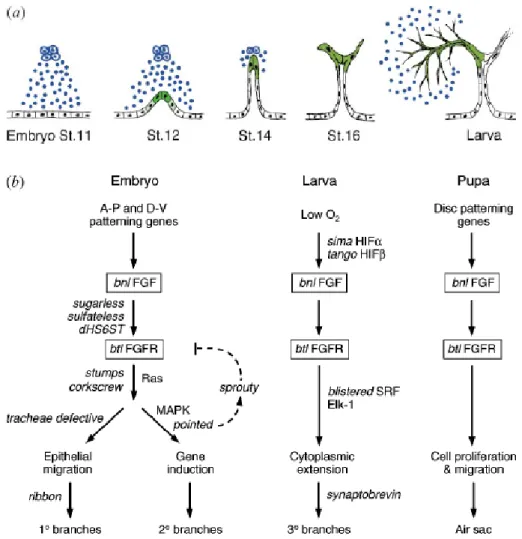
1.2 Tracheal morphogenesis in Drosophila
Tracheal branches are monolayer epithelial cells wrapped into tubes surrounding a central
lumen through which gases flow (C
ASANOVA2007). The tracheal system is bilaterally
symmetric and segmentally repeated in its organisation, which reflects its developmental
origin. The tracheal system comprises of three kinds of branches; the primary, secondary and
the tertiary (terminal) branches, each established according to the sequence of migration and
tube formation. During embryonic development (5hr After Egg Lay), 10 bilaterally
symmetrical ectodermal cell clusters consisting of ~ 80 cells invaginate into an epithelial sac
called the tracheal placode. The tracheal placode develops into interconnected tubes through
a series of events including cell migration, intercalation and fusion (Fig.1). Over the course of
the next few hours, six primary tracheal branches migrate out from each tracheal placode at
stage 12 (Fig.1b, e). The branches bud out with a pair of leading cells followed by a small
number of cells that organise into a tube during the migration process. At stage 15, the
primary branches are ensued by the two-dozen secondary branches (Fig.1d, e). During further
stages of development, the secondary branches sprout terminal branches. Much of the
development of terminal cells takes place during larval phases in response to physiological
oxygen demands.
Introduction
6 Figure 1: Stages of embryonic tracheal development in Drosophila melanogaster
The Drosophila tracheal system develops by sequential branching from a tracheal sac in each hemisegment. (a–
d) Embryonic tracheal development visualised by immunostaining of the tracheal lumen. Embryo stage and age (in hours) are indicated. The first (Tr1) and the tenth (Tr10) tracheal hemisegments are indicated in (a). Brackets indicate position of fifth tracheal hemisegment (Tr5). Lateral views, anterior left, dorsal up. (e) Development of Tr5. The spiracular branch (SB), transverse connective (TC) and the six primary branches (DB, dorsal branch;
DTa and DTp, anterior and posterior dorsal trunk; VB, visceral branch; LTa, anterior lateral trunk; LTp/GB, posterior lateral trunk/ganglionic branch) are indicated. Primary branches at stage 12 and secondary branches at stage 15 are highlighted green. Most secondary branches ramify to form terminal branches in the larval period (not shown). The branches that cease branching and fuse with branches in neighbouring hemisegments are indicated (arrowheads).Adapted from (GHABRIAL et al. 2003).
Branching morphogenesis of the trachea involves the Fibroblast Growth Factor (FGF)
signalling pathway that is used repeatedly to control branch budding and outgrowth
(G
HABRIALet al. 2003; M
ETZGER1999; S
ATO2002; S
KAER1997). branchless (bnl)
(S
UTHERLANDet al. 1996), and breathless (btl) (K
LÄMBTet al. 1992) encode the Drosophila
homologues of the mammalian FGF and the FGF receptor and are critical for the induction
Introduction
7
and maintenance of the tracheal system (Fig.2a). Although the Bnl/Btl signalling provides the cues for development, a different tubulogenesis mechanism is used at each level of branching concurrent with stages of development (Fig.2b).
Figure 2: The Branchless FGF pathway controls each step of branching.
(a) branchless/ FGF (blue) is expressed in clusters of cells surrounding the developing tracheal system, at each position where a primary branch will bud. The secreted growth factor activates the Breathless FGFR on nearby tracheal cells (black), and acts as a chemoattractant that guides outgrowth of primary branches. It also induces expression of secondary branch genes and triggers secondary branch sprouting at the ends of outgrowing primary branches (green; stages 12–16). branchless turns back on again, but in a completely different pattern, during larval life to control outgrowth of terminal branches. The gene is expressed yet again during pupal life where it controls budding of adult air sacs (not shown). (b) The genes that function upstream of Branchless and downstream of Breathless change during development, giving rise to different patterns and structures of branches at each step. Adapted from (GHABRIAL et al. 2003)
Further, different branches within each metamere have a fixed number of cells (S
AMAKOVLISet al. 1996) with each branch featuring characteristic tube dimensions (B
EITELand K
RASNOW2000). Primary branches are composed of multicellular tubes with two to five wedge-shaped
Introduction
8
cells surrounding a lumen and interconnected by intracellular junctions (Fig.3a). In secondary branches, tubes are made up of interconnected cells lying in a single row that form a tube by folding over itself along the long axis and sealing via an autocellular junction (Fig.3b).
However, unlike primary and secondary branches terminal cells form a junctionless and therefore “seamless” lumen (Fig.3c). Also present within the tracheal system are pairs of doughnut-shaped cells (fusion cells) that are derived from primary branches. Two fusion cells extending from the primary branches of each metamere connect to form a fusion anastomosis to give rise to a continuous network. The tube formed by fusion cells is as a result seamless and without intracellular junction (Fig.4). The developmental design of the primary and secondary branch formation is highly stereotyped and pre-programmed unlike branch formation in terminal cells, where branching is highly variable and regulated by oxygen demand.
Figure 3: Types of simple epithelial tubes that constitute the Drosophila tracheal system
Tube walls are formed by polarised epithelial cells with their apical membrane surface (red) facing inward toward the lumen space and their basal surface (green) exposed to the extracellular matrix. (a) A multicellular tube with four curved cells in a cross-sectionof the tube; (b) A unicellular tube formed by a single cell, rolled up to enclose the lumen, and sealed with an autocellular junction; (c) A unicellular tube with the lumen in the cytoplasm of the cell. There is no autocellular junction; the tube is “seamless.”Adapted from (LUBARSKY and KRASNOW 2003).
Introduction
9 Figure 4: Schematic representation of fusion anastomosis
Fusion branches (anastomoses) connect the individual metameres and are made by two cells that, before fusion, are positioned at the tips of the two branches that will fuse. Each fusion cell is doughnut shaped with no autocellular junctions and forms intercellular junctions with its fusion partner and with the following cell in the primary branch Adapted from (UV et al. 2003).
1.3 Terminal cell and branch development
Cells that do not participate in the formation of secondary branches are specified to become terminal cells. Though the specification to become terminal cells occurs in embryonic stages much of the development takes place during larval phases. Terminal cells are outposts of the tracheal system responsible for oxygen delivery to target tissues. Terminal branches originate as cytoplasmic protrusions that develop a lumen intracellularly (Fig.5). Branching morphogenesis in terminal cells is a reiterative process that occurs during the five days of larval development, involving rounds of cytoplasmic extension, followed by lumen formation to create a ramified network that contacts cells of the target tissue (K
EISTER1948). Oxygen carried through the terminal branches is made available at target tissues through diffusion that is facilitated by the close contact between the plasma membrane of the tissue and the blind- end of the terminal branch (M
ANNING1993).
The terminal branches consist of a lumen whose diameter, decreases progressively along with the branch diameter with increasing distance from the nucleus. The average lumen diameter of a terminal is less than 1 µm (G
UILLEMINet al. 1996; L
UBARSKYand K
RASNOW2003;
W
IGGLESWORTH1954). Terminal cells also show apical-basal polarity, with the outer
membrane being the basal membrane, and the membrane facing the lumen being apical which
Introduction
10
shows enrichment of the apical polarity complex Par6/aPKC/Baz along with Crumbs (Jayan N. Nair, PhD thesis) (G
ERVAISand C
ASANOVA2010). Terminal cells on an average have 20 branches and the branching points are regularly spaced and do not cross over one another.
The terminal cell arborisation may span distances over 100 µm. Unlike earlier stages of tracheal branching, terminal cell branching is not stereotyped but governed by oxygen physiology in tissues, resulting in terminal cells with varied branching patterns.
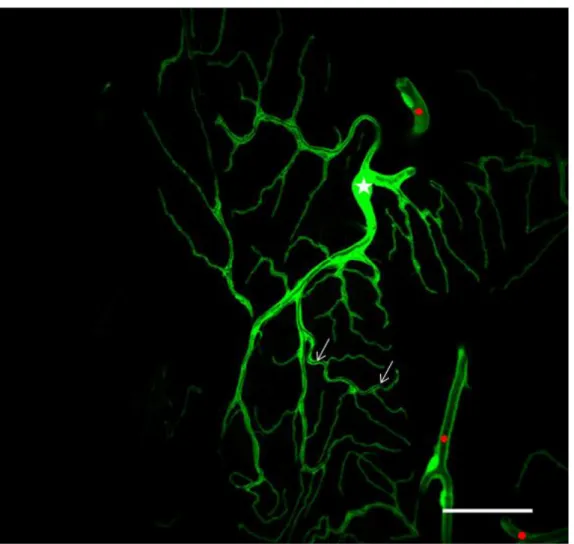
Figure 5: A terminal cells from a third instar larval trachea
The terminal cell is visualised by trachea specific expression of cytoplasmic GFP using btlGal4. The terminal cell extends long processes toward target tissues, called the terminal branches. Each terminal branch bears a lumen within through which air is transported to be supplied to tissues. The terminal cell nucleus is marked with a star (Jayan N. Nair PhD thesis).
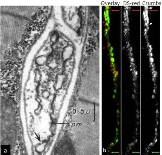
The process of lumen formation within the terminal branches is not quite well understood.
Early work in Drosophila showed the presence of cytoplasmic vesicles in the terminal branches distal from the nucleus (Fig.6a) (S
HAFIQ1963). It is believed that these vesicles fuse to form a larger vesicular body that would coalesce with the already existing tracheal lumen.
This is supported by the presence of Crumbs-positive vesicles at the distal regions of terminal
branches that did not yet possess a lumen (Fig.6b) (Jayan N. Nair PhD thesis). Moreover,
Introduction
11
these Crumbs-positive vesicles appear to line up parallel to each other as if forming a lumen.
Further, work on angiogenesis using cell lines have shown that vacuoles are generated prior to lumen formation and a lumen is formed when these vesicles fuse together (F
OLKMANand H
AUDENSCHILD1980).
Figure 6: Growing ends of the tracheoles show multi-vesicular structures
(a) Electron micrograph of the tip of a developing tracheal branch. Arrows mark vesicular structures within the distil region of the tracheoles. (b) Crumbs-GFP localisation in vesicles at distal regions of a terminal branch where the lumen is beginning to develop. The branch is marked with DsRed expressed using btlGal4. The red and green arrows mark the proximal (to the nucleus) and distal ends of the branch, respectively. Adapted from (a) (SHAFIQ 1963) and (b) Jayan N. Nair (PhD thesis).
However, contrary to the idea of de novo tube formation through vesicular fusion, lumen
formation has also been described to occur in a proximal to distal manner. Work in
embryonic tracheal system showed that lumen in terminal cells formed by the growth of a
new membrane into the cell from the surface contacting the adjacent cell. The membrane
formed was the apical membrane as seen from the accretion of apical polarity complexes,
aPKC/Par6/Baz and Crb/DPatj complexes (G
ERVAISand C
ASANOVA2010). Perhaps lumen
Introduction
12
formation is a combination of both mechanisms, where lumen formation in early stages of tracheal development is predominantly from extensions from previously existing membranes and then during later stages incorporating de novo tube formation in order to hasten the process of tube formation in response to local oxygen demand.
1.4 Factors influencing terminal branch development
Tracheation is modulated by oxygen availabilty in tissues. Both hypoxia and hyperoxia are known to influence tracheal branching (J
ARECKIet al. 1999; W
IGGLESWORTH1954). Under conditions of low oxygen, terminal branch formation is induced, whereas in hyperoxia terminal branch formation is supressed. The Drosophila counterparts of the mammalian hypoxia-inducible factor (HIF), Similar (Sima) and Tango (Tgo) which function as HIF-α and HIF-β homologues, activate the hypoxia induced signalling pathway (L
AVISTA-L
LANOSet al.
2002). Oxygen starved tissues produce a trachaetion signal which induces tracheal branching in regions depreived of oxygen (W
IGGLESWORTH1954). This tracheogenic signal was later identified as branchless (bnl). Bnl is a potent inducer and chemoattractant of terminal branches that results in proliferation of terminal branches (J
ARECKIet al. 1999). Btl signalling through FGFR induces terminal branching (R
EICHMAN-F
RIEDand S
HILO1995) and also the expression of the downstream targets important to terminal cell development, such as srf/blistered/pruned/ (srf/bs). srf encodes the Drosophila homologue of the mammalian serum response factor and is specifically expressed in terminal cells. Mutants of srf fail to develop cytoplasmic outgrowths that later form terminal branches, thus severely affecting branching morphogenesis (A
FFOLTER1994; G
UILLEMINet al. 1996). Srf recruits the co-factor Elk-1( a Ternary Complex Factor, TCF) to the serum response elements (SRE) to form the ternary complex. FGF signalling activates MAPK pathway, activation of the MAPK pathway results in phosphorylation of TCFs which leads to activation of the SRE in response to growth factor signalling (M
ARAISet al. 1993; T
REISMAN1994). Concordant with this observation, expression of the activated forms of Srf and Elk-1 resulted in overgrowth of cytoplasmic extensions and terminal branches.
Screens to identify genes that affect terminal branching have revealed a few genes involved
in maintaining normal branching and terminal cell development. (B
AERet al. 2007; L
EVIet
Introduction
13
al. 2006). Mutants of potential cytoskeletal regulators such as Drosophila talin (rhea), the β- integrin myospheroid (βmys) as well as double mutants of the α-integrins inflated (if) and multiple edematous wings (mew) have been shown to affect maintenance of tracheal tubes within terminal branches (Fig.7). Mutations in ikkε also affected tube formation within terminal cells (O
SHIMAet al. 2006). The authors speculate that these genes contribute to tube stability by regulating the actin cytoskeleton. Further, work from our lab has shown the importance of moesin (moe) in terminal cell development (Jayan N. Nair PhD thesis). Much work remains to be done in order to understand the mechanisms behind tube formation and tube stability in terminal cells.
Figure 7: Multilumen phenotype in talin and integrin mutants
Bright field images of terminal cells showing multilumen phenotypes in mutant of (a) talin (rhea); (b) β- intergrin (mys); (c) and the two α - integrins (mew and if double mutants). a'-c' are enlargements of the boxed regions in a-c respectively. Adapted from (LEVI et al. 2006).
1.5 Moesin is essential for terminal cell development
Moesin (Moesin) is the only Drosophila homologue of the mammalian Ezrin-Radixin- Moesin (ERM) family of proteins. Activated Moesin (p-Moesin) acts as an anchor for F-actin at the apical membrane of polarised epithelia (B
RETSCHERet al. 2002). Moesin is anchored to the membrane via various membrane anchors such as Phosphatidylinositol 4,5-bisphosphate (PtdIns(4,5)P
2), Crumbs and Bitesize (btsz) which are used in a cell specific context (M
EDINAet al. 2002; N
AKAMURAet al. 1999; P
ILOTet al. 2006).
Introduction
14
The activation of ERM proteins is believed to occur upon recruitment to the apical membrane. In Drosophila embryos, btsz is involved in maintaining epithelial integrity and the stabilisation of adherens junctions through its interaction with Moesin (P
ILOTet al. 2006).
Knockdown of btsz in the tracheal system leads to the loss of p-Moesin (Jayan N Nair, unpublished) in terminal cells, indicating that Btsz is an anchor for p-Moesin in the tracheal system. We also know that disruption of Moesin in the tracheal system through RNAi results in tube formation and branching defects within terminal cells (Jayan N Nair, PhD thesis).
Recently, the kinase activating Moesin was identified as SLK and LOK like kinase (slik) (H
IPFNERand C
OHEN2003; H
IPFNERet al. 2004). In order to better understand the relation between anchoring and activation of Moesin at the apical membrane and its importance in tracheal development, I explored the function of slik in terminal cell development.
1.6 slik, a Sterile 20 like kinase involved in Drosophila growth control
SLK (sterile 20 like kinase) is a serine/threonine kinase and a member of the group II germinal centre kinases (GCKs) which includes MST1-3 (Macrophage Stimulating) and the LOK (Lymphocyte Oriented Kinase). SLK is cleaved by Caspase 3 in two domains with distinct activities: an activated N-terminal kinase domain that promotes apoptosis and cytoskeletal rearrangements and a C-terminal domain that disassembles actin stress fibres (S
ABOURINet al. 2000). LOK was identified to be expressed in mammalian lymphocytes and to specifically phosphorylate ERM proteins.
slik (SLK- and LOK like kinase), a Drosophila member of the Ste20 kinase family, was
identified in an overexpression screen as a gene causing increased growth of the posterior
compartment of the wing imaginal disc (H
IPFNERet al. 2002). slik was named on the basis of
its similarity to the human SLK and LOK Ste20 kinases. slik shares high sequence similarity
to the human Slk and Lok which is largely restricted to the N-terminal kinase domain and the
conserved coiled-coil motif bearing C-terminus (Fig.8a). The sequence internal to the
conserved domains of Slik is non-conserved and variable in length, a feature consistent
within members of the same subfamily. The slik transcription unit covers approximately 11
kb and is predicted to have at least 12 exons. There are about 6 predicted transcripts based on
cDNA clones and partial ESTs.
Introduction
15
Two slik mutants, slik
1and slik
KG04837, have been described and used in various studies (H
IPFNERand C
OHEN2003; H
IPFNERet al. 2004; H
UGHESand F
EHON2006). slik
1represents a null allele which is a deletion of exons 2–8 and part of exon 9 of the slik transcript, including the translation start site and the entire kinase domain. slik
KG04837is a partial loss of function allele, an insertion of the P-element KG04837 in the first intron of slik (Fig.8b). slik
1mutant larvae display a striking phenotype through delay in overall growth and developmental timing. The mutant larvae grew at the maximum up to about one-third the size of wild type larvae and rarely progressed beyond third instar larval phase (Fig.9).
Intriguingly, about 5% of the mutant larvae have exceptionally long life spans of 15 days which is three times the normal larval phase of development.
Figure 8: Schematic representation of Slik and the human homologues
(a) Comparison of Slik with human SLK and LOK proteins. Numbers show sequence identity/similarity within the indicated domains. Predicted coiled-coil regions in the C-terminal domain are indicated by hatching; (b) Detailed view of the slik region. The insertion sites of EPg(2)23048 and KG04837 in the first intron and the extent of the slik1 deletion are indicated. Adapted from (HIPFNER and COHEN 2003).
Introduction
16 Figure 9: Growth defect in slik mutant larvae
Heterozygous slik1/+ control and homozygous slik1 mutant larvae after 5d of growth under uncrowded conditions (HIPFNER and COHEN 2003).
1.7 Functional relevance of slik in Drosophila development
Hipfner et al. (H
IPFNERet al. 2004), reported that Slik regulates cytoskeletal organisation during wing disc development by regulating the Drosophila ERM (ezrin/radixin/Moesin) protein Moesin. ERM proteins link cortical actin cytoskeleton to the cell membrane by the N- terminal FERM domain which binds to membrane proteins directly or through adaptor proteins, and the C-teminal actin binding domain which binds to F-actin. Slik was shown to be enriched at the apical membrane in cells of the wing imaginal disc. Also, Slik colocalised with activated Moesin (Phosphorylated Moesin/p-Moesin) and F-actin in these cells. slik
1clones in wing discs showed a considerable reduction of p-Moesin levels in comparison to wild type cells. Further, expression of the kinase domain of Slik (Slik
kin) in slik mutant wing discs restored p-Moesin to normal levels. These results suggest that Slik kinase activates Moesin through its phosphorylation.
Consistent with these results, in an RNAi screen in Drosophila Kc cells to identify kinases
that activate Moesin during mitosis, slik was identified as a positive regulator of Moesin
phosphorylation. During the onset of mitosis, cells retract their actin-based protrusion and
change their morphology from a flattened to a rounded up form. Perturbing P-Moesin through
slik RNAi mimicked the Moesin knockdown phenotype, i.e., failure to withdraw the actin
rich protrusions, highlighting the importance of Slik in activating Moesin during cell division
(K
UNDAet al. 2008).
Introduction
17
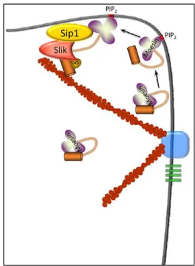
Recent work shows that Slik’s localisation to the apical membrane is mediated by another protein, SRY interacting protein 1 (Sip1) (H
UGHESet al. 2010). Sip1 is the Drosophila homologue of mammalian EBP50 (ERM binding protein 50) that acts as a scaffold linking ERM proteins to transmembrane proteins and other membrane associated cytoplasmic proteins. Sip1 mutant follicle cells show a marked reduction of Slik levels and the subsequent loss of p-Moesin. On the other hand, moesin mutant cells show a drop in overall Sip1 levels, while slik mutant clones showed an increase in Sip1 levels. In addition, Sip1 co- immunoprecipitates Moesin and Slik together, suggesting that both proteins form a complex with Sip1. Though the exact nature of the relationship between Sip1, Moesin and Slik is yet unknown, the authors suggest that Sip1 functions as a scaffold to bring Slik and Moesin in to proximity to phosphorylate Moesin (Fig.10).
Figure 10: Possible model for Sip1 in Slik-dependent activation of Moesin.
Inactive, folded Moesin in the cell cortex might associate with PIP2 in the plasma membrane, inducing a conformational change which results in partial unfolding of Moesin. This event, or other modifications such as phosphorylation of residues in the FERM domain (Krieg and Hunter, 1992), allow Moesin, Sip1 and Slik to form a complex that results in phosphorylation of the C-terminal Threonine residue and full activation of Moesin. Adapted from (HUGHES et al. 2010).
In another study, Merlin (Mer), the Drosophila homologue of the human tumour suppressor
Neurofibromatosis 2 (NF2) was shown to be under the control of Slik kinase (H
UGHESand
F
EHON2006). Merlin shares ~45% sequence similarity with ERM proteins and the similarity
Introduction
18
is restricted to the N and C-terminal regions, which shares feature common to the ERM proteins and other family members. However, unlike ERM proteins that promote maintenance of epithelial integrity through the actin cytoskeleton (S
PECKet al. 2003), Merlin has a distinct role in regulating cell proliferation (R
OULEAUet al. 1993). Moreover, while ERM proteins are activated upon phosphorylation, phosphorylation of Merlin renders it inactive. Merlin mutant clones in eye and wings show overproliferation.
Merlin has a complex subcellular localisation that spans both the apical membrane and the endocytic compartments within the cytoplasm (H
UGHESand F
EHON2006). In wild type wing disc epithelium Merlin is found to localise apically. However, loss of Slik results in an increase in Merlin levels in the mutant cells and bulk of Merlin is redistributed to the basolateral regions with an increased association with punctate structures. Western blotting analysis with extracts from Slik and Slik kinase dead (Slik
kd) overexpressing wing discs showed increased levels of phosphorylated Merlin upon Slik expression while remaining unchanged in Slik
kdexpressing discs (H
UGHESand F
EHON2006). Further, activated Merlin suppressed wing growth, which is suppressed by removal of a copy of slik, suggesting that Slik antagonises Merlin function. The authors also find that Moesin and Merlin are competitive substrates of Slik’s kinase activity.
Additionally, experiments in S2 cells with wild type, phosphomimetic and non- phosphorylable forms of Merlin revealed a dynamic trafficking of Merlin depending on its phosphorylation status. In cells transfected with wild type Merlin, upon induction Merlin initially localised to the membrane but after 2-4 hours became enriched in endocytic compartments. When Merlin expression is induced in cells coexpressing Slik there is a drastic shift in the distribution of Merlin, with the majority localising to the membrane. In contrast, when Merlin expression is induced in Slik
kdtransfected cells, Merlin localisation decreased at cell membranes but increased within endocytic compartments in the cytoplasm. Trafficking of activated/non-phosphorylated Merlin to the endocytic compartments may be of functional relevance in tumour suppression, by facilitating removal of receptors from cell surface that promote cell proliferation. expanded (ex), a Merlin-related tumour suppressor is partially redundant to Merlin in regulating proliferation and differentiation (M
CC
ARTNEYet al. 2000).
Merlin:ex double mutant cells in wing discs present abnormal accumulation of receptors such
Introduction
19
as Notch and EGFR at the plasma membrane, possibly due to the lack of endocytosis of the receptor-bearing plasma membrane along with endocytosing Merlin fraction. Collectively, the activity of Slik kinase results in an orchestrated but opposite regulation of Moesin and Merlin to promote epithelial integrity and cell proliferation (Fig.11).
Figure 11: Schematic diagram of functional relationships between Slik, Merlin, Moesin, and the regulation of tissue integrity and proliferation in developing epithelia.
Slik activity simultaneously promotes Moesin function and inhibits Merlin. Previous results have shown that Moesin functions to negatively regulate Rho activity and promote epithelial integrity (Speck et al., 2003).
Merlin functions to restrict proliferation in the same epithelia. Thus, the net result of Slik activity is to drive proliferation and simultaneously stabilise epithelial integrity (HUGHES and FEHON 2006)
Apart from its kinase activity, Silk also contributes to the growth of wing disc epithelium in a kinase independent manner. Overexpression of Slik in the patched (ptc) domain in wing disc caused wing overgrowth due to increased cell proliferation. However, the Slik driven wing overgrowth is counteracted by an increased number of apoptotic cells. Moreover, overexpression of Slik
kdrecapitulated the Slik driven overgrowth of wings suggesting that the Slik derived cell proliferation is through a kinase-independent mechanism. Tissue overgrowth resulting from Slik overexpression is suppressed through removal of the raf and the wings are restored to normal proportions.
Apoptosis also occurs in slik mutant cells of the wing disc which is mediated by the c-Jun N-
terminal kinase (JNK) and can be suppressed by removing hemipterous (hep/JNKK) which
encodes the activating kinase. Additionally, reduction of Raf levels in slik mutant background
further increased the numbers of apoptotic cells suggesting that Raf is an important
downstream effector of Slik. Since Raf was found to coimmunoprecipitate both Slik and
Slik
kd, the authors conclude that that activation of Raf is through its physical interaction with
Slik, rather than phosphorylation by Slik. Finally, slik mutant cell’s growth defect can be
rescued by expression of activated Raf, but not by activated ERK (MAPK) suggesting a
Introduction
20
signalling independent of the canonical ERK pathway. Briefly, Slik functions to maintain epithelial integrity and promote growth through proliferation in tissues. In conclusion Slik’s activity is comparable to several oncogenes that promote proliferation and apoptosis in parallel.
1.8 Aim
This work aims to investigate the underlying role of Slik in tracheal development. It explores
the contribution of Slik in terminal cell development through the phosphorylation of targets
Moesin and Merlin. In addition, this work addresses the possible signalling input by Slik into
the Btl/MAPK pathway, mediated through its interaction partner Raf.
Materials and Methods
21
2. MATERIALS AND METHODS
2.1 Materials
2.1.1 Drosophila melanogaster stocks
w[1118]; ; embryos were used for Slik antibody stainings
2.1.2 UAS Transgenes
From Bloomington stock centre w[*]; P{w[+mC]=UAS-phl.gof}F179
w[1118]; P{w[+mC]=UAS-Ras85D.V12}TL1 From other sources
w[1118]; ;P{w[+mC]=UAS-Slik
kin/TM6B,Tb [1] (David Hipfner) w[1118]; ;P{w[+mC]=UAS-Slik
kd/TM6B,Tb[1] (David Hipfner)
P{hsFLP}, w[1118]; P{neoFRT}42D P {w[+mC]=tubGal80; P{w[+mC]=btl-Gal4, btl- moesin-mRFP, UAS-CD-GFP} T(2;3)CyO -TM6 (Mirka Uhlirova)
From VDRC stock centre
UAS-IR-btl VDRC 27106 UAS-IR-egfr VDRC 107130
UAS-IR ex VDRC 109281
UAS-IR-Merlin VDRC 7161
UAS-IR-moesin VDRC 37917
UAS-IR-raf VDRC 107766
UAS-IR-slik VDRC 43783
UAS-IR-srf VDRC 100609
UAS-IR-cad 96Cb VDRC 103296
Materials and Methods
22
From NIG Japan
UAS-IR-Ras NIG 9375-2
2.1.3 Gal4 driver lines
w[1118]; P{w[+mC]=btl-Gal4}; +/+
w[1118]; P{w[+mC]=UAS-dicer }; P{w[+mC]= btl-Gal4}, P{w[+mC]= UAS-GFP}
w[1118]; P{w[+mC]=btl-Gal4}, P{w[+mC]=UAS-2XEGFP}; MKRS/TM6B,Tb[1]
w[1118]; If/Cyo; {w[+mC]= btl-Gal4}, P{w[+mC]= UAS-GFP}
w[1118]; slik
1, {w[+mC]= btl-Gal4}, P{w[+mC]= UAS-GFP}/Cyo; MKRS/TM6B,Tb[1]
2.1.4 Mutants
y[1]; P{y[+mDint2] w[BR.E.BR]=SUPor-P}slik[KG04837]/CyO; ry[506]
w[1118]; slik
1/Cyo, P{GAL4-kr}2, P{UAS- GFP};
w[1118]; P{neoFRT}42D slik
1/CyO P{GAL4-kr}2, P{UAS- GFP};
2.1.5 Antibodies
The following primary antibodies were used: guinea pig anti-Slik (1:100, Hipfner et al 2004), mouse anti-SRF (1:200, DSHB), rat anti-E-Cadherin (1:200, DSHB), rabbit anti-Dof (1:200, Vincent et al., 1998), rabbit anti-P Moesin (1:200, S3149 Cell Signaling), rabbit anti-Moesin (1: 5000, François Payre), rabbit anti-GFP (1:500, Torrey Pines Biolabs Inc). Fluorophore conjugated secondary antibodies: Alexa468 and Alexa568 and Alexa647 (Molecular Probes) were used at a dilution of 1:2000.
2.1.6 Oligonucleotides
Btl RT_F tccacacggaaacctcaaggacttc
Btl RT_R acgtcgctctgtgagtcgtacttc
bs_RT_F cgctgcccaacaagaagtctccgcctg
bs_RT_R cagcttgcgcgtggcaaatgtgtaca
Ex_RT_F acttctggggcagcagcagccgaa
Materials and Methods
23
Ex_RT_R gtgggtgtgcgatgatcgccagc
Merlin _RT tgtggctgggcgtcacctccgtg Merlin _RT_R gcaggtgctccatgctcttctccag Moesin_F_500 cctggacaccgacgagcatatcaaggac Moesin _R_SalI acgcgtcgaccatgttctcaaactgatcgacgcg Raf RT_F actgctgtccgcttcaatatgagcag
Raf RT_R ccagttttcctcggaacttttggcgt RpL32 RT_F tcctaccagcttcaagatgacc RpL32 RT_R cacgttgtgcaccaggaact
Slik RT_F gatccgcaggtgaggcccacgacgga Slik RT_R gtttgtcaatgtcttggctctgcagc
2.1.7 Microscopes
Olympus FV1000, Leica TCS SP2, Zeiss Axioplan 2-imaging, Zeiss Apotome and Leica M2 16FA were used for microscopy.
2.1.8 Imaging and data analysis software
Image acquiring softwares FV10-ASW 2.0 Viewer, Leica Confocal Software LCS, Axiovision Rel 4.6 (Zeiss) and Axiovision 1 (Zeiss) were used. Images were edited using Adobe Photoshop (Adobe Systems) and ImageJ softwares. All images are maximum intensity projection unless otherwise mentioned. DNA sequence alignments, analysis and oligonucleotide designing were done using VectorNTI. Imaris (Bitplane) 3D/4D image processing software was used for 3D reconstruction of the tracheal system.
2.1.9 Reagents
TritonX 100, Tween20 and BSA were purchased from Sigma. Vectashield mounting media
for fluorescent samples was from Vector Laboratories. Restriction enzymes used were from
New England Biolabs. Expand High Fidelity PCR system was supplied by Roche
Materials and Methods
24
Diagnostics. Agarose electrophoresis grade was from Gibco BRL. Unless otherwise mentioned, all the other chemicals were purchased from Merck, Sigma or Roth.
2.2 Methods
2.2.1 Collection of embryos
The flies were maintained under standard conditions (Ashburner, 1989; Wieschaus and Nuesslein-Volhard, 1986).
To fix the embryos, an overnight egg lay (16 hr) was collected on an apple juice–agar plate, dechorionated using 50% bleach and washed in tap water. Embryos were fixed in 4%
formaldehyde in PBS:heptane = 1:1 solution at 37°C for 20 minutes, with vigorous shaking, followed by devitellinilisation with methanol:heptane = 1:1 solution by vortexing for 30 seconds. Embryos were washed several times in methanol and stored in methanol at -20°C if not used immediately.
2.2.2 Immunostaining in embryos
The fixed embryos were rehydrated and washed with 0.3% PTX (1XPBS+ 0.3% TritonX 100) three times for 10 min each followed by 30 min incubation in blocking reagent (1X PBS+ 0.1% TritonX 100 + 1% BSA). After blocking, the liquid phase was taken off and the primary antibody was added to the embryos. The samples were left at 4°C overnight, on a rotating wheel. Embryos were washed with 0.3% PTX several times, at room temperature followed by incubation with Alexa coupled secondary antibody at room temperature for 90 minutes. The embryos were washed four times (15 minutes each) in 0.3% PTX at room temperature and mounted in Vectashield before imaging.
2.2.3 RNAi in the tracheal system
Crosses were set up using the UAS-RNAi transgenic line and the btlGal4, UAS-GFP
recombinant driver line, with UAS-dicer or excluding UAS-dicer in cases where
Materials and Methods
25
coexpression of UAS-dicer caused lethality or resulted in extremely small larvae. The crosses were maintained at 29°C unless otherwise mentioned. Wandering third instar larvae were collected and dissected to expose the tracheal system. Larvae were fixed using 4%
paraformaldehyde in PBS for 20 minutes and washed with 0.3% PTX (1XPBS+ 0.3%
TritonX 100) followed by mounting the larval fillets using Vectashield. In case where antibody staining was required the larvae were subjected to the antibody staining protocol described below.
2.2.4 Immunostaining in larvae
Post fixation fillets were washed with 0.3% PTX (1XPBS+ 0.3 % TritonX 100) three times for 10 min each followed by 30 min incubation in blocking reagent (1X PBS+ 0.1% TritonX 100 + 1% BSA). After blocking, the samples were incubated overnight in antibody solution at 4°C. Fillets were washed four times (15 minutes each) in 0.3% PTX after overnight incubation. Fillets were incubated in Alexa fluorophore conjugated secondary antibodies for 2 hours at room temperature. Next, the fillets were washed four times (15 minutes each) in 0.3% PTX at room temperature, mounted in Vectashield and taken for microscopy.
2.2.5 Generation of slik
1MARCM clones in the tracheal system
Crosses were set up using the appropriate stocks and maintained at 25°C. A 24-hr egg lay collection was taken and heat shocked for about 2 hours. After heat shock the tubes were placed at 29°C. Around day 5-6 post egg lay, wandering third instar larvae were collected, dissected and mounted on slides as described in the previous section.
2.2.6 Tracheal cDNA synthesis
Wild type third instar larvae were dissected, the tracheal system was isolated and the tissue
macerated in Trizol (Invitrogen). RNA was isolated using the standard Trizol-based isolation
protocol. RNA isolated was treated with DNase according to manufacturer’s protocol
(Invitrogen). 200 ng of RNA was used for cDNA synthesis using First strand synthesis
Materials and Methods
26
kit/SuperscriptIII according to manufacturer’s instructions (Invitrogen) using a 1:1 mix of both random hexamers and oligo dT.
2.2.7 RT PCRs
1 l of the tracheal cDNA was used as the template for PCR reactions. The reaction
components were: 20 pmols of each primer, 1X Red Taq Readymix (Sigma) in a 20 l PCR
reaction mix. The PCR was carried out on a Thermoblock (Biometra). The PCR programme
included a denaturation step of 2 minutes at 94°C followed by 30 cycles: 15 seconds at 94°C,
30 seconds annealing at 57°C, 30 seconds extension at 72°C. No final extensions were
included in the programme. The PCR products were analysed on a 1.5% agarose gel.
Results
27
3. RESULTS
3.1 Slik is required for tracheal development
Studies on tracheal development and more specifically on terminal cells have outlined the importance of Moesin in tube formation and maintenance of the tube within the branches.
One of the factors that regulate Moesin in other tissues is the Moesin activating kinase Slik, implicated in wing imaginal disc development, mitotic events in S2 cells and normal development of follicle cells of oocytes (H
IPFNERet al. 2004; H
UGHESand F
EHON2006;
H
UGHESet al. 2010). This study explores the function of Slik in terminal cell development in conjunction with Moesin and another signalling molecule, Raf.
3.1.1 Slik is expressed in both the embryonic and the larval tracheal system
To analyse Slik distribution in the tracheal system, I used an antibody against Slik (H
IPFNERand C
OHEN2003). This antibody was previously used to examine Slik localisation in wing discs, oocytes and mitotic S2 cells (C
ARRENOet al. 2008; H
IPFNERet al. 2004; H
UGHESet al.
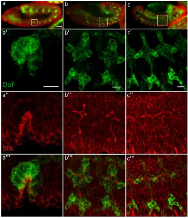
2010). Stainings from these studies showed that Slik was enriched at the apical membrane of cells in the wing imaginal disc and also in the follicular epithelium of oocytes. In mitotic S2 cells Slik is localised at the mitotic cortex of the dividing cells. To mark the tracheal cells antibodies against Dof was used. Dof is an adaptor protein specific to the two FGFRs, breathless (btl) and heartless (htl) which are active in the tracheal system and the migrating mesoderm, respectively (V
INCENTet al. 1998). Dof is expressed through all stages of tracheal development. The distribution of Slik and Dof was visualised using secondary antibodies coupled to fluorescent (Alexa Fluor®) dyes (Fig.12).
At embryonic stage 10, tracheal development commences with the invagination of the
tracheal placodes within each segment. In the invaginating tracheal placode, cells constrict
their apical surface which is the concave side of the depression. Staining for Slik reveals that
Slik is expressed in the invaginating placode with enrichment at the apical side of the cell
(Fig.12a). In stage 12 embryos, cells within the migrating placode continue to have an apical
enrichment of Slik (Fig.12b). At stage 14 of development, cells from each placode migrate
Results
28
and are arranged side-by-side and interdigitate to form a continuous tube. At this stage of tracheal development Slik enrichment persists at the apical regions of cells forming the tube, i.e. the membrane facing the lumen of the developing tube (Fig.12c.) Also, previous studies in wing imaginal discs reported Slik to be enriched at the apical membrane of the cells (H
IPFNERet al. 2004).
Figure 12: Slik expression during embryonic development of the tracheal system
Tracheal cells are visualised by anti-Dof staining (a-c). (a) Slik expression during tracheal placode invagination at stage 10; (b) Outgrowth of primary branches at stage 12 and (c) Stage 14. Slik expression shows strong apical
Results
29 enrichment within the trachea. a', b' and c' are enlargements of the corresponding stages in a, b and c. Scale a, b, c - 50 µm and a', b', c' - 10 µm.
At the end of embryonic development, patterning of the tracheal system is complete with the dorsal trunk, the dorsal branches, the lateral branches and the terminal cells in place.
Terminal cells at this stage are morphologically distinct from those found in larvae as they possesses only a single branch extension with a tube within. Over the larval phases of development these cells generate multiple branches to form a functional terminal cell.
Branching events in a terminal cell alter the morphology of the cell from a simple cell to a cell that is highly ramified and spanning over a large area (Fig.13). The tips of these branches establish direct contact with tissues requiring oxygen.
Figure 13: A terminal tracheal cell from a larva at third instar phase of development
Tracheal cells are visualised by tracheal specific cytoplasmic GFP expressed using btlGal4. The terminal cell is highly branched and carries a tube within each branch (arrows). Oxygen is transported through these tubes to surrounding tissues and organs. Star denotes the cell nucleus. Also seen are parts of the secondary branches from adjacent areas (marked by dots). Scale - 50 µm.
Results
30
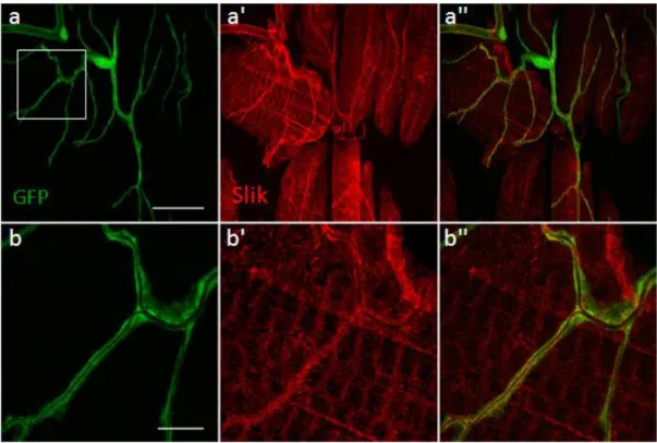
To analyse the distribution of Slik in the terminal branches, Slik staining was performed in larvae expressing the UAS-GFP transgene in the tracheal cells using the tracheal specific btl- Gal4 line. The GFP is expressed in both the nucleus and the cytoplasm and hence is used to highlight the tracheal system. Staining for Slik in third instar larvae showed that Slik is expressed in the terminal cells (Fig.14a). Further, high resolution imaging showed that Slik is enriched at the apical membrane within the terminal branches, with occasional diffused staining at sub-apical regions (Fig.14b).
Figure 14: Slik expression in terminal cell of the tracheal system
Tracheal cells are visualised by tracheal specific cytoplasmic GFP expressed using btlGal4 (a, b). (a) Slik expression in a terminal cell; (b) Enlargement of the corresponding area in image a (boxed) showing Slik enrichment in the apical membrane of the branch. Scale a - 25 µm, b - 10 µm.
Next, I investigated the localisation of Slik within cellular junctions of the tracheal system.
The tracheal terminal cells are junctionless cells except for a junction connecting terminal
cells to the secondary branch. However, the dorsal trunk is a multicellular tube and therefore
suited for studies on junctions within the tracheal system. Of particular ease in studying
junctions within the tracheal system are the fusion cells, a pair of doughnut shaped cells that
form the anastomoses of the dorsal trunk (Fig.4). In epithelial cells E-Cadherin (E-Cad) is
localised to the apicolateral regions where it is a component of the adherens junction. In order
to mark the junctions within the dorsal trunk I performed immunostaining for E-Cad. At the
Results
31
point of fusion of the dorsal trunk E-Cad is expressed in 3 stripes, the central stripe which marks the adherens junction formed at the point of contact between fusion cells and the two other flanking stripes that delineate the junction between the fusion cell and the dorsal trunk at either side (Fig.15b). Analysis of the dorsal trunk for Slik protein expression shows that Slik is expressed in the fusion cells of the dorsal trunk (Fig.15a). A simple maximum intensity z-projection of images of the dorsal trunk was not sufficient to determine the localisation of Slik in relation to the junction. Therefore, I performed 2D reconstruction of the dorsal trunk using Imaris imaging software. 2D reconstruction of the dorsal trunk revealed that Slik expressed in the fusion cell localised to the apical membrane of the fusion cells.
Optical sections across various planes of the dorsal trunk showed that the localisation of Slik was more apical to E-Cad at the lumen (Fig.15c). This result is consistent with the results from a previous study where the authors observed that Slik localised apical to the adherens junction in wing disc epithelium (H
IPFNERet al. 2004)
3.1.2 Slik is required for normal branching, lumen formation and tube stability in terminal cell development
To investigate the function of slik in tracheal development, slik
1mutant larvae were analysed.
slik
1is a null allele as it is a deletion that removes exons 2–8 and part of exon 9. This deletion
removes the translation start site and the entire kinase domain. slik mutant clones of terminal
cells were generated through the MARCM method. Also, I performed tracheal specific
knockdown of slik through RNAi and analysed the outcome. Results from experiments using
various approaches showed similar phenotypes and are discussed in the following sections.
Results
32 Figure 15: Slik expression in fusion cells of the dorsal trunk
Tracheal cells are visualised by tracheal specific cytoplasmic GFP (green), antibodies specific for Slik (red) and E-Cadherin (Cad) (blue). a and b are maximum intensity projections. (a-a''') A segment of the dorsal trunk showing fusion anastomosis (boxed). The fusion cells are two ring shaped cells connecting the multicelluar dorsal trunk from each segment. (a') Slik is expressed at high levels in fusion cells; (a'') E-Cad at the fusion point is seen as three stripes, marking junctions between the fusion cells and that between the fusion cells and the multicellular tube; (a''') overlay of Slik and E-Cad; (a'''') overlay of GFP, Slik and E-Cad. (b-b'') Enlargement of boxed area in a'''; (c-c'') 2D projection of the dorsal trunk showing apical localisation of Slik; view of the dorsal trunk centred at the fusion anastomosis along (c) the XY plane; (c') the YZ plane (sagittal section) shows Slik localisation apical to E-Cad localisation. (c'') View across the XZ (transverse section) plane shows that Slik is localised more apical to E-Cad. Inset- magnified view of indicated region. Scale a - 25 µm, b - 10 µm
Results
33
3.1.2.1 Analysis of the tracheal system in slik
1mutant animals
slik
1homozygous mutants are larval lethal and therefore very few escapers of the homozygous mutant larvae were obtained. The escaper slik
1mutant larvae had a smaller body size (Fig.16), which was consistent with previous observation (H
IPFNERand C
OHEN2003).
slik
1homozygous third instar larvae were filleted and processed. The larvae showed gross abnormalities in the tracheal system. The dorsal branches showed lumen defects. In a wild type dorsal trunk the multicellular tube is composed of cells arranged with their apical surfaces facing the lumen. The apical surface of the tracheal cells expands permitting tube dilation and at the same time secretes chitin to reinforce the expanding tube. A bright field image of the wild type dorsal trunk showed that the lumen diameter spans almost the entire width of the dorsal trunk. By contrast, bright field images of the dorsal trunk from a slik
1mutant larva showed lumen defects. While the outer diameters of the dorsal trunk from both wild type and mutant larvae were comparable, the diameter of the lumen in the mutant was less than half as wide as in the wild type. The presence of a narrow lumen despite of the normal branch size indicated a failure in lumen expansion within the mutant dorsal trunk (Fig.17a', b').
To further analyse tracheal phenotypes in slik
1mutants, the terminal cells from these larvae were compared with the wild type cells. The mutant terminal cells showed defects in branching. Wild type terminal cells on an average had over 20 branches, whereas in slik
1mutant cells branching was restricted to fewer than 10 branches (Fig.13 and 18). In addition to reduction in number of branches the mutant terminal cells also exhibited luminal defects.
Normally in terminal cells each branch bears a single lumen but in the mutant terminal cells multiple lumens were observed, either within a single branch or clustered near the nucleus.
This phenotype is henceforth addressed as the mutilumen phenotype (Fig.18c').
Results
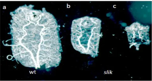
34 Figure 16: slik1mutant larvae show altered body size
Larvae in the image above have been dissected on their ventral side to expose the tracheal system. (a) wild type third instar larva; (b) and (c) are slik1mutant larvae also at third instar larval stage as judged by the presence of completely extruded anterior spiracles with spiracular papillae, but exhibiting smaller body sizes.
Figure 17: slik1 mutant larvae show tube defects within the dorsal trunk
slik1 mutant tracheal system is visualised by tracheal specific cytoplasmic GFP (green) expressed using btlGal4 and tubes within the trachea are visible in bright field (BF) (a, b). (a-a'') Dorsal trunk from a wild type larva;
(b-b'') dorsal trunk from a slik mutant larva. (a') Bright field image of the wild type trachea shows that the dorsal trunk and the tube within are almost of the same diameter and therefore not clearly visible as two distinct components. (b') Dorsal trunk from a slik mutant larva shows that the dorsal trunk and the tube (marked by short and long arrow respectively) are easily identified. The tube within the dorsal trunk is not centrally positioned and has not expanded sufficiently to line the inner wall of the dorsal trunk. Scale - 50 µm.
.
Results
35 Figure 18: Terminal cells from slik1 mutant third instar larvae show branching and lumen defects
Tracheal system is visualised by tracheal specific cytoplasmic GFP (green) expressed using btlGal4. (a-c) Terminal cells from slik1 mutant larvae show reduced branching. (c') Enlargement of the corresponding area in image c (boxed), showing multilumen (multiple tubes within a branch) phenotype. Scale - 25 µm.
3.1.2.2 Analysis of slik
1MARCM clones in the tracheal system
In addition to analysis of the slik
1mutant tracheal system, slik
1mutant clones were generated
in the tracheal system using the MARCM (Mosaic Analysis with a Repressible Cell Marker)
technique. I chose this approach due to the unavailability of sufficient numbers of
homozygous mutant third instar escapers, coupled with the difficulty of dissecting small sized
larvae. The mutant terminal cells express GFP which distinguishes these cells from the
Results
36
neighbouring wild type cells. Very few terminal cells (MARCM clone) of the mutant genotype were obtained indicating that slik is required for cell survival. The few MARCM clones that survived showed abnormal terminal cell development. Like terminal cells from slik
1homozygous mutant larvae, mutant terminal cells from the MARCM clones displayed tube and branching growth defects. The mutant terminal cell had fewer branches (Fig.19a,b).
Clonal slik
1terminal cells also showed the multilumen phenotype (Fig.19a) as observed with slik
1homozygous mutant larvae. These results indicated that slik definitely has a role in branching morphogenesis of terminal cells.
Figure 19: slik1 MARCM clones in the tracheal system from third instar larvae
slik1 mutant clonal terminal cells generated by MARCM technique express cytoplasmic GFP. (a-a'') A terminal cell showing multilumen phenotype (marked by an arrow); (b-b'') mutant terminal cell showing reduced branching phenotype, Scale a -25 µm and b - 10 µm
Given the low frequency of occurrence of the MARCM clones and the difficulty in working
with slik
1mutant larvae that barely grew in size, RNAi mediated knockdown of slik was
employed to study the slik mediated terminal cell development.
Results
37
3.1.2.3 Analysis of slik mutant tracheal system using RNAi
An alternate in vivo strategy to study the function of genes is to deplete the protein by knocking down the gene products within tissues. Slik levels in the tracheal system were depleted using slik RNAi constructs expressed specifically in the tracheal system using btlGal4. Two slik RNAi lines were tested in the tracheal system, VDRC 43783 and VDRC 43784. Since both these lines had a potential off-target, pKC98E, RNAi for this gene in the tracheal system was performed as control. Knockdown for pKC98E did not result in any observable phenotypes; therefore I concluded that knockdown of slik using these lines gives only Slik specific phenotypes. Knockdown of slik using both lines resulted in similar phenotypes; therefore all further experiments were performed using line VDRC 43783.
The knockdown of slik resulted in a similar phenotype as seen in slik
1mutants. The most obvious effect of slik knockdown in the tracheal system was the reduction of the larval body size (Fig.20), but still larger than slik
1mutant larvae. Moreover, the larvae had substantially smaller fat bodies than a wild type animal which gave the larvae a more transparent appearance. Reduction in body size could be an effect of reduced metabolism resulting from a deficit in oxygen supply due a physiologically inefficient tracheal system as a result of defective terminal branching. Next, I investigated if slik knockdown affected terminal cell differentiation.
To address this, immunostainings with antibodies against Drosophila Serum response factor
(Srf) was performed. Srf is a transcription factor expressed only in terminal cells among all
tracheal cells. srf is also expressed in muscle nuclei, but since the tracheal system is marked
by GFP the terminal cells nuclei can be identified by the overlay (Fig.21). Terminal cells
identified by Srf expression were counted in five animals from both wild type and slik RNAi
animals. Terminal cells from the tracheal segments tr3-5 were counted which were located by
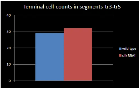
the presence of tracheoblast at the dorsal trunk (Fig.21). The results from these cell counting
experiments showed that the number of terminal cells, both in wild type and slik RNAi, were
comparable. (Fig.22, Appendix Fig.50). The average terminal cells count in both genotypes
were as follows, wild type = 29.2 ± 2.68, slik RNAi = 31± 3.08. The p value from a Student’s
t-test was p = 0.35. Thus branch counting showed that slik knockdown in the tracheal system
Results
38
does not interfere with the specification of terminal cells but rather with the development of terminal cells and branches.
Figure 20: Knockdown of slik in the tracheal system affects larval body size
Comparison of third instar slik knockdown larva with a wild type larva of the same stage. The larvae were raised at 29°C. Third instar larva were identified by the presence of the characteristically exposed spiracular papillae of the anterior spiracles. Tracheal specific knockdown of Slik reduced body size in larvae.
Figure 21: Dorsal terminal cells of the tracheal segments tr3-tr5 from third instar larvae
Tracheal system is visualised by tracheal specific cytoplasmic GFP (green) expressed using btlGal4 and the terminal cells are identified by Srf staining (red). (a) Schematic representation of the tracheal system in a third instar larva, marking tracheal segments tr3, tr 4 and tr5. (b) An assembly of a series of snapshots that cover tr3, the specific tracheal segment is identified by the presence of tracheoblasts (blue dots) while the preceding tracheal segment lacks the tracheoblast. Terminal cells express Srf (marked by arrows) and therefore in this overlay are seen as yellow.