Functional analysis of the role of FOXO in ageing and metabolism in Drosophila melanogaster
Inaugural-Dissertation Zur
Erlangung des Doktorgrades
der Mathematisch-Naturwissenschaftlichen Fakultät der Universität zu Köln
vorgelegt von
Victor Manuel Bustos Parra aus Bogotá, Kolumbien
Gutachter: Prof. Dr. Linda Partridge Prof. Dr. Aleksandra Trifunovic Tag der mündlicher Prüfung: 24.10.2016
ACKNOWLEDGEMENTS ... I ABBREVIATIONS ... III ZUSAMMENFASSUNG ... V SUMMARY ... VII
INTRODUCTION ... 1
1.1AGEINGANDTHEINSULINSIGNALLINGPATHWAY ... 3
1.2.FOXOTRANSCRIPTIONFACTORS ... 6
1.3FOXO ACTIVITY IS REGULATED BY POST-TRANSLATIONAL MODIFICATIONS ... 10
1.3.1 FOXO phosphorylation ... 10
1.3.2 FOXO acetylation ... 11
1.4FOXO IS REGULATED BY PROTEIN-PROTEIN INTERACTIONS ... 14
1.4.1 FOXO functions independent of DNA binding ... 15
1.5METABOLISM REGULATION AND FOXO FACTORS ... 17
1.5.1 dFOXO and metabolism regulation in Drosophila ... 18
1.6AIM OF THE THESIS ... 20
MATERIAL & METHODS ... 21
2.1GENERATION, MAINTENANCE AND CHARACTERIZATION OF TRANSGENIC FLY LINES . 23 2.1.1 Genomic engineering of the dfoxo locus (see also, Results 2.2) ... 23
2.1.2 Fly maintenance ... 25
2.1.3 Fly lines used in this study ... 25
2.1.4 Lifespan and fecundity assays ... 26
2.1.5 Stress assays and fly experiments ... 26
2.1.6 Fly developmental time and body weight ... 27
2.2BIOCHEMISTRY AND MOLECULAR BIOLOGY METHODS ... 28
2.2.1 DNA extraction for genotyping ... 28
2.2.2 RNA extraction and qPCR Analysis ... 28
2.2.3 Chromatin preparation, immunoprecipitation and qPCR ... 29
2.2.4 Lipid assays ... 30
2.2.5 Immunoprecipitation ... 30
2.2.6 Western Blot ... 31
2.2.7 Mass spectrometry ... 32
2.3DROSOPHILA CELL CULTURE METHODS ... 32
2.3.1 Cloning of cell culture plasmids ... 32
2.3.2 Cell culture maintenance, luciferase assay and cell imaging ... 33
2.4STATISTICAL ANALYSIS ... 33
GENOMIC ENGINEERING AND FOXO DNA BINDING ... 35
3.1INTRODUCTION ... 37
3.2RESULTS ... 37
3.2.1 Genomic Engineering of the Drosophila foxo locus ... 37
3.2.1.1 Generation of dfoxo parental knock-out lines ... 37
3.2.1.2 Generation of dfoxo replacement lines ... 42
3.2.2 Dissection of dFOXO functions independent of DNA binding ... 47
3.2.2.1 Identification of DNA binding deficient dFOXO ... 47
3.2.2.2 Generation and validation of in locus dfoxo-DBD mutants ... 49
3.2.2.3 dFOXO regulates body size independent of DNA binding ... 52
3.2.2.6 Is lip3 fundamental for lipid mobilization under starvation? ... 60
3.2.2.7 Generation of a putative dominant negative dFOXO ... 65
3.3DISCUSSION ... 69
3.3.1 Genomic engineering of the foxo locus ... 70
3.3.2 FOXO functions dependent of DNA binding ... 71
3.3.3 Body size and developmental effects mediated by dFOXO ... 72
3.2.4 Lipid metabolism regulation by FOXO ... 73
3.2.5 Lip3 could be a limiting factor for fat mobilization ... 75
3.2.6 Lip3 regulation by dHNF4 ... 76
3.2.7 dFOXO is required for starvation-induced autophagy. ... 77
FOXO ACETYLATION ... 81
4.1INTRODUCTION ... 83
4.2RESULTS ... 83
4.2.1 Mutation of putatively acetylated residues ... 83
4.2.2 In vivo generation of dFOXO acetylation mutants ... 85
4.2.3 Identification of dFOXO post-translational modifications ... 90
4.3DISCUSSION ... 92
CONCLUSION ... 97
5.1CONCLUSIONSANDFUTUREPERSPECTIVES ... 99
REFERENCES ... 101
SUPPLEMENT ... 121
ERKLÄRUNG ... 129
I would like to start by thanking Prof. Linda Partridge for receiving me in her research group, for giving me the freedom to pursue my own project, and for encouraging me throughout the years. Your trust has allowed me to develop personally and scientifically and for that I will always be grateful.
I would also like to thank my committee, composed by Prof. Aleksandra Trifunovic and Prof. Siegfried Roth, for evaluating my thesis. Also, thanks to Dr. Carlos Chacón, for taking the protocol during the PhD defense.
Next, I would like to thank the Max Planck Society and the University of Cologne.
Thanks to the institute, MPI for biology of ageing, and all the facilities that were always happy to provide an answer to any of my questions. Moreover, I thank the Boehringer Ingelheim Fonds (BIF) and its members for the support, not only financial, and all the experiences provided in the form of courses and meetings.
I would like to specially thank Sebastian. He has supervised my work since my first day in the lab and his feedback has always been fundamental for the development of my PhD project. Thank you for all the patience! Also, I would like to thank all other members from the Partridge lab. Special thanks go to René for making our lives easier by cooking the fly food and to Christine for making German bureaucracy a lot easier! Of course, I thank Jacky for all the microinjections, Nazif from the London lab, and all the other people in the Cologne lab that facilitated my work. A big ‘thank you’ also goes to the two Master students I supervised during my PhD: Jabiz and specially Ralf helped me develop the project and formed an amazing FOXO-team. I want to also thank my fellow PhD students Carina, Chirag, Oliver and Paula (aka Lucas) for all the good times and for enduring all my “Victor Questions”. Let there be many more. Also thanks to Luke and Tobias for the quick jokes and the support. A special mention goes to Anchal, writing the thesis at the same time made things easier and less stressful! So thank you for that!!!
I would like to specially thank the ‘linear regression’ group: Timo, Johanna, Pilar, Camilo and Joana. Thank you very much guys, let there be much more fun in the future! – Jo!
Thank you for putting up with me! You are #score #superawesome and I am very lucky to have shared all this time with you.
Germany a place to call home. I am sure I don’t need to specify what you guys mean to me, so I’ll just say thank you!
I thank my friends (el tetra): Pablo, Tesón and Daniel. Despite the distance and the fact that they never really understood what I did, they always supported me and brought a sense of reality when needed. Also, a huge thank you goes to Caro, not only she help me proofread this thesis, she was also always there. Always.
I thank the amazing city of Cologne. It is easy to live far away from your home country when the place you end up is so open and friendly as Cologne is. May there be many more carnivals to celebrate!!! Kölle Alaaf!!!
Last but not least, I would like to thank my family: Victor, Martha, Felipe, Lily, Hailee and Ema. Their constant communication and encouragement made the distance between us feel irrelevant. I love you.
Also, let’s not forget to thank the real MVPs of this thesis, my awesome FOXO flies!
Victor Bustos
You cannot avoid making mistakes, so you better learn from them.
∆ null mutants
4E-BP 4 Eukaryotic initiantion factor Binding Protein 4xFRE 4xFOXO responsive element
AA Amino acid
AcCoAS Acetyl Coenzyme A synthase AMPK AMP-activated protein kinase atg Autophagy related gene ATGL Adipose Triglyceride lipase BLRP Biotin ligase recognition peptide
bmm brummer
CBP CREB binding protein Cdk1 Cyclin-dependent kinase 1 ChIP Chromatin Immunoprecipitation colt congested-like trachea
CREB cAMP-response element-binding protein
da daugtherless
Dah Dahomey
DBD DNA binding domain
dHR96 Hormone receptor-like in 96 DILP Drosophila ILP
DNA Deoxyribonucleic acid
FB Fat body
FFA Free fatty acids
FLP Flippase
FOXO Forkhead Box O
FRT Flippase recognition target G6Pase Glucose-6-phosphatase GcK Glucose carboxykinase HDAC Histone deacetylase hid head involution defective HNF4 Hepatocyte nuclear factor 4 IGF Insulin-like growth factor IIS Insulin/IGF signaling ILP Insulin-like peptide InR Insulin(-like) receptor
IP Immunoprecipitation
IPC Insulin producing cells IRES Internal ribosomal entry site IRS Insulin receptor substrate JNK Jun-N-terminal kinase
Lip3 Lipase 3
Lip4 Lipase 4
Lipa Lysosomal acid lipase
min minute
mTORC1 mammalian TOR complex 1
NID NHR interacting domain NLS Nuclear localization signal O-GlcNac O-linked-N-acetylglucosamine OGT O-GlcNac transferase
P Phosphate / phosphorylated residue PDK1 Phosphoinositide-dependent kinase 1 pepck Phosphoenolpyruvate carboxykinase PH Pleckstrin homology
PI3K Phosphatidylinositol 3-OH kinase PIP2 Phosphatidylinositol 3-phosphate PIP3 Phosphatidylinositol 3-phosphate PKB Protein kinase B
PolyQ Polyglutamine
PTM Post-translational modification
qRT-PCR quantitative Real Time Plymerase Chain Reaction SGK1 Serum- and glucocorticoid-inducible kinase 1
Sirt Sirtuin
TAG Triacylglycerol TNPO1 Transportin 1
TOR Target of Rapamycin
Ups Ultraspiricle
w white
yip2 yippee interacting protein 2
Eine verringerte Aktivität des evolutionär hochkonservierten Insulin/Insulin-ähnlichem Wachstumsfaktor-Signalweges (IIS) verlängert die Lebenszeit von Nematoden, Fruchtfliegen und Mäusen. In Nematoden und Fruchtfliegen ist der Forkhead Box-O (FOXO) Transkriptionsfaktor notwendig für den lebensverlängernden Effekt durch verringerten IIS, und auch erhöhte FOXO Aktivität alleine verlängert das Leben. FOXO Transkriptionsfaktoren sind jedoch nicht nur am Alterungsprozess beteiligt, sondern auch an der Entwicklung und dem Stoffwechsel eines Organismus. Daher ist es essentiell zu verstehen, durch welche molekularen Mechanismen der FOXO Transkriptionsfaktor diese Prozesse steuert.
In meiner Doktorarbeit habe ich mithilfe der Fruchtfliege Drosophila melanogaster untersucht, wie die Aktivität von FOXO reguliert wird und welche unterschiedlichen Funktionen dFOXO hat. Ich habe neue dfoxo-null Mutanten generiert, mit denen ich unterschiedliche Modifikationen von dfoxo in das Genom inserieren konnte. Insertion des Wildtyp-dFOXOs oder von dFOXO mit FLAG-Tag am C-Terminus hatte keinen Effekt auf die Funktion von dFOXO und wurde verwendet, um die in vivo Funktion von dFOXO zu charakterisieren. Außerdem habe ich dfoxo Allele generiert, die das Binden von dFOXO an DNA in vivo verhindern. Diese Mutanten erlaubten die Separierung von dFOXO Funktionen, die abhängig (Fertilität, Resistenz gegen oxidativen Stress und Lebensspanne) und unabhängig (Gewicht und Lipid-Verbrauch während Hungerperioden) von der Interaktion von dFOXO mit der DNA sind. Unsere Ergebnisse weisen darauf hin, dass dFOXO die Genexpression und Autophagie in Hungerperioden unabhängig von der Fähigkeit an DNA zu binden moduliert, vermutlich durch die Interaktion mit einem anderen Transkriptionsfaktor.
Zudem habe ich dfoxo Mutanten erzeugt, die Acetylierung an einem konservierten Lysin- Rest imitieren oder verhindern. Unsere Experimente lassen vermuten, dass diese posttranslationalen Modifikationen auch den Phosphorylierungs-Status von dFOXO beeinflussen. Außerdem scheint dFOXO-Acetylierung eine wichtige Rolle in der Antwort auf den Entzug von Aminosäuren zu spielen, jedoch müssen weitere Experimente den verantwortlichen Mechanismus identifizieren.
zur Modifikation des dfoxo locus und separiert wichtige Funktionen des FOXO Transkriptionsfaktors in Drosophila melanogaster.
Reduced activity of the highly evolutionarily conserved insulin/insulin-like growth factor signalling pathway (IIS) has been shown to increase lifespan in nematode worms, fruit flies and mice. In both worms and flies the single Forkhead Box-O (FOXO) transcription factor is required for increased lifespan from reduced IIS, and increased FOXO activity itself lengthens life. However, FOXO transcription factors are involved in the regulation of diverse cellular and organismal processes besides ageing, including development and metabolism. It is therefore of crucial importance to understand the molecular mechanisms by which this transcription factor acts to regulate those processes.
During my PhD, I used the fruit fly Drosophila melanogaster to investigate how FOXO activity is regulated and tried to dissect the different functions associated with dFOXO. I have generated novel dfoxo-null mutant lines that allow me to reintroduce modified versions of the dfoxo gene. Wild type and C-terminal tagged reinsertion lines seem to have no effect on normal dFOXO function and, hence, were used to better characterize dFOXO regulation in vivo. Next, I generated dfoxo mutant alleles that abolished DNA binding in vivo. dFOXO-DBD mutant flies permitted the separation of dFOXO-functions that are dependent (fecundity, redox stress resistance, and lifespan) or independent (body weight and lipid usage under starvation) of dFOXO-DNA binding. Our results suggest that dFOXO modulates gene expression and autophagy under starvation conditions independent of DNA binding, probably through the interaction with another transcription factor.
On the other hand, I generated dfoxo alleles that either mimic or abolish acetylation within conserved lysine residues. Our results indicate that this post-translational modification regulates dFOXO-phosphorylation state. Moreover, dFOXO-acetylation seems to play a crucial role in the response associated with amino acid starvation. However, further studies should identify the mechanisms at play.
In conclusion, this PhD thesis describes a gene-editing tool for the dfoxo locus and separates some of the important functions associated with FOXO transcription factors in Drosophila.
I NTRODUCTION
1.1 AGEING AND THE INSULIN SIGNALLING PATHWAY
The idea of immortality has intrigued humans for thousands of years. Whether it was alchemists trying to create the philosophers’ stone, explorers trying to find the fountain of youth, or the promise of an afterlife by most religions, the idea of eternal live has shaped humanity. For a long time ageing has been regarded as an immutable process due to damage accumulation over time that leads to functional decline and finally death.
However, this view was challenged by the identification of a mutation in a single gene called daf-2 in the roundworm Caenorhabditis elegans (C. elegans), which resulted in worms that lived twice as long as their wild type counterparts (Kenyon et al., 1993). This observation changed our entire perspective of the ageing ‘process’ and led to the conclusion that longevity is amenable to genetic interventions, just like other biological processes.
Daf-2 encodes the C. elegans homolog of the insulin/insulin-like growth factor (IGF) receptor (Kimura et al., 1997), an upstream component of the insulin/IGF-signalling (IIS) pathway. Similar to the worm daf-2 mutants, reduced IIS can increase lifespan in other animal models, including fruit flies and mammals. In Drosophila, mutations of the insulin-like receptor (InR) extend lifespan (Tatar et al., 2001). Similarly, removal of InR in adipose tissue or full-body heterozygous mutants of the IGF-1 receptor (IGF-1R) in mice is able to extend lifespan when compared to their respective controls (Bluher, 2003;
Holzenberger et al., 2003). Moreover, removal of the insulin receptor substrate in flies (chico) or mice (IRS1) extends lifespan and, at least in mice, it seems to cause a delay in ageing (Clancy et al., 2001; Selman et al., 2008; Tu et al., 2002). These observations make the IIS pathway a prominent target of ageing research. Importantly, the invertebrate models C. elegans and Drosophila are ideal to the study the relationship between IIS and ageing due to their short lifespan and the diverse molecular/genetic tools available (Piper et al., 2005).
The IIS pathway is an evolutionarily conserved nutrient-sensing network that plays key roles in diverse cellular and organismal processes, including growth, metabolism, stress response, reproduction and lifespan (Piper et al., 2005). IIS is activated by insulin-like peptides (ILPs), hormonal signals secreted, for example, in response to nutrient availability. Mammalian genomes code for at least eight ILPs (Wallis, 2009), including insulin and insulin-like growth factor-1 (IGF-1). In worms, 40 ILPs have been identified,
whereas flies express at least eight peptides (Lau and Chalasani, 2014). Interestingly, IIS manipulation at the ligand level can also extend lifespan in flies, as mutation of the Drosophila ILP (DILP) 2, or the triple dilp2-3,5 mutation, produces longer lived flies (Grönke et al., 2010). Unlike worms and flies, where all ILPs seem to transduce the signal through the single insulin-like receptor (the aforementioned Daf-2 and InR), mammals have multiple IIS-related receptors that, in most cases, bind a particular ILP with higher affinity than the rest (Fernandez and Torres-Alemán, 2012). Despite the differences, insulin or ILPs regulation by a favourable nutritional status has similar consequences in different organisms, inducing growth by stimulating protein synthesis, and promoting energy storage in the form of glycogen and fat (Piper et al., 2005).
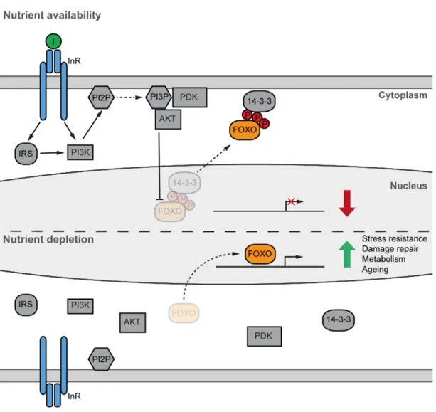
Under nutrient availability, circulating insulin interacts with the insulin receptor, which activates a complex phosphorylation cascade (Figure 1.1) that leads to, among others, glucose uptake by the muscle and fat body, while at the same time inhibiting glucose production by the liver (Boucher et al., 2014; Saltiel and Kahn, 2001). In simplified terms, insulin promotes a conformational change in the InR that allows auto-phosphorylation and phosphorylation of at least nine different targets, including a group of four proteins known as insulin receptor substrates (IRS1-4) (Patti and Kahn, 1998). In Drosophila, there is only one homolog protein of IRS (chico) (Clancy et al., 2001). When phosphorylated, IRS1 functions as a scaffold and allows the interaction with several regulatory proteins, most importantly, Phosphatidylinositol 3-OH kinase (PI3K). Activated PI3K in turn catalyzes the conversion of phosphatidylinositol 2-phosphate (PIP2) to phosphatidylinositol 3- phosphate (PIP3) (Vadas et al., 2011).
Proteins baring a pleckstrin homology (PH) domain can interact with PIP3 and hence be recruited to the cell membrane. This translocation to the membrane activates a series of kinases, of which Phosphoinositide-dependent kinase 1 (PDK1) is best characterized. The interaction between PIP3 and PDK1 leads to the phosphorylation and therefore activation of another fundamental kinase called protein kinase B (PKB or AKT). Importantly, AKT also contains a PH domain, which facilitates its localization to the membrane and therefore activation by PDK1 upon insulin signaling. Among its many targets, AKT phosphorylates the family of transcription factors Forkhead Box O (FOXO). The AKT dependent phosphorylation of FOXO proteins on three highly conserved residues induces their nuclear exclusion, in part by facilitating the interaction with scaffold protein 14-3-3,
and hence the down regulation of FOXO-dependent target genes (Piper et al., 2005). In contrast, under reduced nutrient availability, IIS is reduced, repression of FOXO factors is released and a plethora of target genes are therefore activated (Figure 1.1). While mammalian genomes encode for four different FOXO genes (FOXO1, 3, 4 and 6), invertebrate organisms like C. elegans and Drosophila contain only one homolog, termed daf16 and dfoxo, respectively (Jünger et al., 2003; Lin et al., 1997; Ogg et al., 1997; Puig et al., 2003).
Figure 1.1 Simplified representation of the highly conserved Insulin/Insulin-like growth factor signalling (IIS) pathway. Under nutrient availability (fed state), secreted insulin initiates a phosphorylation cascade that culminates in the repression of FOXO transcription factors by nuclear exclusion. On the other hand, under reduced nutrients (starved state), FOXO repression is released and transcription of multiple target genes can be initiated.
1.2. FOXO TRANSCRIPTION FACTORS
FOXO coding genes were first identified as chromosomal translocations associated with different kinds of cancer, i.e. the genomic fusions between PAX3 and FOXO1 was described in alveolar rhabdomyosarcomas (Galili et al., 1993). Multiple studies have now proven that FOXO proteins are in fact powerful tumour suppressors (Fu and Tindall, 2008).
FOXO proteins belong to the Forkhead family of transcription factors characterized by their evolutionary conserved ~100-amino-acid monomeric DNA binding domain (DBD) (Burgering, 2008) (Figure 1.2A). The DBD, also called winged helix domain, is a variant of the helix-turn-helix motif that consists of three α-helices and two loops or ‘butterfly like wings’ (Brent et al., 2008; Weigelt et al., 2001) (Figure 1.2B). Over 100 proteins belong to the Forkhead family and are classified according to the sequence similarity of their DBDs, into sub-categories denoted by a capital letter (Kaestner et al., 2000), e.g.
Forkhead box subgroup O ! FOXO.
FOXO proteins also have a characteristic lysine-rich nuclear localization signal (NLS) embedded at the 5’ end of their DBD (Brownawell et al., 2001; Zhang et al., 2002). Of note, a key residue phosphorylated by AKT is part of the NLS and it mediates the interaction with 14-3-3 (Obsilova et al., 2005) (Figure 1.2A). This phosphorylation- dependent interaction masks the NLS sequence from the nuclear import machinery and allows FOXO shuffling to the cytoplasm.
In addition to the NLS, mammalian FOXO1, 3 and 4 have a leucine-rich nuclear exclusion signal (NES) that, just like the AKT-phosphorylation/interaction with 14-3-3, is required to mediate FOXO translocation to the cytoplasm under high nutrient availability (Brunet et al., 2002). Interestingly, neither DAF-16 nor dFOXO seem to have a conserved NES, suggesting that FOXO shuttling out of the nucleus is NES-independent in those species.
Similarly, mammalian FOXOs contain a conserved LXXLL motif that mediates interaction with Sirt1 (Nakae et al., 2006) and, possibly, nuclear hormone receptors (NHRs), however, this motif seems to be absent in flies and worms (van der Vos and Coffer, 2008).
Figure 1.2 The FOXO DNA binding domain (DBD) is conserved in evolution.
(A) Amino acid alignment of the DBD of four different FOXO proteins using MUSCLE (Edgar, 2004). Residues highlighted in blue show some degree of conservation between all species.
Orange residues represent the partially conserved nuclear localization signal (NLS). Consensus symbols indicate the degree of conservation: an * (asterisk) indicates perfect conservation whereas a : (colon) or a . (Period) indicate conservation of stronger or weaker similar properties. Within the NLS, the red colon (:) highlights the conserved residue phosphorylated by AKT. (B) Crystal structure of FOXO1-DBD in contact with DNA. Blue structure covers evolutionarily conserved residues, as in the alignment. “Wing 2” points at the position wing 2 would be, since it was not part of the crystal structure. Displayed as stereo pair – PDB code 3CO6 (Brent et al., 2008).
Unlike other FOXO paralogs, Drosophila FOXO (dFOXO) has polyglutamine (polyQ) repeats within its C-terminal domain. PolyQ regions have been well studied in the context of human neurodegenerative diseases (Orr and Zoghbi, 2007). However, we now know that eukaryotic transcription factors are rich in polyQ repeats (Gemayel et al., 2010) and that these repeats modulate the transcription factor activity by tuning its solubility and interactions (Gemayel et al., 2015). These polyQ regions can mediate interaction with other polyQ regions, facilitating co-regulation between transcription factors (Atanesyan et
al., 2012). How the polyQ regions within dFOXO modulate its activity or interactions is still unknown.
In mammals, FOXO proteins are expressed throughout the body, regulate different tissue- specific functions (Salih and Brunet, 2008) and several cellular processes such as apoptosis, stress response and cell cycle (Burgering, 2008). For example, FOXO1 is a key component of energy metabolism and is predominantly expressed in white adipose tissue, liver and muscle (Gross et al., 2008; Kousteni, 2012; Matsumoto et al., 2007).
Additionally, FOXO proteins modulate immune function (Kim et al., 2013; Ouyang et al., 2012) and play important roles in stem cell maintenance in diverse tissues including muscle, neurons and the hematopoietic stem cell pool (Gopinath et al., 2014; Miyamoto et al., 2007; Paik et al., 2009). How exactly FOXO proteins coordinate such a vast range of processes is still largely unknown.
Due to the high degree of conservation, FOXO paralogs virtually share identical DBDs, allowing them to bind the same DNA motifs and therefore have, at least partially, redundant functions. A recent meta-analysis of FOXO-target-genes across tissues found that, even though specific FOXO proteins have tissue specific targets, there is a core set of genes regulated across tissues, and even species (Webb et al., 2016). These conserved target genes are involved in metabolism, proteostasis, stress resistance and growth factor signalling (Webb et al., 2016). This observation indicates that regulation of those core processes, which are fundamental for ageing modulation, is the main function of FOXO factors across evolution.
The presence of only one FOXO orthologue makes C. elegans and Drosophila ideal model systems to study FOXO function independent of redundancy between FOXO paralogues. In both organisms, FOXO factors are mediators of IIS action, and the removal of FOXO function suppresses lifespan-extension upon reduced IIS (Kenyon et al., 1993;
Slack et al., 2011; Yamamoto and Tatar, 2011). Moreover, foxo-null mutants are short- lived in worms and flies, indicating that FOXO proteins are required for normal ageing (Lin et al., 2001; Slack et al., 2011). In addition, DAF-16 over-expression is sufficient to extend lifespan in the worm (S T Henderson and Johnson, 2001). Similarly, muscle- or fat-specific over-expression of dFOXO increases longevity in flies (Demontis and Perrimon, 2010; Giannakou et al., 2004; Hwangbo et al., 2004), suggesting that tissue-
specific functions of dFOXO are important for regulating organismal features, including ageing. These observations are consistent with studies in C. elegans that highlight the gut, which also functions as adipose tissue, as a key factor for modulating ageing (Libina et al., 2003). Even though many of the downstream gene targets of FOXO proteins have been identified in multiple organisms (Webb et al., 2016) , the exact mechanisms by which FOXO factors modulate lifespan are unclear.
Both DAF-16 and dFOXO have different isoforms. In C. elegans there are three isoforms with distinct tissue specificity and impact on ageing (Kwon et al., 2010). In contrast, Drosophila has four isoforms that differ in their 5’UTR, which contains internal ribosomal entry sites (IRES) which are important to mediate translation of dfoxo under low nutritional conditions (Villa-Cuesta et al., 2010). Two of those four dfoxo transcripts produce a 613aa protein (the preferred version for transgene generation), whereas the other two produce a 10aa longer version. Whether these isoforms are preferentially expressed in specific tissues or the amino acid difference is biologically relevant is still unknown.
In summary, FOXO proteins are evolutionarily conserved factors that modulate the organismal response to different kinds of environmental stress. Indeed, FOXO proteins are now seen as fundamental mediators of homeostasis maintenance (Eijkelenboom and Burgering, 2013), even though the mechanisms by which these proteins regulate so many processes are still unclear.
1.3 FOXO activity is regulated by post-translational modifications
FOXO proteins are metabolic nodes where multiple pathways converge. Hence, it is perhaps not surprising that these proteins are regulated by a great number of post- translational modifications (PTMs) (Figure 1.3). In 2008, Calnan and Brunet proposed that different PTM combinations on FOXO could work as a ‘code’ to elicit a specific transcriptional output in response to diverse stimuli (Calnan and Brunet, 2008). FOXO PTMs include phosphorylation, methylation, acetylation, mono- and poly-ubiquitination, O-glycosylation and poly-ADPribosylation (Daitoku et al., 2011; Klotz et al., 2015; Zhao et al., 2011). However, it is not clear to what extent these different PTMs contribute to FOXO-dependent regulation of lifespan. Nevertheless, this FOXO code modulates target genes involved, in grand terms, in three kinds of biological processes: metabolism, stress response and cell proliferation/apoptosis (Calnan and Brunet, 2008).
1.3.1 FOXO phosphorylation
AKT-dependent phosphorylation of FOXO proteins takes place at three evolutionary conserved residues that are part of a motif recognized by this kinase (Figure 1.3). Even though AKT phosphorylation is the best characterized FOXO-PTM, FOXO factors are phosphorylated by a panoply of kinases (Klotz et al., 2015). For example, AKT works in concert with serum- and glucocorticoid-inducible kinase 1 (SGK1), a kinase also activated by PI3K, to phosphorylate at the three conserved residues and inactivate FOXO3a (Brunet et al., 2001) (Figure 1.3A). While AKT prefers to phosphorylate the FOXO3 residue S253, SGK1 prefers S315, suggesting that the combination of both enzymes is what allows full FOXO phosphorylation under high nutrient conditions (Brunet et al., 2001).
AKT/SGK1 phosphorylations do not only reduce DNA binding affinity but also induce the interaction with the scaffold protein 14-3-3. T32 and S253 phosphorylation mediates the 14-3-3 interaction, which changes and probably masks the NLS sequence within FOXO, since S253 is buried within the NLS (Brunet et al., 1999; Obsilova et al., 2005) (Figure 1.3A). Nuclear exclusion of FOXO1 by IGFR1 signalling seems to be 14-3-3 independent, suggesting that at least one additional mechanism can shuttle FOXO1 into the cytoplasm (Rena et al., 2001). On the other hand, Drosophila has an additional scaffold protein that modulates the AKT-phosphorylation of dFOXO, called Melted, which seems to recruit dFOXO to the cell membrane and hence allow dFOXO and AKT to be in close proximity under activated IIS (Teleman et al., 2005). Nevertheless, once in
the cytoplasm, phosphorylated FOXO can undergo ubiquitination followed by proteosomal degradation (Aoki et al., 2004; Huang et al., 2005; Matsuzaki et al., 2003;
Plas and Thompson, 2003). What exactly leads to FOXO degradation or cytoplasmic retention is currently unclear.
Phosphorylation of FOXO proteins can, however, also induce nuclear localization and/or activation (Figure 1.3). For example, cyclin-dependent kinase 1 (Cdk1) phosphorylation induces FOXO1-dependent transcription and causes cell death in neurons (Yuan et al., 2008). In Drosophila cell culture, multiple kinases were shown to modulate dFOXO transcriptional activity (Mattila et al., 2008). However, it is currently unclear whether these phosphorylations actually take place in vivo and whether they have a physiological consequence. Additionally, FOXO proteins are phosphorylated by AMPK (AMP-activated protein kinase) and JNK (Jun-N-terminal kinase) in response to nutritional or oxidative stress respectively (Essers et al., 2004; E. L. Greer et al., 2007; Eric L. Greer et al., 2007).
The control that these two kinases exert over FOXO seems to be evolutionarily conserved.
Moreover, both AMPK and JNK contribute to the modulation of longevity in worms and flies, however, the exact mechanism behind this function are not fully understood (Biteau et al., 2011; Burkewitz et al., 2014).
1.3.2 FOXO acetylation
FOXO activity can also be regulated by acetylation and deacetylation of specific lysine residues. In fact, acetylation was shown to both activate (Motta et al., 2004; Perrot and Rechler, 2005) and repress (Frescas et al., 2005; Fukuoka et al., 2003; Jing et al., 2007;
Matsuzaki et al., 2005; Mihaylova et al., 2011; Wang et al., 2011, 2007) FOXO proteins.
The reasons for such discrepancies are currently under investigation, but one possible explanation is that activating-acetylation seems to occur at the carboxy-terminal region of the protein, whereas repressive-acetylation takes place close to and within the NLS. Of note, Drosophila does not have any lysine residues in its carboxy-terminal region, suggesting that this acetylation is not evolutionarily conserved. Of the two, repressive- acetylation is better understood. This process can reduce FOXO DNA-binding ability and increase its sensitivity to phosphorylation by AKT, which in turn results in reduced transcriptional activity (Brent et al., 2008; Brunet et al., 2004; Matsuzaki et al., 2005;
Qiang et al., 2010).
In mammals, FOXO proteins are acetylated by the co-activators p300 and cAMP response element-binding protein (CREB)-binding protein (CBP) and deacetylated by Sirt1 (Matsuzaki et al., 2005). Additionally, class II histone deacetylases (HDACs) were shown to modulate FOXO acetylation status in both mice and flies in response to nutritional stress (Mihaylova et al., 2011; Wang et al., 2011).
A mouse knock-in study revealed a fundamental role of FOXO1 acetylation in the regulation of glucose and lipid metabolism. An acetylation-mimicking-FOXO1 allele is lethal during early development, whereas the acetylation-null allele has a distinct metabolic phenotype, where mice seem to relay mostly on lipids as an energy source (Banks et al., 2011). The putatively acetylated lysine residues are mostly localized within the NLS of FOXO factors and are evolutionarily conserved. Deacetylation of these conserved residues by HDAC4 was recently suggested to allow dFOXO transcriptional regulation of the starvation response (Wang et al., 2011). Furthermore, dFOXO and FOXO1 seem to be promptly acetylated upon re-feeding of the flies and mice respectively (Banks et al., 2011; Wang et al., 2011). On the other hand, the transcriptional co-factor KDM5 (also known as Lid) seems to also interact with HDAC4 to facilitate FOXO deacetylation in order to elicit a transcriptional response under oxidative stress (Liu et al., 2014). On the other hand, cell culture and xenograft experiments indicate that acetylated FOXO1 is able to interact with ATG7 (autophagy related gene 7) in the cytoplasm to induce autophagy under serum starvation or oxidative stress (Zhao et al., 2010). These observations suggest acetylation may regulate multiple functions of FOXO proteins.
However, the precise role of FOXO regulation by acetylation, and its effect on lifespan, is currently unclear.
In summary, FOXO proteins are regulated by multiple PTMs, however, it is not clear how these modifications regulate each other in vivo nor the biological consequences of their interaction at the cell, tissue or organism level. Thus, it would be important to develop a tool that would allow the in vivo identification and characterization of FOXO PTMs.
Figure 1.3 Phosphorylation and acetylation on mammalian FOXO compared to Drosophila dFOXO.
(A) Repressive-phosphorylation (red P) by AKT takes place at three evolutionarily conserved residues in both mammals and flies. Two of these residues mediate the interaction with scaffold protein 14-3-3. In contrast, many kinases are known to activate (green P) FOXO factors in mammals. Some of these kinases, including AMPK and JNK, can also activate Drosophila FOXO.
However, the exact residues where the modifications take place are unknown. (B) Repressive acetylation of mammalian FOXO proteins was shown to take place at different lysine residues within the NLS. Since some of these residues are evolutionarily conserved, it was proposed that this kind of acetylation also happens in the fly. Conversely, activating-acetylation of FOXO proteins has only been reported in mammals. DBD-DNA binding domain; NLS-Nuclear localization signal; NES-Nuclear exclusion signal; NID-NHR interacting domain; Q-Glutamine rich region.
1.4 FOXO is regulated by protein-protein interactions
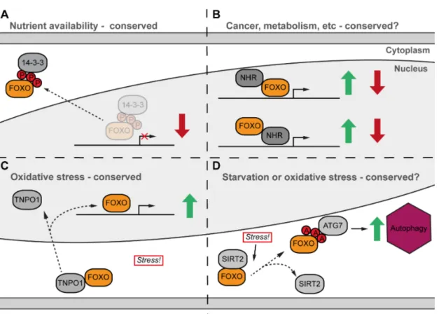
The best-characterised FOXO interactor is the scaffold protein 14-3-3. Under high insulin levels, AKT-dependent phosphorylated FOXO interacts with 14-3-3 and is therefore excluded from the nucleus (Figure 1.4A) (Brunet et al., 2002). However, multiple interaction partners are known to regulate FOXO function, while at the same time FOXO proteins are able to function as co-regulators and modulate the role of other transcription factors (Daitoku et al., 2011; van der Vos and Coffer, 2008).
In mammals, FOXO proteins have been shown to interact with a wide range of nuclear hormone receptors (NHRs), including the androgen, progesterone, glucocorticoid, retinoic acid, peroxisome and thyroid hormone receptors (van der Vos and Coffer, 2008).
Interaction of FOXO with non-steroid receptors leads to co-activation, while biding to steroid NHRs leads to the opposite effect, co-repressing target genes (Zhao et al., 2001).
This kind of interaction can lead to expression alteration of both NHR and/or FOXO target genes (Figure 1.4B). For example, binding of the androgen receptor can suppress FOXO1 transcriptional activity in prostate cancer cells (Li et al., 2003). Moreover, FOXO1 interacts with Hepatocyte Nuclear Factor-4 (HNF4) (Hirota et al., 2003). However, the consequences of this interaction seem to be complex. Under fasting conditions, FOXO binds to HNF4 and represses certain HNF4-target genes while at the same time it has a synergistic effect on HNF4 and FOXO1 shared target genes (Hirota et al., 2008).
However, whether the repressive and/or synergistic interaction is evolutionarily conserved and the exact mechanism by which this kind of regulation takes place is currently unknown.
FOXO factors translocate to the nucleus and are hyper-activated under stressful conditions, such as starvation or oxidative stress (S T Henderson and Johnson, 2001). For example, it was recently reported in mammalian cell culture that FOXO4 is able to interact with a transporter called Transportin1 specifically under Redox stress (Putker et al., 2013).
Of note, this interaction seems to be evolutionarily conserved since DAF-16 is able to interact with Transportin1, and its worm homolog IMB-2, to regulate this translocation (Putker et al., 2013) (Figure 1.4C). On the other hand, a study on a human cancer cell line showed that, upon oxidative stress or serum starvation, cytosolic FOXO1 lost the interaction with NAD-dependent histone deacetylase, SIRT2. This release led to FOXO1 acetylation, which in turn was then able to induce autophagy by interacting with ATG7
(Zhao et al., 2010) (Figure 1.4D). Nevertheless, whether any of these stress-induced interactions has a role in the regulation of ageing through FOXO factors is still unknown.
1.4.1 FOXO functions independent of DNA binding
Besides their prominent role as transcriptional regulators, FOXO proteins also have functions that are independent of the genes they regulate, making the understanding of their functions ever more complex. For example, FOXO1 over-expression in mammalian cell culture inhibits cell cycle progression by down-regulating D-type cyclins and inducing apoptosis. At the same time, inhibition of cell cycle progression is independent of the ability of FOXO1 to bind to DNA. Over-expression of a FOXO1 DNA binding mutant blocked cell cycle, but it did not affect apoptosis, suggesting that some functions of FOXO1 may not require DNA binding ability (Ramaswamy et al., 2002). FOXO1 also regulates progesterone receptor A activity independently of DNA binding (Rudd et al., 2007) and cytoplasmic FOXO1 was shown to mediate autophagy in a human cancer cell line (Zhao et al., 2010) (Figure 1.4D). These results suggest that FOXO proteins may act as transcriptional co-regulators and that DNA binding is not required to fulfil these functions. Interestingly, some of the FOXO1 effects on energy metabolism seem to be DNA-binding-independent, as seen in cell culture (Matsumoto et al., 2006), and liver- specific mouse mutants (Cook et al., 2015). The liver-specific study further suggests that FOXO1 may partially regulate liver lipogenesis, acting as a co-activator of another transcription factor (Cook et al., 2015). The putative interaction partner mediating these effects is still unidentified. Whether any of the dFOXO DNA-binding-independent functions are evolutionarily conserved, and their effect on ageing, is currently unknown.
Figure 1.4 Simplified representation of different FOXO interactions
(A) AKT-dependent phosphorylation of FOXO factors mediates the interaction with scaffold protein 14-3-3, which in turn induces nuclear exclusion. This mode of FOXO regulation is evolutionarily conserved. (B) Nuclear FOXO can interact with multiple NHRs. This interaction can lead to expression or repression of FOXO and/or NHR target genes. (C) Redox stress can induce the interaction of FOXO with TNPO1, which leads to nuclear translocation followed by up- regulation of genes involved in redox stress response. This type of interaction seems to be evolutionarily conserved between worms and mice (see text). (D) Cytosolic FOXO loses the interaction with SIRT1 under starvation or redox stress, which leads to acetylated FOXO accumulation, interaction with ATG7 and induction of autophagy. NHR-Nuclear hormone receptor; TNPO1-Transportin; ATG7-Autophagy related gene 7.
1.5 Metabolism regulation and FOXO factors
Nutrient availability modulates the behaviour and metabolism of all organisms. The IIS pathway plays a fundamental role integrating nutrient sensing and energy homeostasis, for example, by repressing FOXO factors under nutrient abundance and, conversely, releasing said repression upon starvation periods (Saltiel and Kahn, 2001). FOXO proteins play critical roles in metabolism homeostasis; however, it is currently not fully understood how these transcription factors modulate the diverse processes involved in glucose and lipid metabolism.
Among mammalian FOXO proteins, FOXO1 is portrayed as the key regulator of energy metabolism (Kousteni, 2012). This protein is prominently expressed in tissues relevant for glucose homeostasis, such as liver, pancreas and adipose tissue. Moreover, FOXO1 mediates the organismal response to reduced nutrients (low insulin), stimulating hepatic glucose production and inhibiting adipogenesis (Matsumoto et al., 2007; Nakae et al., 2002; Qiao and Shao, 2006). For example, hepatic glucose production is induced by up- regulating pepck, which codes for the phosphoenolpyruvate carboxykinase, the limiting enzyme for gluconeogenesis. In addition, FOXO1 induces the Adipose triacylglycerol lipase (ATGL) expression in both adipose tissue and liver (Chakrabarti and Kandror, 2009; Zhang et al., 2016). ATGL is the limiting rate enzyme regulating lipolysis and its expression can therefore stimulate triacylglycerol (TAG) usage (Zimmermann, 2004).
Moreover, starvation induces FOXO-dependent expression of eukaryotic initiation factor 4E binding protein (4EBP), which in turn dampens translation.
TAG reservoirs are found in lipid droplets, conserved cellular structures that are present across all organisms and have acquired multiple regulatory roles, such as lipid homeostasis, throughout evolution (Murphy, 2012). Consistently, lipid droplets are found across Drosophila tissues and serve mainly as TAG stores (Kuhnlein, 2012). Under fasting conditions, TAG stores are hydrolyzed in the cytosol by lipases, such as ATGL, into free fatty acids (FFA), which in turn are transported into the mitochondria to undergo β-oxidation and serve as an energy source. Fasting also induces autophagy, a process in which a double membrane vesicle, termed autophagosome, grows and engulfs organelles or cytoplasmic entities to later fuse with the lysosome and break its cargo down for energy production (Russell et al., 2014). The autophagy machinery can uptake TAGs and, with
the help of specialized lysosomal-associated-lipases, hydrolyze them to release FFA in a process termed lipophagy (Singh et al., 2009).
FOXO factors promote autophagy in different cell types, such as neurons and hepatocytes, by up-regulating multiple autophagy-related genes and have therefore another layer of control over the starvation response (Webb and Brunet, 2014). A recent study implicated FOXO1 in the regulation of ATG14, a protein that mediates the autophagosome fusion with lysosomes, and hence is fundamental for proper autophagy (Diao et al., 2015; Xiong et al., 2012). While ATG14 knockdown induced lipid accumulation in the liver, its over- expression protected the liver from fat accumulation under high fat diet, suggesting ATG14 is in fact a critical regulator of lipid homeostasis (Xiong et al., 2012).
Furthermore, FOXO1 mediates the expression of lysosomal acid lipase (Lipa) in adipose tissue (Lettieri Barbato et al., 2013). In this study, nutrient restriction stimulated Lipa expression in a FOXO1-dependent manner, and Lipa expression was required for lipophagy induction (Lettieri Barbato et al., 2013). In accordance with these phenotypes, lipa mutant mice are unable to properly mobilize TAGs in the liver (Du et al., 2001).
These observations implicate FOXO factors as key modulators of the starvation response at multiple levels.
1.5.1 dFOXO and metabolism regulation in Drosophila
During the last 15 years, Drosophila has been increasingly used as a powerful model to study the evolutionarily conserved mechanisms of energy homeostasis. The fly has functionally analogous tissues to those found in mammals that mediate energy storage in the form of glycogen and lipids when conditions are favourable (Baker and Thummel, 2007; Kuhnlein, 2012). For example, the fly fat tissue acts both as liver and adipose tissue in mammals, storing energy and modulating its usage under nutrient depravation. Under nutritional stress, these energy stores are mobilized to provide energy for the cells (Baker and Thummel, 2007).
A great number of the proteins involved in the response to energy deprivation have a clear homolog in the fly. For example, the Drosophila homolog of 4ebp (termed thor) is one of the best characterized dFOXO target genes (Puig et al., 2003). Under nutritional stress, dFOXO induces 4ebp expression, which in turn dampens general translation, ensuring careful allocation of energy recourses. Moreover, Drosophila brummer (bmm – the
homolog of mammalian ATGL) is also the rate limiting enzyme during lipolysis (Grönke et al., 2005). bmm was recently characterised as a dFOXO target gene by showing transcriptional induction under starvation in a dFOXO-dependent manner (Wang et al., 2011). These results highlight FOXO as a key mediator of metabolism in flies and mammals. Indeed, it was recently suggested that metabolic regulation by FOXO factors is evolutionarily conserved, at least based on common target genes across species (Webb et al., 2016).
In addition to bmm transcriptional regulation under starvation, dFOXO also stimulates expression of lip4 (lipase 4 – a homolog of mammalian Lipa) by direct binding to its promoter (Vihervaara and Puig, 2008). Moreover, dFOXO regulates the expression of atg8 in the muscle, a key protein involved in autophagy induction (Bai et al., 2013). These results suggest that dFOXO, just like its mammalian counterparts, may in fact be able to regulate autophagy and lipophagy in Drosophila. However, this hypothesis awaits experimental testing.
1.6 Aim of the thesis
FOXO transcription factors are involved in several cellular and physiological processes, such as development, metabolism and ageing. In order to exert control over such diverse functions among different tissues, these proteins are regulated by multiple PTMs and protein-protein interactions. The multiple functions and levels of regulation make the study of FOXO factors complex. Moreover, mammalian genomes encode four FOXO paralogs that are able to target common genes, making it harder to determine the role of FOXO proteins in diverse processes. Drosophila only has one dfoxo gene, making it a simpler model to understand the regulation and functions of FOXO transcription factors.
Hence, I aimed to generate a genetic tool that would allow me to modify the endogenous dfoxo gene. With this, I would be able to dissect the different dFOXO-associated functions by generating in locus mutant alleles of specific regions.
Genomic engineering of the dfoxo locus
Until now, full dfoxo removal or over-expression have been used to characterize the functions related to this transcription factor in Drosophila. However, many of these studies neglect the presence of endogenous dFOXO and overlook the secondary effects of over-expressing proteins in an organism. Therefore, I aimed to generate a genetic tool that would allow me, and others in the future, to modify the endogenous dfoxo gene. This tool would enable us to study in vivo any dfoxo-mutant allele.
Generation and evaluation of dFOXO DNA binding mutants
Preliminary studies suggest FOXO transcription factors may have functions independent of DNA binding. Hence, I aimed to determine which processes could be regulated by FOXO in the absence of DNA binding ability. For this, I planned to generate two dfoxo mutant alleles, using the newly generated gene-editing tool, which would abolish the protein-DNA interaction.
Generation and evaluation of dFOXO lysine acetylation mutants
How acetylation regulates FOXO functions is not fully understood. Thus, I aimed to generate dfoxo mutant alleles to either mimic or abolish acetylation in conserved residues.
Using these mutants, I intend to determine the in vivo roles of dFOXO acetylation.
M ATERIAL & METHODS
2.1 Generation, maintenance and characterization of transgenic fly lines 2.1.1 Genomic engineering of the dfoxo locus (see also, Results 2.2)
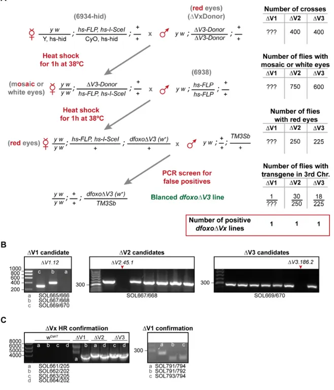
The new dfoxo∆V1, ∆V2 and ∆V3 knockout founder lines were generated by genomic engineering as previously reported (Huang et al., 2009). In a first step, each of the dfoxo target regions, V1, V2, or V3 (Figure 2.3), were substituted by a whitehs marker gene and an attP-site using ends-out homologous recombination. For this, ~4Kb flanking sequences of each target region were cloned into a pBlueScript II SK(+) vector, using ET recombineering (Muyrers et al., 1999; Zhang et al., 1998) and the respective primers SOL572-579 Supplementary Table S1). As template for the ET recombineering, a BAC clone that contains the dfoxo locus (CH321-24|13, BACPAC resource center, Oakland, California) was used. After sequence verification using sequencing primers SOL580-607 and SOL628-635 (Supplementary Table S1), homologous arms were brought into the pGX-attP targeting vector (Huang et al., 2009). To target the V1 region, we cloned a pGXattP vector containing homologous arms 1 and 2; for the V2 region, arms 1 and 4;
and for the V3 region arms 3 and 4 (Figure 2.3). P-element-mediated transformation was done by the BestGene Drosophila embryo injection service (Chino Hills, USA) to generate transgenic flies carrying the pGXattP donor constructs for targeting the V1, V2 and V3 region.
Crosses for ends-out homologous recombination were set for direct targeting as described before (Huang et al., 2008) (Figure 2.4). Subsequently, the whitehs marker gene was mapped to the third chromosome using a TM3 Sb balancer chromosome. Homozygous flies carrying the whitehs marker on the third chromosome were screened by PCR for the absence of the corresponding region of the dfoxo gene using primers SOL665/666 for V1, SOL667/668 for V2 and SOL669/670 for V3 (Supplementary Table S1). We obtained one knockout founder (KO) line for V1, V2 and V3. Subsequently, KO flies were crossed with cre-recombinase expressing flies to remove the whitehs marker gene (Groth et al., 2004).
The generated w[-] lines, denoted dfoxo∆V1w[-], ∆V2w[-] and ∆V3w[-], were brought into a fly line expressing the ΦC31-integrase (Groth et al., 2004), and used as parental lines for any future reinsertion within the dfoxo locus.
To generate the dfoxo gene replacement constructs, the genomic regions V2 and V3 were cloned in the pBlueScript II SK(+) vector by ET recombineering using primers SOL681/682 and SOL683/682, respectively (Supplementary Table S1). Inserts were
sequence verified using primers SOL690/700 (Supplementary Table S1). Subsequently, reinsertion inserts were transferred to the pGEattBGMR vector (Huang et al., 2009) by restriction enzyme cloning using NheI and AscI to cut vector and insert. Ligation was carried out overnight at 18ºC according to standard T4-ligase protocols (NEB). The V1 region was PCR-cloned in the same vector using the In-Fusion system (Clontech) with pBS-V2 as template and primers SOL795/796 (Table 2.1 and Supplementary Table S1).
Mutations on the pBS-V3-3xFLAG construct were introduced using QuickChange II XL site-directed mutagenesis (Agilent Technologies) and the V3-mutant-3xFLAG sequence was subsequently In-Fusion (Clonetech) cloned in the pGEattBGMR vector using the respective primers (Table 2.1 and Supplementary Table S1). pGEattBGMR gene replacement constructs were injected into embryos of the respective KO-parental lines
∆V1w[-] or ∆V3w[-]. Microinjections were done by Jacqueline Dols of the transgenic fly core facility of the Max-Planck Institute for Biology of Ageing.
Table 2.1. Transgenic dfoxo alleles. Transgenic dfoxo flies generated by embryo microinjection.
Injected vector was always pGEattBGMR. InFusion templates labeled V1short, V3short or V3- MAD-3xFLAG were synthesized by Eurofins. Primer sequences are summarized in supplementary Table S1.
dfoxo knock-in lines
Mutagenesis primers on pBS-
V3 InFusion primers InFusion PCR
template Seq primers V3
SOL813-817 pBS-V3
SOL692-700 V3-3xFLAG
SOL728-729
V3short-3xFLAG
V3-Tev-BLRP V3short-Tev-BLRP
V3-mCherry V3short-mCherry SOL692-700 and
SOL717-721 V3-DBD1-3xFLAG SOL809-810
SOL813-824 pBS-V3 with corresponding
mutation
SOL692-700 V3-DBD2-3xFLAG SOL811-812
V3-5KR-3xFLAG
SOL393-394, SOL397-398 and
SOL563-564
V3-5KQ-3xFLAG SOL514-515, SOL516-517 and
SOL561-562 V3-MAD-3xFLAG
SOL813-814 V3-MAD-3xFLAG SOL692
V1 SOL795-796 pBS-V2 SOL690-691
3xFLAG-V1 SOL713-714 3xFLAG-V1short SOL690-691 and SOL717-718
2.1.2 Fly maintenance
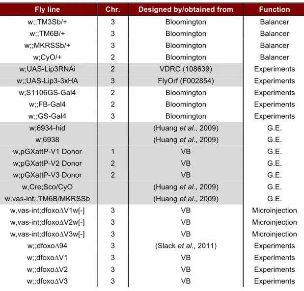
Fly stocks were maintained and experiments were conducted at 25ºC on a 12:12 h light:dark cycle at 65% humidity. To generate experimental flies, larvae were reared at controlled densities by transferring 20µl of eggs ("egg squirts") into a fly stock bottle containing 1SYA food (5% w/v sucrose, 10% w/v brewer’s yeast, 1,5% w/v agar) (Bass et al., 2007). Freshly eclosed adult flies were allowed to mate for 48h before being sorted according to gender. All fly lines used for experiments were backcrossed for at least 6 generations into the outbred, wild-type white Dahomey (wDahT) strain (Grönke et al., 2010). This line was previously treated with tetracycline and does not contain the endosymbiotic bacterium Wolbachia. All fly lines used in this PhD thesis are summarized in Table 2.2.
2.1.3 Fly lines used in this study
Table 2.2. Drosophila stocks used in this study. Balancer and experimental flies were
backcrossed into the wDahT background for at least 6 generations. G.E = Genomic Engineering;
VB = Victor Bustos.
Fly line Chr. Designed by/obtained from Function
w;;TM3Sb/+ 3 Bloomington Balancer
w;;TM6B/+ 3 Bloomington Balancer
w;;MKRSSb/+ 3 Bloomington Balancer
w;CyO/+ 2 Bloomington Balancer
w;UAS-Lip3RNAi 2 VDRC (108639) Experiments
w;;UAS-Lip3-3xHA 3 FlyOrf (F002854) Experiments
w;S1106GS-Gal4 2 Bloomington Experiments
w;;FB-Gal4 2 Bloomington Experiments
w;;GS-Gal4 3 Bloomington Experiments
w;6934-hid (Huang et al., 2009) G.E.
w;6938 (Huang et al., 2009) G.E.
w,pGXattP-V1 Donor 1 VB G.E.
w;pGXattP-V2 Donor 2 VB G.E.
w;pGXattP-V3 Donor 2 VB G.E.
w,Cre;Sco/CyO (Huang et al., 2009) G.E.
w,vas-int;;TM6B/MKRSSb (Huang et al., 2009) G.E.
w,vas-int;dfoxo∆V1w[-] 3 VB Microinjection
w,vas-int;dfoxo∆V2w[-] 3 VB Microinjection
w,vas-int;dfoxo∆V3w[-] 3 VB Microinjection
w;;dfoxo∆94 3 (Slack et al., 2011) Experiments
w;;dfoxo∆V1 3 VB Experiments
w;;dfoxo∆V2 3 VB Experiments
w;;dfoxo∆V3 3 VB Experiments
w;;dfoxo-V3 3 VB Experiments
w;;dfoxo-V3-3xFLAG 3 VB Experiments
w;;dfoxo-V3-Tev-BLRP 3 VB Experiments
w;;dfoxo-V3-mCherry 3 VB Experiments
w;;dfoxo-V3-DBD1-3xFLAG 3 VB Experiments
w;;dfoxo-V3-DBD2-3xFLAG 3 VB Experiments
w;;dfoxo-V3-MAD-3xFLAG 3 VB Experiments
w;;dfoxo-V3-5KR-3xFLAG 3 VB Experiments
w;;dfoxo-V3-5KQ-3xFLAG 3 VB Experiments
w;;dilp2-3,5 3 (Grönke et al., 2010) Experiments
w[DhaT] (Grönke et al., 2010) Experiments
w;;dilp2-3,5, dfoxo∆V3 3 VB Experiments
w;;dilp2-3,5, dfoxo-V3-
3xFLAG 3 VB Experiments
w;;dilp2-3,5, dfoxo-V3-
DBD1-3xFLAG 3 VB Experiments
w;;dilp2-3,5, dfoxo-V3-
DBD2-3xFLAG 3 VB Experiments
w;S1106GS-Gal4;dfoxo∆V3 3 VB Experiments
w;;UAS-Lip3-3xHA,
dfoxo∆V3 3 VB Experiments
2.1.4 Lifespan and fecundity assays
For lifespan assays, 48h mated flies were sorted by sex at a density of 10 flies per small glass vial and 10 vials per genotype (n=100). Flies were tipped to fresh food 3 times per week and dead flies were scored. For fecundity assays, in parallel to the lifespan, 10 vials per genotype with 3 flies per vial were used. During the first 3 weeks, eggs laid per vial were counted after egg-laying periods of ~20h.
2.1.5 Stress assays and fly experiments
For stress assays, flies were sorted at 20 flies per wide plastic vial, 5 vials per genotype (n=100), and kept on 1SYA food for 7 days before starting the stress unless otherwise specified. Starvation food contained 1% w/v agarose, food for oxidative stress assays contained 5% w/v sucrose, 1,5% w/v agarose and 5% v/v H2O2 (Grönke et al., 2010).
Dead flies were scored three times per day. In the case of yeast or sugar starvation, the respective component was omitted during normal 1SYA preparation and dead flies were scored every 2-3 days while tipping flies into fresh food vials.
For over-expression or RNAi-mediated knock down of gene expression the corresponding UAS-lines were mated with Gal4 driver lines (see Table 2.2) for induction of constitutive expression or with GS-driver lines (Roman et al., 2001) for inducible expression. When using an inducible GS-driver line, food was supplemented with RU486 (Sigma) at a concentration of 200µM in 1SYA or at 50µM in starvation food. The respective controls contained equivalent volumes of the drug carrier ethanol.
2.1.6 Fly developmental time and body weight
Fly development and body weight were measured similarly to protocols described previously (Grönke et al., 2010). Briefly, for developmental timing, eggs laid over 3h on grape juice plates were collected and transferred to 1SYA food at a density of 50 eggs per vial and 10 vials per genotype. Upon eclosion of the first flies, their numbers were counted at regular intervals.
For body weight determination, batches of 5 flies were flash frozen in liquid nitrogen and weighted on a ME235S analysis balance (Sartorius Mechatronics). A total of 50 flies per genotype was measured.