The Role of Octopamine
in Attraction and Aversion Behavior in Drosophila melanogaster
Inaugural-Dissertation zur
Erlangung des Doktorgrades
der Mathematisch-Naturwissenschaftlichen Fakultät der Universität zu Köln
vorgelegt von
Gerbera Regina Claßen aus Köln
Köln, 2018
Berichterstatter: Prof. Dr. Henrike Scholz Prof. Dr. Arnd Baumann
Tag der mündlichen Prüfung: 20.06.2018
Et Kölsche Jrundjesetz:
§3 Et hät noch immer jot jejange!
Table of Contents
Abstract ... 1
Zusammenfassung ... 3
1. Introduction ... 5
1.1. The neurotransmitter Octopamine ... 5
1.1.1. Immunoreactivity of OA and Tbh ... 6
1.1.2. The role of OA in Drosophila melanogaster ... 8
1.2. The reinforcing properties of OA ... 13
1.3. OA receptors in adult Drosophila ... 15
1.4. Channelrhodopsins – a tool for light induced neuronal activation ... 18
1.5. Aim ... 22
2. Material and Methods ... 24
2.1. Material ... 24
2.1.1. Fly strains ... 24
2.1.2. Fly husbandry ... 26
2.1.3. Chemicals ... 26
2.2. Methods ... 27
2.2.1. Olfactory two odor choice paradigm ... 27
2.2.2. Feeding of pharmacological substances ... 28
2.2.3. Optogenetic site attraction assay ... 29
2.2.4. LEDs ... 30
2.2.5. All-trans-Retinal ... 31
2.2.5. Cuticle penetration of blue and red light ... 31
2.2.6. Locomotion assay ... 32
2.2.7. Statistics ... 34
3. Results ... 35
3.1. A subset of tyraminergic/octopaminergic/cholinergic neurons mediates site attraction and aversion ... 35
3.1.1. Site attraction elicited by Tdc2-GAL4 targeted neurons is light intensity independent but frequency dependent when using the blue light activatable ChR2 35 3.1.2. Activation of neurons targeted by the Feb15-GAL4 driver did not elicit site attraction ... 38
3.1.3. Activation of neurons targeted by the fru-GAL4 driver elicited site attraction ... 39
3.1.4. Site aversion elicited by 6.2-Tbh-GAL4 targeted neurons is light intensity and frequency dependent ... 40
3.1.5. Site attraction and site aversion are mediated by a subset of acetylcholine co- expressing Tbh positive neurons... 42
3.1.6. Activation of other neurons in the SOG is not sufficient to elicit site attraction ... 45
3.1.7. OA is required to switch the behavioral outcome/response ... 46
3.1.8. norpA
1mutants are impaired in olfaction ... 48
3.2. Octopamine is sufficient and required for site attraction and aversion... 49
3.3. Site attraction and aversion induced by neuronal light activation are transgene independent ... 52
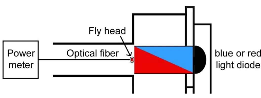
3.3.1. Penetrance of blue and red light through the flies’ cuticle ... 52
3.3.2. The UAS-Chrimson transgene is not suitable for the optogenetic site attraction assay ... 53
3.3.3. Red light activation is not suitable for UAS-ReaChR ... 56
3.3.4. Site attraction elicited by amber light activation in Tdc2-GAL4 dependent neurons is intensity dependent and frequency independent ... 59
3.3.5. Activation of neurons targeted by the Cha-GAL4 driver using ReaChR is not
suitable for inducing site attraction or aversion ... 62
3.3.6. Activation of neurons targeted by the fru-GAL4 driver elicited site aversion . 63
3.3.7. Site aversion elicited by amber light activation in 6.2-Tbh-GAL4 dependent
neurons is intensity dependent and frequency independent ... 64
3.4. OA is required and sufficient for olfactory ethanol attraction ... 67
3.4.1. Activation of OA signaling restores the loss of attraction towards ethanol enriched food odors in Tbh
nM18mutants ... 67
3.4.2. Altered levels of Tbh negatively influence the olfactory attraction while simultaneously altered levels of TA and OA might not influence olfactory attraction behavior ... 70
3.4.3. Tbh
nM18mutants showed no aversion against high concentrations of ethanol 73 3.5. Knock down of OA receptors in olfactory sensory neurons did not result in a clear candidate for involvement in olfactory attraction ... 74
3.6. Tbh mutants display a defect in locomotion ... 77
4. Discussion ... 79
4.1. OA acts as a positive and as a negative reinforcer ... 79
4.2. OA is sufficient and necessary for olfactory ethanol attraction ... 86
4.3. Olfactory attraction is not mediated by the tested OSN OA receptors ... 88
4.4. The interaction of OA and TA is important for the behavioral outcome ... 89
4.5. OA biases the behavioral outcome ... 91
4.6. Blue and amber light activatable channelrhodopsins are both suitable for neuronal activation to elicit site attraction and site aversion ... 93
4.7. Closing remarks ... 98
5. List of Abbreviations ... 99
6. References ...101
Teilpublikationen ...113
Acknowledgement...114
Erklärung ...115
1
Abstract
All animals are exposed to environmental stimuli and influences at any time and place.
They have to decide whether to respond to a stimulus and whether the reaction should be approach or aversion. In this thesis, the role of OA in Drosophila melanogaster as a reinforcer to these kinds of stimuli was investigated. Therefore the optogenetic site attraction assay, the olfactory two odor choice paradigm and feeding of pharmacological active substances were utilized.
So far OA was only known as a positive reinforcer, for example in appetitive olfactory learning and memory, while DA acts as a negative reinforcer in aversive learning and memory (Schwaerzel et al., 2003; Schneider et al., 2012). Here it is shown, that OA can also function as a negative reinforcer. OA is able to mediate attraction and aversion behavior. Optogenetic activation of a Tdc2-GAL4 targeted tyraminergic/octopaminergic/
cholinergic set of neurons elicited site attraction, independent of the used channelrhodopsin transgene. A smaller subset of 6.2-Tbh-GAL4 driven tyraminergic/
octopaminergic/cholinergic neurons, the VUMa4 neurons, which is part of the site
attraction eliciting set of neurons, was able to induce site aversion when activated. Both,
site attraction and site aversion, are due to OA and not to TA, as activation of these two
tyraminergic/octopaminergic/cholinergic subsets in the Tbh
nM18mutant background
abolished site attraction and site aversion, respectively. So the behavioral outcome is
dependent on the combination of activated neurons and OA can function not only as a
positive but also as a negative reinforcer. Furthermore, OA is sufficient and necessary
for olfactory attraction behavior. This was shown by pharmacological experiments. The
loss of olfactory ethanol attraction phenotype in Tbh
nM18mutants (Schneider et al., 2012)
could be restored to control level by feeding OA, while elimination of OA signaling with
epinastine (an OA antagonist) abolished the natural attraction of w
1118control flies
towards ethanol containing food odors. Thus the loss of olfactory ethanol attraction
phenotype of the Tbh
nM18mutants is caused by the lack of OA and not due to the
increased levels of TA. Mutants overexpressing the Tbh enzyme show a similar
phenotype in ethanol attraction and locomotion like the mutants lacking Tbh. Therefore,
it seems like a certain balance or interaction between these two neurotransmitters is
needed for proper regulation of behavior. Furthermore, OA is required to switch a
2
behavioral response. The approaching or aversive response of a fly towards a stimulus is mediated by OA and it is also possible to shift an already existing attraction towards attractive ethanol containing food odor to another, less attractive stimulus. This suppression of a normally positive estimated response by activation of tyraminergic/
octopaminergic neurons indicates that OA is maybe not involved in attraction or aversion itself, but in the initiation and the switch between these two behaviors. Tbh
nM18mutants, which lack OA, consequently fail to show this switch in behavior.
Furthermore it was shown, that the obtained results in the optogenetic site attraction assay are channelrhodopsin transgene independent and thus real. Two different channelrhodopsins (ChR2 and ReaChR) were tested in more detail and neuronal light activation resulted in site attraction (activation of Tdc2-GAL4 targeted neurons) or site aversion (activation of 6.2-Tbh-GAL4 targeted neurons), independent of the used channelrhodopsin. For activation of the different channelrhodopsins it is important to find a suitable wavelength and light intensity. Additionally, neuronal light activation of these tyraminergic/octopaminergic neurons is not frequency dependent. The observed differences are possibly due to the kinetics of the different transgenes.
Taken together, OA is sufficient and necessary for attraction and aversion behavior and
therefore acts as a positive and negative reinforcer. It is probably not involved in these
behaviors itself, but mediates the switch between an approach and an aversive reaction
to a stimulus. Thus OA orchestrates the behavioral outcome by biasing the decision of
Drosophila melanogaster towards different stimuli.
3
Zusammenfassung
Tiere sind überall und zu jeder Zeit den Einflüssen der Umwelt ausgesetzt. Dabei müssen sie sich entscheiden, ob sie auf einen Stimulus reagieren und ob diese Reaktion in Annäherung an den Stimulus oder in Rückzug endet. In dieser Doktorarbeit wurde die Rolle von OA in Drosophila melanogaster als ein Verstärker von dieser Art von Einflüssen untersucht. Dafür wurden der optogenetische Site Attraction Assay, der olfaktorische Two Odor Choice Assay und das Füttern von pharmakologischen Substanzen verwendet.
Bisher war OA nur als positiver Verstärker bekannt, zum Beispiel im appetitiven olfaktorischen Lernen und Gedächtnisbildung, während DA der negative Verstärker in aversivem Lernen und Gedächtnisbildung war (Schwaerzel et al., 2003; Schneider et al., 2012). Hier wird nun gezeigt, dass OA genauso als negativer Verstärker arbeiten kann.
OA ist in der Lage sowohl attraktives, als auch aversives Verhalten zu vermitteln.
Optogenetische Aktivierung von Tdc2-GAL4 getriebenen tyraminergen/
oktopaminergen/cholinergen Neuronen löste eine Seiten-Präferenz aus, unabhängig
vom verwendeten Channelrhodopsin Transgen. Ein kleineres Set von 6.2-Tbh-GAL4
getriebenen tyraminergen/oktopaminergen/cholinergen Neuronen, den VUMa4
Neuronen, welche Teil des Seiten-Präferenz auslösenden Set von Neuronen ist, war in
der Lage bei Aktivierung Seiten-Aversion auszulösen. Beide Verhalten, Seiten-Präferenz
und Seiten-Aversion, werden durch OA und nicht durch TA vermittelt, da eine
Aktivierung dieser beiden tyraminergen/oktopaminergen/cholinergen Sets in einem
Tbh
nM18mutanten Hintergrund zum Verlust der Seiten-Präferenz bzw. der Seiten-
Aversion führte. Daher ist das schließlich gezeigte Verhalten abhängig von der
Kombination and aktivierten Neuronen und OA arbeitet nicht nur als positiver
Verstärker, sondern auch als negativer Verstärker. Des Weiteren ist OA ausreichend und
notwendig für olfaktorische Präferenz. Dies wurde durch pharmakologische
Experimente gezeigt. Der Verlust der olfaktorischen Ethanol Präferenz in Tbh
nM18Mutanten (Schneider et al., 2012) konnte durch Füttern von OA zurück auf Wildtyp
Level gebracht werden, während eine Unterdrückung des OA Signalwegs durch Füttern
von Epinastine (ein OA Antagonist) zum Verlust der natürlichen Präferenz für Alkohol
enthaltende Futterdüfte der w
1118Kontrollfliegen führte. Deswegen ist der Phänotyp mit
Verlust der olfaktorischen Ethanolpräferenz in Tbh
nM18Mutanten auf das Fehlen von OA
4
zurück zu führen und nicht auf den erhöhten TA Spiegel. Mutanten, die eine Überexpression des Tbh Enzyms aufweisen, zeigen einen ähnlichen Phänotyp in Ethanol Präferenz und Lokomotion, wie die Mutanten, denen das Tbh Enzym fehlt. Daher scheint es, dass ein Gleichgewicht und ein Zusammenspiel dieser beiden Neurotransmitter notwendig ist, um normales Verhalten zu zeigen. Darüber hinaus wird OA auch für den Wechsel zwischen zwei Verhaltensantworten benötigt. Die annähernde oder zurückweichende Reaktion einer Fliege auf einen Stimulus wird durch OA vermittelt und es ist sogar möglich, eine schon bestehende Präferenz für einen attraktiven, Alkohol enthaltenden Futterduft durch eine Präferenz für einen anderen, weniger attraktiven Futterduft zu ersetzen. Diese Unterdrückung von einer normalerweise als positiv bewertete Reaktion, hervorgerufen durch die Aktivierung von tyraminergen/
oktopaminergen Neuronen, deutet daraufhin, dass OA eventuell nicht selbst in Präferenz- oder Aversionsverhalten involviert ist, sondern eher an der Einleitung oder dem Wechsel zwischen diesen zwei Verhalten beteiligt ist. Tbh
nM18Mutanten, welche keine OA besitzen, versagen daher diesen Wechsel zwischen zwei Verhalten zu zeigen.
Außerdem wurde gezeigt, dass die mit dem optogenetischem Site Attraction Assay erzielten Ergebnisse nicht abhängig vom verwendeten Channelrhodopsin sind und daher echt sind. Es wurden zwei verschiedenen Channelrhodopsine (ChR2 und ReaChR) genauer untersucht und die neuronal Lichtaktivierung hatte im Fall von Tdc2-GAL4 getriebenen Neuronen Seiten-Präferenz und im Fall von 6.2-Tbh-GAL4 getriebenen Neuronen Seiten-Aversion zur Folge, und das unabhängig vom Channelrhodopsin. Für die Aktivierung der verschiedenen Channelrhodopsine ist eine passende Wellenlänge und Lichtintensität notwendig. Zudem ist die neuronale Aktivierung dieser tyraminergen/oktopaminergen Neurone nicht von einer Frequenz abhängig. Die beobachteten Unterschiede sind eher auf die verschiedenen kinetischen Eigenschaften der unterschiedlichen Channelrhodopsine zurück zu führen.
Zusammengefasst bedeutet das, dass OA ausreichend und notwendig für attraktives und
aversives Verhalten ist und somit sowohl als positiver, als auch als negativer Verstärker
funktionieren kann. Es ist vermutlich nicht direkt in diese Verhalten involviert, sondern
vermittelt den Wechsel zwischen einer annähernden oder einer zurückweichenden
Reaktion auf einen Stimulus. Daher verändert OA die Verhaltensantwort durch das
Beeinflussen der Entscheidung, die von Drosophila melanogaster als Reaktion auf einen
Stimulus gefällt wird.
5
1. Introduction
1.1. The neurotransmitter Octopamine
Octopamine (OA) is a biogenic monoamine which is named after its place of discovery:
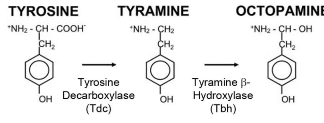
the salivary glands of the octopus, whereas its function there is still unknown (Erspamer and Boretti, 1951). The synthesis of OA is shown in Figure 1. The first step of the OA synthesis is the decarboxylation of the amino acid tyrosine to tyramine (TA) by the tyrosine decarboxylase (TDC) (Livingstone and Tempel, 1983). In Drosophila there are two genes encoding for TDC: Tdc1 and Tdc2. Tdc1 is expressed in non-neuronal tissues and Tdc2 in neuronal tissues (Cole et al., 2005). In the second step, TA is converted to OA by the tyramine β-hydroxylase (Tbh) (Livingston and Tempel, 1983; Monastirioti et al., 1996). The Tbh is encoded by the Tbh gene (Monastirioti et al., 1996), which encodes for at least five transcripts resulting in different isoforms of Tbh (Manuela Ruppert, 2013).
Figure 1: Synthesis of OA.
The amino acid tyrosine is decarboxylated to TA by the TDC enzyme. Then TA is hydroxylated to OA by the Tbh enzyme (modified after Cole et al., 2005).
OA and its precursor TA are present in invertebrates and are structurally related to the
mammalian adrenaline and noradrenaline (NA) (Roeder, 1999). Both, OA and TA are
independent neurotransmitters (Roeder, 2005). OA is thought to function as a
6
homologue of NA, since NA, an important neurotransmitter in vertebrates, has no physiological role in invertebrates and OA, an important neurotransmitter in invertebrates, has no physiological function in vertebrates (Roeder, 1999). Tbh and Dbh (dopamine β-hydroxylase, the enzyme that catalyzes the final step in noradrenaline synthesis) are highly likely to be functionally homologous and therefore evolutionary related (Wallace, 1976), which supports the homologues function of the octopaminergic and adrenergic systems in invertebrates and vertebrates (Monastirioti et al., 1996). This suggests an early evolutionary origin of the adrenergic/tyraminergic/octopaminergic system. Furthermore, they share functions in behavior, like fight or flight response, learning and memory, motivation and aggression (Roeder, 1999 and 2005). OA and TA are the only neuroactive non-peptide transmitters whose physiological role is restricted to invertebrates (Roeder, 1999). Nevertheless OA exists also in vertebrates (David and Coulon, 1985). OA is not only a neurotransmitter, but also functions as a neurohormone and a neuromodulator and is present in non-neuronal and neuronal tissue in relatively high concentrations in almost all invertebrates (Roeder, 1999). In the brain of Drosophila melanogaster OA is present in high concentrations, while its precursor TA is less abundant by the factor 30 (Monastirioti et al, 1996). The normal concentration of OA in nervous tissue is >10mg/g tissue, while in the insects’ flight muscles or oviducts concentrations of 1mg/g were detected (Roeder, 1999).
1.1.1. Immunoreactivity of OA and Tbh
The distribution of octopaminergic neurons has been investigated in insects and it was
observed that only a small number of OA containing neurons exist, but these few
neurons supply almost every neuropil in the insect brain (Bräuning, 1991; Kreissl et al.,
1994). The mushroom bodies (MB) and the optic lobes possess the densest innervation
with octopaminergic neurons (Homberg, 1994). The most prominent and important
octopaminergic neurons are the DUM (dorsal unpaired median) and VUM (ventral
unpaired median) neurons (Konings et al., 1988), which are present at the dorsal and
ventral midline in the suboesophageal, thoracic and abdominal ganglia and contain and
release OA (Hoyle, 1975; Hoyle and Barker, 1975; Evans and O'Shea, 1977; 1978).
7
In the central nervous system (CNS) of adult Drosophila OA is synthesized and released in only a small number of 100 neurons organized in 15 clusters (Sinakevitch and Strausfeld, 2006), and these few neurons have an enormous field of innervation and cover all neuropil areas in the fly brain (Monastirioti et al., 1995; Monastirioti, 1999;
Cole et al., 2005; Busch et al., 2009). Octopaminergic cells have been detected in six neuronal clusters at different levels along the dorsoventral axis. There are 12-14 OA positive cells in the suboesophageal ganglion (SOG), 4-5 cells in the antennal lobe (AL) cluster, 4-5 cells in the dorsal anterior cluster (DAC), four cells in the dorsal medial cluster (DMC), 12-16 cells in the dorsal posterior cluster (DPC) and two cells in the lateral protocerebrum (LP) (Monastirioti et al., 1995). In the thoracico-abdominal ganglion 17-19 cells were observed in a single cell and four clusters (Monastirioti et al., 1995) (see Figure 2).
Figure 2: Schematic representation of octopaminergic neurons in the CNS of adult Drosophila Octopaminergic cells are represented by dark circles in the larval CNS. In the adult brain, the filled circles indicate posterior position, circles with dots are medial and open non-filled circles are anterior cells. The stippled areas represent neuroactive neuropil (Monastirioti, 1999).
8
Busch and colleagues (2009) investigated the expression pattern of the Tdc2-GAL4 driver line, a GAL4-line which contains a part of the Tdc2 promoter and therefore targets neurons which should be tyraminergic/octopaminergic. 137 cells are targeted by the Tdc2-GAL4 line, but not all of them are also octopaminergic. According to Schneider and colleagues (2012), the Tdc2-GAL4 driver line targets about 78 Tbh expressing neurons.
Therefore, 59 targeted neurons are not Tbh positive. An overlap of GFP and OA was found in 21-27 cells in the ventromedial (VM) cluster, seven in the AL2 cluster, two cells in the ventrolateral (VL) cluster and 2-8 cells in the anterior superior medial (ASM) cluster. In the supraoesophageal ganglion (SPG) and SOG 27 types of octopaminergic neurons were observed. Next to the 11 types of VUM neurons, also five VPM (ventral paired median) neurons in the VM cluster were described (Busch et al., 2009).
1.1.2. The role of OA in Drosophila melanogaster
To investigate the role of OA in invertebrates, a certain Drosophila melanogaster mutant
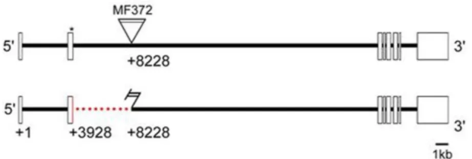
was generated by Monastirioti et al. (1996) – the Tbh
nM18mutant. These mutants have a
deletion in the Tbh gene and therefore a disrupted OA synthesis (Figure 3). TA cannot be
converted to OA, thus there is a lack of OA and six to eightfold increased TA level
(Monastirioti et al., 1996). This mutant is supposed to be a null mutant as no Tbh
immunoreactivity in the CNS of Drosophila larvae and no Tbh protein were detectable
anymore (Monastirioti et al., 1996). But in a semi quantitative RT-PCR it was shown that
there is no significant difference between the w
1118control and the Tbh
nM18mutants and
with a quantitative RT-PCR there are 1.5% of the amount of Tbh left in Tbh
nM18mutants
compared to the w
1118control (Manuela Ruppert, 2010). Thus, the Tbh
nM18mutant is not
a null mutant. It is suggested that there are several Tbh isoforms and the deletion in the
Tbh
nM18mutant does only affect some and not all of the isoforms. The phenotype of the
Tbh
nM18mutants did not differ from wild type flies in some aspects: they survived until
adulthood and had a normal appearance. But they showed a reduced viability under
unfavorable, crowded conditions and female flies were sterile while male flies were
fertile (Monastirioti et al., 1996; Roeder, 2005). The females retain their fertilized eggs,
but this defect could be rescued by feeding OA, so the female sterility is a direct
consequence of the missing OA caused by the deletion in the Tbh locus (Monastirioti et
al., 1996). Additional existing Tbh mutants are the Tbh
Del3and the d01344 mutant. The
9
Tbh
Del3mutant carries a larger mutation and is missing the complete first and second exon of the Tbh gene, but still has detectable transcripts of Tbh (Manuela Ruppert, 2013). Both Tbh mutants are not impaired in ethanol sensitivity but display a reduced ethanol tolerance (Scholz, 2005; Manuela Ruppert, 2013). The d01344 mutant has a 160% upregulation of Tbh and displays increased sensitivity and normal tolerance towards ethanol (Manuela Ruppert, 2013).
Figure 3: Deletion mapping of the TbhnM18 mutant
The Tbh gene consists of eight exons. The TbhnM18 mutant was generated through P-element mutagenesis and lacks 32 base pairs of the coding sequence of the second exons (Manuela Ruppert, 2010)
The expression pattern of the wild type Tbh was described by Monastirioti et al. (1996) and is very similar to the OA expression pattern (Monastirioti et al., 1995). Schneider and colleagues (2012) used another antibody against Tbh (Zhou et al., 2008) to identify Tbh positive neurons, which also might be OA positive. They found 112 Tbh positive neurons in the adult brain and 39 Tbh expressing cells in the ventral nerve cord (VNC).
There was a match in the G0b, G3a/AL2 and VMI-III clusters. The clusters G2b and G4a showed even more Tbh positive cells, but the clusters G3b and G0 had less Tbh positive cells. Furthermore, a new neuron has been identified, which was named G0 posterior.
Hence, the used antibody labels Tbh positive cells similar to the previously described octopaminergic pattern, but on average there are more Tbh positive neurons in the labeled clusters (Schneider et al., 2012).
The function of OA has already been investigated in different species and in Drosophila
melanogaster the Tbh
nM18mutants are a good opportunity to investigate the role of OA. It
is involved in the regulation of many different behaviors like sleeping (Crocker and
10
Sehgal, 2008), egg laying (Monastirioti et al., 1996; Monastirioti, 2003; Middleton et al., 2006), stress response (Möbius and Penzlin, 1993), motivation (Roeder, 1999; 2005), associative appetitive and aversive learning and memory (Dudai, 1988; Schwaerzel et al., 2003; Honjo and Furukubo-Tokunaga, 2009; Sitaraman et al. 2010; Iliadi et al., 2017), fight or flight response (Brembs et al., 2007), aggression (Baier et al., 2002; Hoyer et al., 2008; Zhou et al., 2008), decision making between aggression and courtship behavior (Certel et al., 2007; 2010), response to olfactory and gustatory stimuli (Scheiner et al., 2014), locomotion (Saraswati et al., 2003), starvation induced hyperactivity (Yang et al., 2015), development of tolerance towards alcohol (Scholz et al., 2000; Scholz, 2005) and innate attraction to odors such as ethanol (Schneider et al., 2012). Thus, OA modulates almost every physiological process in peripheral or sense organs in invertebrates which are studied until now (Roeder, 1999; 2005). Furthermore, OA is involved in the modulation of sensory input and the outcome of the olfactory pathway in the insect brain, which is processed in the olfactory lobes (for olfaction) and MB (for learning and memory) (Farooqui et al., 2003; Schwaerzel et al., 2003). Sombati and Hoyle (1984) postulated the “Orchestration Hypothesis” which describes the function of OA and which says that for every set of behavior there is a neural network and those can be selectively activated or inhibited by the release of OA, which thus allows the suppression of opposing behaviors. Stress factors like heat, starvation, mechanical or chemical influences lead to an increase in OA concentration in many organisms (Davenport and Evans, 1984; Hirashima and Eto, 1993).
In the Drosophila larvae, Saraswati and colleagues (2003) investigated the locomotion phenotype of Tbh
nM18mutants and compared it to their genetic control. Tbh
nM18mutants showed a reduced speed and track length and more pausing and directional changes.
This severe phenotype could be partially rescued by feeding OA. Simultaneously feeding
of TA nullified the rescue, while feeding only yohimbine (TA antagonist) also partially
rescued the locomotion phenotype. When the larvae were fed with OA and yohimbine at
the same time, the rescue was further improved. From these experiments, they
concluded that OA and TA both influence locomotion in Drosophila larvae and that they
possibly have antagonistic effects. Thus, for a normal locomotion behavior a balance
between these two neurotransmitters is needed (Saraswati et al., 2003). An interaction
of both neurotransmitters is also required in the flight initiation and maintenance of
Drosophila, as Tbh
nM18mutants are impaired in this behavior (Brembs et al., 2007). This
11
phenotype could be restored to control levels by substituting OA with heat shock inducible Tbh or blocking TA receptors. Simultaneously feeding yohimbine and giving a heat shock even improved the performance over control levels. Feeding of OA did not rescue the mutant phenotype. Elimination of all tyraminergic and octopaminergic neurons by using a Tdc2 mutant resulted in the same phenotype like in Tbh
nM18mutants (Brembs et al., 2007).
The right amount of TA and OA also is important in the courtship behavior of male flies.
OA is necessary to respond appropriately to presented information. Male flies lacking
OA showed courtship behavior towards both, female and male flies, instead of showing
aggression towards other males (Certel et al., 2007; 2010). A small subset of three
octopaminergic VUM neurons in the SOG, which also expresses Fru
M(the male form of
neural sex determination factor) is involved in this behavior (Certel et al., 2007). The
SOG is the primary taste-processing center. The sensory information which is sent to
this neuropil includes the female pheromone recognition cues, which are necessary for
males to identify females and show courtship behavior (Bray and Amrein, 2003). Hence,
OA is needed in this neuronal subset to accurately transmit contact gustatory
pheromone information, as without OA the same information can lead to two different
behaviors – aggression and courtship (Certel et al., 2007). Too low and too high levels of
OA have the same effect, which is why the level of OA signaling has to be in a certain
range. An increase in OA levels or activation of octopaminergic neurons also led to
elevated courtship behavior of male flies towards other males (Certel et al., 2010). But
the subset of VUM neurons described by Certel et al. (2007; 2010) is not the same subset
of octopaminergic neurons in the SOG as mentioned by Zhou et al. (2008) in their
aggression studies with Drosophila. Here, a reduction of OA signaling led to a decrease in
aggression behavior in both males and females, while locomotion, olfaction, sexual
discrimination and courtship behavior were unaffected. Tbh
nM18mutants did not initiate
fighting and did not fight other males even when provoked. An increase in OA levels
(feeding OA agonist, overexpression of Tbh or activation of OA neurons) resulted in
elevated aggressive behavior in grouped flies, but not in socially isolated flies. By
combining the Tdc2-GAL4 driver line with the Cha-GAL80 driver, Zhou et al. (2008)
narrowed the number of involved neurons down to five neurons in the SOG and
indicated that neural OA is needed for aggression. This small subset of octopaminergic
12
neurons seems to mediate aggression in a direct way, as other behaviors were not affected (Zhou et al., 2008).
OA is also involved in appetitive and aversive learning, where Tbh
nM18mutants displayed severe defects (Schwaerzel et al., 2003; Iliadi et al., 2017). Habituation (a simple form of learning) of Drosophila melanogaster is indirectly affected by OA (Scheiner et al., 2014).
Tbh
nM18mutants are less responsive to sucrose and thus showed a faster proboscis extension response (PER) habituation than their controls. This phenotype could be rescued by feeding OA or inducing Tbh expression in suboesophageal neurons in the VM cluster. So the mechanism of habituation is intact in Tbh
nM18mutants, only the gustatory responsiveness and therefore the evaluation of the sweet component of sucrose reward in associative appetitive learning is impaired (Scheiner et al., 2014).
The role of OA was also shown for mediating ethanol related behavior, like tolerance
development (Scholz et al., 2000; Scholz, 2005) or innate olfactory ethanol attraction
(Ogueta et al., 2010; Schneider et al., 2012). Tbh
nM18mutants are impaired in tolerance
development towards alcohol (50-60% compared to genetic control), but the ethanol
sensitivity is normal (Scholz et al., 2000). Furthermore, the Tbh
nM18mutants had a
repression in the initial startle response and a prolonged and increased hyperactivity
phase when exposed to ethanol (Scholz, 2005). Thus, Tbh is necessary for proper
regulation of locomotor activating and repressing effects of alcohol, OA is required for
tolerance development and TA is involved in the regulation of ethanol-activating effect
(as Tbh
nM18mutants already are more active without being exposed to alcohol) but not
in ethanol sensitivity or startle response (Scholz et al., 2000; Scholz, 2005). Regarding
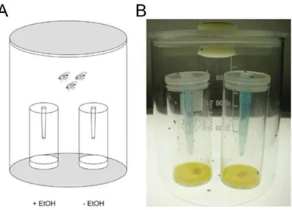
the attraction of Drosophila towards ethanol, wild type flies show an attraction towards
natural alcohol concentration, when offered a choice between 5% ethanol containing
food odor and alcohol free food odor (Ogueta et al., 2010; Schneider et al., 2012). This
concentration of up to 5% of ethanol can be found in the natural environment of the fly,
for example in rotten fruits (Dudley, 2002). Ethanol in food odor mixtures is a key
odorant, which regulates food attraction (Giang et al., 2017). But Tbh
nM18mutants failed
to show attraction towards alcohol, but preferred food odor over water and ethanol over
water, so the odor perception is not impaired and they just might be less sensitive to
ethanol (Schneider et al., 2012). The mutant phenotype could be rescued by expressing
Tbh in a Tdc2-GAL4 dependent manner, which targets 78 Tbh positive neurons. So the
13
function of Tbh in these 78 neurons is required for olfactory alcohol attraction. Reducing the number of targeted neurons by using the Cha-GAL80 driver line did not restore the mutant phenotype. But this subset of neurons is involved in aggression (Zhou et al., 2008), so it is known that olfactory ethanol attraction and aggression are mediated through different kinds of subsets. By testing further GAL4-driver lines, Schneider and colleagues (2012) could narrow down the number of ethanol attraction mediating neurons to 26 Tbh expressing neurons in the G3a/AL2 cluster and in the VMI-III, whereas the Tbh positive neurons in the VNC are not involved in mediating ethanol attraction. From this it can be concluded, that the lack of attraction towards ethanol is not due to deficits in execution of motor tasks, as the VNC is the main region involved in locomotor output and is not involved in attraction behavior. Additionally, they assumed that Tbh
nM18mutants are able to sense environmental changes and to perform motor related tasks, but they are unable to respond to these stimuli in an appropriate way, so the function of Tbh is probably at the interface between sensory information and response selection and acts as a reward center. So far it is still unknown, how exactly the reinforcer works (Schneider et al., 2012).
1.2. The reinforcing properties of OA
Reinforcement systems drive the synaptic plasticity within neuronal circuits that form memories (Waddell, 2013) and more similar in flies and mammals than thought (Burke et al., 2012). Earlier studies showed that OA acts as a positive reinforcer for reward/appetitive learning and DA acts as the negative reinforcer in aversive learning (Schwaerzel et al., 2003). But more recent studies revealed that DA is also involved in appetitive learning and memory (Burke et al., 2012) and that OA can also mediate aversive learning (Iliadi et al., 2017).
The reinforcing properties of OA have been investigated and shown in different
experiments. For the first time it was revealed in the honey bee Apis mellifera, where it
was shown that activation of the octopaminergic VUMmx1 neuron was able to substitute
for the unconditioned stimulus (US) (Hammer, 1993). In Drosophila larvae, it was
14
discovered, that pairing an olfactory stimulus with light activation of tyraminergic/octopaminergic neurons induced appetitive memory formation, while activation of dopaminergic neurons elicited aversive learning. From these findings it was concluded, that these two modulatory systems act antagonistically and are moreover sufficient to substitute for appetitive and aversive reinforcement in an olfactory learning and memory paradigm (Schroll et al., 2006). The same observation was made in adult flies. Schneider et al. (2012) showed that activation of tyraminergic/octopaminergic neurons in a Tdc2-GAL4 dependent manner in adult Drosophila melanogaster is sufficient to elicit site attraction and to increase locomotor activity. Activation of dopaminergic neurons in a TH-GAL4 dependent manner instead led to site aversion, but also to an increase in locomotor activity in a similar way as activation of the tyraminergic/octopaminergic neurons. With this result, they confirmed that the observed site attraction which resulted from the activated tyraminergic/octopaminergic neurons is not due to hyperactivity of the flies. Furthermore, they concluded that tyraminergic/octopaminergic neurons act as a positive reinforcer, while dopaminergic neurons have the opposing effect and act as a negative reinforcer. In experiments with the OA deficient Tbh
nM18mutant (Monastirioti et al., 1996), the effect of missing OA on sugar reward learning was investigated in adult flies. Tbh
nM18mutants displayed normal aversive learning scores, but were severely impaired in sugar learning (Schwaerzel et al., 2003). By blocking dopaminergic neurons using UAS-shi
ts, electric shock learning was severely impaired (Schwaerzel et al., 2003). So they concluded that positive association of an external odor stimulus depends on OA signaling, while negative association is mediated by DA. This is conform to the results of Schroll et al., (2006) and Schneider et al., (2012). But new findings revealed that OA is also involved in aversive learning, as Tbh
nM18mutants showed a reduced performance index in electric shock test. This phenotype was not rescuable with feeding OA, but by expressing Tbh in a Tdc2-GAL4 dependent manner and is not due to sensorimotor defects, as Tbh
nM18mutants showed normal sensorimotor abilities and taste perception (Iliadi et al., 2017).
In addition to the fact, that OA functions as a positive and negative reinforcer, it was
discovered, that DA also is involved in aversive and reward learning. Next to three
different dopaminergic pathways of forming aversive memories with different temporal
stabilities (Aso et al., 2012), there has to be a way to form appetitive memories including
dopaminergic neurons. It was shown that OA signaling in aversive learning requires
15
signaling via the OAMB receptor located on dopaminergic neurons in the MB (Burke et al., 2012). Activation of dopaminergic neurons can also substitute for sugar in an appetitive learning process and form a robust memory, even if the flies are lacking OA and dopaminergic neurons are also involved in the short-term reinforcing effect of OA (Burke et al., 2012). The fact that OA receptors on dopaminergic neurons are involved in memory formation shows that DA signaling is downstream of OA in the appetitive learning process (Burke et al., 2012). Regarding the DA receptors, the receptor dDA1 is the key receptor for aversive and appetitive learning in Drosophila and is located in the MB (Kim et al., 2007). The MB is the main integrative center for learning and memory (Menzel et al., 1988; 1990; Roeder, 1999) and both, appetitive and aversive olfactory memories are localized to the same neuropil in the MB (Schwaerzel et al., 2003). Hence, both neurotransmitters are involved in positive and negative reinforcement and interact which each other to form appetitive and aversive memories.
1.3. OA receptors in adult Drosophila
Mediation of this broad range of behavior in invertebrates by OA signaling requires
receptors specific for OA to pass on the signal from one neuron to the next cell – the OA
receptors. Drosophila has four known OA receptors: OAMB and three OctβRs (Han et al.,
1998; Balfanz et al., 2005; Maqueira et al., 2005) and all of them are G protein-coupled
receptors (GPCRs) (Roeder, 1999 and 2005; Evans and Maqueira, 2005; Maqueira et al.,
2005). These receptors are similar to mammalian adrenergic receptors (Dudai and Zvi,
1982; Han et al., 1998) and are classified into the OA1 and OA2 receptor family (Evans
and Robb, 1993, Balfanz et al., 2005, Maqueira et al., 2005). The OA1 receptor family
contains α-adrenergic-like receptors (OctαRs) (e.g. OAMB) and is known to increase
intracellular Ca
2+levels, while the OA2 receptor family contains β-adrenergic-like
receptors (OctβRs) and is able to stimulate the adenylyl cyclase which results in an
increase of intracellular cAMP (Evans and Robb, 1993, Balfanz et al., 2005, Maqueira et
al., 2005). The three OctβRs (Octβ1Rs, Octβ2Rs and Octβ3Rs) and the OAMB receptor
show a strong preference for OA over TA (Blenau and Baumann, 2001; Maqueira et al.,
2005; Balfanz et al., 2005; 2014). The group of OctβRs mediates a major amount of
16
octopaminergic functions during the development of Drosophila (Ohhara et al., 2012).
There are also receptors which are sensitive to OA and TA or which are even more sensitive to TA than OA. These receptors are called octopamine/tyramine (Oct-Tyr) receptors or tyraminergic receptors (TyrR, TyrRII and TyrRIII) (Monastitiroti, 1999; El- Kholy et al., 2015; Evans and Maqueira, 2005; Roeder, 2005; Bayliss et al., 2013). Just like the OA receptors, the Oct-Tyr and TA receptors are also G protein-coupled, but both neurotransmitters mediate their effect through different GPCRs (Saudou et al., 1990;
Blenau and Baumann, 2001). These receptors belong to the α2-adrenergic receptors and can be divided in two classes: the Type 1 TA receptor (Oct-Tyr receptor) and the Type 2 TA receptor (TA receptor) (Blenau et al., 2017). The Type 1 TA receptor is better activated by TA than OA and has the opposite effect of the OA receptors, as it inhibits the adenylyl cyclase and therefore decreases the intracellular cAMP level, while the Type 2 TA receptor can be almost only activated by TA and is able to mediate Ca
2+and cAMP levels (Blenau et al., 2000; 2017). On the other hand, TA is also able to bind to and activate an OA receptor at high concentrations (Han et al., 1998; Blenau et al., 2000;
Blenau and Baumann, 2001; Balfanz et al., 2005; Maqueira et al., 2005).
All OA receptors are expressed throughout the whole development in the CNS of the fly, especially in the MB (Ohhara et al., 2012; El-Kholy et al., 2015). Additional, expression in other organs varies depending on the OA receptor type.
In adult flies the Octβ1R was found additionally in the ovaries and testis, the muscles, the intestine, the trachea, the pars intercerebralis, the AL and the thoracic-abdominal ganglion, while the optic lobes were innervated by Octβ1R neurons (Ohhara et al., 2012;
El-Kholy et al., 2015). But compared to the other OctβRs it shows only low levels in the female reproductive organ (Li et al., 2015). It was shown, that Octβ1R is required for acute changes in synaptic structure in response to OA and for the increase in locomotion velocity due to starvation (Koon and Budnik, 2012).
The Octβ2R could be found in adult flies in the male and female reproductive system in
the skeletal muscles, the trachea, the intestine, the fat body, the salivary glands, the
malpighian tubes, the maxillary muscular system, the third antennal segment, the pars
intercerebralis, the AL, the optic lobes and with only a few cells in the thoracic-
abdominal ganglion (Ohhara et al., 2012; El-Kholy et al., 2015; Li et al., 2015). The
Octβ2R plays a pivotal role in the fertilization and ovulation of female flies (Lim et al.,
17
2014; Li et al., 2015). The homozygous Octβ2R mutant females display normal pre- and post-mating behavior, but they are unable to lay eggs (Lim et al., 2014). They have a delay in copulation rate and enlarged ovaries compared to w
1118and Tbh deficient flies and the few laid eggs do not develop as they are not fertilized, which indicates a sperm delivery problem (Li et al., 2015). This phenotype could be (partially) rescued by expression of Octβ2R or ectopic expression of the other three OA receptors (Lim et al., 2014). It was suggested, that there might be an interaction of Octβ2R and OAMB in the female reproductive system (Lim et al., 2014; Li et al., 2015).
In the adult fly, the Octβ3R was detected in the ovaries and testis and in the AL and the mechanosensory center in the CNS. The trachea, the malpighian tubes, the muscles and the pars intercerebralis showed only a weak expression and the optic lobes and the thoracic-abdominal ganglion almost none (Ohhara et al., 2012; El-Kholy et al., 2015).
The expression levels in the female reproductive organs were slightly decreased compared to Octβ2R or OAMB (Li et al., 2015). It was shown that Octβ3R is involved in the regulation of ecdysone synthesis in the prothoracic gland, a hormone which is essential for the metamorphosis in Drosophila. A knock down of Octβ3R in the prothoracic gland leads to an arrested metamorphosis in the stage between larva and prepupa. As a knock down of TA synthesis resulted in a similar effect, it is likely that not only OA but also TA is able to activate Octβ3R (Ohhara et al., 2014).
The OAMB belongs to the OctαR family and is a α1-like receptor (Kim et al., 2013; Lim et
al., 2014). In the adult fly, it was detected in in the pars intercerebralis, the ellipsoid
body of the central complex, in some skeletal muscles in the legs, in the reproductive
organs (epithelium of the oviduct), in the trachea, in the intestine and in the thoracic-
abdominal ganglion. The outer and inner medulla and lobula of the optic lobes were
innervated by OAMB neurons (Strauss and Heisenberg, 1993; Han et al., 1998; Lee et al.,
2003; El-Kholy et al., 2015; Li et al., 2015). The OAMB seems to be involved in several
functions in Drosophila. It is involved in synaptic modulation underlying behavioral
plasticity (Han et al., 1998), in associative learning (Heisenberg et al., 1985; de Belle and
Heisenberg, 1994; Davis, 1996; Kim et al., 2013), in motor activities (Strauss and
Heisenberg, 1993) and ovulation in female flies (Lee et al., 2003; Lim et al., 2014; Li et
al., 2015). Comparable to Octβ2R mutants, OAMB mutant females exhibit normal
courtship and copulation behavior, but have a defect in ovulation and thus retain the
18
eggs in their abdomen (Lee et al., 2003; Lim et al., 2014; Li et al., 2015). But it was shown, that only expression of OAMB in the body and not the high expression levels in the brain are required for normal ovulation (Lee et al., 2003). Furthermore, the OAMB is also involved in appetitive (but not aversive) olfactory learning in Drosophila, as OAMB null mutants have a severely impaired learning phenotype (Kim et al., 2013). They showed that the αβ-lobe and the γ-lobe are the functional sites of OA signaling in appetitive olfactory learning and that the OAMB is the key molecule.
1.4. Channelrhodopsins – a tool for light induced neuronal activation
To investigate the role of tyraminergic/octopaminergic neurons in attraction and aversion behavior in Drosophila melanogaster, the genetic tool of neuronal light activation via three different channelrhodopsins was used.
Microbial-type rhodopsins are found in archaea, prokaryotes and eukaryotes and have some structural similarities to the rhodopsin in animals, but no sequence homology (Nagel et al., 2003). They are also found in fungi and algae (Bieszke et al., 1999;
Hegemann et al., 2001). The prototype of these microbial-type rhodopsins is the light- driven proton pump bacteriorhodopsin (Oesterhelt and Stoeckenius, 1971). Other ion channels are Channelrhodopsin-1 (ChR1) and the related Channelrhodopsin-2 (ChR2).
ChR1 mediates the high-intensity response, whereas ChR2 is responsible for low-
intensity photocurrents (Sineshchekov et al., 2002). Both were isolated from the green
algae Chlamydomonas reinhardtii (Nagel et al., 2003) and are involved in generating
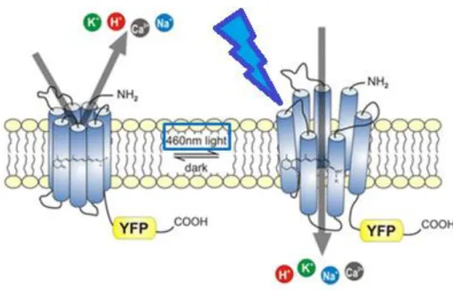
photocurrents (Sineshchekov et al., 2002). The ChR2 is a blue light gated cation channel
with seven transmembrane domains (α helices) and has a covalently bound all-trans
retinal (ATR), which is necessary for opening and closure of the ChR2 (Nagel et al.,
2003). After absorption of a photon with a wavelength of 460-480nm, the retinal
changes from all-trans to cis-conformation, which leads to the opening of the channel
and cations can get inside the cell (Figure 4). This leads to a depolarization and thus
activation of the cell. But ChR2 has only a low conductance and therefore a higher
expression of ChR2 is required for stronger depolarization (Nagel et al., 2003; Pulver et
19
al., 2009). Furthermore it is said, that blue light does not penetrate the cuticle as well as red or amber light (Eichler et al., 1977; Inagaki et al., 2014). Inagaki and colleagues (2014) measured the penetration of different wavelength through the cuticle of a fruit fly and showed that blue light penetrance is much weaker (about 2%) than green (5%), amber (8%) or red (7%) light. To avoid the high absorption and scattering problem of blue light activatable ChR2, modified channelrhodopsins were created and tested (Lin et al., 2013; Dawydow et al., 2014; Inagaki et al., 2014; Klapoetke et al., 2014). One of these new channelrhodopsins is Chrimson from the algae Chlamydomonas noctigama, which has a spectral peak at 590 nm (Klapoetke et al., 2014). A new variant of the channelrhodopsin from Chlamydomonas reinhardtii was engineered by Lin and colleagues (2013) – the red-activatable channelrhodopsin (ReaChR), which has an optimum excitation wavelength of 590-630 nm, which is orange to red light (Lin et al., 2013) and does not interfere with normal visual function (Inagaki et al., 2014). But additionally, it is also activatable with green light (Inagaki et al., 2014; Krause et al., 2017). ReaChR has faster kinetics, higher photocurrents and better membrane trafficking compared to other red-shifted channelrhodopsins. A slow channel closure rate after the ending of stimulation is the only limitation of this new red light activatable channelrhodopsin (Lin et al., 2013).
Figure 4: Schematic drawing of Channelrhodopsin-2.
Closed (left) and open (right) state of the blue light gated ChR2 as an exemplary presentation of all channelrhodopsins. Upon light exposure, the ATR changes its conformation and leads to an opening of the channel which allows cations to enter the cell and which thus is activated. (Modified after http://www.biophys.mpg.de/en/bamberg. html).
20
Using light stimulation to activate neurons has advantages and disadvantages. Due to their characteristics, channelrhodopsins are a non-invasive, rapid and reliable tool with a good spatial and temporal resolution (Nagel et al., 2003, 2005; Boyden et al., 2005;
Wang et al., 2007). Compared to temperature sensitive channels like dTRPA1 (Drosophila transient receptor potential A1) (Hamada et al., 2008; Viswanath et al., 2003), it is possible to switch the light activation on and off in the range of milliseconds and therefore create a faster and more precise stimulation of the neurons. Experiments with different channelrhodopsins have been done a lot in Drosophila melanogaster, regarding different developmental stages and behaviors (Schroll et al., 2006; Zhang et al., 2007; Crisp et al., 2008; Pulver et al., 2009; Schneider et al., 2012; Dawydow et al., 2014; Inagaki et al., 2014; Klapoetke et al., 2014; Hsiao et al., 2015; Xu et al., 2016; Saras et al., 2017).
Light activation of neurons via a channelrhodopsin can be done with constant or pulsed light. In Drosophila larvae, constant light led to a higher spike frequency adaptation than light pulses, thus pulsed light might be more effective for long time stimulation (Pulver et al., 2009). Pulsed light was also used by Schneider et al. (2012) and Xu et al. (2016).
They used an activation pattern of 2s 40Hz, 16s 8Hz, 2s 0Hz, which derives from a study
performed by Hammer (1993) with the honey bee Apis mellifera. In this experiment, the
conditioning of the PER occurs after a single pairing of an odor (CS – conditioned
stimulus) with a food odor US (in this case sugar). Through electrophysiological
recordings, a single neuron attracted attention because of its specific response and
unique morphology – the octopaminergic VUMmx1 neuron (ventral unpaired median
cell of maxillary neuromere 1) – which responded to the US with a long burst of action
potentials and fired in the pattern mentioned above. To test its qualities of substituting
sugar as the US, a supra-threshold depolarization of the VUMmx1 was elicited instead of
offering sugar. There were no differences between the results of eliciting the PER, no
matter if using either sugar or depolarization of the VUMmx1, so stimulation of the
VUMmx1 neuron is equally effective as an US as sugar. So Hammer (1993) showed that a
single neuron is sufficient to mediate reinforcement in associative learning and that OA
might be the neuronal representative of the US. Since stimulation of this single neuron
could substitute for the US, it is possible that the used frequency is a reward firing
pattern. Furthermore, the VUMmx1 neurons has similarities to the VUMa2 neuron in the
Drosophila brain and both innervate olfaction related structures like the AL, the lateral
21
horn (LH) and the MB (Hammer, 1993; Busch et al., 2009), therefore it was decided to use this frequency pattern for our experiments.
In addition to the blue light activatable ChR2 used for example by Schneider and
colleagues (2012), experiments with Chrimson and ReaChR, two red light activatable
channelrhodopsins, have been done. These red activatable channelrhodopsins represent
an additional possibility of neuronal light activation and might be a more convenient
one. Klapoetke et al. (2014) investigated the function of Chrimson. They used pulsed
light for neuronal activation and showed that not only orange (617 nm) or red light (720
nm) stimulation could activate Chrimson, but also blue light (470 nm). Blue and orange
light were able to elicit the investigated behavioral response in larvae and adults even at
short light pulse durations and low intensities, while red light stimulation needed longer
pulses and a higher intensity (Klapoetke et al., 2014). The ReaChR was tested by Inagaki
et al. (2014) with pulsed red (627nm), green (530nm) and blue (470nm) light. Green
and red light are both sufficient to activate ReaChR and achieved higher scores in all
experiments at all times compared to blue light activation of the blue light activatable
ChR2. Green light even has the strongest capacity to activate ReaChR when the intensity
was not normalized (Inagaki et al., 2014), although it is a red light activatable
channelrhodopsin. Consistent with the results obtained by Pulver and colleagues (2009)
with the blue light activatable ChR2, Inagaki and colleagues (2014) also observed a
dependence of light intensity and pulse frequency in the performance of the flies with
the red light activatable ReaChR. Therefore, the broad dynamic range of frequencies and
intensities which can be used for ReaChR stimulation leads to a more quantitative and
temporally controlled approach to investigate neuronal controlled behavior (Inagaki et
al., 2014).
22
1.5. Aim
The aim of this thesis was to investigate the role of OA in attraction and aversion behavior of Drosophila melanogaster. So far it is known that OA is involved in mediating reward learning and memory (Schwaerzel et al., 2003) and in innate odor attraction (Ogueta et al., 2010; Schneider et al., 2012). But it is not known, which kind of set of tyraminergic/octopaminergic neurons is involved in mediating this behavior and if this set is sufficient and necessary to elicit attraction. Furthermore, there is only little information about OA being involved in aversive behavior (Iliadi et al., 2017). These questions are investigated in this thesis.
To answer the first question, which subset of neurons mediates site attraction, the optogenetic site attraction assay was used. Using neuronal light activation, different tyraminergic/octopaminergic sets of neurons were activated via two different channelrhodopsins – a blue (ChR2) and a red (ReaChR) light activatable one. Activation of 78 Tbh positive neurons targeted by the Tdc2-GAL4 driver line elicited site aversion (Schneider et al., 2012). To narrow down the number of neurons, additional GAL4 driver lines were tested (Feb15-GAL4, fru-GAL4 and 6.2-Tbh-GAL4). The achieved results should give further insight into which set of neurons is responsible for attraction behavior and if there probably might be also a Tbh positive subset of neurons which is able to elicit aversive behavior. This would answer the next question, whether OA is also capable of mediating site aversion. To further verify the role of OA, the experiments were repeated in a Tbh
nM18mutant background with lacking OA levels (Monastirioti et al., 1996). By combining the TH-GAL80 or Cha-GAL80 driver line to the Tbh positive neurons targeting driver lines, putative effects of dopaminergic or cholinergic neurons were ruled out.
Furthermore it was tested, if OA is able to switch an already existent decision by shifting
the reinforcer to another option. For all these experiments, an activation pattern of 2s
40Hz 16s 8Hz 2s 0Hz (successfully used by Schneider et al. (2012) and Xu et al., (2017))
was used to stimulate the flies and thus to elicit the desired behavior. Regarding this
activation pattern, it was also tested if modifications or only parts of it were able to elicit
the same behavior as the original light stimulation pattern. Additionally to the potential
frequency dependence, the influence of different light intensities was investigated. From
these results, it could be told whether the observed behaviors are really due to neuronal
activation or if they are just artefacts caused by the channelrhodopsin transgenes. The
23
results also give the opportunity to compare the functionality of the two different channelrhodopsin with each other.
The results of the optogenetic site attraction assay revealed that OA is sufficient to elicit
site attraction. To answer the question about the necessity, the role of OA in innate
olfactory attraction was investigated. The Tbh
nm18mutants and their genetic control
w
1118were tested in the two odor choice paradigm (Ogueta et al., 2010). Tbh
nm18mutants
fail to show attraction towards ethanol containing food odors in natural concentrations
(Ogueta et al., 2010; Schneider et al., 2012). The flies were pre-fed with different
pharmacological substances to either activate or block the OA or TA receptors. Next to
OA and TA, two OA agonists (clonidine and naphazoline), an OA antagonist (epinastine)
and a TA antagonist (yohimbine) were administered to the flies. Furthermore, different
Tbh mutants (Tbh
nm18and Tbh
Del3with decreased OA levels and d01344 with increased
levels of OA) and a Tdc2 mutant (which lacks both OA and TA) were compared to each
other and the effect of an overexpression of Tbh was investigated. This should also
provide evidence about the relation of OA and TA in attraction behavior. In a last step,
the four different OA receptors were eliminated in the olfactory sensory neurons (Orco-
GAL4) by using RNAi. In the two odor choice paradigm it was investigated which OA
receptor is involved in mediating olfactory attraction towards ethanol containing food
odors.
24
2. Material and Methods
2.1. Material
2.1.1. Fly strains
Name of fly stock
(named in this thesis) Origin Chromo-
some
Stock list
w
1118Lindsley & Zimm X #4
w
1118; Tdc2
RO54/CyO (Tdc2
RO54) Cole et al., 2005 II #535
w
1118, Tbh
nM18/FM7(Tbh
nM18) Monastirioti et
al., 1996 X #1
w
1118, Tbh
R3-XPdel/FM7 (Tbh
Del3) Manuela Ruppert,
2013 X #536
w
1118, XP
d01344(d01344) Exelixis
Collection at HMS X #16 w
1118;; UAS-Tbh/TM2 Henrike Scholz III #22 norpA
1; UAS-ChR2; UAS-ChR2 (UAS-ChR2) Nuwal, 2010 X + II + III #318 w
1118, Tbh
nM18; UAS-ChR2; UAS-ChR2 Gerbera Claßen X + II + III #40 w
1118; UAS-ReaChR; UAS-ReaChR (UAS-
ReaChR) (original Stocks: w; UAS-ReaChR and w;;
UAS-ReaChR)
Gerbera Claßen (original stocks:
Inagaki &
Anderson)
II + III #536
w
1118, UAS-CS Chrimson (UAS-Chrimson) Vivek Jayaraman X #506 w
1118; dTdc2-GAL4 (Tdc2-GAL4) Cole et al., 2005 II #35 w
1118;; 6.2_2-Tbh-GAL4 Hampel, 2007 III #278
w
1118; Feb15-GAL4 Siegmund &
Korge, 2001 II #217
25
w
1118;; NP0021/TM6 (fru-GAL4) Kimura et al.,
2005 III #392
w
1118; dTdc2-GAL4/CyO; Cha
3,3kb-
GAL80/TM6,Tb1 (Tdc2-GAL4; Cha-GAL80) Andrea Schneider II + III #30 w
1118; TH-GAL80; 6.2-Tbh-GAL4 Gerbera Claßen II + III
norpA
1; UAS-ChR2; fru-GAL4 Gerbera Claßen X + II + III norpA
1; UAS-ChR2; 6.2-Tbh-GAL4 Gerbera Claßen X + II + III norpA
1; UAS-ChR2; Cha-GAL80 Gerbera Claßen X + II + III w
1118;; Orco-GAL4 11.17 (Orco-GAL4) Vosshall, 2008 II #89
BDSC = Bloomingtion Drosophila Stock Collection
w[1118]; P{y[+t7.7] w[+mC]=GMR29H06- GAL4} attP2 (GMR29H06-GAL4)
#49506, Pfeiffer
et al., 2008 III #394
w[1118]; P{y[+t7.7] w[+mC]=GMR47A10- GAL4} attP2 (GMR47A10-GAL4)
#50289, Pfeiffer
et al., 2008 III #395
w[1118]; P{y[+t7.7] w[+mC]=GMR51D07- GAL4} attP2/TM3, Sb[1] (GMR51D07-GAL4)
#48186, Pfeiffer
et al., 2008 III #393
y[1] v[1]; P{y[+t7.7] v[+t1.8]=
TRiP.JF01673}attP2 (UAS-OAMB-RNAi)
#31171, Ni et al.,
2008 III #457
y[1] v[1]; P{y[+t7.7] v[+t1.8]=
TRiP.JF01571}attP2 (UAS-Octβ1R-RNAi)
#31106, Ni et al.,
2008 III #455
y[1] sc[*] v[1]; P{y[+t7.7] v[+t1.8]=
TRiP.HMS0115 1}attP2 (UAS-Octβ2R-RNAi)
#34673, Ni et al.,
2011 III #459
y[1]v[1];P{y[+t7.7] v[+t1.8]=TRiP.JF01573}
attP2/TM3,Sb[1} (UAS-Octβ3R-RNAi)
#31108, Ni et al.,
2008 III #456
26