Analysis of complex stability and allosteric interaction in the
imidazole glycerol phosphate synthase complex
DISSERTATION ZUR ERLANGUNG DES
DOKTORGRADES DER NATURWISSENSCHAFTEN (DR. RER. NAT.) DER FAKULTÄT FÜR BIOLOGIE UND VORKLINISCHE MEDIZIN
DER UNIVERSITÄT REGENSBURG vorgelegt von
Alexandra Holinski aus Lindau (Bodensee)
im Jahr 2017
Das Promotionsgesuch wurde eingereicht am:
13.01.2017
Die Arbeit wurde angeleitet von:
Prof. Dr. Reinhard Sterner
Unterschrift:
This work was done in the period from August 2012 to January 2017 in the group of Prof.
Dr. Reinhard Sterner (Biochemistry II, Institute of Biophysics and Physical Biochemistry,
University of Regensburg).
Table of contents I
Table of contents
TABLE OF CONTENTS ... I LIST OF FIGURES ... V LIST OF TABLES ... VII FORMULA INDEX ... VIII LIST OF ACRONYMS AND ABBREVIATIONS ... IX
ABSTRACT ... 1
ZUSAMMENFASSUNG ... 3
1 INTRODUCTION ... 6
1.1 P
ROTEIN-
PROTEIN INTERACTIONS... 6
1.2 A
LLOSTERY... 9
1.3 T
HE IMIDAZOLE GLYCEROL PHOSPHATE SYNTHASE... 11
1.4 A
NCESTRAL SEQUENCE RECONSTRUCTION... 16
1.4.1 Theory and application of ASR ... 16
1.4.2 Previous phylogenetic studies on ImGPS ... 19
2 OBJECTIVES OF THE THESIS ... 21
3 MATERIALS ... 22
3.1 I
NSTRUMENTATION... 22
3.2 C
ONSUMABLES... 24
3.3 C
HEMICALS... 25
3.4 K
ITS... 26
3.4.1 Kits for molecular biology ... 26
3.4.2 Kits for protein crystallization ... 26
3.5 E
NZYMES... 26
3.6 B
ACTERIAL STRAINS... 27
3.7 V
ECTORS... 27
3.7.1 pET vectors ... 27
3.7.2 pQE vectors (Qiagen, Hilden) ... 30
3.7.3 pACYCDuet-1 (Novagen) ... 30
3.8 O
LIGONUCLEOTIDES... 31
3.8.1 Vector specific amplification and sequencing primers ... 31
3.8.2 Amplification and mutagenic primers for hisF ... 31
3.8.3 Amplification and mutagenic primers for hisH ... 33
3.9 L
ADDERS AND MARKERS... 33
3.10 B
UFFERS AND SOLUTIONS... 34
3.10.1 Buffers and solutions for molecular biology ... 34
3.10.2 Buffers and solutions for working with E. coli ... 35
3.10.3 Buffers and solutions for working with proteins ... 35
3.10.4 Buffers and solutions for SDS-PAGE ... 36
3.11 B
ACTERIAL GROWTH MEDIA... 36
3.12 S
OFTWARE... 37
4 METHODS ... 39
4.1 P
REPARATION OF INSTRUMENTATION AND SOLUTIONS... 39
4.2 M
ICROBIOLOGICAL METHODS... 39
4.2.1 Cultivation and storage of E. coli strains ... 39
4.2.2 Preparation of chemically competent E. coli cells (Inoue et al., 1990) ... 39
4.2.3 Transformation of chemically competent E. coli cells ... 40
4.3 M
OLECULAR BIOLOGY METHODS... 40
4.3.1 Isolation and purification of plasmid DNA from E. coli ... 40
4.3.2 Determination of DNA concentration ... 40
4.3.3 Agarose gel electrophoresis ... 41
4.3.4 Isolation of DNA fragments from agarose gels ... 42
4.3.5 Enzymatic manipulation of dsDNA ... 42
4.3.5.1 Cleavage of dsDNA by restriction endonucleases ... 42
4.3.5.2 Ligation of DNA fragments ... 42
4.3.6 Amplification of DNA fragments by standard polymerase chain reaction ... 42
4.3.7 Colony PCR ... 44
4.3.8 QuikChange site-directed mutagenesis ... 44
4.3.9 Overlap Extension PCR (Ho et al., 1989) ... 46
4.3.10 DNA sequencing ... 46
4.3.11 Gene synthesis ... 47
4.4 P
ROTEIN BIOCHEMISTRY METHODS... 47
4.4.1 Gene expression ... 47
4.4.1.1 Gene expression at analytical scale ... 47
4.4.1.2 Gene expression at preparative scale ... 48
4.4.2 Protein purification ... 48
4.4.2.1 Heat step ... 48
4.4.2.2 Metal affinity chromatography ... 48
4.4.2.3 Ion exchange chromatography ... 49
4.4.2.4 Ammonium sulfate precipitation ... 51
4.4.2.5 Preparative size exclusion chromatography ... 52
4.4.3 Buffer exchange by dialysis ... 52
4.4.4 Concentrating protein solutions ... 53
4.4.5 Storage of purified proteins ... 53
4.4.6 Synthesis of ProFAR ... 53
4.4.7 Peptide synthesis ... 54
Table of contents III
4.5 A
NALYTICAL METHODS... 54
4.5.1 Protein concentration determination via absorption spectroscopy ... 54
4.5.2 SDS-polyacrylamide gel electrophoresis (SDS-PAGE) ... 55
4.5.3 Analytical size exclusion chromatography ... 56
4.5.4 Circular dichroism spectroscopy ... 56
4.5.5 Fluorescence titration ... 57
4.5.6 Steady-state enzyme kinetics ... 58
4.5.6.1 Ammonia-dependent cyclase activity ... 58
4.5.6.2 Glutamine-dependent cyclase activity ... 59
4.5.6.3 Glutaminase activity ... 60
4.5.7 HPLC-analysis for the determination of basal glutaminase activity ... 61
4.5.8 Protein crystallization and X-ray structure determination ... 62
4.5.9 NMR spectroscopy ... 63
5 RESULTS AND DISCUSSION ... 65
5.1 A
SSESSING THE BINDING OF A PEPTIDE TO THEH
ISF:H
ISH
INTERFACE BY MEANS OF[
1H-
15N] HSQC
SPECTROSCOPY... 65
5.1.1 Preliminary work and aim of this project ... 65
5.1.2 Expression and purification of
15N-tmHisF ... 67
5.1.3 HSQC titration experiments of
15N-tmHisF with peptide ... 67
5.1.4 Conclusion ... 71
5.2 A
NALYSIS OF PROTEIN-
PROTEIN INTERACTION AND ALLOSTERY INI
MGPS
WITH THE HELP OF CONTEMPORARY AND PRIMORDIAL PROTEINS... 72
5.2.1 General concept and initial interaction and allosteric studies with contemporary HisF and HisH proteins ... 72
5.2.2 Reconstruction and characterization of a putative glutaminase subunit from the LUCA era ... 76
5.2.2.1 Wild type LUCA-HisH ... 76
5.2.2.1.1 Reconstruction of LUCA-HisH ... 76
5.2.2.1.2 Cloning, heterologous expression and purification of LUCA-HisH ... 77
5.2.2.1.3 Structural integrity and thermal stability of LUCA-HisH ... 78
5.2.2.1.4 Complex formation of LUCA-HisF and LUCA-HisH ... 79
5.2.2.1.5 Activity tests with LUCA-HisF:LUCA-HisH complex ... 80
5.2.2.2 Putatively constitutively active LUCA-HisH mutants ... 81
5.2.2.2.1 Mutagenesis of LUCA-HisH, heterologous expression and purification of LUCA-HisH mutants ... 81
5.2.2.2.2 Structural integrity and thermal stability of LUCA-HisH mutants ... 82
5.2.2.2.3 Complex formation of LUCA-HisF and LUCA-HisH mutants ... 84
5.2.2.2.4 Activity tests with complexes of LUCA-HisF and LUCA-HisH mutants ... 85
5.2.2.3 Basal glutaminase activity of LUCA-HisH ... 86
5.2.2.4 Conclusion ... 87
5.2.3 Reconstruction of primordial HisF proteins ... 89
5.2.4 Studies on protein-protein interaction with the help of primordial HisF proteins ... 90
5.2.4.1 Concept ... 91
5.2.4.2 Cloning, heterologous expression and purification of HisF and HisH proteins used for interaction
studies ... 93
5.2.4.3 Identifying a protein-protein interaction hot spot in HisF on the basis of ASR and in silico as well as experimental mutagenesis ... 94
5.2.4.4 Analysis of physicochemical principles of complex stability with the help of in silico mutagenesis and coevolution analysis... 100
5.2.4.5 Conclusion ... 102
5.2.5 Studies on allosteric regulation with the help of ancient HisF proteins ... 103
5.2.5.1 Concept, mutational studies and computational approach ... 103
5.2.5.2 Crystal structure of LUCA-HisF wt:tmHisH with and without bound glutamine ... 108
5.2.5.3 Conclusion ... 110
6 FINAL DISCUSSION AND OUTLOOK ... 112
6.1 I
MPLICATIONS FOR PROTEIN-
PROTEIN INTERACTION AND ALLOSTERY INI
MGPS ... 112
6.2 ASR
AS AN EFFECTIVE METHOD FOR DISENTANGLING PROTEIN-
PROTEIN INTERACTION AND ALLOSTERY... 113
7 REFERENCES ... 116
8 APPENDIX ... 125
8.1 P
HYLOGENETIC TREE FOR RECONSTRUCTING INTERMEDIATE PRIMORDIALH
ISF
SEQUENCES... 125
8.2 N
UCLEOTIDE AND AMINO ACID SEQUENCES OFLUCA-H
ISH, A
NC1
PA-H
ISF, A
NC1
TM-H
ISF
ANDA
NC2
TM-H
ISF ... 126
8.3 S
TRUCTURAL INTEGRITY OFH
ISF
VARIANTS USED FOR INTERACTION STUDIES... 131
8.4 S
TRUCTURAL INTEGRITY OF ZMH
ISH
MUTANTS... 133
8.5 S
TEADY-
STATE KINETIC CHARACTERIZATION OF RECONSTRUCTED AND MODERNH
ISF
ANDH
ISH
PROTEINS... 134
8.5.1 General remarks ... 134
8.5.2 Ammonia-dependent cyclase activity ... 135
8.5.3 Glutamine-dependent cyclase activity ... 137
8.5.4 Glutaminase activity ... 139
8.6 S
TRUCTURAL INTEGRITY OFA
NC2
TM-H
ISF_A2 ... 142
8.7 C
ALIBRATION CURVES... 143
8.8 D
ATA COLLECTION AND REFINEMENTS STATISTICS... 145
9 ACKNOWLEDGEMENTS ... 147
List of figures V
List of figures
Figure 1: Structure and reaction of the ImGP synthase from Thermotoga maritima (HisF:HisH complex). .... 12
Figure 2: Putatively functional important residues and allosteric pathway in tmImGPS. ... 14
Figure 3: An example of a phylogenetic tree. ... 17
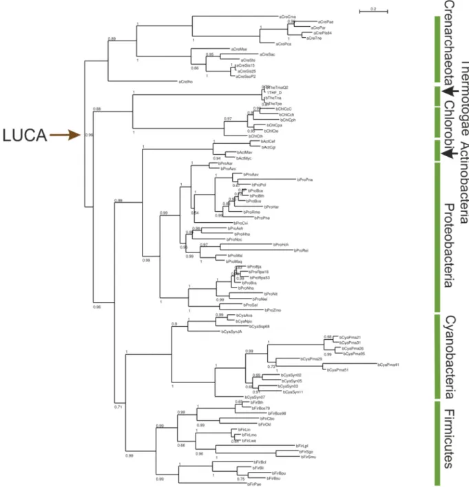
Figure 4: Phylogenetic tree used for the reconstruction of LUCA-HisF and LUCA-HisH. ... 20
Figure 5: DNA and protein ladder and marker. ... 34
Figure 6: Overview of the QuikChange site-directed mutagenesis method. ... 45
Figure 7: Scheme for standard OE-PCR. ... 46
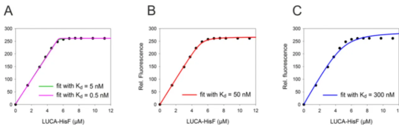
Figure 8: Comparison of data of tight binding LUCA-HisF:zmHisH fitted with increasing K
d-values. ... 58
Figure 9: [
1H-
15N] HSQC spectra for the identification of a putative interaction site for the binding of the peptide ligand to tmHisF. ... 69
Figure 10: Total chemical shift changes (Δδ) of tmHisF induced by the peptide ligand. ... 70
Figure 11: Putative binding site of the peptide ligand on tmHisF as determined in [
1H-
15N] HSQC titration experiments. ... 71
Figure 12: Fluorescence titration experiments to determine dissociation constants (K
d) for the interaction of present-day HisF with HisH proteins. ... 73
Figure 13: Detection of glutaminase HisH reaction in a NAD
+coupled assay. ... 74
Figure 14: SDS-PAGE (12.5% polyacrylamide) for the analysis of the purity of LUCA-HisH. ... 77
Figure 15: Structural integrity of LUCA-HisH. ... 78
Figure 16: Thermal denaturation of LUCA-HisH. ... 79
Figure 17: Fluorescence titration experiment to determine the dissociation constant (K
d) for the interaction of LUCA-HisF with LUCA-HisH. ... 80
Figure 18: SDS-PAGE (12.5% polyacrylamide) for the analysis of the purity of LUCA-HisH mutants. ... 82
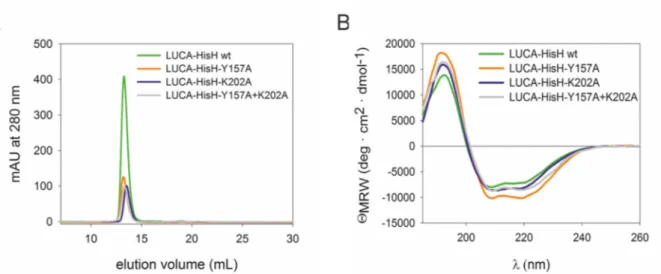
Figure 19: Structural integrity of LUCA-HisH-Y157A, LUCA-HisH-K202A and LUCA-HisH- Y157A+K202A in comparison with LUCA-HisH wt. ... 82
Figure 20: Thermal denaturation of LUCA-HisH mutants in comparison with wild type. ... 83
Figure 21: Fluorescence titration experiments to determine dissociation constants (K
d) for the interaction of LUCA-HisF with LUCA-HisH mutants. ... 84
Figure 22: HPLC assay for the determination of the basal glutaminase activity of LUCA-HisH wt. ... 87
Figure 23: Phylogenetic tree of HisF and HisH. ... 90
Figure 24: Fluorescence titration experiments to determine dissociation constants (K
d) for the interaction of LUCA-HisF with modern HisH proteins. ... 91
Figure 25: SDS-PAGE (12.5% polyacrylamide) for the analysis of the purity of HisF and HisH proteins used in interaction studies. ... 94
Figure 26: 3D model of the LUCA-HisF:zmHisH complex. ... 95
Figure 27: Identification of interface residues determining the affinity of LUCA-HisF and Anc1pa-HisF for zmHisH by means of in silico design. ... 96
Figure 28: Fluorescence titration experiments to determine dissociation constants (K
d) for the interaction of
various HisF proteins with zmHisH. ... 98
Figure 29: Stepwise identification of a HisF hot spot for binding to zmHisH. ... 99
Figure 30: Fluorescence titration experiments to determine dissociation constants (K
d) for the interaction of Anc1pa-HisF or LUCA-HisF-F74S with zmHisH mutants. ... 102
Figure 31: Dynamic cross correlation analysis (DCC) of HisF:tmHisH complexes with bound PRFAR. ... 106
Figure 32: SDS-PAGE (12.5% polyacrylamide) for the analysis of the purity of Anc2tm-HisF_A2. ... 107
Figure 33: Crystallization of LUCA-HisF wt:tmH. Crystals were obtained in in 0.1 M sodium citrate pH 5.0 and 15% PEG 4000. ... 108
Figure 34: Superposition of tmHisF:tmHisH with bound glutamine (pdb: 3ZR4, cahin AB; List et al. 2012, cell) and LUCA-HisF:tmHisH with bound glutamine. ... 109
Figure 35: Proximity of position 74 to putatively allosterically important motifs in HisF. ... 112
Figure 36: Phylogenetic tree used for reconstruction of ancestral HisF sequences after optimization with FastML. ... 125
Figure 37: Analytical size exclusion chromatography of HisF proteins used for fluorescence titration with zmHisH. ... 131
Figure 38: Far-UV CD spectra of HisF used for fluorescence titration with zmHisH. ... 132
Figure 39: Structural integrity of zmHisH-A28R, zmHisH-L202R and zmHisH-A28R+L202R in comparison with zmHisH wt. ... 133
Figure 40: Ammonia-dependent cyclase activity. ... 135
Figure 41: Glutamine-dependent cyclase activity. ... 137
Figure 42: Glutaminase activity. ... 139
Figure 43: Structural integrity of Anc2tm-HisF_A2. ... 142
Figure 44: Calibration of analytic Superdex 75 in 50 mM Tris/HCl pH 7.5, 300 mM KCl for determination of MW
appof LUCA-HisH and LUCA-HisH mutants. ... 143
Figure 45: Calibration of analytic Superdex 75 in 50 mM Tris/HCl pH 7.5, 300 mM KCl. ... 144
List of tables VII
List of tables
Table 1: Plasmids used in this thesis. ... 29
Table 2: Sequencing primers used in this thesis. ... 31
Table 3: Amplification and mutagenic primers for hisF used in this thesis. ... 31
Table 4: Amplification and mutagenic primers for hisH used in this thesis. ... 33
Table 5: Protocol for protein purification with HisTrap column. ... 49
Table 6: Protocol for protein purification with Mono Q column. ... 50
Table 7: Protocol for protein purification with Resource S column. ... 51
Table 8: Composition of 12.5% SDS-PAGE gel. ... 55
Table 9: Protocol for qualitative HPLC assay. ... 62
Table 10: Dissociation constants (K
d) for the interaction of various present-day HisF proteins with HisH proteins. ... 74
Table 11: Apparent molecular weights (MW
app) of LUCA-HisH mutants and wild type (wt) determined via size exclusion chromatography on an analytical Superdex 75 column. ... 83
Table 12: Dissociation constants (K
d) for the interaction of various HisF with HisH proteins. ... 92
Table 13: Dissociation constants (K
d) for the interaction of various HisF proteins with zmHisH. ... 99
Table 14: Amino acid distribution at coevolving interface positions in different HisF and HisH subunits. ... 101
Table 15: Overview of allosteric activation in chimeric HisF:HisH complexes. ... 104
Table 16: Kinetic parameters of the ammonia-dependent cyclase activity of isolated HisF proteins. ... 136
Table 17: Kinetic parameters of the glutamine-dependent cyclase activity of various HisF:HisH complexes. ... 138
Table 18: Kinetic parameters of the glutaminase activity of various HisF:HisH complexes. ... 140
Table 19: Proteins used for calibration of analytical Superdex 75. ... 143
Table 20: Proteins used for calibration of analytical Superdex 75. ... 144
Table 21: Crystal structure determination for glutamine bound LUCA-HisF wt:tmHisH. ... 145
Table 22: Crystal structure determination for LUCA-HisF wt:tmHisH. ... 146
Formula index
Equation 1: Calculation of Gibbs free energy. ... 7
Equation 2: Determination of DNA concentration. ... 41
Equation 3: Calculation of the melting temperature of oligonucleotides. ... 43
Equation 4: Calculation of the optimum annealing temperature of a primer. ... 43
Equation 5: Determination of the molar extinction coefficient ε
280. ... 54
Equation 6: Determination of the specific extinction coefficient
0.1%A
280. ... 54
Equation 7: Determination of the protein concentration by using the specific extinction coefficient
0.1%A
280. . 55
Equation 8: Calculation of mean molar ellipticity per amino acid. ... 57
Equation 9: Quadratic function for K
ddetermination. ... 57
List of acronyms and abbreviations IX
List of acronyms and abbreviations
0.1%
A
xspecific extinction coefficient at x nm
A absorbance, adenosine
Å Ångström (10
-10m)
Ac acetate
Ala alanine
APS ammonium persulfate
Arg arginine
ASP ammonium sulfate precipitation
ASR ancestral sequence reconstruction
ASU asymmetrical unit
ATP adenosine triphosphate
bp base pair
c concentration
C cytosine
°C degree Celsius
cAMP cyclic adenosine monophosphate
CAP catabolite activator protein
CD circular dichroism
cm centimeter (1·10
-2m)
C-terminal carboxy-terminal end of a polypeptide chain
CV column volume
d pathlength [cm]
Da dalton [g/mol]
DCC dynamic cross correlation
DMSO dimethyl sulfoxide
DNA deoxyribonucleic acid
dNTP deoxyribonucleotide triphosphate (N = A, C, G or T)
dsDNA double-stranded DNA
DSS 4,4-dimethyl-4-silapentane-1-sulfonic acid
DTE 1,4-dithioerythritol
DTT 1,4-dithithreitol
E enzyme
E. coli Escherichia coli et al. and other authors (et alii)
EtBr ethidium bromide
EtOH ethanol
f dilution factor
g gram
G guanosine, Gibbs free energy
GDH glutamate dehydrogenase
GREMLIN Generative Regularized ModeLs of proteINs
H enthalpy
h hour
HisA ProFAR isomerase
HisF cyclase subunit of ImGPS
HisH glutaminase subunit of ImGPS
HisF
ext+ HisH
extMSA of concatenated HisF and HisH sequences HisF:HisH imidazole glycerol phosphate synthase complex
(His)
6-tag hexahistidine-tag
HPLC high pressure liquid chromatography
IDA iminodiactetic acid
ImGPS imidazole glycerol phosphate synthase
indels insertions and deletions
INEPT insensitive nuclei enhanced polarization transfer
IPTG isopropyl-ß-D-thiogalactopyranoside
kb kilobase pair
k
catturnover number
k
cat/K
Mcatalytic efficiency parameter
kDa kilodalton (1·10
3g/mol)
K
ddissociation constant for protein-protein interaction K
idissociation constant for an enzyme-inhibitor complex K
MMichaelis-Menten constant, equivalent to the substrate
L ligand
lacZ gene coding for the enzyme β-galactosidase
LB Luria-Bertani (-medium)
Leu leucine
List of acronyms and abbreviations XI
LUCA Last universal common ancestor
µ micro (1·10
-6)
m milli (1·10
-3);
M molar [mol/l]
MCS multiple cloning site
MD molecular dynamics
mg milligram
min minute
mL milliliter
ML maximum likelihood
mm millimeter
mM millimolar
MPa megapascal
mpa most probable ancestor
µs microsecond
ms millisecond
MSA multiple sequence alignment
MW molecular weight
MWCO molecular weight cut off
n nano (1·10
-9); number of nucleotides
NAD
+nicotinamide adenine dinucleotide (oxidized form)
NADH nicotinamide adenine dinucleotide (reduced form)
nm nanometer
NMR nuclear magnetic resonance
ns nanosecond
N-terminal amino-terminal end of a polypeptide chain
OE-PCR overlap extension PCR
P pellet (insoluble cell fraction)
p pico (1·10
-12)
P. arsenaticum (pa) Pyrobaculum arsenaticum
PAGE polyacrylamide gel electrophoresis
PCR polymerase chain reaction
PDB protein data bank
PEG polyethylene glycol
pH negative decadic logarithm of the proton concentration
Phe phenylalanine
PRFAR N´-[(5´-phosphoribulosyl)formimino]-5-
aminoimidazole-4-carboxamide-ribonucleotide
ProFAR N´-[(5´-phosphoribosyl)formimino]-5-
aminoimidazole-4-carboxamide-ribonucleotide
PRPP phosphoribosyl pyrophosphate
ps picosecond
QCM QuikChange mutagenesis
r.m.s.d. root mean square deviation
rbs ribosome binding site
rpm revolutions per minute
RT room temperature
s second
S supernatant (soluble cell fraction); substrate
concentration, entropy
S. cerevisiae Saccharomyces cerevisiae
Ser serine
SDS sodium dodecyl sulfate
ssDNA single-stranded DNA
T temperature; thymidine
T. maritima (tm) Thermotoga maritima
T
Aannealing temperature
TBE Tris-Borat-EDTA buffer
TEMED N,N,N’,N’-tetramethylethylenediamine
T
Mmelting temperature of primers; temperature at which 50% of the protein is in a non-native state
Trp tryptophan
Tyr tyrosin
U Unit, 1U is equivalent to the amount of enzyme that
converts 1 μmol substrate per minute at standard conditions
UV ultraviolet
V volt
List of acronyms and abbreviations XIII
v
iinitial velocity
v
maxmaximum velocity
Z. mobilis (zm) Zymomonas mobilis
Abstract 1
Abstract
Imidazole glycerol phosphate synthase (ImGPS) is a bi-enzyme complex that consists of the glutaminase subunit HisH and the cyclase subunit HisF. HisH hydrolyzes glutamine to glutamate and ammonia, which is transported through a channel to the active site of HisF where it reacts with N´-[(5´-phosphoribulosyl)formimino]-5-aminoimidazole-4- carboxamide-ribonucleotide (PRFAR) to imidazole glycerol phosphate (ImGP) and 5- aminoimidazole-4-carboxamide ribotide (AICAR). ImGP and AICAR are further used in histidine and de novo purine biosynthesis, rendering ImGPS a key metabolic enzyme. The sequential HisH and HisF reactions are tightly coupled: glutaminase HisH activity is allosterically induced by the binding of PRFAR to the active site of HisF. The structural bases for complex formation between HisH and HisF and for the coupling of their catalytic activities are poorly understood. Thus, HisF:HisH is a paradigm for the study of protein- protein interactions and allosteric regulation. Moreover, only plants, fungi, bacteria, and archaea are able to synthesize histidine. Thus, the inhibiton of ImGPS might be a potential therapeutic strategy to fight pathogenic microorganisms. In this context, recently, a peptide was identified that impedes the glutaminase activity in ImGPS from Thermotoga maritima (tm). It has been hypothesized that the peptide inhibits the catalytic activity of HisF:HisH by binding to tmHisF in the complex interface, however, the exact HisF:peptide interaction sites and the mode of inhibition remained elusive.
Within the first part of this thesis, nuclear magnetic resonance (NMR) titration experiments demonstrated that the inhibitory peptide mainly interacts with structural elements and residues around positions 71-77 and 90-99 in HisF. Parts of this set of residues belong to the HisF:HisH interface and are thought to be involved in allosteric signal transduction, based on previous NMR and molecular dynamics data. This suggests that the peptide inhibits glutaminase activity by perturbing the interaction and allosteric communication between the HisF and HisH subunits.
Within the second part of this thesis, residues of HisF that are crucial for its structural and functional interaction with HisH should be identified. For this purpose, it was planned to analyze the interaction of various combinations of HisF and HisH enzymes.
However, these experiments could not be performed due to the insolubility of most of the
tested proteins. In order to produce proteins that can be characterized, the primordial HisF
and HisH enzymes from the last universal common ancestor (LUCA) were resurrected by
ancestral sequence reconstruction (ASR). LUCA-HisF and LUCA-HisH formed a high- affinity complex; however, LUCA-HisH was catalytically inactive, probably due to inaccuracies of ASR. In contrast, LUCA-HisF was catalytically active and could be used for further analysis, which was performed as follows: Initial experiments showed that HisH from Zymomonas mobilis (zmHisH) tightly binds to LUCA-HisF but not to the present-day HisF from Pyrobaculum arsenaticum (paHisF), which are separated by 103 residues.
Following the characterization of a reconstructed evolutionary intermediate linking LUCA- HisF and paHisF and the inspection of the ImGPS interface, the number of candidate HisF residues crucial for binding to zmHisH could be narrowed to nine. Subsequent in silico mutagenesis based on homology modeling indicated that a single phenylalanine at position 74 in HisF was most important for binding to zmHisH. The decisive role of this “hot spot”
residue for complex formation between HisF and zmHisH was confirmed by extensive experimental site-directed mutagenesis. Subsequently, primordial HisF proteins were also utilized to disentangle mechanistic principles of allosteric communication with HisH. In this context, no glutaminase activity was observed for tmHisH when bound to LUCA-HisF with PRFAR. However, the crystal structure of LUCA-HisF:tmHisH with bound glutamine revealed no significant differences compared to the catalytically active tmHisF:tmHisH complex. LUCA-HisF and tmHisF are separated by 79 residues. Although the number of potentially important residue differences could be reduced to 69 with the help of a primordial enzyme that links LUCA-HisF and tmHisF and by means of computational analysis, residues that are decisive for allostery could not be identified by this approach.
Taken together, the results of this thesis show that peptides interrupting allosteric
inter-subunit communication and molecular fossils being resurrected by ASR can
contribute to unraveling the structure-function relationship of multi-enzyme complexes
such as ImGPS.
Zusammenfassung 3
Zusammenfassung
Die Imidazolglycerolphosphat-Synthase (ImGPS) ist ein Bienzymkomplex, der aus der Glutaminase-Untereinheit HisH und der Zyklase-Untereinheit HisF besteht. HisH hydrolisiert Glutamin zu Glutamat und Ammoniak, das durch einen intermolekularen Kanal zum aktiven Zentrum von HisF diffundiert und dort mit [(5‘Phosphoribulosyl)formimino]- 5-aminoimidazol-4-carboxamid-Ribonukleotid (PRFAR) zu Imidazolglycerolphosphat (ImGP) und 5‘-Aminoimidazol-4-carboxamid-Ribonucleotid reagiert. ImGP und AICAR fließen in die Histidin- und de novo Purinbiosynthese, was die ImGPS zu einem Schlüsselenzym des Metabolismus macht. Die Reaktionen, die von HisH und HisF katalysiert werden, sind eng aneinander gekoppelt: Die Glutaminaseaktivität von HisH wird durch die Bindung von PRFAR an das aktive Zentrum von HisF allosterisch induziert. Die strukturellen Grundlagen der HisF:HisH Interaktion und der Kopplung ihrer enzymatischen Aktivitäten ist bisher noch weitestgehend ungeklärt. Deshalb stellt der HisF:HisH Komplex ein Musterbeispiel zur Untersuchung von Protein-Protein Interaktionen und allosterischer Regulation dar. Außerdem sind nur Pflanzen, Pilze, Bakterien und Archaeen in der Lage Histidin herzustellen. Deshalb stellt die Inhibition der ImGPS eine potentielle Strategie zur Bekämpfung pathogener Mikroorganismen dar. In diesem Zusammmenhang wurde vor Kurzem ein Peptid identifiziert, das die Glutaminaseaktivität der ImGPS von Thermotoga maritima (tm) hemmt. Es wurde angenommen, dass das Peptid die katalytische Aktivität inhibiert, indem es in der Kontaktfläche der tmImGPS an die HisF Untereinheit bindet, jedoch blieb unklar wo genau das Peptid an HisF bindet und wodurch die Inhibition speziell zustande kommt.
Im ersten Teil dieser Arbeit, konnte mit Hilfe von Kernspinresonanzspektroskopie (nuclear magnetic resonance, NMR) gezeigt werden, dass das inhibitorische Peptid vor allem mit Strukturelementen und Aminosäuren im Bereich der Positionen 71-77 und 90-99 in der HisF-Untereinheit interagiert. Einige dieser Reste befinden sich in der HisF:HisH- Interaktionsfläche und aufgrund vorheriger Studien, die auf NMR und Moleküldynamik beruhen, wird angenommen, dass diese an der Weiterleitung des allosterischen Signals beteiligt sind. Dies legt nahe, dass das Peptid die Glutaminaseaktivität hemmt, indem es die Interaktion und allosterische Kommunikation zwischen HisF und HisH stört.
Im zweiten Teil der Arbeit sollten Reste von HisF, die sowohl für die strukturelle
als auch für die funktionelle Interaktion von Bedeutung sind, identifiziert werden. Zu
diesem Zweck sollte die Interaktion verschiedenster Kombinationen von HisF- und HisH- Enzymen untersucht werden. Da jedoch die meisten der ausgewählten Proteine nicht löslich exprimierbar waren, konnten diese Experimente nicht durchgeführt werden. Um Proteine herzustellen, die charakterisiert werden können, wurden HisF und HisH aus dem letzten gemeinsamen Vorläufer der zellulären Organismen (last universal common ancestor, LUCA), mittels Sequenzrekonstruktion „auferweckt“. LUCA-HisF und LUCA-HisH bildeten zwar einen hoch affinen Komplex aus, jedoch war LUCA-HisH katalytisch inaktiv, was möglicherweise auf Ungenauigkeiten bei der Sequenzrekonstruktion zurückzuführen ist. Im Gegensatz dazu war LUCA-HisF katalytisch aktiv und konnte für weitere Analysen herangezogen werden. Diese erfolgten folgendermaßen: Erste Untersuchungen zeigten, dass die HisH-Untereinheit aus Zymomonas mobilis (zmHisH) stark an LUCA-HisF, jedoch nicht an das rezente HisF aus Pyrobaculum arsenaticum (paHisF) bindet. Beide HisF- Proteine unterscheiden sich an 103 Positionen. Die Reste in HisF, die für das unterschiedliche Bindeverhalten an zmHisH verantwortlich sind, konnten mit Hilfe der Charakterisierung eines HisF-Vorläuferproteins, das auf dem phylogenetischen Pfad zwischen LUCA-HisF und paHisF liegt, und mit Hilfe der Analyse der ImGPS Kontaktfläche, auf neun Aminosäuren eingeschränkt werden. In anschließenden in silico Mutagenesestudien, basierend auf Homologiemodellen, zeigte sich, dass vor allem ein einzelnes Phenylalanin an Position 74 in HisF entscheidend für die Bindung an zmHisH ist.
Die herausragende Bedeutung dieses „hot spots“ für die Komplexbildung von HisF und zmHisH wurde in einer umfassenden gerichteten Mutagenese experimentell bestätigt.
Rekonstruierte HisF-Proteine wurden anschließend ebenfalls verwendet um den Mechanismus der allosterischen Kommunikation mit HisH näher zu beleuchten. In diesem Zusammenhang konnte in Aktivitätsmessungen keine Glutaminaseaktivität für tmHisH, das mit LUCA-HisF mit gebundenem PRFAR interagiert, beobachtet werden. Die Kristallstruktur des LUCA-HisF:tmHisH Komplexes mit gebundenem Glutamin wies jedoch keine signifikanten Unterschiede zum katalytisch aktiven tmHisF:tmHisH Komplex auf. LUCA-HisF und tmHisF unterscheiden sich an 79 Positionen. Obwohl mit der Hilfe eines rekonstruierten HisF-Vorläuferenzyms, das auf dem phylogenetischen Pfad zwischen LUCA-HisF und tmHisF liegt und bioinformatischer Analyse die potentiell bedeutenden Unterschiede auf 69 Reste reduziert werden konnten, konnten keine Reste identifiziert werden, die eine wichtige Rolle für die Allosterie spielen.
Zusammengefasst zeigen die Ergebnisse dieser Arbeit, dass Peptide, die die
Kommunikation zwischen Komplexuntereinheiten stören, sowie molekulare Fossilien, die
Zusammenfassung 5
durch Sequenzrekonstruktion „zum Leben erweckt“ wurden hilfreich sein können bei der
Untersuchung von Struktur-Funktions-Beziehungen von Multienzymkomplexen wie der
ImGPS.
1 Introduction
1.1 Protein-protein interactions
Nowadays, it is generally accepted that the complexity of an organism is not defined by the total number of its genes but by the number of interactions between its cellular compounds.
For example, while for Saccharomyces cerevisiae about 18000-30000 binary interactions were determined, for humans about 600000 were estimated (Merkl, 2015). Among these interactions, protein-protein interactions are of great importance as they are involved in many biological processes such as metabolic pathways and signal transduction cascades.
This makes clear how important the understanding of protein-protein interactions and complexes is in order to understand biological systems in general.
Protein complexes can be grouped on the basis of different features. They can be categorized into homo-oligomers and hetero-oligomers according to the polypeptide chains that form the subunits. In a homo-oligomer the polypeptide chains are identical, while the polypeptide chains in a hetero-oligomer are different (Zhang et al., 2013). Apart from that, complexes can be distinguished according to the duration of protein-protein interaction. In permanent complexes interactions are tight and stable once the complex has formed, whereas in transient complexes interactions can be broken by external influences. The first category involves, for example, antigen-antibody complexes, for which a permanent interaction is of great biological importance. The second category comprises proteins that regulate signaling pathways and that are required to bind to their partners only at a certain time (La et al., 2013). The assembly of proteins to oligomers has various biological functions. For example, it is often observed that oligomerization increases thermal stability (Jaenicke & Böhm, 1998; Sterner & Liebl, 2001; Vieille & Zeikus, 2001; Walden et al., 2001; Schwab et al., 2008). Moreover, the assembly of proteins to complexes enables the transport of highly reactive and volatile substances under separation from the environment between two or more active sites that are located at different subunits (Miles et al., 1999;
Huang et al., 2001; Raushel et al., 2003). Finally, oligomerization allows for regulative coupling of enzymatic functionalities via an allosteric mechanism (Perica et al., 2012).
The stability of a complex is determined as the difference in Gibbs free energy G
between the isolated monomers and the monomer assembly. This difference ΔG at the
temperature T is defined as follows (Equation 1):
Introduction 7
∆ ∆ ∆
Equation 1: Calculation of Gibbs free energy.
The free energy has two components: ΔH and ΔS. ΔH describes the change in enthalpy that is linked to the change in intermolecular, non-covalent interactions during complexation such as Van der Waals interactions, hydrogen bonds or Coulomb interactions. ΔS is the entropy term that either contributes to a destabilization during complex formation through desolvatation of polar groups at the protein surface or results in a stabilization in the form of an increase of the hydrophobic effect (Hilser et al., 1996; Chandler, 2005). In general, the hydrophobic effect makes the largest contribution to complex stability (Chothia &
Janin, 1975; Young et al., 1994). Complex formation and stabilization results in a ΔG value
< 0. Destabilization of the complex is accompanied by an increase in ΔG.
Most often, protein-protein interfaces (PPIs) exhibit geometric and electrostatic complementarity and are densely packed. Although they are commonly large and expand from 700-1500 Å
2(Reichmann et al., 2007), not all residues in the interface contribute equally to binding affinity. In fact, complex stability critically depends on few residues in the interface, so called hot spots (Clackson & Wells, 1995). The replacement of such hot spots by alanines significantly destabilizes the free energy of complex formation (ΔΔG > 2 kcal /mol) (Bogan & Thorn, 1998). Hot spots are commonly located in the center of the interface, are enriched in Trp, Arg, and Tyr and often show a higher level of conservation compared to other interface residues (Zhang et al., 2013).
The identification of hot spots is essential for the mechanistic understanding of protein-protein interactions. For example, abnormal protein-protein interactions are often associated with diseases like cancer. Thus, for medicinal science, protein-protein interfaces are attractive drug targets. As the restoration of protein-protein interactions is often difficult to realize (Fry & Vassilev, 2005), the disruption of abnormal protein-protein interactions with the help of small molecules has achieved much success during the last years (Watanabe & Osada, 2016). As protein-protein interfaces are large in size and rather flat, the identification of small molecules that tightly bind to the interface is often difficult.
However, the knowledge of interface hot spots allows for the selective inhibition of key
interactions in the interfaces (Fry & Vassilev, 2005). For example, an interesting target for
cancer therapy is the interaction face of the complex of the tumor suppressor p53 and the
MDM2 protein. The assembly of both proteins results in an inhibition of the transcription
activation of p53 and promotes p53 degregation. As MDM2 is overexpressed in many
tumor cells, the inhibition of p53-MDM2 complex formation is a potential therapeutic strategy. In this context, small organic molecules (nutlins) were identified that effectively mimic specific hydrophobic key interactions that are essential for p53-MDM2 binding (Vassilev et al., 2004; Fry & Vassilev, 2005). Moreover, protein-protein interactions have been blocked with the help of peptides that were directly derived from epitopes of the corresponding interaction face. This strategy was, for example, successfully applied to the herpesvirus ribonucleotide reductase (Cohen et al., 1986).
Various methods can be used for the systematic identification and analysis of protein-protein interactions. For example, PPIs can be identified by means of crystal structure analysis. However, PPIs deduced from crystal packing contacts may be artifacts and the interactions may not be of biological relevance (Janin & Rodier, 1995). Alanine scanning mutagenesis, which is the replacement of particular residues by alanines, is a common way to experimentally test the energetic contribution of interface residues to complex stability. This approach, however, is often laborious and expensive (Moreira et al., 2007). Thus, various computer algorithms have been developed that predict PPIs, identify hot spots or asses the energetic contribution of particular residues to protein-protein interaction by in silico mutagenesis (Moreira et al., 2007; Aumentado-Armstrong et al., 2015). These algorithms are based on empirical functions or force fields. The KFC2 server (Zhu & Mitchell, 2011), for example, identifies hot spots by means of geometric and biochemical features. Another example is FoldX, which is a force field that predicts the effect of mutations on the stability of proteins with known 3D structure. Moreover, this program was successfully applied to calculate ΔΔG-values, which are a measure for the effect of mutations on complex stability.
Another computational method which allows for the analysis of PPI is residue
coevolution. Coevolution describes the evolutionary interdependency of two or more
positions in protein chains. This interdependency implies that a mutation at one position in
the protein results in selective pressure on another position leading to a compensatory
amino acid change and vice versa (Lovell & Robertson, 2010). Intermolecular coevolution
is a crucial phenomenon in the evolution of protein complex interfaces as it guarantees the
maintenance of the biological functionality of the complex by mutual adaptation of the
subunit interfaces (Zhang et al., 2013). Along these lines, Aakre and co-workers (2015)
successfully utilized GREMLIN (Generative Regularized ModeLs of proteINs)
(Balakrishnan et al., 2011) for the identification of interaction hot spots in the toxin-
antitoxin system. GREMLIN is a method that learns a global statistical model of the amino
Introduction 9 acid compositions in an MSA. The model accounts for conservation and correlated mutation statistics between sequential and long-range pairs of residues, and thus allows for the determination of residue contacts in a complex interface.
1.2 Allostery
Often, the binding of a ligand to a certain site in a protein induces functional, structural and/or dynamic modifications at another site, which can be even 20 to 30 Å apart from the initial binding site. This regulatory mechanism has been named allostery (Monod et al., 1963) and the act of conveying these modifications between the two binding sites has been denoted as allosteric communication. More general, allostery can also be induced by other factors such as mutations or light (Nussinov et al., 2013). Two types of allostery can be distinguished depending on the parameter that is modified during the allosteric mechanism.
While, in K-type allostery the binding of substrate to the active site is affected, V-type allostery describes an alteration of reaction velocity (Manley et al., 2013).
The view on the mechanistic principles of this phenomenon has changed during the past 50 years (Motlagh et al., 2014) and is still under debate (Nussinov & Tsai, 2015). In the infancies of allosteric studies, allosteric regulation has been described for oligomeric proteins. The binding of a ligand, often denoted as effector, to the oligomer resulted in a shift from one discrete conformational state into another (Monod et al., 1965) or in a sequential conformational rearrangement (Koshland et al., 1966). These models allow, for example, the description of principles of cooperativity in hemoglobin (Perutz et al., 1998).
Improvement of experimental technologies and the emergence of data from nuclear magnetic resonance (NMR) spectroscopy and computational methods like molecular dynamics (MD) simulations have attracted notice to intrinsic protein flexibility and thus the notion of allostery has been revised. The new view assumes that proteins populate a continuous conformational and dynamic ensemble. The effector may bind to more than one state and the binding results in a shift of the population distribution via multiple conformational and/or dynamic pathways throughout the protein (Goodey & Benkovic, 2008; del Sol et al., 2009; Perica et al., 2012; Motlagh et al., 2014). These structural pathways that energetically couple two binding sites are often denoted as allosteric pathways (Perica et al., 2012). The new view implies that allostery must not be necessarily accompanied by conformational changes but can only be of dynamic nature (Cooper &
Dryden, 1984). This was observed for the catabolite activator protein (CAP). CAP is a
transcriptional activator which forms a homodimer in solution. Each subunit comprises a cAMP binding site. Two cAMP molecules bind with negative cooperativity to CAP. The binding of cAMP results in an enhanced affinity of CAP to DNA. Studies on changes in CAP structure and dynamics, based on NMR and ITC measurements, revealed that the binding of the first cAMP does not affect the conformation at the second cAMP binding site. Instead, negative cooperativity is solely driven by changes in protein motions between the two binding sites (Popovych et al., 2006). This dynamically mediated allostery is, however, still subject of discussion (Nussinov & Tsai, 2015). Interestingly, many allosterically controlled systems are protein assemblies. Actually, oligomerization allows for the communication between subunits via additional, novel concerted motions, thus enabling highly sophisticated regulation (Perica et al., 2012).
According to the current view, intra- and inter-molecular allosteric communication is based on various protein motions and conformational changes from atomic fluctuations and loop motions in the picosecond (ps) to nanosecond (ns) range, domain motions in the ns to microsecond (µs) range and larger conformational rearrangements to which a whole network of amino acids contributes and that occur in the µs to millisecond (ms) range (Goodey & Benkovic, 2008; Manley et al., 2013).
Analogous to protein-protein interactions, allostery can be modulated with small molecules. Thus, the allosteric mechanism of an enzyme is, beside PPIs, a further target for therapeutic drugs (Nussinov & Tsai, 2013). Allosteric drugs bind to a site at the enzyme that is distal to the active site. Therefore, the use of allosteric drugs in therapy can have advantages in contrast to drugs that target the active site: For example, they are often more specific due to the fact that allosteric sites generally exhibit a lower degree of conservation than active sites. Moreover, allosteric drugs allow for an optimal regulation of the pharmacological effect. As they do not compete with the substrate the enzyme cannot only be simply switched on and off by it, but can variously be modified (Conn et al., 2009;
Nussinov et al., 2011).
An example of eligible targets for allosteric drugs are G-protein coupled receptors (Conn et al., 2009), as they are allosterically regulated and often involved in a multitude of human diseases. In this context, allosteric drugs have been used in the therapy of AIDS:
Maraviroc (Selzentry), for example, is an allosteric modulator of the chemokine receptor
CCR 5, which is used by HI-virus to enter and infect host cells (He et al., 1997). Maraviroc
stabilizes a conformation of the CCR5 receptor that has a lower affinity to the virus and
thus prevents its entry into the cell (Conn et al., 2009).
Introduction 11 Other examples demonstrated that the design of allosteric modulators can also fail. For example, allosteric drugs that were designed for the suppression of proteinkinase C, an essential regulatory enzyme in cell signaling pathways, rather led to an activation (Nussinov et al., 2011). This makes clear that a detailed mechanistic understanding of an allosteric mechanism is needed in order to develop effective allosteric modulators.
Various approaches help to strengthen this understanding: Mutational studies led to the identification of residues that are involved in allosteric pathways. For example, double mutant cycles are an experimental approach to determine the energetic coupling of two distal residues and thus to estimate the degree to which they are functionally coupled (Horovitz & Fersht, 1990; Sadovsky & Yifrach, 2007). Furthermore, X-ray crystallography was successful in capturing structural rearrangements upon ligand binding in the case of hemoglobin (Perutz et al., 1998) or ferric citrate membrane transporter FecA (Ferguson &
Deisenhofer, 2004). Moreover, a detailed understanding of allosteric signal transduction with fast time scales requires sophisticated methods that allow for the observation of motions and dynamic fluctuations at atomic scales such as NMR spectroscopy and MD simulations. While NMR techniques cover all time scales that play a role in allosteric mechanisms, transitions on the ps to ns range can be studied with standard MD simulations.
In particular, analyses of correlated motions in MD simulations allow for the identification of allosteric transitions of that kind (Manley et al., 2013).
1.3 The imidazole glycerol phosphate synthase
The imidazole glycerol phosphate synthase (ImGPS) belongs to the family of glutamine amido-transferases (GATases), which are the most widespread enzymatic ammonia donors in the biosynthesis of nucleotides, amino acids and coenzymes (Zalkin & Smith, 1998).
GATases consist of glutaminase and synthase domains. The glutaminase hydrolyzes
glutamine to glutamate and ammonia, which is added to a specific substrate by the synthase
subunit. While synthase subunits markedly differ in their topology and catalytic
mechanisms, GATases can be divided into two unrelated classes according to the active site
residues of the glutaminase subunit (Massiere & Badet-Denisot, 1998; Zalkin & Smith,
1998). Class I glutaminases adopt an α/β-hydrolase fold and are characterized by a catalytic
triad in their active site (Ollis et al., 1992). Class II glutaminases are N-terminal nucleophile
(Ntn)-type GATases, which have an N-terminal catalytic cysteine (Brannigan et al., 1995).
Figure 1: Structure and reaction of the ImGP synthase from Thermotoga maritima (HisF:HisH complex).
The catalytic residues in the active site of HisF and HisH are depicted as spheres. The conformational transition that is induced by binding of PRFAR to the active site of HisF is illustrated as a red arrow. The blue arrow indicates the way of nascent ammonia through the tunnel in HisF. Details of the catalytic mechanism are outlined in the text. The crystal structure is taken from the pdb (1gpw) (Douangamath et al., 2002).
ImGPS is a class I GAT that consists of the glutaminase domain HisH and the synthase domain HisF (Klem & Davisson, 1993) (Figure 1). In plants and fungi, the glutaminase and synthase active sites are located on a single polypeptide chain (Chittur et al., 2000), whereas in bacteria and archaea HisF and HisH are two separate subunits.
ImGPS is, however, not present in mammals (Chaudhuri et al., 2001) . Structures of the ImGPS complex or of isolated HisF from Saccharomyces cerevisiae (Chaudhuri et al., 2001; Chaudhuri et al., 2003), Pyrobaculum aerophilum (Banfield et al., 2001), Thermus thermophilus (Omi et al., 2002) and Thermotoga maritima (Douangamath et al., 2002;
Korolev et al., 2002; List et al., 2012) show that HisF adopts a (βα)
8-barrel fold, which consists of eight parallel β-sheets, which are surrounded by eight α-helices thus forming a tunnel along the central barrel axis. The (βα)
8-barrel belongs to the most ancient observed folds (Caetano-Anolles et al., 2007). The C-terminal face of the barrel bears the cyclase catalytic site and the N-terminal face forms the complex interface with HisH.
ImGPS from T. maritima (tmHisF:tmHisH or tmImGPS) is a hetero-obligomer, which is an obligatory permanently interacting bi-enzyme complex (Ofran & Rost, 2003).
In tmHisF:tmHisH the interface has a dimension of 1100 Å
2and is characterized by
electrostatic and shape complementarity, ensuring a tight docking of the subunits to each
other (Douangamath et al., 2002; Amaro et al., 2005).
Introduction 13 The active site of HisH (catalytic triad Cys84, His178, Glu180, numbering according to tmImGPS, Figure 1), which provides ammonia, is located in the interface near the opening of the tunnel in HisF. This opening is framed by four invariant residues that form a salt bridge cluster [Arg5(HisF), Glu46(HisF), Lys99(HisF), Glu167(HisF)]. From here, nascent ammonia is channeled over a distance of 25 Å to the active site of HisF (Figure 1) where it is added to N´-[(5´-phosphoribulosyl)formimino]-5-aminoimidazole-4-carboxamide- ribonucleotide (PRFAR). Thus, ammonia is sequestered from the solvent and not protonated to nonreactive ammonium ions. In a cyclization reaction PRFAR is cleaved into imidazole glycerol phosphate (ImGP) and 5-aminoimidazole-4-carboxamide ribotide (AICAR), which are essential components of histidine and de novo purine biosynthesis (Figure 1) (Beismann-Driemeyer & Sterner, 2001). By linking amino acid and nucleotide metabolism, ImGPS serves as a metabolic key enzyme. Noteworthy, glutaminase activity of HisH is contingent upon the binding of a ligand to the active site of HisF. Therefore, in the absence of HisF and in the presence of HisF without a bound ligand, glutaminase activity of HisH is too low to be measured. This phenomenon has been ascribed to a ligand-induced V-type allosteric mechanism (Beismann-Driemeyer & Sterner, 2001; Myers et al., 2003;
Myers et al., 2005; Amaro et al., 2007; Manley et al., 2013). This tight functional coupling ensures that glutamine is only hydrolyzed when PRFAR is present in the cell, thus preventing an unnecessary waste of glutamine. HisF is able to use ammonia salts as an ammonia source at basic pH values. As a consequence, HisF is active in the absence of HisH (Klem & Davisson, 1993; Beismann-Driemeyer & Sterner, 2001).
In an attempt to shed light on the principles of the allosteric mechanism in ImGPS
computational methods have been combined with NMR experiments, X-ray crystallography
and mutational studies. These studies were predominantly conducted with ImGPS from
yeast, Escherichia coli or T. maritima. Some of the observations that have been made on
the basis of these investigations are outlined in the following. The residue numeration is
according to tmHisF:tmHisH. In Figure 2 (panel A) the most important residues and
structure elements that might be involved in the allosteric mechanism and that are
mentioned here are projected on a schematic model of tmHisF:tmHisH.
Figure 2:


![Figure 9: [ 1 H- 15 N] HSQC spectra for the identification of a putative interaction site for the binding of the peptide ligand to tmHisF](https://thumb-eu.123doks.com/thumbv2/1library_info/4125485.1551618/89.892.142.817.205.935/figure-spectra-identification-putative-interaction-binding-peptide-ligand.webp)
![Figure 11: Putative binding site of the peptide ligand on tmHisF as determined in [ 1 H- 15 N] HSQC titration experiments](https://thumb-eu.123doks.com/thumbv2/1library_info/4125485.1551618/91.892.149.818.168.344/figure-putative-binding-peptide-ligand-determined-titration-experiments.webp)