Dynamic interactions between
PAR-aPKC complex and Crb complex in Drosophila epithelial cells
DISSERTATION ZUR ERLANGUNG DES
DOKTORGRADES DER NATURWISSENSCHAFTEN (DR.RER. NAT.) DER FAKULTÄT FÜR BIOLOGIE UND
VORKLINISCHE MEDIZIN DER UNIVERSITÄT REGENSBURG
vorgelegt von
Rui Sun
aus
Tengzhou, China im Jahr 2017
Das Promotionsgesuch wurde eingereicht am:
Die Arbeit wurde angeleitet von:
Prof. Dr. Dr. Michael P. Krahn
Unterschrift:
Declaration of Publications
1. Arnab Sen #, Rui Sun # and Michael P. Krahn (2015). Localization and Function of Pals1-associated Tight Junction Protein in Drosophila Is Regulated by Two Distinct Apical Complexes. Journal of Biological Chemistry 290(21): 13224-13233. (#: equal contribution, contributed to 45% results)
2. Rui Sun, Arnab Sen, and Michael P. Krahn. PAR-6 prevents degradation of Stardust by inhibiting the proteasomal receptor Rpn13 in Drosophila. (Revision with Development, contributed to 70% results)
3. Leonie Koch, Sabine Feicht, Rui Sun, Arnab Sen and Michael P. Krahn (2016).
Domain-specific functions of Stardust in Drosophila embryonic development. Royal Society Open Science 3(11): 160776. (Contributed to 30% results)
Table of contents
Acknowledgements ... 1
Summary... 2
Introduction ... 3
1.1 Cell polarity models in Drosophila ... 3
1.2 Epithelial cell polarity ... 5
1.3 Epithelial polarity determinants and complexes ... 8
1.3.1 The Crumbs complex... 8
1.3.2 Baz/PAR-3-aPKC complex ... 13
1.4 Dynamic interactions between apical polarity regulators ... 13
1.4.1 Dynamic composition of protein complexes ... 13
1.4.2 Protein modifications ... 15
1.5 Epithelial polarity and signaling pathways ... 17
1.5.1 Wnt signaling and epithelial polarity ... 17
1.5.2 Notch signaling and epithelial polarity ... 17
1.5.3 Hippo pathway and epithelial polarity ... 18
1.5.4 Other pathways related to epithelial polarity ... 19
1.6 Aim of the study ... 20
Chapter 1. Upstream regulators of PATJ ... 21
Chapter 2. PAR-6 stabilizes Sdt via Rpn13 ... 42
Chapter 3. Sdt structure-function analysis ... 61
Discussion... 83
1. PATJ as a component in two distinct apical complexes ... 84
2. PAR-6 regulates the stability of the Crb complex ... 86
References ... 90
Acknowledgements
First of all, I would like to thank my supervisor Prof. Dr. Dr. Michael P. Krahn for offering this fantastic opportunity to do my Ph.D. and his close supervision during the last three years.
I always feel very grateful and pride that I am involved in so many diverse projects which broaden my knowledge and make me generated new research ideas in between.
My gratitude goes to my other mentors, Prof. Dr. Stephan Schneuwly and Prof. Dr. Eugen Kerkhoff, for their insightful inputs to my projects. I am also very grateful to our chair Prof.
Dr. Ralph Witzgall for his comments and discussions during my regular progress reports.
I would like to thank all the past and current members in Krahn lab. Without any of you, I would never manage to go through the last three years. I thank Arnab and Giada for their helps especially during the start of my Ph.D. in and out of the lab. I appreciate the discussions together with Lars and Olga about projects, and fun times with Sabine and Ina. I would also like to mention our excellent technicians, Vroni and Lucy for their great helps in mouse work.
Thank you! Special thanks to Sabine, Olga, Lars, Giada and Ina for sharing their friendships and generous helps in my daily life.
Last but not least, I want to thank my dear parents for their continuous love and supports.
Without these, I could not imagine how I can manage all these things in the last years. I love
you forever. 我还要感谢我的爸爸妈妈:感谢他们无私的爱,感谢他们的抚养和教育,感
谢他们对我学业的默默支持和付出。我爱你们。
Summary
Epithelial cell polarity is one of the key prerequisites for the establishment of multicellular organisms. The apical-basal polarity of epithelia is controlled by evolutionarily conserved complexes which determine an apical versus a basolateral domain. The PAR-aPKC complex (Baz/PAR-3-PAR-6-aPKC) defines together with the Crumbs complex (Crb-Sdt/Pals1-PATJ) the apical plasma membrane domain of the cell. Interestingly, both complexes are highly dynamic during development. The activity and stability of these two complexes are counterbalanced by basolateral protein complexes, mainly the Scribble-Lethal giant larvae-Discs large complex.
In this study, the apical targeting mechanism of PATJ, which has been described to form a complex with Crb-Sdt, was investigated in detail. In the embryonic epidermis of Drosophila, the PATJ-Sdt heterodimer is not initially recruited to the apical cell-cell contacts by binding to Crb but depending on forming a new complex with Bazooka (Baz). The Baz-Sdt-PATJ complex together with Crb-Sdt-PATJ complex co-exist in the mature epithelium of the embryonic epidermis, and might play a redundant role in myosin activity modulation.
As a core component of the PAR-aPKC complex, PAR-6 was shown to regulate the stability of the Crb complex. Indeed, from a loss-of-function study, we found that PAR-6 promoted the stability of the Crb-adaptor protein Sdt, thereby regulating the Crb complex assembly. Further data showed that PAR-6 associated with the proteasomal receptor Rpn13 and prevented the degradation of Sdt indirectly. Importantly, the Sdt degradation phenotype by loss of PAR-6 could be rescued to some extent by downregulation of Rpn13. Thus, we identified a new mechanism of PAR-6 inhibiting proteasomal degradation of Sdt to maintain proper apical-basal polarity.
Lastly, a structure-function analysis of the Crb-adaptor protein Sdt was performed and the localization and function of the mutant proteins in the embryonic epidermis were evaluated.
Our in vivo data confirmed that the PDZ domain of Sdt is crucial for Crb binding and localization. Interestingly, the conserved N-terminal regions (ECR1 and ECR2) are not crucial for epithelial polarity. In contrast, the GUK domain plays an important role in epithelial polarity, which is independent of Crb stabilization, and the L27N domain is essential for epithelial polarization independently of PATJ binding.
Introduction
Cell polarity is the fundamental characteristic of cells and is featured by the asymmetric distributions of cellular components, including lipids, proteins or RNAs.
The asymmetric organization produces distinct cellular functions. Polarized cells such as neurons can propagate the electric signals from dendrites to axons while migrating cells sense the extrinsic chemical signals and migrate in the defined directions.
Besides, one type of tissues exhibiting the extreme polarity is the epithelial tissue, which consists of specialized cells that tightly interconnect with each other and form single or multiple layered epithelia.
1.1 Cell polarity models in Drosophila
Over thirty-year work proved the fruit fly Drosophila melanogaster to be an excellent model to study fundamental biological issues due to a vast number of state-of-art genetic approaches and the relatively short life cycle. In contrast to mammalian cell culture which only focuses on a certain amount of cell types or mouse model which is time-consuming and has many ethical issues, Drosophila allows us to answer questions with in vivo data: not only focusing on one cell type or one gene but also having a whole picture on the entire organism.
Up to date, there are many models developed for cell polarity studies.
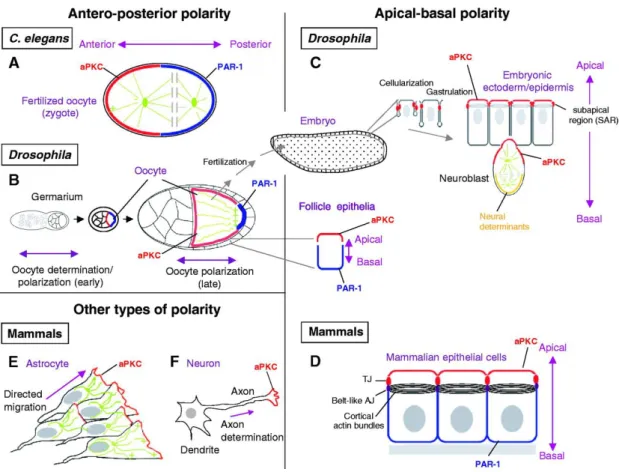
1. Egg chamber. Drosophila egg chamber is a versatile system to study many different polarity issues. A typical egg chamber consists of the monolayer epithelia organized by follicular cells, germ cell derived nursing cells and one oocyte. Follicular cells are polarized cells with apical facing towards inside and basal facing to outside of the chamber. They are the largely used model to study epithelial polarity and planar cell polarity. Oocyte has an anterior-posterior axis (from nursing cells’ side to the next egg chamber).
Figure 1.1 Various types of cell polarity (Suzuki and Ohno 2006).
2. Ectodermal epithelia. Ectodermal epithelia can be subdivided into embryonic epidermis, gut and trachea systems. Embryonic epidermis provides an excellent model to study the de novo epithelial polarity establishment, polarity maintenance as well as complicated morphogenesis.
3. Imaginal discs. Imaginal discs are sac-like epithelial structure found inside in larva.
Along with the metamorphosis, imaginal discs turn into the external structure of many body parts of Drosophila. There are 19 discs in total. However, only the wing discs and eye discs are widely used as an epithelial model due to their relatively large size.
They are nice models to study PCP and many different cross-talking pathways of polarity proteins, such as Notch signaling and Wnt pathway.
4. Other polarized models. Neural stem cells (Neuroblasts) are a great model to study the stem cell biology, especially focusing on the asymmetric cell division related issues. They start to massively proliferate from embryonic stage nine. Photoreceptor
cells are also polarized and appropriate for polarity studies.
Figure 1.2 Epithelial polarity is highly conserved from Drosophila to vertebrates.
Epithelial polarity is regulated by various evolutionarily conserved protein complexes.
At the apical region, there are apical polarity regulators (APR), namely the Baz/PAR-3-aPKC complex and the Crb complex. The basolateral membrane is marked by the basolateral polarity regulators (BLPR), such as the Scrb-Dlg-Lgl complex. APR and BLPR are counterbalancing each other to ensure the integrity of cell polarity.
1.2 Epithelial cell polarity
Polarized epithelia outline the cavities and surfaces of the body of animals, functioning as the selective and physical barrier between the tissue compartments or between environment and inner space (Muthuswamy and Xue 2012; Tepass 2012). To achieve this function, epithelial cells are highly polarized and featured by the differentiation of plasma membranes, namely the apical membrane, the lateral membrane and the basal membrane (Laprise and Tepass 2011; Thompson 2013). The apical membrane faces towards the outside environment or luminal space of internal
organs, while the lateral membrane connects adjacent cells within the epithelium and the basal membrane is linked to the basement membrane and further to the connective tissues. The lateral and basal membranes are often summarized as the basolateral domain.
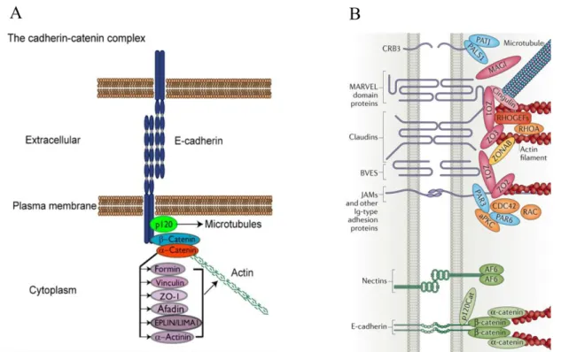
Figure 1.3 Schemes of adherens junctions and tight junctions. (A) E-Cadherin undergoes trans-homophilic interactions to form Cadherin clusters. The E-Cadherin intracellular domain contains binding sites for the catenins p120 and β-catenin.
β-catenin binds α-catenin, which in turn binds actin and several actin-associated proteins (Baum and Georgiou 2011). (B) A brief overview of types of tight junction proteins with only representatives of the main groups shown. For details, check review (Zihni, Mills et al. 2016).
Apical-basal axis establishment and maintenance require cell-cell contacts and junction formation. The distinct apical and basolateral membrane domains are separated by the belt-like structure, called zonula adherens (ZA). The ZA (also known as adherens junctions, AJs) consists of Cadherin-Catenin complexes and Actin filaments (F-actin) (Baum and Georgiou 2011; Takeichi 2014). Specifically, the extracellular domain of E-Cadherin can dimerize with trans-homophilic interactions to establish Cadherin clusters while the intracellular domain contains binding sites for
the Catenins p120 and β-catenin, thereby forming the Cadherin–Catenin complex.
β-catenin binds α-catenin, which in turn binds F-actin directly or indirectly via several actin-associated proteins, including α-actinin, Vinculin, Afadin and Formin-1 (Fig. 1.3 A). A bundle of F-actin runs in parallel to cell borders and provides the strength to single cells gathering all of them into the epithelium. The maturation of this structure provides a barrier which is necessary for the differentiation of distinct apical and basolateral domains. Below the ZA, cadherin proteins can also be detected throughout the lateral membranes, however, with a rather low intensity in these regions (Takeichi 2014).
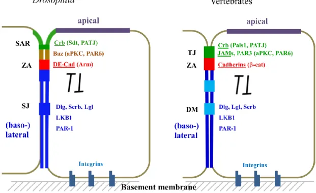
Apical to the ZA, Tight Junctions (TJs) are formed between adjacent epithelial cells in vertebrates, functioning as the main paracellular diffusion barrier and controlling the diffusion of ions and solutes (Anderson and Van Itallie 2009; Zihni, Mills et al. 2016).
Electron microscopy revealed that TJs form close focal contacts between two neighboring cells leaving pores of different sizes for water and ions in between (Zihni, Mills et al. 2016). Many TJ associated transmembrane proteins are identified from intensive molecular studies. Transmembrane proteins include claudins which are considered to be most critical for defining TJ functionality, junctional adhesion molecules (JAMs), Occludin and Crumbs3 (Crb3). Similar to Cadherins, these TJ-associated proteins also exhibit a homophilic interaction pattern while their intracellular adaptors are highly diverse and dynamic (Fig. 1.3 B). In Drosophila epithelium, instead of forming TJs, the corresponding region (named as the subapical region, SAR) is featured by enrichment of the single pass transmembrane protein Crb (Bulgakova and Knust 2009). However, the function of Crb homophilic interaction is rather obscure. The paracellular barrier function which is accomplished by TJ in vertebrates is largely achieved by formation of a Drosophila-specific junction called Septate Junction (SJ). SJs localize basally to the ZA and contain many evolutionarily conserved proteins, like Claudin related proteins, e.g. megatrachea, Kune-kune and Sinuous (Behr, Riedel et al. 2003; Genova and Fehon 2003; Wu, Schulte et al. 2004;
Wu, Marcus et al. 2007; Nelson, Furuse et al. 2010).
In vertebrates, the basolateral desmosomes (DMs) connect the intermediate filament cytoskeleton with the cell cortex.. DMs are widely found in epithelial tissues of specialized organs or systems which need strong adhesion, such as skin and heart (Garrod and Chidgey 2008). Similar homophilic bindings of the Cadherin family proteins bridge the extracellular spaces and provide platforms to assemble intracellular protein complexes. DMs are not found in Drosophila lateral membrane of the epithelial cells because the genes encoding core DM components (such as Desmogleins and Desmocollins, Plakoglobins and Plakophilin) are not conserved in Drosophila.
Epithelia are anchored to the basement membrane through the interaction between integrins and extracellular matrix (ECM) (Campbell and Humphries 2011). Integrins are transmembrane receptors, which form heterodimers from different protein isoforms. By linking the cytoskeleton and ECM, integrins allow cells to perceive and response properly to a dynamic microenvironment.
1.3 Epithelial polarity determinants and complexes
Since the 1980s, the genetic screenings in Caenorhabditis elegans and Drosophila led to identification of a wide range of polarity proteins (Tepass 2012). After intensive studies for around four decades, researchers figure out that multiple distinct and evolutionarily conserved groups of proteins constitute the epithelial cell polarity program, including: the Crb complex, Baz/PAR-3-aPKC complex, and the Lgl-Scrib-Dlg complex.
1.3.1 The Crumbs complex
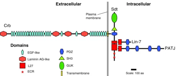
The canonical Crumbs complex consists of the transmembrane protein Crb (Crb1-3 in mammals), its intracellular adaptor Stardust (Sdt, protein associated with Lin7 1 Pals1/Mpp5 in mammals), Pals1 associated tight junction protein (PATJ) and Lin-7 (Bulgakova and Knust 2009; St Johnston and Ahringer 2010). The formation of the Crumbs complex is achieved by direct interactions among these different proteins.
Sdt/Pals1 acts a hub, on one hand, binding the C-terminal ERLI motif of Crb with its PSD-95/Discs-large/ZO-1 (PDZ) domain and stabilizing Crb at the apical junctional region, the SAR in Drosophila (Bachmann, Schneider et al. 2001; Hong, Stronach et al. 2001; Li, Wei et al. 2014). On the other hand, PATJ and Lin7 bind to the L27-N and C domains of Sdt/Pals1 respectively (Roh, Makarova et al. 2002; Bachmann, Timmer et al. 2004).
Figure 1.4 Scheme diagram shows the core proteins of the Drosophila Crumbs complex. The canonical Crumbs complex consists of the transmembrane protein Crb, its intracellular adaptor Sdt, Pals1 associated tight junction protein PATJ and Lin-7 (Bulgakova and Knust 2009).
1.3.1.1 Crumbs
Crb is a type I transmembrane protein and is conserved from C. elegans and Drosophila to human (Tepass, Theres et al. 1990; Pocha and Knust 2013). Crb was initially identified as the apical determinant from loss-of–function studies in Drosophila epithelia (Tepass, Theres et al. 1990). Drosophila Crb has a large extracellular domain composing of 29 epidermal growth factor (EGF) like domains and four laminin A globular-domain like repeats. The intracellular part of Crb which is rather small (only 37 amino acids in total) contains two functionally important and conserved motifs: The C-terminal PDZ binding motif (also designed ERLI motif in Drosophila according to the last four amino acids) and 4.1/ezrin/radixin/moesin
(FERM)-binding motif (Tepass, Theres et al. 1990).
Mutation of crb abolishes the formation of the apical domain, whereas overexpression increases the apical membrane size at the expense of the basolateral domain (Wodarz, Grawe et al. 1993; Wodarz, Hinz et al. 1995). Similar defects were observed in mammalian systems, for instance in MDCK cells (canine kidney tubular cells) which are widely used as an in vitro model for polarity studies (Roh, Fan et al. 2003;
Lemmers, Michel et al. 2004). Interestingly, crb mutant phenotype can be fully rescued by only expression of the transmembrane domain and the intracellular tail, highlighting the crucial functions of the PDZ-binding motif (mediating binding of the PDZ domain-containing proteins, e.g. Sdt/Pals1) and FERM-binding motif (anchoring Crb to cytoskeleton through FERM containing proteins) (Pocha and Knust 2013).
These mechanisms are thought to be very important in inhibiting the endocytosis of Crb (Pocha and Knust 2013).
In contrast, the function of the large extracellular domain in cell polarity regulation is rather obscure. One study indicated that the trans-homophilic dimmer formation of Drosophila extracellular domains was able to rescue the localization of Crb-extra variant in a crb mutant background (Letizia, Ricardo et al. 2013). However, this observation was only found in the boundary between wild-type cells and mutant ones.
Nevertheless, the extracellular domain seems to be important in some special cells or under particular conditions during development. It may also be involved in the regulation of several different signaling pathways, such as Notch signaling (Pellikka, Tanentzapf et al. 2002; Muschalik and Knust 2011; Thompson, Pichaud et al. 2013).
1.3.1.2 Stardust/Pals1
Sdt/Pals1 belongs to the membrane-associated guanylate kinase (MAGUK) family, containing two N-terminal evolutionary conserved regions (ECR), two L27 domains, a PDZ domain, a Src homology 3 domain (SH3) and a catalytically inactive GUK domain (Roh, Makarova et al. 2002). Sdt was first identified in a genetic screen in Drosophila and loss of sdt phenocopies the crb mutant (Wieschaus, Nüsslein-Volhard
et al. 1984; Tepass and Knust 1993).
Drosophila sdt employs different splicing strategies, resulting in four different isoforms: Sdt-B, F, H and D (Bulgakova, Rentsch et al. 2010). However, only Sdt-F is expressed throughout the entire embryonic development, while Sdt-B is only expressed at the early stages of embryos (Horne-Badovinac and Bilder 2008).
Intriguingly, apart from the apically localized Sdt protein, the sdt mRNA also exhibits the apical localization in embryonic epidermis and follicular cells (Horne-Badovinac and Bilder 2008).
Recent structural analysis by crystallization revealed that not only the PDZ domain but also the PDZ-SH3-MAGUK tandem is crucial for Crb binding and in vitro cysts formation in an MDCK cell culture model (Li, Wei et al. 2014).
1.3.1.3 PATJ and Lin7
PATJ and Lin7 exhibit an N-terminal L27 domain and subsequent PDZ domains (four in Drosophila PATJ and one in Drosophila Lin7). PATJ was initially described as discs lost (Dlt) by a mistake (Bhat, Izaddoost et al. 1999). Loss-of-function studies indicate that PATJ seems to be not important for the early embryonic development as evidence that PATJ mutant embryos do not show obvious defects in either Crb-Sdt stabilization and localization or junction formation (Pénalva and Mirouse 2012; Sen, Nagy-Zsvér-Vadas et al. 2012; Zhou and Hong 2012). However, PATJ is indeed very important in the photoreceptor cells to stabilize Crb and Sdt at the stalk membrane and prevent strong light-induced degeneration of rhabdomeres (Nam and Choi 2006;
Richard, Grawe et al. 2006, Zhou and Hong 2012). Our group has recently shown that PATJ regulates myosin phosphorylation by directly binding to myosin binding subunit (MBS) thereby inhibiting the phosphatase activity (Sen, Nagy-Zsvér-Vadas et al.
2012). This function is even more obvious in the Drosophila epidermis with weaken AJs, shedding light on the redundant function between PATJ and Cadherin complex in modeling cytoskeleton architecture (Sen, Nagy-Zsvér-Vadas et al. 2012).
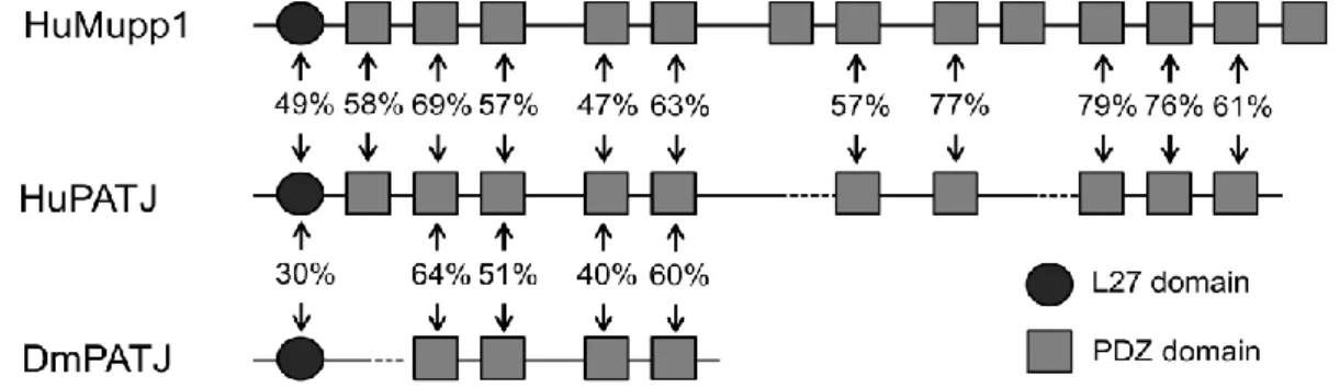
Figure 1.5 Scheme of the structural conservation between PATJ and Mupp1.
PATJ and Mupp1 are highly conserved in the protein domains and sharing several common binding partners (adapted from Roh, Makarova et al. 2002).
Two homologous of Drosophila PATJ are encoded in mammal systems: PATJ (also named INADL in mouse) and multiple PDZ protein 1 (Mpdz or Mupp1) (Hamazaki, Itoh et al. 2002; Adachi, Hamazaki et al. 2009). Mammalian PATJ has ten PDZ domains and Mupp1 is a bit larger in size, exhibiting thirteen PDZ domains. Although PATJ and Mupp1 share several common binding partners such as JAM1, ZO-3, Pals1, PAR6 and nectins and have similar TJ localization, their functions seem to be differentiated: In mammalian epithelial cells (at least in the analyzed MDCK cells), PATJ is indispensable for the TJ establishment and cell polarization, while the function of Mupp1 seems to be rather mild (Shin, Straight et al. 2005; Adachi, Hamazaki et al. 2009). Interestingly, knock-down of PATJ impairs the in vitro cyst formation and attenuates the migrating ability of MDCK cells (Shin, Straight et al.
2005; Shin, Wang et al. 2007). Moreover, PATJ is shown to be important for the proper cytokinesis in a mammary cell line, presumably through regulating (inhibiting) the phosphorylation of myosin regulatory light chain (MRLC) (Bui, Lee et al. 2016).
Lin7 exhibits a similar structure as PATJ, despite the number of PDZ domains.
Surprisingly, lin7 mutant flies are viable and fertile without any defects regarding epithelial polarity, except light-dependent degeneration of photoreceptor cells (Bachmann, Grawe et al. 2008). In contrast, mammalian Lin7 appears to stabilize Pals1 and further ensure TJ formation in MDCK cells (Straight, Pieczynski et al.
2006).
1.3.2 Baz/PAR-3-aPKC complex
Another apical protein complex is the Baz/PAR-3-aPKC complex, consisting of two PAR (partitioning defective) proteins (PAR-3/Baz and PAR-6), atypical PKC (aPKC) and a small GTPase Cdc42. Baz, a homolog of mammalian PAR-3, was the first PAR protein characterized in Drosophila and was shown to be important in regulating epithelial polarity as well as the asymmetric division of neural stem cells (Kuchinke, Grawe et al. 1998; Wodarz, Ramrath et al. 1999; Wodarz, Ramrath et al. 2000). Baz acts together with PAR-6/aPKC in the establishment and maintenance of epithelial polarity through forming trimeric complex (Lin, Edwards et al. 2000; Tepass 2012).
PAR-6 binds to aPKC through the interaction of their PB1 domains, and the semi-CRIB domain of PAR-6 mediates the binding of Cdc42. It is proposed that binding of Cdc42 by PAR-6 is able to activate the kinase activity of aPKC, phosphorylating Baz/PAR-3 and therefore releases Baz/PAR-3 from the complex (Horikoshi, Suzuki et al. 2009; Graybill et al., 2012; Yamanaka et al., 2001).
Although Baz is thought to act as one of the initiating cues for cellularization of Drosophila blastoderm epithelium, a recent study shows that it is dispensable for apical-basal polarity in the follicular epithelium of Drosophila (Harris and Peifer 2004;
Laprise and Tepass 2011; Shahab, Tiwari et al. 2015).
1.4 Dynamic interactions between apical polarity regulators
Apico-basal polarity is a hallmark of epithelial tissues and is regulated by several evolutionary conserved protein complexes. Many components of these protein complexes are highly dynamic and regulated by various modifications along different developmental stages and in distinct tissues or cell types.
1.4.1 Dynamic composition of protein complexes
The founding members of the major apical polarity regulators were identified in the
screening with worms and flies and led to the common concept of two independent complexes, namely as Baz/PAR-3-aPKC complex and Crb complex (Tepass 2012).
However, the last two-decade studies indicate that these two protein complexes are highly dynamic in compositions and far more complicated.
One way to increase this complexity is by encoding more than one isoforms of the polarity gene. As the more precise genome annotations are released, it is predicted that nearly all Drosophila polarity genes have more than one isoforms (see FlyBase for more information). A recent study demonstrates the importance of different isoforms of Crb by characterizing a helicase gene mutant obelus (Vichas, Laurie et al.
2015). Interestingly, they found that in obelus mutant, one crb RNA is upregulated during development and the obelus phenotype can be mimicked by overexpression of a certain crb isoform but not by the other two. This study raises the possibility that different Crb complexes are existing in parallel by simply exchanging the Crb isoforms. Besides, Sdt has four isoforms and these isoforms seem to have different functions and different expression patterns as described above (Bulgakova, Rentsch et al. 2010).
In the light of evolution, things can be more complicated. In mammalian systems, apart from the different splicing strategies, some genes are tending to duplicate themselves, resulting in more than one homologue. PAR-6 is a typical example. In Drosophila there is only one PAR-6 gene, while in mammals the number becomes four (Gao and Macara 2004). These different PAR-6 genes appear to have distinct functions in polarity regulation at least in MDCK cells. A recent study shows that PAR6G is a major suppressor of tumorigenesis, but not the other homologous (Marques, Englund et al. 2015).
A second way to modify the complex components is achieved by promiscuous interactions. During gastrulation of Drosophila embryonic epidermis, Sdt is able to interact with Baz directly, though the PDZ domain of Sdt and the aPKC binding domain of Baz (Krahn, Bückers et al. 2010). This interaction is proposed to be the important mechanism stabilizing Sdt before the onset of Crb expression during early
embryogenesis. PAR-6 seems to be another versatile protein. It can directly interact with the C-terminal PDZ binding motif of Crb via its PDZ domain and with the third PDZ domain of PATJ through its PB1 domain (Nam and Choi 2003; Hutterer, Betschinger et al. 2004; Kempkens, Médina et al. 2006). Moreover, PAR-6 interacts with Pals1/Sdt. This interaction is mediated by the semi-CRIB-PDZ tandem of PAR6 and the evolutionarily conserved regions (ECRs) of Pals1/Sdt by in vitro GST pull-down assays (Hurd, Gao et al. 2003; Wang, Hurd et al. 2004). Apart from these, aPKC binds to the intracellular tail of Crb and PATJ (Sotillos, Díaz-Meco et al. 2004).
Binding of aPKC to Crb indices the phosphorylation of Crb and is essential for the maturation of the Crb complex (Sotillos, Díaz-Meco et al. 2004). However, most of these findings have been achieved by using overexpression systems in cultured cells and it still remains to be investigated, whether these different complexes are stably formed in vivo and whether they contribute to the apical-basal polarization of epithelial cells.
Additionally, along with intensive studies with respect to cell polarity, novel players with diverse functions are identified. The PDZ binding motif of Crb can interact with α-adaptin which is a component of the AP-2 complex (Lin, Currinn et al. 2015). Upon this interaction, the endocytosis of Crb is strengthened and thought to be important for the degradation of Crb and the dynamic regulation of epithelial polarity. As the Sdt competes for the same binding site and crucial for Crb functionality, it is reasonable to propose that Crb stability and function is balanced by the different intracellular binding partners.
The network becomes even more complicated when all the interactions and dynamic compositions are linked with the specific developmental issues as different developmental stages and different cells behave differently: harboring distinct and dynamic polarity regulators.
1.4.2 Protein modifications
Epithelial polarity regulators are frequently modified in various means.
Phosphorylation is one of the important modifications. It has been reported that in Drosophila, phosphorylation of Baz by the basolateral localized kinase PAR1 at Ser151 and Ser1085 prevents the binding of aPKC and inhibits the dimerization of Baz and this phosphorylation can be counteracted by the protein phosphatase 2A (PP2A) (Benton and St Johnston 2003; Krahn, Egger-Adam et al. 2009). Another example is that aPKC phosphorylates Baz at the aPKC binding region (S980) and weakens the binding ability of Sdt and Baz, therefore leading to the dissociation of Sdt from Baz and forming the Crb complex (Krahn, Bückers et al. 2010; Morais de Sa et al. 2010; Walther and Pichaud 2010). Phosphorylations by different kinases seem to provide crucial cues mediating the dynamic changes of protein complex components.
Another way to control protein complex stability lays to the protein quality control, specifically the ubiquitylation dependent protein degradation (Bórquez and González-Billault 2011). To date, many E3 ligases, which directly bind the specific substrates and tag them with a degradation mark, are shown to be important in regulating cell polarity. The F-box E3 ligase Slmb restricts and modulates the activity and stability of aPKC-PAR-6 in Drosophila epithelial cells as well as in oocytes (Morais-de-Sá, Mukherjee et al. 2014; Skwarek, Windler et al. 2014). Moreover, another E3 ligase PDZRN3 can induce the poly-ubiquitination of Mupp1 and impair the endothelial cell-cell junction stability in the corresponding overexpression mouse model (Sewduth, Kovacic et al. 2017). Apart from the well characterized E3 ligases, the notch pathway regulator Ebi physically binds to the extracellular domain of Crb and results in the ubiquitin-dependent downregulation of Crb in the dorsoventral (DV) boundary of Drosophilaimaginal discs (Nguyen, Vuong et al. 2016).
Lastly, the ubiquitination-like modification, sumoylation is also involved in polarity regulation. For instance, Nup358 dependent SUMO modification of aPKC controls the kinase activity of aPKC, which might play a role in the epithelial cell polarity (Yadav, Magre et al. 2016).
1.5 Epithelial polarity and signaling pathways
Epithelial polarity is closely connected to many important signaling pathways.
Through these cross-talks, epithelial cells are able to react to extrinsic and intrinsic signals and control the homeostasis and remodeling of tissues as well as the epithelial cells themselves. Here, the signaling pathways which are regulated/regulating epithelial polarity are briefly summarized.
1.5.1 Wnt signaling and epithelial polarity
Wnt proteins are secreted short-range signals which control a variety of developmental processes in all metazoans (Kikuchi, Yamamoto et al. 2011; Niehrs 2012; Swarup and Verheyen 2012; Wang, Sinha et al. 2012). Based on studies in Drosophila, Xenopus, and zebrafish, Wnt signaling can be briefly divided into the canonical pathway which involves β-catenin mediated transcription and the β-catenin-independent noncanonical Wnt pathways composing of Wnt-planar cell polarity (PCP) and Wnt-Ca+2 signaling.
The best-known cross-talking mechanisms between Wnt signaling and epithelial polarity are established on studies into PCP (Yang and Mlodzik 2015; Chu and Sokol 2016). PCP signaling regulates the tissue organization through remodeling the cytoskeleton networks. Dishevelled (Dvl), the adaptor protein of Wnt receptor, can active the kinase activity of aPKC by directly binding, therefore playing important roles in axonal differentiation and polarized migration of mammalian cells (Schlessinger, McManus et al. 2007; Zhang, Zhu et al. 2007). Additionally, PATJ can recruit aPKC to the Wnt receptor Frizzled through direct interactions, resulting in enhanced phosphorylation of Frizzled and inhibited PCP in Drosophila wing (Djiane, Yogev et al. 2005). This effect can be further balanced by expressing Baz (Djiane, Yogev et al. 2005). Thus, aPKC mediates many connections between PCP and cell polarity.
1.5.2 Notch signaling and epithelial polarity
Notch signaling defines an evolutionarily conserved mechanism that regulates various developmental events, including growth, cell fate determination and many other functions (Artavanis-Tsakonas, Matsuno et al. 1995; Artavanis-Tsakonas, Rand et al.
1999). Notch signaling is activated by the binding of Notch receptor to its transmembrane ligands and this binding leads to the shedding of the intercellular domain of Notch receptor cutted by γ-secretase, which enters the nucleus and leads to gene transcription (Mumm and Kopan 2000).
Studies in Drosophila show that Crb genetically interacts with Notch receptor as demonstrated by larger wing size in heterozygous double mutants (Richardson and Pichaud 2010). This function is achieved presumably by direct interaction between the extracellular domains of Crb and Notch, therefore inhibiting Notch receptor binding to its ligands (Nemetschke and Knust 2016). Apart from Crb, aPKC-iota inhibits stem cell self-renew and promotes differentiation by downregulating Notch signaling in mouse neural stem cells (Mah, Soloff et al. 2015). Moreover, loss of PAR-3 cooperates with Notch signaling in tumorigenesis and enhances tumor invasion and following up metastasis in a Notch receptor intracellular domain (NICD) overexpression model (McCaffrey, Montalbano et al. 2012). The basolateral marker, Lgl negatively regulates Notch signaling by inhibiting the endocytosis of Notch receptor, which contributes to tumorigenesis upon loss of Lgl in mammals (Parsons, Portela et al. 2014; Portela, Parsons et al. 2015).
1.5.3 Hippo pathway and epithelial polarity
The Hippo pathway was first discovered in a screening for regulators of organ size in Drosophila (Grusche, Richardson et al. 2010). Follow-up studies showed that this pathway is highly conserved and plays a central role in controlling cell proliferation, apoptosis and stemness in response to various extracellular and intracellular signals, such as cell-cell contacts, cell polarity, mechanical and energy status (Yu, Zhao et al.
2015). The Hippo pathway involves a core kinase cascade which ultimately results in the phosphorylation of the transcriptional coactivators Yorkie (Yki, Yap in mammals)
and TAZ (not conserved in flies), promoting their cytosolic retention (Yu, Zhao et al.
2015).
The upstream activating protein complex, Expanded-Merlin-Kibra, binds to Crb and propagates the Hippo signaling (Chen, Gajewski et al. 2010; Ling, Zheng et al. 2010;
Robinson, Huang et al. 2010). Thus, the loss of Crb leads to dephosphorylation of YAP/Yki and enhanced proliferation. In addition, in mammals, the tight junction associated protein Angiomotin (AMOT) directly binds YAP and sequesters it in cytosol independent of the YAP phosphorylation status (Chan, Lim et al. 2011; Zhao, Li et al. 2011). Since AMOT also binds to Pals1, it is interesting to investigate how Pals1 regulates Hippo pathway (Varelas, Samavarchi-Tehrani et al. 2010).
Additionally, a cell adhesion molecule Echinoid (Ed) acts as a tumor suppressor by directly binding the core kinases and enhancing their activity and Yki phosphorylation in Drosophila (Yue, Tian et al. 2012).
1.5.4 Other pathways related to epithelial polarity
TGF-β signaling is featured by harboring the serine/threonine kinase receptors and controls cell polarity and cancer progression (Ozdamar, Bose et al. 2005). Upon TGF-β stimulation, TGF-β type II receptor can bind to and phosphorylate PAR6. This phosphorylation creates a docking site for an E3 ligase Smurf1, which mediates ubiquitylation and degradation of RhoA. Loss of RhoA causes cell remodeling and epithelial-to-mesenchymal transition (EMT). This mechanism seems to play an important role in the cancer metastasis. A recent study further shed light on the Pals1 mediated cross-talk between TGF-β signaling and Hippo pathway, emphasizing the central role of cell polarity proteins in linking different pathways (Weide, Vollenbröker et al. 2017).
Polarity proteins are also linked to immune signaling. One study using a mouse mammary cell line shows that loss of PAR-3 leads to enhanced NF-κB signaling mediated by boosted aPKC kinase activity and this cascade finally results in the activity of JAK-STAT axis (Guyer and Macara 2015). However, it is an open question
whether this observation has a biological relevance or not especially upon pathogen invasions.
1.6 Aim of the study
Apico-basal polarity is a significant feature of epithelial tissues and it is regulated by several evolutionarily conserved protein complexes. Among them, PAR-aPKC complex and Crb complex are defining the apical domain of the cell membrane.
Interestingly, both complexes are highly dynamic in components during Drosophila embryonic development. Hence, it is intriguing to understand how these dynamics are mediated and the impacts on the epithelial cell polarity.
Firstly, the apical targeting mechanism of the core component of the Crb complex PATJ was investigated. Through biochemistry analysis and genetic studies, we identified the existence of the Baz-Sdt-PATJ complex, which is important for apically localization and functionality of PATJ in Drosophila.
Secondly, we focused on the role of PAR-6 on regulating the stability of Crb complex.
PAR-6 has been shown to interact with the Crb complex in previous studies with overexpression systems of cell lines and/or bacterially purified proteins. In our study, we established a new mechanism of PAR-6 in regulating epithelial polarity, which links protein quality control to the maintenance and establishment of cell polarity in Drosophila.
Lastly, a structure-function analysis of Crb-adaptor Sdt was performed due to limited in vivo information of protein-interaction features of Sdt. Several genetically modified flies harboring domain-deletion variants of Sdt were generated and the detailed phenotypes were analyzed.
Chapter 1. Upstream regulators of PATJ
Localization and Function of Pals1 Associated Tight Junction Protein in Drosophila is Regulated by Two Distinct Apical Complexes
Arnab Sen*, Rui Sun* and Michael P. Krahn
*These authors contributed equally to the work.
This project aims to understand the apical targeting mechanism of the core component of the Crb complex PATJ. In the embryonic epidermis of Drosophila, PATJ is not initially recruited to the apical cell-cell contacts by binding to Crb-Sdt as Crb is not expressed in the early embryogenesis, but depending on forming a new complex with Bazooka (Baz). We found that the Baz-Sdt-PATJ complex exists in parallel to the Crb-Sdt-PATJ complex and is important for the apically targeting of PATJ. These two complexes might play a redundant role in myosin activity modulation.
Author contributions:
Rui Sun: mostly the biochemistry studies, partially the genetic studies and manuscript writing
Status:
Published in Journal of Biological chemistry on April 2015
Localization and Function of Pals1 Associated Tight Junction Protein in Drosophila is Regulated by Two Distinct Apical Complexes
The transmembrane protein Crumbs (Crb) and its intracellular adaptor protein Pals1 (Stardust, Sdt in Drosophila) play a crucial role in the establishment and maintenance of apical-basal polarity in epithelial cells in various organisms. In contrast, the multiple-PDZ-domain-containing protein PATJ, which has been described to form a complex with Crb/Sdt, is not essential for apical-basal polarity or for the stability of the Crb/Sdt complex in the Drosophila epidermis. Here we show that in the embryonic epidermis Sdt is essential for the correct subcellular localization of PATJ in differentiated epithelial cells but not during cellularization. Consistently, the L27-domain of PATJ is crucial for the correct localization and function of the protein.
Our data further indicate that the four PDZ domains of PATJ function to a large extent in redundancy regulating the protein’s function. Interestingly the PATJ-Sdt heterodimer is not only recruited to the apical cell-cell contacts by binding to Crb but depends on functional Bazooka (Baz). However biochemical experiments show that PATJ associates with both complexes, the Baz-Sdt and the Crb-Sdt complex in the mature epithelium of the embryonic epidermis, suggesting a role of these two complexes for PATJ’s function during development of Drosophila.
Keywords: Cell polarity, Adherens junctions, Drosophila, Tight junction, Myosin
INTRODUCTION
Apical-basal polarization of epithelia is regulated by conserved complexes determining the apical versus the basolateral domain (Tepass 2012; Roignot, Peng et al. 2013): Apical to the Adherens Junctions (AJ), the membrane-associated PAR (partitioning-defective) - aPKC (atypical protein kinase C)-complex regulates assembly of the Crumbs(Crb)-complex, which assembles more apically in the so-called subapical region (SAR). The activity of these two complexes is counterbalanced by proteins such as Scribble-Lethal(2) giant larvae-Discs large(Dlg), which localize to the basolateral domain. In the past, various studies have demonstrated that both apical complexes are rather dynamic and that their composition might be tissue-dependent and temporally and/or developmentally regulated (Hurd, Gao et al. 2003; Nam and Choi 2003; Penkert, DiVittorio et al. 2004;
Sotillos, Diaz-Meco et al. 2004; Wang, Hurd et al. 2004; Kempkens, Medina et al.
2006; Krahn, Buckers et al. 2010).
In Drosophila, the multiple PDZ-domain-containing protein PATJ has been described to function in a complex with Crb and Stardust (Sdt, the Drosophila homologue of Partner of Lin-7 one, Pals1) to regulate apical-basal polarity in follicle epithelial cells and photoreceptor cells (Tanentzapf, Smith et al. 2000; Nam and Choi 2006; Richard, Grawe et al. 2006).
In mammalian and Drosophila epithelial cells, Pals1/Sdt is recruited by the cytoplasmic tail of Crb to the “subapical region” and in turn stabilizes Crb (Bachmann, Schneider et al. 2001; Hong, Stronach et al. 2001; Makarova, Roh et al. 2003; Roh, Fan et al. 2003; Straight, Shin et al. 2004). The L27 domain of Pals1 has been shown to heterodimerize with the L27-domain of PATJ, thereby tethering PATJ to tight junctions (TJ) (Roh, Makarova et al. 2002; Li, Karnak et al. 2004; Feng, Long et al.
2005).
Recently, we and others reported that loss of PATJ in Drosophila does not affect apical-basal polarity in the embryonic epidermis or in follicle epithelial cells but
rather modulates Myosin activity to support AJ stability (Penalva and Mirouse 2012;
Sen, Nagy-Zsver-Vadas et al. 2012; Zhou and Hong 2012). Exclusively in photoreceptor cells and to some extent in the follicular epithelium, PATJ seems to be essential for the correct subcellular localization of the Crb-Sdt complex, either directly by stabilizing this complex or indirectly by regulating photoreceptor morphology/development (Sen, Nagy-Zsver-Vadas et al. 2012; Zhou and Hong 2012).
Two mammalian orthologues of PATJ are expressed in epithelia: mammalian PATJ (mPATJ, encoded by INADL in mice) and Multiple PDZ-domain protein 1 (MUPP1).
Both proteins are very similar to DmPATJ: In addition to an N-terminal L27 domain, they exhibit several PDZ domains (DmPATJ four, mPATJ ten, MUPP1 thirteen) and localize to the TJ in mammalian epithelial cells (Adachi, Hamazaki et al. 2009).
However, Abachi et al. showed that despite its domain similarity, mPATJ but not MUPP1 regulates TJ stability (Adachi, Hamazaki et al. 2009). These data are in line with previous findings describing TJ-formation delay or defects upon loss of mPATJ in cultured epithelial cells (Michel, Arsanto et al. 2005; Shin, Straight et al. 2005).
Other studies describe a role of mPATJ in Myosin-driven processes like apical constriction and cell migration (Shin, Wang et al. 2007; Ernkvist, Luna Persson et al.
2009; Nakajima and Tanoue 2011).
In this study, we report that in the embryonic epidermis of Drosophila PATJ assembles with the Crb-Sdt complex but additionally associates with the Baz-Sdt-complex we described previously (Krahn, Buckers et al. 2010). Notably, deletion of Baz and Sdt but not of Crb leads to mislocalization of PATJ during gastrulation and in fully differentiated epithelia of the embryonic epidermis. In contrast, localization of PATJ at the basal junction next to the tip of the invaginating plasma membrane during cellularization is independent of Baz/Sdt. Consequently, deletion of the L27-domain of PATJ leads to an abolished apical accumulation and impaired function of the protein. Studies with chimeric proteins further suggest that targeting of PATJ’s PDZ-domains to the Baz-(Sdt) and Crb-(Sdt) complex are sufficient for PATJ’s function. Finally, we investigated the functionality of PATJ’s four
PDZ domains and provide evidence that under close to endogenous expression levels, none of these domains is essential.
EXPERIMENTAL PROCEDURES
Drosophila genetics - The following mutant alleles were used: PATJ1 (Sen, Nagy-Zsver-Vadas et al. 2012), baz815-8 (McKim, Dahmus et al. 1996; Krahn, Klopfenstein et al. 2010), bazXR11 (Shahab, Tiwari et al. 2015), sdtK85 (Berger, Bulgakova et al. 2007) and crb11A22 (Tepass and Knust 1990). Germ line clones were generated with the mutant alleles recombined with FRT using the dominant female sterile technique (Chou, Noll et al. 1993). Homozygous mutant embryos were identified using antibody stainings. Follicle cell clones were generated with the FRT-Flp system as described before (Sen, Nagy-Zsver-Vadas et al. 2012).
Ubi::PATJ-GFP (mutant/chimeric) constructs were generated using phiC31-mediated germ line transformation using attP40.
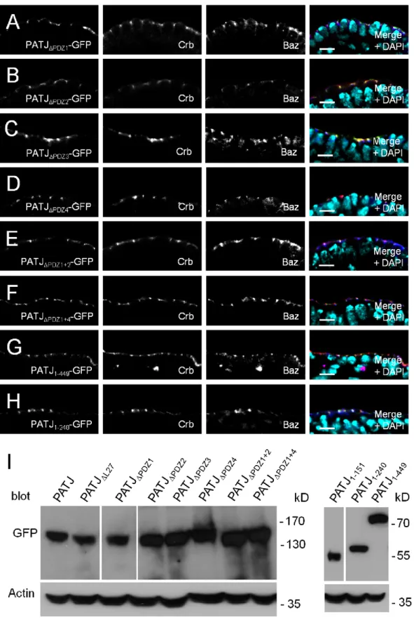
DNA and constructs - The QuickChange Site-Directed Mutagenesis Kit (Stratagene) was used to generate domain deletions with full-length PATJ cDNA in pENTR (Sen, Nagy-Zsver-Vadas et al. 2012) as a template. The following oligonucleotides were used for mutagenesis:
PATJL27: 5’- GCGGATATTTCCAGCTCCAT-GTTGCCCAAC-3’; PATJPDZ1:
5’-GCCATA-GAGCTGGTCCGTCCCGTTGAGCAG-3’; PATJPDZ2:
5’-GAAACGGAGAAGCTTCGC-TACCTGAGGGGC-3’; PATJPDZ3:
5’-GGCT-CCGATGTGGAGTGCGGTCGCAACAGG-3’;
PATJPDZ4: 5’-ATGTGGTCGTCCCAACGC-ATTGGTGTGGCC-3’;
To generate truncated versions of PATJ, the following primers were used: PATJ-F:
5‘-CACCATGCACCTCAGCGCGGA-3’; PATJ-151-R:
5‘-CTCTATGGCCTGGATCTGAGC-3‘;
PATJ-256-R: 5’-CAGGGCGTACTGGGG-3’;
PATJ-449-R: 5’-TGATGGTGTAGTTGTGGC-3‘
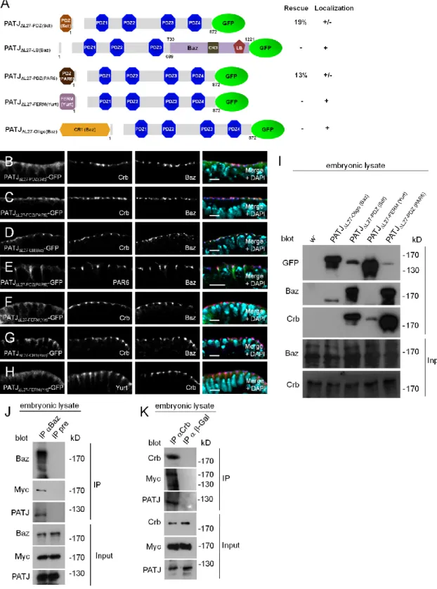
For PATJL27 PDZ(Sdt), the PDZ domain of Sdt was amplified with Sdt-PDZ-F:
5’-GCGGCCGCCCCCTTCACCATGCGTATCATCCAGATCGAG-3’ and
Sdt-PDZ-R: 5’-GCGGCCGCCGGTGGACTACCCGCTGG and inserted with NotI (underlined) into PATJL27 pEntry. Similarly, the PDZ domain of PAR6, the oligomerization domain of Baz and the FERM-domain of Yurt were cloned into NotI of PATJL27 pEntry using the following oligonucleotides: PAR6-PDZ-F:
5’-GCGGCCGCCCCCTTCACCATGAGAAGAGTGCGGCTACTG-3’,
PAR6-PDZ-R: 5’-GCGGCCGCCTTCACGGTGATTATCAGATTG-3’, Yrt-FERM-F:
5’-GCGGCCGCCCCCTTCACCATGGTGCTCGGAAAGGATGGC-3’,
Yrt-FERM-R: 5’-GCGGCCGCTTTGACCGGCGCCC TAA-3’; Baz-CR1-F:
5’-GCGGCCGCCCCCTTCACCATGAAGGTCACCGTCTGCTTCGGC-3’,
Baz-CR1-R: 5’- GCGGCCGCATCTCCGCC TCCTTGC-3’. Baz733-1221 was cloned into an endogenous SacII site (aa 633) of PATJL27. All constructs were recloned into destination vectors (modified UWG, Murphy lab, DGRC) using the gateway technology (Life technologies).
Immunoprecipitation and Western blotting - For immunoprecipitations, w- embryos from an overnight collection were dechorionated and lysed in lysis buffer (1% Triton X-100, 150mM NaCl, 1mM CaCl2, 1mM MgCl2, 50mM TRIS-HCl pH 7.5) supplemented with protease inhibitors. After centrifugation, 2 l of rabbit anti-Baz (Wodarz, Ramrath et al. 1999) or 2 µl of the corresponding preimmune serum were added to the embryonic lysate, corresponding to 500 µg total protein. Immune complexes were harvested using protein A-conjugated agarose (BioVision).
GFP-binder (Chromotek) was used to immunoprecipitate Crb-GFP. Wild-type flies served as control. Beads were washed five times with lysis buffer and boiled in 2x SDS sample buffer before SDS-PAGE and Western blot. Western blotting was done according to standard procedures. Primary antibodies used for Western blotting were as follows: mouse anti Crb (Cq4, 1:50, developed by E. Knust (Tepass, Theres et al.
1990) was obtained from the Developmental Studies Hybridoma Bank (DSHB),
created by the NICHD of the NIH and maintained at the University of Iowa, Department of Biology, Iowa City, IA 52242), guinea pig anti PATJ (1:1000, Sen, Nagy-Zsver-Vadas et al. 2012), mouse anti Sdt (1:20, Berger, Bulgakova et al. 2007), rabbit anti-Baz (1:2000, Wodarz, Ramrath et al. 1999) and mouse anti-GFP (B2, 1:500, Santa Cruz, sc-9996).
Immunohistochemistry - Embryos were fixed in 4% formaldehyde, phosphate buffer pH 7.4 as described previously (Krahn, Egger-Adam et al. 2009). Primary antibodies used for indirect immunofluorescence were as follows: rabbit anti-Baz (1:1000, Wodarz, Ramrath et al. 1999), mouse anti Crb (Cq4, 1:50, DSHB), mouse Dlg (1:50, DSHB (Parnas, Haghighi et al. 2001)), guinea pig anti PATJ (1:500, Sen, Nagy-Zsver-Vadas et al. 2012), mouse anti Sdt (1:20, Berger, Bulgakova et al. 2007), guinea pig anti PAR6 (Kim, Gailite et al. 2009), guinea pig anti Yrt (Laprise, Beronja et al. 2006), rabbit anti-GFP (#A11122, 1:1000, Life technologies), rabbit anti-GFP (sc-8334, Santa Cruz Biotechnology) and chicken anti-GFP (1:2000, Aves Laboratories). Secondary antibodies conjugated with Alexa 488, Alexa 568 and Alexa 647 (Life technologies) were used at 1:400.
Images were taken on a Zeiss LSM 710 Meta confocal microscope and processed using Adobe Photoshop.
RESULTS AND DISCUSSION
PATJ is recruited by Sdt to a complex with Baz at the apical junctions in the embryonic epidermis - Upon the formation of apical AJ in late cellularization/early gastrulation in Drosophila, PATJ is recruited to the apical cell-cell contact region whereas staining at the basal membrane ceases (Sen, Nagy-Zsver-Vadas et al. 2012).
Studies in Drosophila and cultured mammalian epithelial cells showed that PATJ associates with Sdt/Pals1 which in turn binds to the transmembrane protein Crb, which targets the complex to TJ in vertebrates and in the corresponding “subapical region” (SAR) in Drosophila (Klebes and Knust 2000; Roh, Makarova et al. 2002).
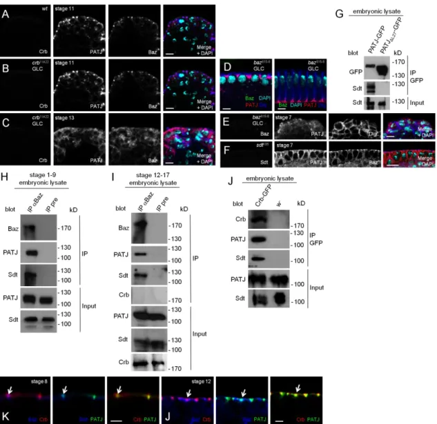
Figure 2.1 A. Endogenous PATJ colocalizes with Crb and Baz at the apical junctions in epithelial cells of the embryonic epidermis. Green: Crb, red: PATJ, blue: Baz. B.
Endogenous PATJ localizes at the apical junctions in the absence of Crb expression during early embryogenesis. Green: Crb, red: PATJ, blue: Baz. C. Upon disruption of epithelial integrity in crb-mutant embryos (maternally and zygotically mutant, derived from GLCs) of later stages, Baz as well as PATJ are mislocalized to the cytoplasm.
Green: Crb, red: PATJ, blue: Baz. D. PATJ localization during cellularization is not affected in baz-mutant embryos derived from baz815-8 germ line clones. Green: Baz, red: PATJ, blue: Dlg. E. Loss of maternal and zygotic Baz in gastrulation (baz815-8 germ line clones) results in a disturbed apical-basal polarity and cytoplasmic PATJ localization. Green: Baz, red: PATJ, blue: Dlg. F. PATJ is not recruited to the apical junctions in the absence of Sdt. Green: Sdt, red: PATJ, blue: Baz. G. Endogenous Sdt
co-immunoprecipitates with PATJ- -GFP. H and I.
Endogenous PATJ co-immunoprecipitates with Baz in early stages (stage 1-9, H) and late stages (stage 12-17, I). Preimmune serum (pre) served as negative control. J.
Co-immunoprecipitation of PATJ with Crb-GFP expressed from its endogenous promoter from embryonic lysates. Wild-type flies served as negative control. K and L.
High magnifications of stainings of wild-type embryos with PATJ, Crb and Baz reveal a segregation of Baz and Crb, which starts in earlier embryonic stages (stage 8, K) and gets more pronounced in later stages (stage 12, L). Remarkably, PATJ overlaps with both proteins in both stages (arrows). Scale bars = 5µm in A-F, 2µm in K and L.
We recently found that in the embryonic epidermis of Drosophila, Sdt is initially localized to the apical junctions in early gastrulation before Crb is expressed and even remains at the junctional region of epithelial cells when Crb is absent (Krahn, Buckers et al. 2010). This is accomplished by a direct interaction of the PDZ-domain of Sdt with Baz. Upon phosphorylation of Baz by aPKC at Serine 980, Sdt is released from Baz and becomes available to stabilize the Crb complex (Krahn, Buckers et al. 2010).
We, therefore, tested whether the subcellular localization of PATJ is dependent on Crb or Baz or both. In maternal and zygotic crb-mutant embryos (crb11A22 GLC), PATJ shows a correct localization not only during cellularization (data not shown) but also after gastrulation as long as apical-basal polarity is still intact (stage 6-11, Fig. 2.1B, compared to wild-type embryos in Fig. 2.1A). In later stages (from stage 11/12 on), the apical-basal polarity is impaired due to the loss of Crb, finally resulting in a multilayered epithelium. In these cells, PATJ staining is cytoplasmic or in aggregates (Fig. 2.1C). Notably, the loss of cortical PATJ in these embryos is accompanied by a loss of membrane-associated Baz (Fig. 2.1C).
In contrast, in maternal and zygotic baz-mutant embryos (baz815-8 GLC), the accumulation of PATJ at the basal junction next to the tip of the furrow canal during plasma membrane invagination is not affected (Fig 2.1D) but the targeting of the protein to the apical junctional region after cellularization is abolished (Fig. 2.1E).
Furthermore, we found endogenous PATJ and Sdt to coimmunoprecipitate with endogenous Baz from embryonic lysates (Fig. 2.1H and I). Consequently, in embryos lacking Sdt, PATJ is correctly localized during cellularization (data not shown) but fails to relocalize to the apical junctions during gastrulation (Fig. 2.1F), indicating that PATJ is recruited by Sdt to the apical junctions. Furthermore, deletion of the L27