El-Merzabani, El-Aaser and Zakhary: Determination of inorganic phosphorus without deproteinization 715 J. Clin. Chem. Clin. Biochem.
Vol. 15,1977, pp. 715-718
A New Method for Determination of Inorganic Phosphorus in Serum without Deproteinization
»»
By Mahmoud Mohamed EI-Merzabani, Abdelbaset Anwer-El-Aaser and Nadia hkandar Zakhary Cancer Chemotherapy and Cell Chemistry Units, Cancer Institute, Cairo University, Cairo-Egypt (Received July 6,1976/January 26,1977)
Summary: A simple method was developed for the determination of inorganic phosphorus in serum without deproteinization. The method is based on the use of formic acid as protein solubilizer and glycerol as stabilizer for the assay system. The optimal conditions for colour development were determined. The results obtained with the new method correlate well with those obtained after deproteinization of serum with trichloroacetic acid. The present method could be fully mechanized, and its application to the determination of serum phosphatases is discussed.
Eine neue Methode zur Bestimmung von anorganischem Phosphat im Serum ohne Enteiweißung
Zusammenfassung: Eine einfache Methode zur Bestimmung von anorganischem Phosphat im Serum ohne Entei- weißung wurde entwickelt. Die Methode benutzt Ameisensäure zur Lösung der Proteine und Glycerin zur Stabili- sierung des Bestimmungssystems. Die optimalen Bedingungen für die Farbentwicklung wurden bestimmt. Die mit der neuen Methode erhaltenen Ergebnisse korrelieren gut mit den nach Enteiweißung des Serums mit Trichloressigsäure erhaltenen. Die Methode könnte voll mechanisiert werden. Ihre Verwendung zur Bestimmung der Phosphatasen im Serum wird diskutiert.
Introduction
Inorganic phosphorus, present in serum as phosphate, forms a phosphomolybdate complex with molybdic acid. The latter can be easily reduced to a blue colour, which can be measured colorimetrically. This reaction is applied widely in clinical chemistry (1, 2).
In our preliminary investigations to determine serum phosphorus without deproteimzation, it had been found that formic acid, together with glycerol, could solübilize serum proteins even in the presence of ammonium molybdate. When glycerol was omitted serum proteins were reprecipitated after the addition of ammonium molybdate. Ethylene and propylene glycols, in higher concentration, gave the same effect as glycerol in the presence of formic acid.
Therefore it was of interest to fully investigate the optimal conditions for the determination of serum phos- phorus without deproteinization using formic acid as protein solubüizer and glycerol as stabilizing agent for the assay system.
Material and Methods
Reagents required for the established method mentioned in table l,are:
1. Protein solubilizer
Use pure formic acid 1220 g/1 (density 1.22, Prolabo) 2. Stabilizer
Mix 300 ml glycerol (density 1.263, Prolabo) with 700 ml distilled water, this will give a concentration of 378 g/1. Keep in closed brown container.
3. Ammonium molybdate
Dissolve 10 g ammonium molybdate (BDH) in one litre of distilled water. The solution is stable at room temperature.
4. Reagent mixture for use (formic acid/glycerol/molybdate) Mix solution 1, 2 and 3 in a ratio of 200 ml + 200 ml + 100 ml respectively. Keep in closed brown container. Solution is stable for up to three weeks.
5. Reducing agent
Use freshly prepared stannous chloride (BDH), 1.5 g/1 in 1 mol/1 hydrochloric acid.
6. Standard phosphorus
Dissolve 0.0878 g potassium dihydrogen phosphate (BDH) in one litre distilled water. Add few drops of chloroform and keep at 4°C. This solution contains 20 mg/1 phosphorus (Pj).
Phosphorus determinations
Phosphorus was determined in serum without deproteinization using formic acid and glycerol according to the optimal conditions outlined in Table 1. For comparison, parallel determinations were carried out after deproteinization with trichloroacetic acid according to Kuttner & Lichtenstein (3): 0.2 ml serum, in final
I din. Chem, Qin. Biochem, / Vol. 15,1977 / No,12
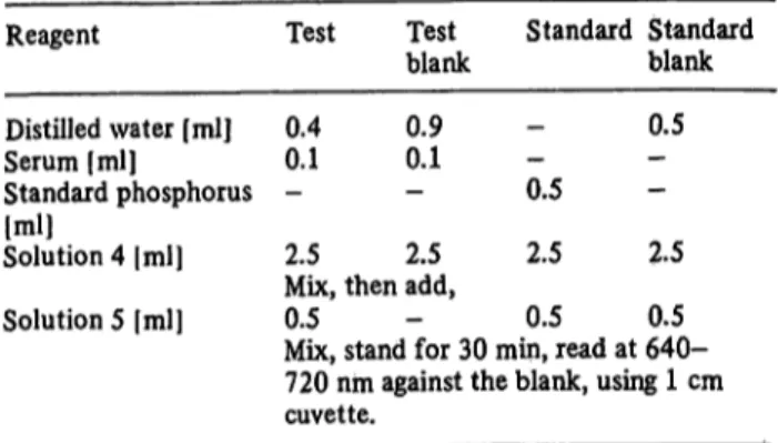
716 El-Marzabani, El-Aaser and Zakhary: Determination of inorganic phosphorus without deproteinizätion Tab. 1. Standard formic acid-glycerol procedure:
Reagent
Distilled water [ml]
Serum [ml]
Standard phosphorus Solution 4 |ml][ml]
Test 0.40.1
2.5
Testblank 0.90.1
2.5
Standard Standard blank 0.5 0.5
2.5 2.5 Mix, then add,
Solution 5 [ml] 0.5 - 0.5 0.5 Mix, stand for 30 min, read at 640- 720 nm against the blank, using 1 cm cuvette.
volume of one ml, was treated with 5 ml of 160 g/1 trichloro- acetic acid; after centrifugation a 3 ml aliquot (equivalent to 0.1 ml serum) was mixed with 0.5 ml of 10 g/1 ammonium molybdate, followed by 0.5 ml 1.5 g/1 stannous chloride. After standing for 30 minutes, the colour was measured at 640 nm, using a Unicam SP 500 and a 1 cm cuvette.
Calculation
Phosphorus, mg/dl serum:
A64°teSt
A640 standard
Phosphorus, mmol/1 serum:
mg/dl X 0,01292
100 (per hundred) 0.1 (mi of serum used)
Results
Establishment of optimal conditions Effect of formic acid and glycerol
As shown in table 2, the assay of 2.85 mg/1 phosphorus in the presence of a constant concentration of formic acid (348 g/1) and different concentrations of glycerol (88 to
126 g/1 in the final assay mixture) showed an optimal optical density of 1.05 at 640 nm, irrespective of the presence or absence of serum. This value correlates well Tab. 2. Effect of different concentrations of formic acid and
glycerol.
Formic acid Glycerol 2.85 mg/1 P; 2.85 mg/1 P; Recovery- ·-- - ·- -/~\ * /U\ * ^+ serumW % 348348
348348 348 170244 317348
10788 126145 164 126126 126126
1.081.08 1.050.87 0,54 1.051.00 1.001.05
1.021.02 0.991.05 0.66 1.021.02 1.051.05
94.494.4 100.0
102.097.1 105.0 100.0 Trichloroacetic acid
method 1.10 1.10 100.0
- Data represented as A640 nm
- Ammonium molybdate concentration in the assay mixture was 1.43 g/1.
(a) Formic acid and glycerol as final concentration (g/1) in the assay medium.
(b) Serum blank was subtracted.
with that obtained after deproteinizätion with trichloro- acetic acid. However, colour development was abolished when glycerol was used at concentration of more than 126 g/1 in the absence or presence of serum.
On the other hand, when glycerol was used at a concen- tration of 126 g/1, arid the concentration of formic acid was increased from 170 to 348 g/1, colour development, was not affected arid the results correlated well with those obtained with trichloroacetic acid.
Accordingly, 348 g/1 formic acid and 107 g/1 glycerol as a final concentration in the assay mixture were selected as the optimal concentrations for the present work.
Effect of ammonium molybdate
As shown in table 3, the amount of ammonium molyb- date required for optimal colour development increases with increasing glycerol concentration. Iri the presence of serum and 101 g/1 glycerol, ammonium molybdate in a concentration of 1.12 g/1 is optimal for colour develop- ment. This concentration is identical to that required after deproteinizätion with trichloroacetic acid. In the absence of serum, and at the same concentration of glycerol, a minimum concentration of 1.68 g/1 is required for optimal colour development. However the concentration used in the method outlined in Table 1 was 1.43 g/1.
Tab. 3. Effect of different concentrations of ammonium molybdate and glycerol on phosphorus determination in the presence of 348 g/1 formic acid.
Assay medium
Glycerol + 2.85 mg/1 ^
Glycerol + 2.85 mg/1 ^ + serum Trichlprpacetic acid + 2.85 mg/1 ?{
Trichloroacetic acid + 2.85 mg/1 ^ + serum
Glycerol Ammonium molybdate [g/1]
[g/11 10163 139176 10163 176139
0.56 0.480.06 0.030.03 0.870.51 0.090.06 0.30 0.60
1.12 1.140.90 0.540.06 1.051.11 0.601.17 1.05 1.05
1.68 1.111.11 0.990.66 1.291.14 1.111.14 1.05 1.05
2.24 1.141.14 1.141.14 ppt1,15 1.111.17 1.05 1.05
2.80 1.141.14 1.081.11 ppt1.13 1.111.08 1.05 1.05
Data represented as A640 nm Serum blank was subtracted.
Effect of the type of reducing agent
As shown in figure 1, both stannous chloride and ascorbic acid could be used as reducing agents in a concentration of 114-285 mg/1. Stannous chloride at a final concentration of 214 mg/1 was used for colour development through out the present study. However,
J. Clifi. Chem. Cliii. Bioehem. / Vol. 15,1977 / No.12
EL-Marzabani, El-Aaser and Zakhary: Determination of inorganic phosphorus without deproteinization 717
1.2 1.0
|0.8 Jo.6
0.4 0,2
57 114 171
Reducing agent [mg/l]
22Θ
Fig. 1. Effect of type and concentrations of reducing agent.
Stannous chloride.
• Formic acid-glycerol method
* Trichloroacetic acid method Ascorbic acid.
ο Formic acid-glycerol method Δ Trichloroacetic acid method
the absorbance values obtained with stannous chloride were approximately three times those obtained when ascorbic acid was used.
Effect of serum concentration on phosphorus determination
Determination of phosphorus in the presence of up to 0.2 ml serum (double the volume required for the assay), did not interfere with intensity and stability of colour development. Jaundiced specimens did not represent any problem since the yellow colour due to bilirubin did not absorb at 640 nm.
Characteristics of the colour reaction Rate and stability of colour reaction
When phosphorus was determined as outlined in Table 1 in the presence or absence of serum, the optimal colour was developed after 30 and 20 minutes at 20°C and 37 °C respectively. The colour was found to be stable for a period of up to six hours.
Maximal absorbance of the colour
The colour of the reduced phosphomolybdate, developed under the described conditions, absorbs maximally in the range of 640-720 nm irrespective of the reducing agent used.
Sensitivity of formic acid-glycerol method
To test the validity and accuracy of the formic acid- glycerol method, phosphorus determinations were carried out over a wide range of concentrations. The results were compared with those obtained after removal
1.2 1.0
§0.8
f0.6 0.4 0.2
0 0,57 1.14
P, N/l)171 2.2Θ 2.85
Fig. 2. Standard phosphorus-curve.
Formic acid-glycerol method.
• in presence of serum
* in absence of serum Trichloroacetic acid method, o in presence of serum Δ in absence of serum
of protein by trichloroacetic acid. As shown in figure 2 linearity could be obtained up to 2.85 mg/l phosphorus.
When a concentration of 0.285 to 2.85 mg/l phosphorus was added to serum, the percentage recovery was found to be 100 ± 5 with the formic acid-glycerol method, compared with 98.6 ±5.4 after deproteinizatiofi with trichloroacetic acid.
Specificity of formic acid-glycerol method for inorganic phosphorus
The specificity of the reaction for inorganic phosphorus was checked using various organic phosphates. With 5 mmol/1 adenosine-S'-mono- and tri-phosphate, glucose-6-phosphate, 0-glycerophosphate and p-nitro- phenyl phosphate, absorbance values in the range of zero to 0.15 were obtained, whereas 32 μπιοΐ/ΐ inorganic phosphate gave absorbance values in the order of unity (see tab. 4).
Tab. 4. Specificity of formic acid-glycerol method for inorganic phosphorus.
Method Substrate
5'-AMP (5 mmol/1) Glucose-6-phosphate (5 mmol/1)
p-Nitrpphenyl phosphate (5 mmol/1) ATP (5 mmol/1) 0-Glycerophosphate (5 mmol/i)
Pi (32 μιηρί/ΐ)
Trichloroacetic acid
I h 0.000.00 0.11 0.090.00 1.05
Formic acid- glycerol 3h
0.000.00 0.11 0.000.11 1.08
I h 0.020.01 0.01 0.110.01 1.00
3h 0.010.02 0.11 0.020.12 1.04 Data represented as A$4o nm
J. Clin. Chem. Clin. Biocfaem. / Vol. 15,1977 / No. 12 SO
718 El-Marzabani, El-Aaser and Zakhary: Determination of inorganic phosphorus without depioteinization Clinical application of formic acid-glycerol
method for serum phosphorus determination The level of phosphorus in the serum of normal Egyptian subjects (100 cases) determined by the formic acid- glycerol method outlined in Table 1 was found to be 4.3 ± 0.7 (range 3-6) mg/dl serum. This correlates well with the values obtained after deproteinization with trichloroacetic acid which were 4.8 ± 0.8 mg/dl serum.
Discussion
Development of a simple method for the determination of serum inorganic phosphorus without deproteinization is of great importance in routine analysis of phosphorus and phosphatase in serum. The deproteinization step represents the main difficulty in full mechanisation.
Different methods have been previously reported that deal with phosphorus determination in serum without deproteinization. Some are based on colorimetric meas- urement of the phospho-molybdate complex, either in its reduced form in alkaline medium (4), or its unreduced form in acid medium (5). Another depends on spectro- metric measurements (6).
However, colorimetric determination of the reduced phosphomolybdate complex in acid medium without removal of serum protein has not hitherto been reported.
This has been achieved in the present work, using formic acid and glycerol.
Neither formic acid nor glycerol was enough to keep serum protein in solution after the addition of molybdate ions. Formic acid in a concentration of 348 g/1 and glycerol in a concentration of 107 g/1 was found to be convenient for colour development with phosphate.
Glycerol at a higher concentration (145 g/1) abolished
the colour development. However, this effect on colour development could be prevented if serum was added, or its concentration was reduced to 107—125 g/1.
The mechanism involved in solubilization of serum protein by the combined action of formic acid and glycerol may involve two steps. The first step is solubilization of protein by formic acid, which also acidifies the medium, a step necessary for colour development. The second step is stabilization of the system through the chelation of molybdenum with the hydroxyl group of glycerol, thus preventing protein precipitation without interfering with the formation of phosphate-molybdate complex. This speculation was supported by the data presented in Table 3.
With stannous chloride äs the reducing agent, the sensitivity of the method was three times higher than with ascorbic acid. Stannous chloride does not interfere with the stability of the system, or with colour develop- ment.
The present method proved specific for inorganic phosphorus. The fact that organic monophosphate esters are not affected by formic acid made it possible to use this method for the determination of serum phos- phatases without deproteinization (this will be reported in a separate publication). The levels of phosphorus in the serum of normal Egyptian subjects determined by this method correlate well with the values obtained after deproteinization with trichloroacetic acid reported here and in the literature (1, 7—9).
The stability of the colour and the ease of measuring the blue colour give the method great advantages over the trichloroacetic acid method. By avoiding precipitation it is possible to fully mechanize the phosphorus determ- ination.
References
1. Fiske, C. H. & Subbarow, Y. (1925), J. Biol. Chem. 66, 375-400.
2. Heppel, L. A. & Hilmoe, R. J. (1951), J. Biol. Chem. 255, 471-474.
3. Kuttner, T. & Lichtenstein, L. (1930), J. Biol. Chem. 86, 671-675.
4. Raabe, S. (1955), Rec. Trav. Chim., 74, 651-660 5. Daly, J. A. & Ertingshansen, G. (1972), Clin. Chem. 18,
263-265.
6. Baginski, E. S., Marie, S. S. & Zak, B. (1974), Microchem. J., 19, 285-294.
7. Birggs, A. P. (1920), J. Biol. Chem. 53,13-18.
8. King, E. J. & Wootton, I. D. P. (1956), Microanalysis in bio- chemistry 3rd ed. J. & Churchill, p. 76.
9. Guirgis, F. K. & Habib, . A. (1971), Clin. Chem. 17, 78-81.
Dr. M. M. El-Merzabani Cancer Chemotherapy Unit Cancer Institute
Cairo, Egypt.
J. din. Chem. Clin. Biochem. / Vol. 15,1977 / No.12