Arkesteijn: A kinetic method for serum S'-nucleotidase 155 J. Clin. Chem. Clin. Biochem.
Vol. 14, 1976, pp. 155-158
A Kinetic Method for Serum 5'-Nucleotidase Using Stabilised Glutamate Dehydrogenase By C. L. M. Arkesteijn
Biochemistry Laboratory, Diaconessenhuis, Leiden, The Netherlands (Received March 3/August 1, 1975)
Summary: A modification of a kinetic determination of 5'-nucleotidase (EC 3.1.3.5) activity is described. Special attention has been paid to the stabilisation of glutamate dehydrogenase (EC 1.4.1.2) by L-leucine, optimal NADH concentration and the influence of endogeneous ammonia. The optimal concentrations of the other constituents of the reagent were checked with the optimal values given in the literature. Normal values were determined. The proposed method shows a good correlation with a colorimetric reference method.
Ein kinetisches Verfahren zur Bestimmung von 5*-Nucleotidase im Serum unter Verwendung stabilisierter Glutamat- dehydrogenase
Zusammenfassung: Die Modifikation einer kinetischen Bestimmung von 5'-Nucleotidase (EC 3.1.3.5) wird beschrieben.
Besonders wurde auf die Stabilisierung der Glutarnatdehydrogenase (EC 1.4.1.2) durch L-Leucin, optimale NADH- Konzentration und den Einfluß von endogenem Ammoniak geachtet. Die optimalen Konzentrationen der übrigen Bestandteile des Reagenz wurden mit den in der Literatur angegebenen optimalen Werten überprüft. Die vorgeschla- gene Methode zeigt eine gute Korrelation mit einer kolorimetrischen Bezugsmethode.
Introduction
Recently, kinetic methods for the determination of S'-nucleotidase have been described using bovine gluta- mate dehydrogenase in a coupled assay (1, 2). Briefly, adenosine liberated by the action of S'-nucleotidase is converted to inosine and ammonia with adenosine deaminase (EC 3.5.4.4). In a coupled reaction cata- lysed by glutamate dehydrogenase ammonia forms glutamate in the presence of 2-oxoglutarate and NADH. The overall reaction is monitored by the de- crease in extinction at 340 nm caused by conversion of NADH to NAD. A fairly high concentration of NADH is required to compensate for side reactions such as removal of endogeneous ammonia and pyruvic acid during a pre-incubation period. NADH however, will inactivate glutamate dehydrogenase especially at higher concentrations (3). The inactivation promoted by NAD can be abolished by adding allosteric modifiers such as ADP (3, 4, 5) orZ-leucine (6, 7). Preliminary experi- ments revealed that ,-Jeucirie, unlike ADP (1), does riot affect the measurement of 5'-nucleotidase activity (Persijn, personal communication). The present report describes a kinetic assay with glutamate dehydrogenase protected against inactivation under conditions optimal for the measurement of 5'^nucleotidase activity.
Materials and Methods Materials and Methods
The enzyme reaction rates were measured at 340 nm on a Photo- volt Era I reaction rate analyzer at 37°C. The following solutions were prepared:
Solution 1: Dissolve in 900 ml aqua dest. 17.28 g triethanolamine/
HCI (0.115 mol/1), 3.02 g t-leucine (23 mmol/l), 847 mg Na-2- oxoglutarate (5.75 mmol/l) and 12.53 g Na-0-glycerophosphate (57.5 mmol/l). Adjust to pH 7.6, add 259 mg MnSO4 · 2H2O (1.15 mmol/l) and dilute to 1000 ml.
Stable for 2 weeks at 4°C.
Solution 2: Dissolve 144 mg NADH (40 mmol/l) in 5 ml aqua dest. Stable for 1 week at 4°C.
Solution 3: Adenosine deaminase in glycerol, 400 kU/1, Boeh- ringer 15069 EAAT.
Solution 4: Glutamate dehydrogenase in glycerol, 900 kU/1, Boehringer 15324 EGAH.
Solution 5: Dissolve 10 mg 5'-AMP (23 mmol/l) in 1 ml water.
Prepare fresh daily.
Working solution: Prior to use, mix solutions 1, 2, 3 and 4 in the ratio 10 ml + 0.1 ml + 0.01 ml + 0.1 ml. Stable for 4 hr at room temperature. The final concentrations in the cuvet are according the selected values of table 1.
Procedure
Pipet 0.2 ml of fresh serum into a test tube and add 2 ml working solution, mix and incubate the tube for 30 min in a waterbath at 37°C. Add then 0.1 ml of solution 5 and mix.
Measure the /min after 2 min temperature equilibration.
J. Clin. Chem. Clin. Biochem. / Vol. 14, 1976 / No. 3 11 A*
156 Arkesteijn: A kinetic method for serum 5'-nucleotidase
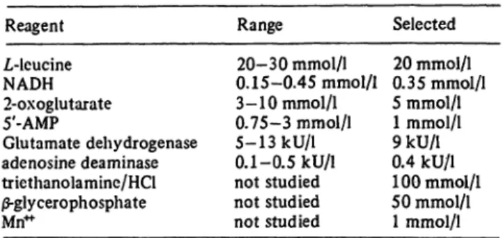
Tab. 1. Optimal concentrations of reagents in the cuvet Reagent
L-leucine NADH 2-oxoglutarate 5'-AMP
Glutamate dehydrogenase adenosine deaminase triethanolamine/HCl
|S-glycerophosphate Mn"
Range 20-30 mmol/1 0. 15-0.45 mmol/1 3- 10 mmol/1 0.75-3 mmol/1 5-13 kU/1 0.1-0.5 kU/1 not studied not studied not studied
Selected 20 mmol/1 0.35 mmol/1 5 mrnol/1 1 mmol/1 9kU/l 0.4 kU/1 100 mmol/1 50 mmol/1 1 mmol/1
To obtain the activity in U/l, multiply the ΔΕ/min by the factor 1850. This factor is calculated as follows:
Factor = Vt= total voume, Vs = sample volume
e X Vs ' S
and e is the molar absorption coefficient of NADH at the wave length used (6.22 αη2/μιηο1).
Results and Discussion
Optimal L-leucine concentration
To establish the optimal concentration of I-leucine for the activity of glutamate dehydrogenase, the following experiment was carried out. Five working solutions were prepared with i-leucine concentrations of 5,10, 15, 20, and 30 mmol/1. Five min after reagent prepara- tion glutamate dehydrogenase activity was measured at 340 nm after adding 0.07 μιηοΐ NH4C1 to 2 ml aliquots of the working solution. According to figure 1 a /,-leu- cine concentration of 20 mmol/1 and more is optimal.
This result disagrees with the literature where a L-leu- cine concentration of 10 mmol/1 is already optimal (4, 7). An explanation of this different result is the high NADH concentration in the working solution. In the
0.07-
Ξ 0.06
l r 0.05
Γ·
-S 3 0.04ο*'ε3 0.03
10 15 20
L-Leucine [mmol/l] 25 30
proposed method: 0.35 mmol/1, Persifn et al (7):
0.19 mmol/1, Jung et al (4): 0.25 mmol/1. A L-leucine concentration of 20 mmol/1 was chosen in the proposed
method and used in further experiments.
Glutamate dehydrogenase stabilisation To evaluate the glutamate dehydrogenase stabilising effect of L-leucine, and the influence of Mri**, working solutions containing all reagents in their optimal con- centration (sol. A), with Mn**" (1 mrnol/1) and no i-leucine (sol. B) and no L-leucine and Mri* (sol. C) were prepared and allowed to age for 60 min at 25 °C. Directly following reagent preparation and then at 15 min intervals, gluta- mate dehydrogenase activity was assayed by adding 0.07 μήιοί NH4C1 to 2 ml aliquots of the solutions and measuring the decrease in absorption at 340 nm during the first min. The results shown in figure 2 indicate a rapid inactivati n of glutamate dehydrogenase in the absence of L-leucine. Although the presence of Mn4* in a concentration of 1 mmol/1 slightly inhibits the glutamate dehydrogenase activity (1), it is necessary for activation of 5'-nucleotidase (8, 9). In addition it also has a small stabilising effect on glutamate dehydrogenase. This is in accordance with the findings of Ellis & Goldberg (I).
When the working solutions were aged at 37 °C essen- tially the same results were obtained. At this tempera- ture however, sol. A exhibited after 2.5 hr a glutamate dehydrogenase activity of 80% of the initial value. This has no practical consequences because of the broad optimal range of glutamate dehydrogenase activity (tab. 1). Absence of Mn4* in solution A in this experi- ment had no effect on the stability. It is evident that incorporation of L-leucine in the working solution con- tributes considerably to its stability at room and assay temperature.
20 30 40 50 Aging of reagent [min] 60
Fig. 1. Effect of different L-leucine concentrations on gluta- mate dehydrogenase activity.
Fig. 2. Effect of Zrleucine (20 mmol/1) and Mn" (1 mmol/1) on
» glutamate dehydtogenase stability. Working solutions A, B, and C contained Ζ,-leucine and Mri" (o o), no I-leucine (x x) and no I-leucine and Mri" (Δ Δ) resp.
J. Clin. Chem. Clin. Biochem. / Vol. 14,1976 / No. 3
Arkesteijn: A kinetic method for serum 5'-nucleotidase 157
Optimal NADH concentration and effect of endogeneous ammonia
The optimal NADH concentration in the working solu- tion was established with 3 serum samples with a normal, an elevated and a high S'-nucleotidase activity resp.
The NADH concentration ranged from 0.15—0.45 mmol/1 (fig. 3), the other constituents had the selected values as given in Table 1. The concentration of 0.35 mmol/1 was chosen for the proposed method to ensure an opti- mal NADH concentration during the assay, even at high endogeneous ammonia levels and, as a consequence, high NADH consumption during the pre-incubation time. Figure 4 illustrates the decrease in absorbance during a pre-incubation of 3 serum samples with 5'- nucleotidase activities of 25, 63 and 125 U/l resp. The serum sample of 63 U/l was enriched with ammonia to a concentration of 250 μπιοΐ/ΐ. After a rapid drop in absorbance, initial decrease in absorbance approaches zero after approximately 25 min and corresponds to a S'-nucleotidase activity of less than 0.5 U/l, even in a sample with a high ammonia concentration. The results indicate that a pre-incubation time of 30 min effectively eliminates side reactions causing non-specific NADH con- sumption. To demonstrate that after the pre-incubation the NADH concentration is still high enough to carry out the assay, 2 serum samples with different amounts of ammonia were pre-incubated, followed by the determina- tion of the 5'-nucleotidase activity. The results are shown in table 2. It is obvious that ammonia up to a concen-
Tab. 2. Effect of endogeneous ammonia.
ammonia added
lMmol/l] 0 50 125 250 5'-nucleotidase activity [U/1J
sample A 29 30 29 29 sample B 63 62 62 59
190 -
170
= 150
tj 130
1S
20
I ^ 0 0.10 0.20 0.30
NADH [mmol/l]
Fig. 3. Optimal Zrleucine concentration.
ΟΛΟ 0.50
0.10
2 3 4 5 1 0
Pre-incubation time (mm) 30
Fig. 4. NADH consumption during pre-incubation. S'-nucleo- tidase activity of serum samples: 154 U/l (x x), 63 U/l (Δ Δ) and 25 U/l (o o). Sample (Δ Δ) contained 250 /umol added ammonia.
tration of 250 μπιοΐ/ΐ has no effect on the determination of S'-nucleotidase activity.
Optimal concentrations of reagents
The optimal concentrations of 2-oxoglutarate, 5'-AMP, glutamate dehydrogenase, adenosine deaminase and the pH value given in the literature were checked in the proposed method. Experiments with sera having a normal and an elevated S'-nucleotidase activity resp. revealed that maximal and constant activities were obtained when final reagent concentrations in the reaction mix- ture were within the ranges shown in table 1. These ranges are in good agreement with the optimal ranges given in the literature (1, 2, 4, 7). For the additional reagents Na-jS-glycerophosphate, Mn4* and the buffer
40 -
0 2 10 20 30 40 5'-Nucleotidase (method of Persijn et al) fU/ll(37°C)
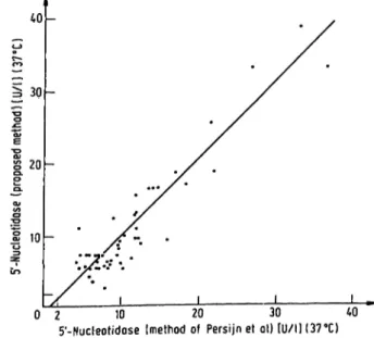
Fig. 5. Comparison of proposed method with reference method.
J. Clin. Ghem. Clin. Biochem. /Vol. 14, 1976 / No. 3
158 Arkesteijn: A kinetic method for serum 5'-nucleotidase
composition the recommendations of Ellis & Goldberg were followed (1). For the routine procedure final concentrations in the cuvet were selected as given in table 1.
Comparison with other method
This method was compared with the technique of Per- sijn et al. (10) by parallel analyses of 51 serum samples.
Hie results are plotted in figure 5. The regression equa- tion (y = l.OSx - 0.69, Sy · χ = 4.8 and the coefficient of correlation is 0.937) indicates a good correlation between the two methods.
Precision and normal values
The interrun precision in the normal range was 6.5%
(x - 9.8, ri = 22) and in the elevated range 4.9%
(x> 23.6, η = 22). The normal range, compiled from values of 35 healthy persons, was found to be 2.1-10.6 U/l (95% confidence limits).
Acknowledgements
The auther wishes to express his gratitude to Dr. J.-P. Persijn for his interest and for providing the serum samples for the parallel analyses and to miss K, AI ting and miss E. C. P.
Videler for their technical assistance.
References
1. Ellis, G. & Goldberg, D. M. (1972). Analyt. Lett. 5, 65-73.
2. Bootsma, J. & Wolthers, B. G. (1972). Clin. Chim. Acta 41, 219-222.
3. Frieden, C. (1959). J. Biol. Chem. 25^808-814.
4. Jung, K., Sokolowski, A. & Egger, E. (1972/1973).
Enzyme 14, 44-54.
5. Ellis, G. & Goldberg, D. M. (1970). J. Lab. Clin. Med. 76, 505.
6. Yielding, K. L & Tomkins, G. M. (1961). Proc. Nat. Acad.
Sei. 47, 983-989.
7. Persijn, J.-P., Van der Slik, W., Timmer, C. J. & Riethorst, A.
(1970). Clin. Chim. Acta 30, 377-386.
8. Ellis, G. & Goldberg, D. M. (1970). Spectrovision -23, 8-12.
9. Belfield, A, & Goldberg, D; M. (1969). J. Clin. Path. 22, 144 10. Persijn, J.-P. & Van der Slik, W. (1970). Clin. Enzym. 2,
108-112.
Hofpoort Ziekenhuis Klin. Chem. Lab.
Utrechtsestraatweg 52 Woerden
The Netherlands
J. Clin. Chem. Clin. Biochem. / Vol. 14,1976 / No. 3