Persijn, van der Siik and Engelsman: A New Method for the Determination of Serum Nudeotidase 77
Z. klin. Chem. u. klin. Biochem.
10. Jg. 1972, S. 77—85
A New Method for the Determination of Serum Nucleotidase
1)
V (Final Part). Evaluation of the Response of Serum Nucleotidase to Bone Metastases and Therapy in Cancer Patients
By J.-P. PERSIJN, W. VAN DER SLIK and E. ENGELSMAN
From the Department of Clinical Chemistry (Head Dr. J.-P. Persijn), Netherlands Cancer Institute, Amsterdam, and the Department of Clinical Chemistry (Head Drs. W. van der Slik) State University Hospital Leiden, The Netherlands
(Eingegangen am 24. August 1971)
This paper deals with comparative studies on the levels of alkaline phosphatase, the presence of bone phosphatase as measured electro- phoretically, and levels of S'-nucleotidase, as measured in the presence of phenylphosphate in sera of patients suffering from bone me- tastases.
Secondly, the levels of 5'-nucleotidase and other serum enzymes were studied in cancer patients during various kinds of therapy.
A classification of serum enzymes is proposed, based on their preferential response to either liver metastases or toxic liver injury or both.
Es wird über vergleichende Untersuchungen der Aktivität der alkalischen Phosphatase, das Vorkommen von Knochenphosphatase durch elektrophoretischen Nachweis und der Aktivität der S'-Nucleotidase, bestimmt in Gegenwart von Phenylphosphat, im Serum von Pa- tienten mit Knochenmetastasen berichtet.
Die Aktivität der 5'-Nucleotidase und anderer Enzyme im Serum wurden bei Karzinom-Patienten während verschiedener therapeutischer Verfahren bestimmt. Eine Einteilung der Serumenzyme entsprechend ihrem bevorzugten Auftreten entweder bei Lebermetastasen oder bei toxischen Leberschäden bzw. beiden wird vorgeschlagen.
Introduction
In 1968 we introduced a method for the measurement of serum S'-nucleotidase utilizing adenosine deaminase to generate ammonia during incubation. After a certain period the BERTHELOT colour reaction for ammonia was applied which terminated the incubation without deproteinization. The extinction of the colour was found to be a sensitive measure of 5'-nucleotidase activity (1,2)·
A modification was presented in a following paper: the addition of phenylphosphate as an inhibitor of the nucleotidase effect of bone phosphatase (3).
Evidence was presented in ref. (3) for the ability of phenylphosphate to inhibit the hydrolysis of AMP by bone phosphatase. In this study, however, sera with elevated levels of alkaline phosphatase were used from patients suffering from non-malignant bone diseases.
The present paper deals with comparative studies of 5'-nucleotidase and alkaline phosphatase of patients with malignant diseases affecting bone or bone and liver. For this purpose sequential studies of the enzymes were made and a greater insight was obtained into the clinical significance of consecutive S'-nucleotidase assays using phenylphosphate as an inhibitor of interference by alkaline phosphatase. In addition, attention was paid to the influence of hepatotoxic therapy on the levels of these enzymes.
x) Enzyme. 5'-Nucleotidase = S'-Ribonucleotide phosphohydro- lase (EC 3.1.3.5); Aspartate aminotransferase = L-Aspartate:
2-oxoglutarate aminotransferase (EC 2.6.1.1); Alanine amino- transferase = kL-Alanine: 2-oxoglutarate aminotransferase (EC 2.6.1.2); Alkaline phosphatase = Orthophosphoric monoester phosphohydrolase (EC 3.13.1); Glutamate dehydrogenase = fc-Glutamate: NAD oxidoreductasc (deaminating) (EC 1.4.1.2).
An attempt was made to evaluate the differential diag- nostic significance of serum enzymes in the follow-up of cancer patients by comparison with the response of other enzymes such as serum aspartate-aminotransferase and alanine aminotransferase. This report concludes our series: "A Method for the Determination of Serum Nucleotidase".
Materials and Methods
5'-Nucleotidase
A complete description of the procedure for S'-nucleotidase can be found in ref. (4). The reader can also find all the particulars by combining the procedure from ref. (1), (2) and (3). Readings were done with a Gilford 300 microspectrophotometer.
Alkaline pbospbatase
The measurement was performed with the Auto-Analyzer using /Miitrophenyl phosphate as a substrate and 2-amino-2-methyl- propanol-1 as a buffer (5). The advantage of this buffer is that a 100 U/l sample (high normal) gives a chart reading of ± 32 scale units (transmission, full scale = 100 scale units, 8 min. in- cubation), which is about twice as high as with glycine buffer.
Dilution of serum is necessary above ± 500 U/l. In addition the substrate produces its own chromogen. Real incubation time was regularly checked and if necessary corrected for by comparison with results from the manual method. This was done particularly after renewal of the manifold.
Aspartate aminotransferase
The measurement was done with Boehringer reagent kit (non- optimal test).
Alanine aminotransferase
This enzyme was assayed with Boehringer reagent kit (non- optimal test).
Both transferases were assayed either with the aid of a mechanical cuvet changer (at 25°) with ten minutes intermittent registration or with an LKB reaction rate analyzer (at 35°). In the latter case the automatic dispenser was set for 2-oxoglutarate so-
Z. klin. Chem. u. klin. Biochem. /10. Jahrg. 1972 / Heft 2 11
lution, since smaller volumes occasionally gave erratic results.
The 2-oxoglutarate solution from the reagent kit was diluted so as to obtain the prescribed final concentration. The values were reguraly checked by comparison with data obtained from meas- urements at 340 nm with the Zeiss PMQII.
Electropboretic serum phosphatase fractionation
Patterns of iso-enzymes of alkaline phosphatase were obtained with agar gel electrophoresis according to HAYE and DE JONG (6), with the exception that a solution of naphthylphosphate and Fast Blue BB (Dajac) and MgSO4 (0.07M) in borate buffer (pH 9.0) were used for the demonstration of enzyme activities (30 to 60min. incubation at 37°). The HAYE and DE JONG technique was found to be superior to other methods tried and to give very reliable results. As a control each slide with the serum to be treated electrophoretically \vas placed between two slides, treated identically, on which serum from patients suffering unmistakably from liver diseases had been applied. These latter specimens were selected on the basis of elevated alkaline phosphatase levels from clear-cut cases of liver disease (gallstone etc.) without bone being involved. After electrophoresis samples and controls were simul- taneously subjected to the procedure to locate the iso-enzymes.
By dilution experiments it was found that below approximately 200—150 U/l bone phosphatase could not be detected clearly in the iso-enzyme pattern.
Serum glutamate dehydrogenase
This enzyme was assayed according to SCHMIDT (7) or to PERSIJN and Co workers (8).
Note on automation of 5'-nucleotidase assay
In view of the increasing interest in 5;-nucleotidase as a clinical test a procedure for automated assay would be useful. Attempts to realise a sensitive automated procedure for our method using either the Auto-Analyzer system or the LKB reaction rate ana- lyzer have failed. With the latter design the BERTHELOT colour reaction was replaced by the reductive amination of 2-oxogluta- rate catalysed by bovine glutamate dehydrogenase in the presence of NADH. It was assumed that the concentration of S'-nucleo- tidase was rate determining and that the rate of decrease in ab- sorbance at 340 nm caused by the conversion of NADH to NAD would be a measure of S'-nucleotidase. To stabilize glutamate dehydrogenase L-leucine was added (8). It was striking that without serum a decrease in absorbance was observed as soon as AMP was added to initiate the reaction.
The rate of decrease was markedly lower in the absence of L- leucine and strongly suppressed by diethylstilboestrol. Since
these substances are known respectively to enhance and to sup- press bovine glutamate dehydrogenase (9) it seems reasonable to assume that bovine glutamate dehydrogenase and not a conta- mination is able to catalyse some kind of reaction between the above mentioned constituents while transforming NADH to NAD. This would make an accurate measurement of a kinetic glutamate dehydrogenase linked S'-nucleotidase assay impossible.' Using glutamate dehydrogenase from Clostridium (known to have a much lower Km for ammonia (10)) gives an identical reaction.
The main problem when using the Auto-Analyzer was the long incubation time necessary to produce sensitive recordings.
Results
5'-Nucleotidase and alkaline phosphatase
It seemed interesting to accumulate data which would allow us to make statements about the amount of bone phosphatase present in sera with elevated alkaline phos- phatase levels. With this in view the following was done.
a) The iso-enzyme pattern of an arbitrarily chosen num- ber of sera with elevated alkaline phosphatase and normal 5'-nucleotidase was studied.
b) A number of sera, in which the S'-nucleotidase was less elevated than the alkaline phosphatase, was examined electrophoretically for the presence of bone phospha- tase.
The results of these studies are given in Tables 1 and 2.
In these tables pregnant female patients are not included.
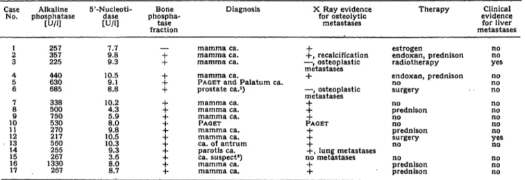
Table 1 shows that in practically all cases bone phospha- tase is present if S'-nucleotidase level is normal. As a typical illustration case no. 16 is described here in detail.
Figure 1 represents' a patient suffering from breast carcinoma with osteolytic metastases confirmed by bone biopsy. During the course of time, as indicated on the abcissa no clinical indication of the existence of liver metastases was found.
During the first two weeks of therapy, only 5'^-nucleo- tidase and aspartate and alanine transaminase levels were elvated. Prednison therapy was especially succesf ul,
Tab. 1
Results of electrophoretic examination of sera on the presence (-f-) or absence (—) of alkaline bone phosphatase fraction in the case of normal 5'-nucleotidase levels
Case Alkaline No. phosphatase
[U/l]
21 3 45 6 78 109 1112 1314 1516 17
257357 225 440630 685 338500 750530 270217 560255 1330267 267
5'-Nucleoti- [U/l]dase
7.79.8 9.3 10.59.1 8.8 10.24.3 5.98.0 10.59.8 10.39.3 3.68.0 8.7
Bone Diagnosis phospha-
fractiontase
— mamma ca.
+ mamma ca.
+ mamma ca.
-f mamma ca.
4- PAGET and Palatum ca.
+ prostate ca.1) + mamma ca.
4- mamma ca.
4- mamma ca.
4- PAGET -f- mamma ca.
4- mamma ca.
+ ca. of antrum + parotis ca.
4- ca. suspect2) -f mamma ca.
-f- mamma ca.
X Ray evidence for osteolytic
metastases
+
+, recalcification
— , osteoplastic metastases_j_
— , osteoplastic metastases
4-4-
PAGET +-j-
+4-, lung metastases no metastases
-j-
+
Therapy
estrogen
endoxan, prednison radiotherapy endoxan, prednison nosurgery
noprednison nono prednison surgery no prednisonno prednison
Clinical evidence for liver metastases
nono yes nono no nono nono yesno no nono no Acid phosphatase level elevated (41 U/l).
Young girl suspect for malignant disease but found to have no malignancy.
Z. klin. Chem. u. Win. Biochem./ 10. Jahrg. 1972/Heft2
Persijn, van der Slik and Engelsman: A New Method for the Determination of Serum Nucleotidase 79
Tab. 2
Results of electrophoretic examination of sera on the presence (-f) or absence (—) of alkaline bone phosphatase fraction in case of elevated 5'-nucteotidase levels
CaseNo.
21 34 56 78 109 1112 1314 1516 1718 2019 21
Alkaline phosphatase
[U/l]
412625 245568 1720344 3100660 1140595
470880 375630 388540 21501720
485339 398
5'-Nucleoti- [U/l]dase
13.717.9 49.017.4 26.611.3 193.013.4
32.815.4 69.418.8 21.636.8 12.619.1 64.825.5 23.012.8 23.6
Bone phos- phatase fraction
+ 4-+
— —4.
-h+ -j~+ .4.4- 4--j-
4-4~4- -f.4-
—
Diagnosis
mamma ca.
mamma ca.
bronchus ca.
M. HODOKIN mamma ca.
mamma ca.
rhabdomyosarcoma mamma ca.
mamma ca.
mamma ca.
chordoma bronchus ca.
oesophagus ca.
bronchus ca.
mamma ca.
ca. of the bladder ca. of the bladder mamma ca.
mamma ca.
osteosarcoma mamma ca.
X Ray evidence for osteolytic metastases
— , osteoplastic metastases -f-, recalcification -j-•f
osteoplastic metastases
•f, recalcification
— , osteoplastic metastases
— , osteoplastic metastases
-j- .μ4--j-
— , osteoplastic metastases
+
Therapy
radiotherapy androgens noradiotherapy surgery estrogen radiotherapy estrogen radiotherapy 5-fluoruracil leukeran nono
radiotherapy prednison actinomycin noandrogens prednison androgensno
Clinical evidence for liver metastases
yesno yesno nono noyes yesho yesyes yesno yes1) nono nono noyes
^Elevated 5'-nucIeotidase 1 month before.
Estrogen
Prednison 1600
1400
§1200
>>
liooo
t
600400 200
- =2
;40»
I
120
Androgens
b.f.+
" m " 1 2 3 4 5
Time [months]
Fig. 1
Variation, with time, of alkaline phosphatase (o—o) and 5'-nu- cleotidase (·— ·) levels. Patient Ze. Q
The dotted line represents upper normal level of alkaline phosphatase and 5'-nucleotidase. The sign b. f. + indicates, that a bone fraction
could be demonstrated
since it resulted in normalization of the osteolytic areas.
This was revealed by
a) X ray investigation showing recalcification;
b) a rise of alkaline phosphatase level in the serum, which was shown dectrophoretically to contain bone phospha- tase (right arrow).
This graph illustrates that even if alkaline phosphatase level rises to a value 28 times its mean normal level, 5'- nucleotidase can remain in the normal range.
Table 2 shows that in the majority of cases a bone phos- phatase fraction is present if 5'-nucleotidase levels are elevated, but to a lesser degree than alkaline phosphatase.
A typical illustration (case No. 18) has been given in Figure 2. Patient Ni (fig. 2) and patient Ze (fig. 1) pre-
2800 2600 2400- 2200 2000 51800
•51600o
-21400
-C
1.1200
.£O)
11000
Estrogen
800 600 400 200
- 100 -=80
-540 -= 20 0L
H. Androgens. Prednison
b.f.+
\
0 1 2 3
Time [months]
Fig. 2
Variation, with time, of alkaline phosphatase (o—o) and 5'-nucleo- tidase (·—·) levels. Patient Ni. 9
The dotted line represents upper normal level of alkaline phosphatase and 5'-nucleotidase. The sign b. f. -f indicates, that a bone fraction
could be demonstrated
seated similar clinical pictures. Estrogen therapy was stopped because of hypercalcaemia and replaced by prednison therapy. It is interesting to note that amino-
2. klin. Gbem. u. klin. Biochem./ 10. Jahrg. 1972 / Heft 2 11*
transferases fluctuated closely parallel to the 5'-nucleo- tidase level during the first month indicated. The fraction from bone could be demonstrated electro- phoretically (see arrow) in a serum sample with a 5'- nucleotidase level of 25.5 U/l. A repeated assay of this sample for S'-nucleotidase but omitting phenylphosphate gave a 5'-nucleotidase value of 45.9 U/l.
There is no indication that the sharp increase of alkaline phosphatase would elevate the level of 5'-nucleotidase.
In fact S'-nucleotidase level starts to rise just when alkaline phosphatase begins to fall.
From this point liver phosphatase may be expected to increase at some time or other. Apparently this increase is not sufficient to neutralize the decrease of alkaline phosphatase to an amount of 1700 U/l within 35 days.
In this respect it should be noted that return of increased alkaline phosphatase levels due to bone repair usually takes several months (see for example fig. 5 ref. 11).
In a previous paper (11) we reported evidence that the 5'-nucleotidase level rises before the alkaline phospha- tase level if the liver is primarily involved. After studying this phenomenon more extensively in many hundreds of patients the following characteristics of S'-nucleo- tidase and alkaline phosphatase can be described: serum S'-nucleotidase level will rise to a greater degree than the alkaline phosphatase level and before the rise of the alkaline phosphatase level. A typical illustration is given in Figure 3. This figure shows plots of serum alkaline
clear-cut cases of osteoblastic processes such as recal- cification of osteolytic areas or the presence of osteo- blastic metastases. Two cases will serve as an illustration.
Patient M (fig, 4) was suffering from breast carcinoma.
The liver was found to be enlarged on palpation three weeks before onset of therapy. Evidence for the recalci- fication of osteolytic areas was found at the time in- dicated by arrow A. Subsequently progression of osteo- lytic areas took place. Note the. ,transient increase of alkaline phosphatase level above that of 5'-nucleotidase during a period in which there was evidence for recalcification. At the time indicated by arrow B a bone phosphatase fraction could not be detected electro- phoretically. Aminotransferases rose parallel to the in- crease of 5'-nucleotidase.
Figure 5 shows serum enzyme levels of a patient suffering from breast carcinoma with osteolytic metastases. At the
Prednison
Time [months]6 10
Fig. 3
Variation, with time, of alkaline phosphatase (o—o) and S'-nu- cleotidase (·—·) levels. Patient De. 9
The dotted line represents the upper normal limits
phosphatase and 5'-nucleotidase levels in a patient suffering from M. HODGKIN. Aminotransferase levels were hardly elevated. X ray examination detected no bone metastases. The data of Figure 3 indicate that the 5'- nucleotidase level is increasing before that of alkaline phosphatase.
On the other hand it can occur that the alkaline phospha- tase level rises before that of 5'-nucleotidase and even more than the latter. Such a phenomenon occurs in
Endoxan 600r 60
:> 400
i.200-"520.2
1 2 3 4 5 6 7
Time [months]
Fig. 4
Variation, with time, of alkaline phosphatase (o—o) and 5'-nucIeo- tidase (· — ·) levels. Patient M. 9
The dotted line represents the upper normal limits.
Androgens
J —-800 Prednjson.
:E600 1-400
••200
6 10Time [months]
Fig. 5
Variation, with time, of alkaline phosphatase (o—o), 5'-nucleo- tidase (·—·), aspartate (25°, ^—^) and alanine (25°, z±—Δ)
transaminase levels. Patient S. 9
The dotted lines represent the normal limits of aspartate and alanine transaminase (upper) and alkaline phosphatase and 5'-nucleotidase
(lower).
Z. klin. Chem. u. klin. Biochem./10. Jahrg. 1972/Heft 2
Persijn, van der Slik and Engelsman: A New Method for the Determination of Serum Nucleotidase 81 time indicated by arrow A there was progression of the«
osteolytic metastases. A report of X ray investigation mentions the cessation of progression and recalcification at time B.
Recalcification was confirmed at time C. Evidence for progression of osteolytic metastases in bone and liver indicating the end of the remission phase was found at the time indicated by arrow D. Note that alkaline phos- phatase increases above 5'-nucleotidase only during recalcification and that S'-nucleotidase level rises prior to the level of alkaline phosphatase before therapy started and at the end of the remission phase.
S'-Nucleotidase and transaminases
The sequential course of 5'-nucleotidases and amino- transferases was studied in 50 patients suffering from malignant diseases known to give liver metastases. Care was taken to exclude patients with apparent toxic liver damage as described below. We cannot provide proof that among these patients some may not have an elevated S'-nucleotidase from a nonmalignant cause, such as biliary obstruction or cirrhosis. Study of the variation in serum enzymes over a period and their response to therapy made the occurrence of non-malignant liver disfunctions improbable. In 39 cases the S'-nucleo- tidase level rose prior to the rise of the aspartate amino- transferase level and generally to a much higher value.
In 2 cases S'-nucleotidase and aspartate aminotransferase activity rose equally. Examples have been given in a previous paper (ref. (11), fig. 8, 12 and 13) and in this paper (fig. 5).
Additional information on the response of aspartate aminotransferase in comparison with 5'-nucleotidase was obtained by assembling data from autopsy or surgery reports confirming the existence of liver metastases (tab. 3). The reports were selected at random. Each case in table 3 is used only once in this paper. The highest
level of each serum enzyme mentioned was chosen for use in table 3. This was done because in the final stage transaminases probably tend to fall. Since the table in addition records S'-nucleotidase and alkaline phospha- tase elevations, a fair comparison of transaminases to these enzymes would not be possible if the last values available before death had been given. From the data of table 3 it seems evident that aspartate aminotransferase is a less sensitive parameter for the existence of liver metastases than S'-nucleotidase, but that omitting aspartate aminotransferase from routine examination is not advisable.
Alanine aminotransferase is a less sensitive monitor of liver metastases, which agrees with data from many reports in the literature (see ref. (11)). The foregoing studies provided a basis for evaluating the significance of assays of S'-nucleotidase in relation to some other serum enzymes in cases of metastatic liver and bone. Cancer patients however often receive therapy which would be reflected by changes in the pattern of serum enzymes.
It seemed therefore interesting to study the response of S'-nucleotidase to such therapy in relation to other serum enzymes. While these studies were being carried out it became evident that there is no "standard" response to a given therapy. In fact a therapy may be without measurable effect on the levels of any enzyme in one case while effecting a variety of transient rises of serum enzymes in other cases.
Studies were carried with about 40 patients whose serum enzymes showed at least a partial response to therapy.
In some cases these patients received several subsequent therapeutic treatments which permitted us to study the effect of possible accumulation of the hepato-toxic effects of serum enzymes. The results can be summarized as follows:
a) If there is a response, the transient rise of S'-nucleo- tidase is generally between a few U/l and about 20 U/l.
Tab. 3
Classification of aspartate and alanine aminotransferase, S'-nucleotidase and alkaline phosphatase in cases of confirmed metastatic liver1)
') Notations
db+ -
+ ++ + + - 4- + + H- «
Case 5'-Nucleoti- Alkaline No. dase phosphatase
1 + + + + +
• 2 + ± 3 normal normal 4 4- + + +*) 5- + + + + + + + +1) 6 +-f + + + 7 + ± 8 + + + + + + 9 ± ±
10 ++ ++»)
Π + + + + 12 + + + + + + 13 ++ + 14 normal + 15 + + + + + 16 + + + + + 17 ++ + 18 normal normal 19 normal normal 20 ++ ±
Aspartate aminotrans-
ferase + + + + + + + + normal
+ + +
•f+ + normal
+ + + H- + 4-± H-+ + normal+ normal+ ++ + +
±+
between upper limit normal to twice elevated between twice elevated to 4 χ elevated between 4 χ elevated to 8 χ elevated between 8 χ elevated to 16 χ elevated more than 16 times elevated.
Alanine Diagnosis aminotrans-
ferase
-|- mamma ca.
+ + liver ca.
normal reticulosarcoma + mamma ca.
4- mamma ca.
normal rectum ca.
normal cardia ca.
+ mamma ca.
normal mamma ca.
-f mamma ca.
± rnamma ca.
+ seminoma 4- ca. of the bladder normal lung ca.
normal mamma ca.
normal ovarium ca.
± mamma ca.
-(- parotis ca.
normal mamma ca.
± mamma ca.
*) Recalcification of osteolytic areas
Metastatic liver confirmed by autopsy autopsy autopsy autopsy autopsy autopsy laparotomy autopsy laparotomy autopsy autopsy liver scintigraphy autopsy1) autopsy5) autopsy autopsy laparotomy autopsy autopsy0) laparotomy7) (X Ray evidence).
') Osteoplastic metastases (X Ray evidence).
4) A few metastases of ± 0,5 cm.
6) One liver metastase of ± 0.3 cm.
e) y-glutarnyltranspeptidase ^ -f -f.
7) y-glutarnyltranspeptidase: -f··
Z. klin. Chem. u. klin. Biochem./ 10. Jahrg. 1972/Heft2
Higher elevations seldom occur but can be found e. g.
following splenectomy (one case has been mentioned in ref. (3)). Elevations of alkaline phosphatase are of the same order up to the upper normal limit,
b) In contrast alanine aminotransferase and aspartate aminotransferase levels showed a much greater variety in response. Extensive elevations could be found, whereas 5'-nucleotidase and alkaline phosphatase did not show significant alterations.
For S'-nucleotidase, alkaline phosphatase, aspartate aminotransferase and alanine aminotransferase the length of time from the onset of therapy till the return of serum engines to their previous values was approxi- mately equal, i. e. 20—40 days.
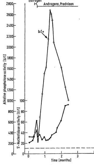
Figure 6 describes 4 cases where the response of 5'- nucleotidase, alkaline phosphatase and aminotransferases was weak to moderate. Figure 6 a concerns a patient
800r 80r
a testis tumour. Two days after the resection aspartate aminotransferase value rose to 58 U/l while alanine aminotransferase was only reached the upper normal value. Figure 6d shows the levels of serum enzymes of a patient with liposarcoma in the leg, who received, intraveneous injections of both vincristine and ctino- mycine (see arrows). It would appear that alanine amino- transferase attains high'er values than aspartate amino- transferase. Additional evidence is presented in figures 7,
20 40 0
Time(days! 20 40
Fig. 6
Variation, with time, of alkaline phosphatase (o—o), 5'-nucIeotidase (e—.); aspartate (35°, ^ — ^) and alanine (35°, /x—-Δ.) trans-
aminase levels
a = Patient G. Q b = Patient Wa. cf c = Patient St. cf d = Patient K. Cf
The upper dotted line represents upper normal limit of aspartate and alanine transaminase, the lower dotted line those of 5'-nucleotidase
and alkaline phosphatase
suffering from M. HODGKIN who had been treated by splenectomy (left arrow). During splenectomy a biopsy was taken from the liver which did not reveal the pre- sence of any metastatic tissue. At the time indicated by the right arrow the patient received intravenous chemo- therapy involving vincristine, natulan and prednison;
alanine aminotransferase only shows a clear reaction after the second treatment. Figure 6b concerns a patient suffering from a carcinoma of the stomach. Examination of the liver during partial gastrectomy (arrow) failed to detect the presence of metastases.
Figure 6c describes the variation of the serum enzymes following surgical therapy of lung metastases from
Metofhrexate Androgens
^
— ^
^
-"ξ
tsα
CD CDCD CDZ^ «XI1 3SD}oqdsoL|d
O)c:
"o
3· r·< (
- 160 -Il40
~
"o
" E
120 'ec-jflOOσ cucr
- §80
σ-ζ}
Cσ
-S 6001
σ σ
I I -Nucleotidose Γ*0 *- CD CD 10
i nL- U
• 1 I
α
1
111 I1
_
JA I tj
, . ,
~:-l\~
— 4τ^^4τ^ —
0 30ι ι
*!
' i l l
-
II,'/
IIII
/\!
Time [d.oys]0 30
Fig. 7
Variation, with time, of alkaline phosphatase (o—o), 5'-nucleotidase (.—,); aspartate (35°, -A.—^) and alanine (35°, ^—Δ), transaminase
levels
a = Patient R. cf b « Patient Z. 9
The upper dotted line represents upper normal limit of aspartate and alanine transaminase, the lower dotted line those of S'-nucleotidase
and alkaline phosphatase Metothrexafe
£^o=20Ur:g=2ZOr
ffioohlfiohE^
f-g o
Lf * o
1— ^»,
10 30 50 70
<c "^ Time [days]
Fig. 8
Changes of 5'-nucleotidase (·—·), alkaline phosphatase (o—a), aspar- tate (35°, Α.-Τ-Α) and alanine (35°, -Δ.—Δ) transaminase levels during several courses of intermittent metothrexate treatment.
Patient Be. 9
The dotted lines represent the upper normal limits
2. klin. Chem. u. klin. Biochem./ 10. Jahrg. 1972/Heft2
Persijn, van der Slik and Engclsman: A New Method for the Determination of Serum Nucleotidase 83 Prednison
2000-
Timeldoys]30 60
Fig. 9
Variation, with time, of aspartate (35°, A.—^) and alanine (35°, Δ—Δ) transaminase (top). The dotted line represents the upper normal limits. Variation, with time, of alkaline phosphatase (o—o), 5'-nucleotidase (·—·) and glutamate dehydrogenase ( x — x ) (bottom). The upper dotted line represents the upper normal limits of alkaline phosphatase and 5'-nucleotidase, the lower dotted line the
upper normal limit of glutamate dehydrogenase. Patient No. 9
8 and 9. Patient R (fig. 7 a) was suffering from a tumour in the lower jaw. A intraarterial catheter (for infusion therapy) was placed during general anaesthesia.
Figure 7 a shows changes in the level of enzymes in the blood as a response to infusions of 3H-thymidine and metothrexate at the time indicated by the arrow, plus metothrexate over 7 days. Patient Z. (fig. 7b) received five injections of androgens. The treatment was stopped because of clinical evidence of toxicity. Ovarectomy was performed two months before, when examination of the liver did not reveal the presence of metastases. Note the weak response of 5'-nudeotidase in both cases.
Figure 8 is an example of variations of serum enzymes during intermittent courses of therapy. Note the variety in response of the transaminases and the weak response of S'-nucleotidase and alkaline phosphatase. This figure concerns a patient suffering from chorionepithelioma.
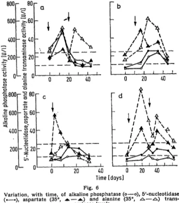
An interesting illustration of the weak response of 5'-
nucleotidase and alkaline phosphatase to toxic effects of therapy as compared to aspartate aminotransferase and alanine aminotransferase is given in Figure 9. This figure concerns a patient suffering from melanoma. At the time indicated by the arrow inguinal lymph nodes were excised under general anesthesia using halothane (fluothane). The pronounced response of aspartate aminotransferase, alanine aminotransferase and gluta- mate dehydrogenase immediately after surgery is apparently a hypersensitivity reaction to this hepato- toxin, which is known to cause histological changes in a few cases indistinguishable from viral hepatitis (12).
The serum enzyme with the highest response was aspar- tate aminotransferase (3000 U/l, 35°). It will be noted that the S'-nucleotidase level did not rise more than 12 U/l at most. Note that glutamate dehydrogenase, a liver specific enzyme, increases to about twenty times the normal limit. Nineteen days later the patient received prednison therapy which evoked again a small rise of S'-nucleotidase. It would appear that the response of the aminotransferases to therapy is overshadowed by their response to halothane. It is of interest to note that serum bilirubin did increase to 170 mg/1 (283 μηιοΐ/ΐ) at the 19th day after surgery. Fever was noted during the five postoperative days. The data of the other cases are illustrated in table 4. The greatest changes of serum enzymes as a response to therapy are classified, using the same symbols as in table 3.
Some responses of alkaline phosphatase are not given since their rapid change, caused by a process of recal- cification, overshadowed any change due to toxicity.
Discussion
The serum enzymes studied in this investigation are characterised by a variety of response to changes in liver due to malignant growth or therapy. It is therefore of great importance to know what kind of a change in a serum enzyme level each of these two factors may produce if laboratory data are used for evaluation of the therapy applied. Such knowledge can be made complete only if the results of consecutive assays of sera enzymes for a given period are studied in relation to clinical data from the patients involved. Reliable results are not ob- tained by plotting levels of one enzyme against levels of a second enzyme for a number of patients and concluding which enzyme corresponds to the greatest number of plots on a side of the tangent. This procedure, which masks potential-hepatotoxic effects, is occasionally followed (see e. g. ref. (13)). One conclusion to be drawn from our data is that application of phenylphosphate in the assay of 5'-nucleotidase in sera of cancer patients leads to 5'-nucleotidase values unaffected even at high levels of bone phosphatase. Further evidence for this phenomenon presented here and in ref. (11) combined with the data about the presence of bone phosphatase in cases of elevated alkaline phosphatase (tab. 1,2; fig. 1,2 and 4) allow the following conclusions to be drawn in the case of patients suffering from malignant disease known to affect bone.
2. klin. Gbem. u. klin. Biochem. /10. Jahrg. 1972 / Heft 2
Tab. 4
Classification of elevations of alkaline phosphatase, S'-nucleotidase, aspartate and Canine aminotransferase as^
planation of symbols see table 3. Thus an elevation of 15 U/l for aspartate aminotransferase and 5;nucleotidase is noted as normal ana ± respectively. If no change occurred this has been indicated by no
CaseNo.
23 45 67 89 1011 1213 1415 1617 1819 2021 2223 2425 2627 2829 3031
3.
343536
Diagnosis
ca. of tongue ca. of testis mamma ca.
ca. of testis mamma ca.
mamma ca.
M. HODGKIN chorionepithelioma ca. of nasal cavity mamma ca.
ca. of the jaw ca. of tongue ca. of the bladder mamma ca.
ca. of oesophagus ca. of antrum ca. of the cheek M. HODGKIN M. HODGKIN ca. of the cheek chorionepithelioma chorionepithelioma melanoma mamma ca.
chorionepithelioma chorionepithelioma mamma ca.
M. HODGKIN reticulosarcoma ca. of the bladder ca. of the jaw palatum ca.
mamma ca.
mamma ca.
ca. of the cheek chorionepithelioma
5'-NucIeotidase
nonormal no-f normal normal nono nonormal nonormal normal normal no1
normal normal
± normal± normal± normal_k normal normal no± normal normal
Alkaline phospha-
r **τ
tase nono nonormal normal normal normal normal normalno nonormal normalno normal normal no_|_
normal±
normal normalno normal± no
——normal_|_
nonormal no
—no normal
Aspartate amino- transferase
normal
±± normal normal normal
±±
-j 1 J-
4" 4*
±
H — h ~h
normal
± -j-
*i — h
± normal normal
±± normal-f-
444--j. -j_ .j. _j_
4- normal normal +
Alanine amino- transferase
normal -j — (- 4--J- 4- normal
±± 4- + 4 4-4-4- + 4Η — 1 — 1 — f~
normal 4-4-4- 4 +
~i f~
±
±
normal4-
±+ n rmal-j-
H- 4- + 4-4-4- 4-4-+ 4- +
±44- 4 +
Therapy
surgery actinomycin metothrexate radiotherapy estrogen§
surgery bleiomycin metothrexate metothrexate surgery metothrexate metothrexate surgery endoxan radiotherapy metothrexate 5-fluoruracil natulan radiotherapy o^fluoruracil metothrexate metothrexate surgery estrogen actinomycin metothrexate estrogen surgery metothrexate surgery metothrexate metothrexate radiotherapy radiotherapy metothrexate surgery
Period
response „of f (days)
157 60? 1517 216 23? 24? 24? 1618 6015 1315 3013 3012 5011 2212 2321 19 6012 13
a) If a patient has an elevated alkaline phosphatase activity and a normal S'-nucleotidase activity (measured in the presence of phenylphosphate) the amount of elevated alkaline phosphatase consists entirely of bone phosphatase.
b) If both alkaline phosphatase and S'-nucleotidase are elevated, but alkaline phosphatase to a greater extent, bone phosphatase is present in the serum.
c) If alkaline phosphatase and S'-nucleotidase are elevated, but alkaline phosphatase to a lesser degree, bone phosphatase may be present in a number of cases.
A statement can be made only after a separate electro- phoretic investigation.
The clinical significance of assays of S'-nucleotidase, alkaline phosphatase andJaminotransferases in cancer patients can be summarized as follows:
The S'-nucleotidase level is a useful parameter for the existence of liver metastases. Consecutive assays enable one to evaluate the effect of therapy on liver metastases even if therapy is prolonged.
Increases of 5 '-nucleotidase as a response to therapy can easily be detected, since they are small and accompanied by an increase of alanine aminotransferase .that is in many cases much stronger. A normal S'-nucleotidase value does not exclude the existence of liver metastases, which however can be ascertained with a higher degree of sensitivity by determining S'-nucleotidase in com- bination with aspartate aminotransferase.
Consecutive alkaline phosphatase assays inform us about the course of osteoblastic processes even during hepa-
totoxic therapy. Using alkaline phosphatase/5'-nucleo- tidase ratios statements can be made about the presence of bone phosphatase in serum. Alanine aminotrans- ferase is a useful monitor of the hepatotoxic effects of therapy especially if it is more elevated than aspartate aminotransferase.
The reverse (aspartate aminotransferase elevated more than alanine aminotransferase) cari be an indicator of metastatic liver or may be a sign of hepatotoxic liver damage. A higher increase of aspartate aminotransferase compared to alanine aminotrarisferase could also be explained by more widespread tissue damage including the liver, such as may occur in the case of surgery.
Nevertheless alanine aminotransferase and aspartate aminotransferase are useful parameters detecting early toxic effects from therapy or adapting therapy during its course to the condition of the liver of the patient.
For a discussion of data about S'-nucleotidase and serum transaminase in the literature the reader should consult ref. (11). On the basis of evidence presented there and in this paper we propose to divide serum enzymes into three groups according to their clinical value as a liver test. Although the number of serum enzymes used as a liver test is not large, we consider a classification useful for clinical evaluation.
Group I Serum enzymes which are a reliable monitor for the existence of liver metastases, since their values rise little or not at all in cases of toxic liver damage (S'-nucleo- tidase, alkaline phosphatase).
Group II Serum enzymes which may increase consider- ably both in cases of liver metastases and toxic damage
Z. klin. Chem. u. klin. Biocketn./ 10. Jahrg. 1972/Heft2
Persijn, van der Slik and Engelsman: A New Method for the Determination of Serum Nucleotidase 85 of the liver. An example of this group is aspartate amino- Preliminary investigations in our laboratories have pro- transf erase. * duced evidence that serum y-glu tarnjrl-transpep tidase and Group III Serum enzymes which react mainly to hepa- glutamate dehydrogenase might belong to group II. If so, totoxic therapy and which are of little or no diagnostic this means that y-glutamyl-transpeptidase could replace value in cases of metastatic liver. An example is alanine aspartate aminotransferase, but not 5'-nucleotidase as has
aminotransferase. beensuggested(13).Furtherinvestigationsareinprogress.
References
1. PERSIJN, J.-P., W. VAN DER SLIK, K. KRAMER and C. A. DE in H. U. BERGMEYER (Ed), Methoden der enzymatischen Analyse, RUYTER, this Journal 6, 442 (1968). — 2. PERSIJN, J,-P., W. VAN Verlag Chemie, Weinheim, 752 (1962). — 8. PERSIJN, J.-P., W. VAN DER SLIK, C. J. TIMMER and A. W. M. BON, this Journal 7, 199 DER SLIK, C. J. TIMMER and A. RIETHORST, Clin. Chim. Acta (1969). — 3. PERSIJN, J.-P., W. VAN DER SLIK and A. W. M. BON, (Amsterdam) 30, 377 (1970). — 9. BITENSKY, M. W., K. L. YIEL- this Journal 7, 493 (1969). — 4. PERSIJN, J.-P. and W. VAN DER DING and G. M. TOMKINS, J. biol. Chemistry 240, 1077 (1965). — SLIK, 7th. int. Congr. clin. Chem., Geneva 1969; vol 2: Clinical 10. WINNACKER, E. L. and H. A. BARKER, Biochim. biophysica Enzymology, p 108 (Karger Ed) 1970. — 5. MORGENSTERN, I., Acta, (Amsterdam) 2/2,225 (1970). —11. VAN DER SLIK, W,, J.-P.
G. KESSELER, J. AUERBACH, R. FLOR and B. KLEIN, Gin. Chem., PERSIJN, E. ENGELSMAN and A. RIETHORST, Clin. Biochem. 3, 59 New York //, 876 (1965). -— 6. HAYE, W. G. and M. DE JONG, (1970). — 12. AACH, R. and J. KISSANE, Amer. Med. J. 45', 589 Clin. Chim. Acta (Amsterdam) 8, 620 (1963). — 7. SCHMIDT, E. (1968). — 13. ZEIN, M. and G. DISCOMBE, Lancet 1970, 748.
Dr. J.-P. Persijn Amsterdam C.
Sarphatistraat 106—108
Z. klin. Chem. u. klin. Biochem. / 10. Jahrg. 1972 / Heft 2 12