398 Persijn, van der Slik, Timmer and Reijntjes: Determination of serum nucieotidase
Z. klin. Chem. u. klin. Biochem.
8. Jg., S. 398-^102, Juli 1970
A New Method for the Determination of Serum Nucieotidase
IV. Evaluation of the conditions for assays using adenosine deaminase1} ' '
By J.-P. PERSIJN, W. VAN DER SLIK, C. J. TIMMER and C. M. REIJNTJES
From the Department of Clinical Chemistry (Head dr. J.-P. Persijn), Netherlands Cancer Institute, Amsterdam and the Department of Clinical Chemistry (Head drs. W. van der Slik), State University Hospital, Leiden, Holland
(Eingegangen am 27. Februar 1970)
Several conditions affecting the assay of serum 5'-nucleotidase2) using the Berthelot reaction were examined. The results are used in an evaluation of the methods for 5'· nucieotidase using the enzyme adenosine deaminase2).
Verschiedene Bedingungen, die die Bestimmung der Serum-S'-Nucleotidase2) mit der Berthelot-Reaktion beeinflussen, wurden unter- sucht. Die Ergebnisse werden f r eine Bewertung der Methoden f r S'^-Nucleotidase, die mit Adenosindesaminase2) arbeiten, verwendet.
In 1968 we introduced a new method for the assay of serum-5'-nucieotidase in which the adenosine liberated from the substrate adenosine-5-monophosphate (AMP) was determined using the enzyme adenosine-deaminase.
5'nucleotidase
AMP
(2) adenosine + H2O
adenosine + PI
adenosine deaminase inosine + NH3
Reaction (1) caused by serum-5'-nucieotidase is followed immediately by reaction (2) since adenosine deaminase is present in the assay mixture during incubation. The enzymatically liberated ammonia, which is proportional to the 5'-nucieotidase activity, is measured by the indo- phenol reaction (BERTHELOT).
For the experimental details the reader should consult references 1. c. (1), (2) and (3). In brief, the assay is started by the addition of 100 μ/ serum to 1.0 m/ incu- bation medium (veronal buffer pH 7.5 at 21° C) con- taining 5μΜο1 AMP, 25μΜο1 MgSO4 and 500 mU adenosine deaminase at 37° C. To inhibit non-specific bone-phosphatase 8 μΜο! phenylphosphate must also be present. The pH of the final mixture is 7.33—7.41 at 37°C; after 1 hour incubation it rises to 7.43—7.51 at 37° C For the blank the AMP is omitted. After incu- bation for 60 or 75 min the phenol and hypochlorite reagent can be added. In the phenol reagent EDTA (5.3μΜο1/πι/) is present to prevent precipitation of Mg salts which would interfere in the measurement of the colour. The final indophenol blue colour is read at 625 nm after a further 30 min incubation. In references 1. c. (1), (2) and (3) evidence has been given of the sensitivity of this method and of the fact that patho- logical sera do not interfere with the Berthelot-reaction.
In reference 1. c. (4) additional information is given about the type of buffer, effect of incubation time, and
!) Part III see 1. c. (3).
2) Enzymes: S'-Nucleotidase (EC 3.1.3.5), Adenosine deaminase (EC 3.5.4.4), Alkaline Phosphatase (EC 3.1.3.1), AMP-deaminase (EC 3.5.4.6).
precision of the method as shown by data derived from routine control, etc.
However a number of questions have arisen concern- ing
a) substrate and serum^protein concentrations, presum- ably in reference to those used in kinetic assays for 5'-nucleotidase based on the decrease of absorbance at 265 nm as a consequence of the conversion of AMP to inosine (reaction (1) and (2)).
b) the influence of EDTA on the final extinction of the indophenol blue reaction.
This paper provides a more detailed presentation of the answers to these questions. Some results will be dis^
cussed in connection with the aspects of the kinetic method for 5'-nucieotidase as mentioned sub a).
Materials und Methods
AMP and adenosine deaminase (stabilised with 50% glycerol) were obtained from Boehringer Mannheim. The other chemicals were obtained from BDH in England.
The S'-nucleotidase activity was measured according to references 1. c. (1) and (2), and the variations are described in the text. Total protein content was measured with the biuret reaction; the con- version factor used was derived from Kjehldahl estimations in a standard serum. The measurement of optical density in the ultra- violet range was performed with a Zeiss PMQ II spectrophoto- meter.
Results
In the previous papers (1, 4) evidence has been given of the direct relationship between optical density at 625 nm and the amount of enzyme in the range 0 to WO μι serum. Figure 1 shows the extent to which the final indophenol-blue colour as a measure of 5'-nucleo- tidase activity is proportional to the quantity of serum added. The two curves refer to serum pools of different total protein content and different 5'-nucieotidase activity.
As can be seen from figure 1 a deviation from the linear relationship between optical density and amount of Z. klin. Chem. u. klin. Biochem. / 8. Jahrg. 1970 / Heft 4
Persijn, van der Slik, Timmer and Reijntjes: Determination of serum nucleotidase 399
serum becomes apparent if the quantity of serum is about 150μ/. The deviation from linearity is small; at 200 μ/ it amounts to only about 9%.
The effect of the EDTA present in the phenol reagent is shown in figure 2. In the range 0—3μΜο1/ιη/ some
serum [
Fig. 1
Relationship between volume of serum and extinction of Berthelot reaction produced after incubation
a) pooled serum with protein content of 6.6 g/100 m/, and 5'-nucleo- tidase activity of 30.4 mU/m/
b) pooled serum with protein content of 7.8 g/100 m/, and 5'-nucleo- tidase activity of 58.9 mU/m/
100'
90
= 60
70
60 10 15
EDTA [μΜοΙ/mU 20 25 Fig. 2
Effect of the concentration of EDTA in the phenol reagent on the ex- tinction of the Berthelot reaction as performed in the 5'-nucleotidase
assay according to PERSIJN and VAN DER SLIK
I J_ I ..
1.0 2.0 3.0 4.0
AMPlmMol/ll 5,0 6.0
precipitate was formed which was centrifuged off. In separate experiments it was confirmed that the precipi- tate did not adsorb colour to any significant extent.
As can be seen from figure 2 on increasing the concen- tration of EDTA in the phenol reagent beyond ΙΟμΜοΙ/
m/ the extinction of the final mixture is progressively diminished. The actual concentration of EDTA in our method is 5.3μΜο1/ηι/, thus EDTA does not affect the sensitivity of the assay for S'-nucleotidase under our conditions.
Figure 3 illustrates the measured S'-nucleotidase activ- ity with substrate concentration up to 6 mMol//. In fact activity remains constant at concentrations up to 10 mMol// (not tested further). The experiment was performed with a pooled serum showing a relatively low alkaline phosphatase level (360 mU/m/, normal range till 116 mU/m/).
Attention was paid to S'-nucleotidase activity at AMP concentrations of 0.100 and 0.125 mMol// in order to obtain data to compare with the statement of BECKMANN and coworkers (5) and GOLDBERG (6) that substrate inhibition takes place at concentrations above 0.120 mMol//. This was not found to be the case in our method, in which a concentration of 5.0 mMol// incubation me- dium was chosen, according to SCHWARTZ and BODAN- SKY (7).
Discussion
In figures 1, 2 and 3 evidence is provided to confirm earlier (unpublished) findings that the concentration of substrate or serum or EDTA in the colour reagent in our standard procedure as described in reference 1. c.
(1) is satisfactory. The serum protein content in cases of liver diseases seldom exceeds 8 g/100 m/. In view of this, the application of 125 μΐ serum cannot be expected to cause interference from serum proteins. Generally speaking serum proteins will not affect our method for S'-nucleotidase provided that the amount of protein does not exceed 10 g/100 ml.
With regard to the substrate concentration a remarkable discrepancy in AMP concentration can be found be- tween colorimetric and kinetic assays for S'-nucleoti- dase.
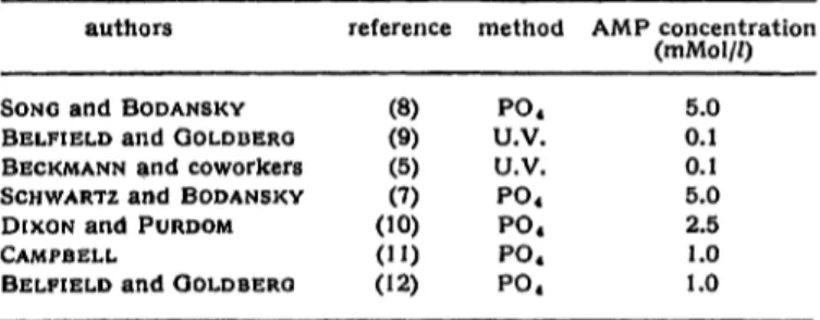
Table 1 summarizes the AMP concentrations used by different authors in the incubation media for serum S'-nucleotidase. The colorimetric methods for S'-nucleo-
Tab. 1
AMP concentrations, used for the determination of S'-nucleotidase activity
authors reference method AMP concentration (rnMol//)
Fig. 3
Activity as function of substrate concentration
SONG and BODANSKY BELFIELD and GOLDBERG BECKMANN and coworkers SCHWARTZ and BODANSKY DIXON and PURDOM CAMPBELL
BELFIELD and GOLDBERG
(8)(9) (5)(7) (10) (") (12)
P04
U.V.
U.V.
P04
P04
P04
P04
5.0 0.1 0.1 5.0 2.5 1.0 1.0
Z. klin. Chem. u. klin. Biochem. / 8. Jahtg. 1970 / Heft 4
400 Persijn, van der Slik, Timmer and Reijntjes: Determination of serum nucleotidase tidase based on the determination of inorganic PO4
released from AMP are designated by "PO4", the kinetic methods based on measurements at 265 nm by the symbol U.V. It is interesting to note that the authors 1. c. (12) and (9) raise the AMP concentration while changing from the kinetic to a colorimetric method.
The answer to the question why in the kinetic method appreciably lower concentrations are used seems to be that AMP contributes to a relatively high extent to the final optical density of the assay mixture. This is illus- trated in Table 2. In this connection it should be pointed
Tab. 2
Optical density at 265 nm of AMP solutions in Tris-buffer at pH 7.5 concentration
(mMol//). (at 265 nm)O. D.
5.0 1.0 0.12 0.10
00 oo
1.65 1.33
out that serum can contribute considerably to the final O.D. of the assay mixture; in the medium of BECKMANN and coworkers this contribution to O.D. may be 1.3 to 2.0. In view of these facts it would probably be difficult to find the optimal substrate concentrations for the kinetic method for serum 5'-nucleotidase. The de- velopment of kinetic methods for 5'-nucleotidase dates back to 1947, in which year KALCKAR introduced a method for the determination of adenosine (13). He found that the addition of adenosine deaminase to a solution of adenosine resulted in a rapid decrease of O.D. at 265 nm to approximately 40 per cent. His quan- titative method for adenosine is based on this change.
The suggestion of KALCKAR (14) to use such a reaction for the assay of 5'-nucleotidase was followed up by SEGAL and BRENNER (15) in their study of 5'-nucleo- tidase in rat liver microsomes. After a certain incubation period the enzymatic reaction was terminated and the adenosine released was determined according to KAL- CKAR.
In 1969 two detailed methods for the assay of S'-nucleo- tidase in serum involving measurements at 265 nm were presented by LEYBOLD and coworkers (16) and BELFIELD and GOLDBERG (17). The principles of the two methods were essentially the same, except for the addition of glycerophosphate in the latter, which is not a critical point in the present discussion. A salient point of differ- ence with the SEGAL and BRENNER assay is that in the two former methods for serum 5'-nucleotidase the rate of decrease in O.D. at 265 nm is measured during the incubation period itself. The change of O.D. in kinetic methods is the results of the consecutive reactions (1) and (2):
(3) AMP + H2O > inosine + PO4 -f NH3
and the decrease in O.D. following the conversion of AMP to inosine is ä measure of the 5'-nucleotidase activity involved. The measurement of reaction (3) is
made possible by the presence of adenosine deaminase in the incubation mixture and the fact that the spectral difference between AMP and inosine is similar to that between adenosine and inosine.
For the present discussion both the Segal-Brenner method and the kinetic method will be referred to as the u.v. method. · t
In this discussion we propose to consider two points : a) the effect of the contamination of adenosine deaminase with AMP-deaminase in the u.v. method and our method using the Berthelot reaction.
b) the sensitivity of the u.v. method and our method for S'-nucleotidase.
In an assay for 5 '-nucleotidase using adenosine de- aminase a second reaction can take place as a con- sequence of the contamination of. adenosine deaminase with AMP-deaminase:
(4) AMP - -+ IMP +
This type of reaction is accompanied by a change in O.D. at 265 nm which is greater than in the case of reaction (3). The presence of AMP-deaminase will there- fore interfere in both the u.v. method and our method.
The extent to which in such a case the accuracy of the assay for S'-nucleotidase activity will be diminished de- pends of course on the ratio of serum to adenosine deaminase present, but will be less in the Segal-Brenner method. The conversion of adenosine in the latter by adenosine deaminase added in excess after the incu- bation takes place in a very short time (a few minutes)
— therefore with a relatively long incubation time for S'-nucleotidase assay, AMP-deaminase present as con- tamination will presumably contribute to the overall change in O.D. only to a very minor extent — unlike the kinetic method and our method where the S'-nucleo- tidase and AMP-deaminase work simultaneously on their substrates. At low activity of S'-nucleotidase the effect of AMP-deaminase can even prevail in the meas- ured S'-nucleotidase activity. This has not been taken into account by the authors 1. c. (16) and (17).
The following calculation is intended to show the im- portance of including corrections for AMP-deaminase activity in the results of kinetic measurements. It should be emphasized that the actual dependence on substrate concentration for AMP-deaminase has not been con- sidered. The interpretation of the calculations should be accepted with some reserve. Several commercial batches of adenosine deaminase delivered in 1968 were found to be contaminated with AMP-deaminase. In the absence of serum after 60 min incubation time the O.D. at 625 nm in our method for S'-nucleotidase was fre- quently found in the range 0.060 — 0.090, the highest value being 0.230 (60 min incubation time). These values were not due to contaminations of AMP with adenosine. Such values were subtracted from total ex- tinction in the presence of the serum tested to obtain real serum S'-nucleotidase levels.
The calculation of the interfering AMP-deaminase activ- ity is facilitated by the fact that both S'-nucleotidase and Z. klin. Chem. u. klin. Biochem. / 8. Jahrg. 1970 / Heft 4
neues ietekf ionssysfem Wir SäuleDi-CBirontäfografE®
Flüssig-Chromatograf für präparative und analytische Arbeitsweise aus dem Programm PYE UfMICAM
Ein sehr kleiner Teil der aus der Säule ausströmenden Flüssigkeit wird als dün- ner Film auf einen Draht aufgebracht, beim Transport durch den Ofen ther- misch zerlegt und mit dem Trägergas in den Detektor gespült (als Detektoren dienen die aus der Gaschrpmatografie bekannten, hochempfindlichen Systeme).
Während die bisherigen Methoden zum ' Nachweis der aus der Säule austreten·*
den Fraktionen aufwendig und zeitrau- bend waren, sorgt das neue Detektions-
system dafür, daß die Flüssig-Chromato- grafie mit der Gas-Chromatografie ohne weiteres konkurrieren kann und ihr in manchen Fällen sogar überlegen ist, wie . bei der Analyse von organischen Ver- bindungen mit extrem niedrigen Dampf- druck wie Fettsäuren, Pestiziden, Ste- roiden, Lipiden u. a.
Die Anzeige wird durch das Lösungs- mittel nicht beeinflußt. Das System hat eine große Empfindlichkeit bei kleinem Bauschpegel. Die Ergebnisse werden
schnell und kontinuierlich aufgezeichnet.
Die Detektionssysteme sind auswechsel- bar: Alle diese Eigenschaften sichern dem neuen Flüssig-Chromatografen ei- nen breiten Anwendungsbereich.
Ausführliches Informationsmaterial liegt für Sie bereit. Bitte fordern Sie es an.
Philips Elektronik Industrie GmbH, 2000 Hamburg 63, Rönt- genstraße 22, Postfach 630111, Telefon (0411) 501031.
Telefon-Nummern der Büros in: Berlin (0311) 245908, Düssel- dorf (0211) 346051, Dortmund (0231) 41961, Frankfurt (0611) 79131, Hannover (0511) 16601, Hamburg (0411) 782557, Mün- chen (0811) 76791, Stuttgart-Fellbach (0711) 589081, Bielefeld (0521) 23081, Bremen (0421) 310041, Nürnberg (0911) 464763.
PHILIPS! Analysentechnik
PHILIPS
Wir interessieren uns für die beschriebenen Liquid-Chromatografen und bitten um Q] Zusendung ausführlicher Unterlagen
£] ein Angebot
Q Besuch Ihres Beratungsingenieurs Gewünschtes bitte ankreuzen oder ergänzen
(S5)
BULLETIN
DE LA
DE CHIMIE BIOLOGIQUE
(„Berichte der Gesellschaft für biologische Chemie")'
Unter Mitwirkung des
„CENTRE NATIONAL DE LA RECHERCHE SCIENTIFIQUE"
(National-Centrum für wissenschaftliche Forschung) veröffentlicht
J.-P. EBEL R. PERLfiS Y. RAOUL Secr6taire G n ral Secrotaire G6n6ral Radacteür en Chef
(Relations F. PERCHERON F. GROS
exterieures) Rodacteur en Chef Adjoint Rddacteur en Chef Adjoint
Sekretariat und Redaktion: A, avenue de lObservatoire, Paris (6e) Herausgeber: MASSON et CIE, 120, Boulevard Saint-Germain, Paris (6e)
Der „BULLETIN DE LA SOCIETE DE CHIMIE BIOLOGIQUE" veröffentlicht jährlich 11 Hefte; diese enthalten die Arbeiten der französischen Biochemiker, welche der „SOCIETE DE CHIMIE BIOLOGIQUE" (Gesellschaft für
biologische Chemie) angehören.
- \ Abonnementspreis 1969:
Frankreich und „Franc-Zone" . . . 150 ffrcs Belgien 1684 bfrcs Andere Länder 165 ffrcs
l *
l
jACTA BIOCHIMICA ET BIOPHYSICA ACADEMIAE SCIENTIARUM HüNGARICAE
Zeitschrift der Ungarischen Akademie der Wissenschaften
BAND 5 INDEX NUMMER l G. Cseh: Nucleotide Antagonism in the Hormone-sensitive Lypolysis of Fat Tissue
Gy. Bot, Edit F. Koräcs, Edit N. Polyik: The Influence of Allosteric Effectors on the Conversion of Phosphorylase-b
F. Hajos, S. Kerpel-Fronius, fiva Schay: Electron Microscopic Demonstration of Differences between Succinic Dehydrogenase Activ- ities of Brain Mitochondria in Homogenates and Mitochondrial Fractions
I. Alkonyi, E. Palfi, D. Szabo: ATP-Dependent Enzymic Splitting of Mesitene Lactone and Triacetic Acid Lactone I. Mile, V. Csanyi, Ilona Ferencz: Reversible Change in Iodine Sensitivity of Penicellinase by Alkali Treatment J. Hajdu, V. Csanyi: The Influence of Concentrated Electrolytes on the Constitutive Penicellinase Synthesis of B-ceteus
(Short Communication)
M. Bälint, A. Schaefer, N. A. Biro: The Subunit Structure of Light-Meromyosin (Short Communication)
Susan Libpr, P. Elödi: Selective Reaction of Tyrosyl Side Chains With Iodine in Glyceraldehyde-3-phosphate pehydrogenase. . Spec- ificity of the Reaction in Urea. Determination of Reacting Histidine and Tryptophan (Short Communication)
L. Polgar: Transformation of a Serine Protease of Aspergillus oryzae into a Thiol Enzyme (Preliminary Communication) E. Ernst: Bound Water in Physics and Biology
A. Pellipnisz: Computer Simulation of the Pattern Transfer of Large Cerebellar Neuronal Fields F. Aradi, Z. Futo: Quantitative Researches into the Volume Decrease of the Muscle, I
F. Aradi: Quantitative Researches into the Volume Decrease of the Muscle, G. Biro, K. Gabor, J. ÖrkonyirFrequent Excitation of the Nerve and Muscle
B. Tanko, T. Karsay, F. Teichmann: Employment of Collagen for Constructing a Mechano-chemical Machine
I. Bojtor, K. Dosay: Investigation on a Method Measuring Low Energy X-ray Doses with Energy-dependent Film Dosemeter (Short Communication)
Gy, Koczkas, K. Dosay: Physical Condition andDosimetry Problems of Experimental Irradiations inRadiobiology (Short Communication)
Book Review
ACTA BIOCHIMICA ET BIOPHYSICA erscheint vierteljährlich in Heften zu einem Band von etwa 400—500 Seiten. Format: 17 X 24 cm.
Abonnementspreis pro Band: $ 16.00; DM 64,—
Vertrieb: KULTURA, Budapest 62 . Postfach 149; Auslieferung für das Gebiet der Deutschen Bundesrepublik:
KUNST UND WISSEN, Erich Bieber, Stuttgart S, Wilhelmstraße
AKADßMIAI KIADO, Verlag der Ungarischen Akademie der Wissenschaften, Budapest 502. Postfach 24 *
Persijn, van der Slik, Timmer and Reijntjcs: Determination of serum nucleotidase 401
AMP-deaminase are measured with the same conversion factor. If we take as an example the batch of adenosine.
deaminase showing in our method a false 5'-nucleotidase activity as expressed in O.D. at 625 nm of 0.060 the amount of AMP-deaminase present amounts to:
0.060 0.375 A C C TT
χ —77-- χ 103 = 0.55 mU.
0.680 60
where 0.060 = extinction in absence of serum against blank at 625 nm.
0.680 = extinction standard adenosine at 625 nm.
0.375 = amount of adenosine standard (μΜο!) in test mixture.
60 = to express activity per minute.
103 = to convert to mU.
When 500 mU adenosine deaminase are present the contamination is about 0.1%. This means that if in our method a sample with a real S'-nucleotidase activity of 10 mU/m/ is tested the test mixture will contain 1.0 mU S'-nucleotidase activity and 0.55 mU AMP-deaminase activity.
Thus a 10 mU/m/ sample will give a reading of 15.5 mU/
m/, i. e. pathological.
In the kinetic method of 1. c. (17) the ratios of serum 5'-nucleotidase to adenosine deaminase present in the assay are extremely unfavourable as far as contamination with AMP-deaminase is concerned. Here 20 μι serum are incubated in the presence of 1300 mU adenosine deaminase. In the investigation of a serum with 10 mU/
m/ 5'-nucleotidase activity the assay mixture contains 0.2 mU S'-nucleotidase and 1.4mU AMP-deaminase (approx. 0.1% contamination). In this case the serum S'-nucleotidase activity will correspond to an O.D. at 265 nm amounting to:
0.2 χ 20
10 X 8.0 χ ΙΟ6 χ 10-9 = 0.011
where 0.2 = amount S'-nucleotidase (mU) pres- ent in assay mixture.
20 == incubation time in minutes.
3.0 = total volume (m/) assay mixture.
8.0 χ 106 = molar extinction coefficient for reac- tion (3) according to 1. c. (17).
10-~9 = to convert to nMol/m/.
Since according to KALCKAR (13) the molar extinction coefficient at 265 nm for reaction (4) is 8.75 χ 10β
Mol""1 · cm2, a similar calculation will show that 1.4 mU AMP-deaminase will cause a change in O.D. of 0.082. This change corresponds (according to 1. c. (17)) to 0.082 χ 18,700 χ 0.05 = 76.9 mU/m/of falsely assayed S'-nucleo- tidase activity. This example shows that with a con- tamination of only 0.1% the real S'-nucleotidase activity (10 mU/m/) is only 12% of the totally measured activity (86.9 mU/m/). For the kinetic method according to BECKMANN and coworkers the ratios of serum and adenosine deaminase are more favorable resulting in a real participation of S'-nucleotidase of 50%.
Summarizing, it can be said that slight contamination of AMP-deaminase has little if any effect in the Segal- Brenner method, but probably gives increasing inter- ference in the Persijn-van der Slik method, and the kinetic methods according to BECKMANN and coworkers and BELFIELD and GOLDBERG in that order. Our method is placed first because it allows the use of a relatively large volume (100 μι) of serum (Fig. 1). Corrections for AMP-deaminase as standard procedure are therefore indispensable. This is not a groundless demand since the authors of the kinetic methods presumably ob- tained their adenosine deaminase from the same com- mercial source as we did in 1968. It should be noted that at present Boehringer (Mannheim) or British Drug Houses (England) are marketing at our request adeno- sine deaminase preparations in which contaminations with AMP-deaminase are generally extremely low (± 0.02—0.04%) corresponding to O.D. at 625 nm in the absence of serum of 0.010—0.020.
To evaluate the sensitivity of the different methods the O.D. for a given S'-nucleotidase activity (10 mU/m/) are tabulated (Tab. 3). For the calculations the data given by the authors mentioned are used, with the exception of the method of BECKMANN and coworkers.
These authors use a factor derived from reaction (2), but the actual concentration of adenosine during the kinetic assay approximates to zero, a point which has been overlooked in the calculation of the factor to convert results of the kinetic measurements to S'-nucleo- tidase activities. In this case the factor of 1. c. (17) is applied. In the Segal-Brenner method we have replaced the volume of suspended microsomes by an equal volume of serum. This method seems to be the most sensitive with a final concentration of AMP of 1 mMol//.
Other criteria for evaluation such as results of recovery experiments or data about reproducibility or accuracy
Tab. 3
Methods of assay of S'-nucleotidase using adenosine deaminase
Decrease in O. D. calculated from data in the references mentioned for a sample of 10 mU/m/
method
1. PERSIJN and VAN DER SLIK 2. SEGAL and BRENNER1) 3. BECKMANN and coworkers 4. BELFIELD and GOLDBERG
ref.
(1), (3) (13), (15) (5), (16) (Π)
wavelength (nm)
625265 265265
incubation time (Min.) at 37°
6060 204
amount of serum («0 100100 10020
O.D.
0.108 0.261 0.011 0.011 modified
2. klin. Chem. u. klin. Biochem. / 8. Jahrg. 1970 / Heft 4 51
402 Persijn, van der Slik, Timmer and Reijntjes: Determination of serum nucleotidase '
in the 0—20 mU/m/ range have not been presented for For biochemical studies the application of u.v. methods the kinetic methods but have been given for our method have the advantage of a greater choice of type of buffer in 1. c. (1), (3) and (4). Nevertheless it seems justifiable while in our method there is only a limited choice (4).
to conclude that for a serum having a 5'-nucleotidase It is in the field of biochemistry that u.v. methods have in the transition range of normal to elevated level the value, especially the Segal-Brenner method with its kinetic methods have only limited value in contrast with minimal interference* from slig'ht contamination of the method using the Berthelot reaction. adenosine deaminase with AMPncIeaminase.
References
1. PERSIJN, J.-P., W. VAN DER SLIK, K. KRAMER and C. A. DE RUY- (London) 219, 73 (1968). — 10. DIXON, I. E. and M. PURDON, TER, This journal 6, 441 (1968). — 2. PERSIJN, J.-P., W. VAN DER J. Clin. Path. (London) 7, 341 (1954). — 11. CAMPBELL, D. M., SLIK, C. J. TIMMER and A. W. M. BON, This journal 7,199 (1969). Biochem. J. 82, 34 P (1962). —12. BELFIELD, A. and D. M. GOLD- 3. PERSIJN, J.-P., W. VAN DER SLIK, and A. W. M. BON, This BERG, J. Clin. Path. (London) 22, 144 (1969). — 13. KALCKAR, journal 7, 493 (1969). — 4. VAN DER SLIK, W., J.-P. PERSIJN, H. M., J. biol. Chemistry 167, 445 (1947). 14. KALCKAR, H. M., E. ENGELSMAN and A. RIETHORST, Clin. Biochemistry 3, 59 J. biol. Chemistry 167, 461 (1947). — 15. SEGAL, H. L. and B. M.
(1970). — S. BECKMANN, J., K. LEYBOLD and L. WEISBECKER, BRENNER, J. biol. Chemistry 235, 471 (1960). — 16. LEYBOLD, K., This journal 7,18 (1969). — 6. GOLDBERG, D. M., private cornmu- J. BECKMANN and L. WEISBECKER, This* journal 7, 25 (1969). — nication. — 7. SCHWARTZ, M. K. and O. BODANSKY, Cancer 18, 17. BELFIELD, A. and D. M. GOLDBERG, Clin. Chem. New York 886 (1965). — 8. SONG, C. S. and O. BODANSKY, Biochem. J., 101, 15, 931 (1969).
5c (1966). — 9. BELFIELD, A. and D. M. GOLDBERG, Nature
Dr. J.-P. Persijn Amsterdam C Sarphatistraat 106
Z. klin. Chem. u. klin. Biochem. / 8. Jahrg. 1970 / Heft 4