Heinz and Beushausen: A new enzymatic method for the determination of glucose 977 J. Clin. Chem. Clin. Biochem.
Vol. 19,1981, pp. 977-978
A New Enzymatic Method for the Determination of Glucose
By F. Heinz and Th. W. Beushausenl)
Zentrum Biochemie, Medizinische Hochschule Hannover (Received March 26/September 8,1980)
Summary: A method for the determination of glucose is described. H202, produced by the action of glucose oxidase, is measured from the change in absorbance due to oxidation of NAD(P)H in the presence of catalase, aldehyde de- hydrogenase and a high concentration of ethanol. The quality data of the method are equivalent to those of the hexokinase-glucose-6-phosphate dehydrogenase method used as reference.
Eine neue enzymatische Methode zur Bestimmung von Glucose
Zusammenfassung: Bei der beschriebenen Glucosebestimmung wird das durch Glucoseoxidase entstandene H202 mit Katalase und Aldehyddehydrogenase in Anwesenheit von hohen Ethanolkonzentrationen bestimmt; dabei dient NAD(P)H als Meßgröße. Die Qualitätsmerkmale der Methode entsprechen denen der Hexokinase-Glucose-6-phos- phatdehydrogenase Referenzmethode.
Introduction
Glucose determination is one of the most frequently used methods in clinical chemistry, and is based mainly on three enzymatic-photometric assays: the NADP dependent hexokinase-glucose-6-phosphäte dehydro- genase method (1), the NAD-dependent glucose dehy- drogenase method (2) and the H202 -producing glucose oxidase-peroxidase method coupled with leuco dyes (3).
Our assay is based on the glucose oxidase reaction, with measurement of the produced H2O2 , using catalase and NAD(P)-dependent aldehyde dehydrogenase in the presence of high ethanol concentrations. This principle (4), which has already been evaluated for uric acid (5) and cholesterol (6) avoids the known difficulties arising from the use of peroxidäse coupled with leuco dyes.
Reactions (1) P-glueose
(2) H202 + ethanol (3)
aldehyde dehydrogenase
2H2O
acetic acid + NAD(P)H
*) This work contains part of the doctor thesis of 7%. W, Beu*
hausen
Materials and Methods
KCl-diphosphate buffer (pH 8.0): Dissolve 11.15 g tetrasodium- diphosphate-10-hydrate (Merck No. 6591) and 11.85 g KC1 (Merck No. 4936) in 400 ml H2O; adjust pH to 8.0 with HC1 and add H2O to 500 ml.
Aldehyde dehydrogenase solution: Dissolve a 10 U equivalent of aldehyde dehydrogenase, K* activated from baker's yeast, grade II (No. A-6758, Fa. Sigma, St. Louis) in 1 ml H2O.
NAD-solution: Dissolve 10 mg NAD (No. 15298, Fa. Boehringer, Mannheim) in 1 ml H2O.
Glucose oxidase solution: Dissolve an 1100 U equivalent of glucose oxidase lyophilisate (No. 15426, Fa. Boehringer, Mann- heim) in 0.5 ml H2O.
Buffer/ethanol mixture: Mix 10 ml buffer and 1 ml ethanol.
Catalase: Catalase 1.43 GU/1 (Fa. CalBiochem., Lot. 600660).
Reaction mixture (for 10 determinations): Take 8.5 ml buffer/
ethanol mixture, add 1 ml NAD-solution, 1 ml aldehyde dehy- drogenase solution, 0.5 ml glucose oxidase solution, and 10 catalase.
This mixture is incubated at room temperature for 1 h. At 4 °C, it is stable for approximately 24 h.
Manual procedure
Pipette 1 ml of the reaction mixture into a semimicro cuvette with 10 mm optical path length, then read absorbance for
1-3 min at 334 or 340 nm Hg and add 5 sample. Mix, then read absorbance when the reaction has come to the end (reac- tion temperature: 25 °C). Before and after the'end of the reac- tion a slight endogenous slope is found. By extrapolation back to the starting point the absorbance difference is determined.
The glucose concentration is calculated from the absorbance difference. By using NADP instead of NAD identical results were obtained.
034(M)76X/81 /0019-0977 $02.00
©by Walter de Gruyter & Co. - Berlin · New York
Results
The accuracy of the method was established by deter- mining the glucose concentration in aqueous solutions of known glucose concentrations, in control sera con- taining known concentrations and by determination of the glucose concentration in patients' sera with our method and the hexokinase-glucose-6-phosphate dehy- drogenase method (1). The results of both methods were compared with the paired-t-test, which did not show significant differences. The regression analysis for patients' sera is shown in figure 1.
40
CO £
<D*—
o 30
3 20
10
0 5 10 20 30 4F
Glucose (hexokinose / glucose -6 - phosphot e-
dehydrogenose method) [mmol/U
Fig. 1. Linear regression for glucose concentration in 29 patients' sera determined with the glucose oxidase-aldehyde de- hydrogenase method (y) and the hexokinase-glucose-6- phosphate dehydrogenase method (x).
y = 1.00-0.03 r = 0.999 ç = 58 Paired t-test:
f = 5 7 á = 0.05 t0 =1.43
The precision from day to day for control sera was CV < 3.08% (n = 55), which almost meets the figure of the American College of Pathologist* (CV < 2.2%) (7), and which is well below the requirements of the guide- lines of the Medical Society of the Federal Republic of Germany (CV < 5%) (7).
For normal sera or those with slight haemolysis, hyper- lipidaemia or hyperbilirubinaemia, the absorbance value of the sample is negligible. For strongly haemolytic samples, turbid hyperlipidaemic samples or samples with well elevated bilirubin levels (> 115 ìðéïÀ/ß), the
absorbance value of the sample volume has to be deter- mined in a reaction mixture without glucose oxidase, and then subtracted.
Discussion
The described method is suitable for the determination of glucose in human sera. The linear range of the method was verified for concentrations from 2-38 mmol/1, thus permitting determination of almost any normal or pathologic glucose concentration.
Relevant inhibitor substances for glucose oxidase or aldehyde dehydrogenase are not known in human sera.
For the evaluation of a uric acid determination method, which has the same indicator system, a number of sub- stances often used clinically were tested for possible interference. No interference by these substances was noted (5).
Compared to other glucose oxidase methods coupled with leuco dyes, our method has several advantages: The reaction time is 10-15 min shorter. The glucose con- centration can be calculated directly from the absorb- ance difference using the coefficient of absorbance for NAD(P)H. Stabilisation of the indicator is not necessary.
The manual procedure for our method is rather simple and it can be easily automatized.
References
1. Bergmeyer, H.-U. & Gawehn, K. (1974) in: Methoden der enzymatischen Analyse (Bergmeyer, H.-U. ed.) Verlag Chemie, Weinheim, 3rd ed., p. 1241-1246.
2. Banauch, D., Brummer, W., Ebeling, W., Metz, H., Rindfrey, H., Lang, H., Leybold, K. & Rick, W. (1975) this J. 13,101- 3. Bergmeyer, H.-U. & Bernt, C. (1974) Methoden der enzymati-107.
schen Analyse (Bergmeyer, H.-U. ed.) Verlag Chemie, Wein- heim, 3rd ed., p. 1250-1257.
4. Haeckei, R. & Heinz, F. (1975) this j. 13, 244.
5. Haeckcl, R. (1976) this j. 14,101-107.
6. Haeckei, R. & Perlick, M. (1976) this j. 14,411-414.
7. Haeckei, R. (1975) Qualit tssicherung im klinischen Labor, Dtsch. rzteverlag K ln, p. 43.
Prof. Dr. Fritz Heinz
Zentrum Biochemie -r 4310 - Medizinische Hochschule Hannover Karl-Wiechert-Allee 9
D-3000 Hannover 61
J. Clin, Chem. Clin. Biqehem. / Vol. 19,1981 / No. 9
Puschendorf, Grunicke, Jentsch and Berger: Urinary cGMP in children with malignant tumours 979
J. Clin. Chem. Clin. Biochem.
Vol. 19,1981, pp. 979-981
Urinary Excretion of Cyclic Guanosine 3'.5'-nrionophosphate in Children with Malignant Tumours
1) By A Puschendorf \H. Grunicke
Institut f r Medizinische Chemie und Biochemie (Vorstand: Prof. Dr. H. Grunicke) der Universit t Innsbruck J. Jentsch and Berger
Kinderklinik (Vorstand: Prof. Dr. H. Berger) der Universit t Innsbruck
(Received November 27,1980/March 13,1981)Dedicated to Professor Dr. Helmut Holzer on his 60th birthday
Summary: The present study shows an increased urinary cyclic guanosine S'.S'-monophosphate (cyclic GMP) excre- tion rate in children of all age groups bearing malignant tumours or lymphomas. The incidence of increased cyclic GMP excretion was highly significant (79%). Follow-up studies of up to three years have revealed that during periods of remission of malignant disease the urinary cyclic GMP excretion drops to near normal values, whereas recurrences are accompanied by a new increase of cyclic GMP excretion.
Ausscheidung von zyklischem Guanosin-3'.5'-monophosphat im Urin von Kindern mit malignen Tumoren
Zusammenfassung: Die vorliegende Studie zeigt eine erh hte Ausscheidung von zyklischem Guanosin-S'.S'-mono- phosphat (zyklisches GMP) im Urin von Kindern aller Altersgruppen mit malignen Tumoren oder Lymphomen. Das Auftreten dieser zyklischen GMP-A sscheidung im Urin war mit 79% hoch signifikant. Verlaufsstudien bis zu 3 Jah- ren zeigten w hrend der Remission der malignen Erkrankung eine Ausscheidung, die bis zu Normalwerten abfiel, hin- gegen waren Rezidive von einer erneuten Erh hung der Ausscheidung von zyklischem GMP begleitet.
Introduction Material and Methods
Earlier Observations (1,2) have suggested that an eleva- Urine was collected at room temperature and was stored at tion Of guanosine 3'.5'-monophpSphate (cyclic GMP) - 20 °C until assayed. The urinary cyclic GMP was measured by
^ j n j . . . ** j ÷ é /^\ j · ÷ ' ÷ - j a radioisotope dilution test with cyclic GMP binding protein promoted cell division. Murad et al. (3) demonstrated (Boehringer, Mannheim, Germany) (5-7). Urinary levels of an increased urinary excretion of cyclic GMP in rats creatinine were determinated by the picric acid method (8). The bearing tfansplantable liver and kidney tumours. The values reP°rted for urinary <*°1ßï GMP e*<*eti°n «the mean
. ^ ,. ' ^\.« . V · é j " Ë values of duplicate de terminations and are expressed as ìðéïß
excretion of cyclic GMP in these animals decreased
per g creati
ni
ne.
after radiotherapy, chemotherapy, or excision of the tumours and correlated well with the tumour size
and growth. The same group (4) found that urinary Results and Discussion
cyclic GMP, but not cyclic denosine 3'.5'-monophos- Several reported data demonstrate a positive correlation phate (cyclic AMP), increased progressively with the between the intracellular level of cyclic GMP and the growth of several, but not all transplantable liver and
ceu proliferation (1, 2). Therefore, it seemed possible kidney tumours studied. Stimulated by these findings that newborn children would have an elevated excretion we made an attempt to correlate the urinary cyclic
of cyclic GMP due to their higher rate of cell prolifera- GMP excretion rate (/imol per gram ereatinine) with the tion. Figure 1 shows the urinary excretion of cyclic clinical course of malignant tumours in children. GMP by children as a function of age. The youngest
children were premature births (36 weeks of pregnancy).
:
—~--~· The excretion rates of cyclic GMP were considerably
*) Parts of this paper were presented as a preliminary communi- higher in young children (below 2 years) and decreased
with
·». ™- -ult is probably due to the normally 1979, Salzburg, Austria. decreased excretion of creatmine in young children (9,
0340-076X/81 /0019-0979$02.00
© by Walter de Gruyter & Co. · Berlin - New York
I 4
f 3
36 38 40
Weeks of pregnancy (premature births)
2 4 6 8 10 12 2 4 6 8 10 12 14 16
months Age
yeors
Fig. 1. Urinary cyclic GMP excretion rates in children, as a func- tion of age.
10), rather than by an increased level of cyclic GMP.
The upper limit of urinary excretion of cyclic GMP in children younger than two years, is 5 ìðéïÀ/g creatinine;
whereas the normal range in children older than 2 years
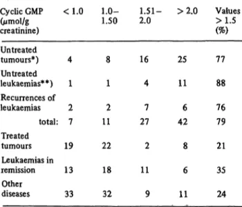
Tab. 1. Urinary cyclic GMP excretion rates in children with tumours and other diseases (Pediatric Clinic of the Uni- versity of Innsbruck/Austria; observation period: 197Ô-
É 979).
Cyclic GMP (ìðéïÀ/g creatinine) Untreated tumours*) Untreated leukaemias**) Recurrences of leukaemias
total:
Treated tumours Leukaemias in remission
Other diseases
<1.0
4 1 2 7 19 13 33
1.0-1.50
8 1 2 11 22 18 32
1.51- 2.0
16 4 7 27 2 11 9
>2.0
25 11 6 42 8 6 11
Values
77 88 76 79 21 35 24
*) Total 53 determinations; number of cases in brackets: brain tumours (13) (ependymoma (2), craniopharyngioma (2), pineal tumour (1), medulloblastoma (3), spongioblastom (1), brainstem tumour (1), astrocytoma (2), unclassified (2)); lymphomas (7) (M.Hodgkin: stage I (1), stage III (1), AurfaYMymphoma (1), non-/fod£fci>i-lymphoma stage IV (4));
Wilms tumours (5) (stage II (3), stage III (1), stage. IV (1));
neuroblastomas stage IV (3) sarcomas (3) (stage IV (1), sarcoma botryoides (1), angiosarcoma (1)); osteosarcomas without metastases (2).
2 determinations were performed at different time points in 5 cases (brain tumour, Wilms tumour, neuroblastoma, sarcoma and lymphoma), and 15 values are determinations during the recurrences of the tumours (brain tumours, Wilms tumours, neuroblastoma, sarcoma and lymphomas).
**) acute lymphatic leukaemias (12), acute myeloic leukaemias (2), and acute monocytic leukaemia (1). 2 determinations were performed at different time points in 2 cases (acute lymphatic leukaemia).
"55^
*? „ 8.0
*«
« 0 0 0
9 ÏÄÏ
363840 2 4 6 8 10 12 2 4 6 8 10 12 14 16 18 20
iumonths
pregnancy (premature births)
A
9
eFig. 2. Urinary cyclic GMP excretion rates in children bearing malignant tumours and lymphoproliferative diseases, as a function of age.
ð untreated tumours, ï treated tumours
Ä brain tumours.
is 0.5-1.2 ìðéïÀ/g creatinine. The normal level of cyclic GMP in plasma has been determined as 1.2 nmol/1; this level is extremely constant.
As shown in figure 2, elevated excretion rates of cyclic GMP were found in children with treated and untreated malignant tumours. The excretion rates of some untreated tumour patients were ten times the normal level as shown in figure 1. The mean increase in cyclic GMP excretion was lower in patients who were already under radio- or chemotherapy when the first analysis was made (fig. 2). Brain tumours, with 2 exceptions, yielded little or no elevation of the cyclic GMP excretion rate.
Table 1 shows 186 determinations of the excretion rates of cyclic GMP in 70 children older than 2 years bearing malignant tumours or lymphoproliferative diseases. The highest percentage of elevated rates was found in patients with untreated malignant tumours (69 of 87), while in patients with malignant tumours undergoing treatment as well as with malignant tumours in remission the ratio was considerably lower (10 of 51 and 17 of 48, respecti- vely). In a control group of 85 patients with other non- proliferative diseases, 20 also had elevated excretion rates.
Follow-up studies up to 3 years revealed that urinary cyclic GMP excretion drops to near normal values in patients during remission of their disease, while recur- rences are accompanied or preceded by a rise of cyclic GMP excretion. In cases of Wilms tumour (fig. 3) nephrectomy was followed by a drastic drop of cyclic GMP excretion. Another patient (fig. 4) with acute myelosis gave a similar response after chemotherapy. A recurrence of the disease was accompanied by a second increase of cyclic GMP excretion.
J. Clin. Chem. Clin. Biochem. / Vol. 19,1981 / No. 9
Puschendorf, Grunicke, Jentsch and Berger: Urinary cGMP in children with malignant tumours 981
12
1977
1978Time [month,year]
4 6 8 10 12*
1979 I Fig. 3. Follow-up study of urinary cyclic GMP excretion rates:
Wilms tumour (stage II) (M.R.) 8. 11.1977 nephrectomy right, subsequent radiotherapy (18 Gy (1800 rad)) and usual chemotherapy (actinomycin & vincristine) (11) for 2 years.
Our results for children with malignant tumours should not be generalized to include adults. Adult tumour patients (total 61) also show an increase in 61% of treated and untreated malignant tumours, but an increase is also found in 38% of other diseases — especially in cases of myocardial insufficiency (Puschendorf, /?., Michelmayr, G. &Dienstl, F., unpublished results). Only
26% of patients (total 152) with untreated gynaecologi- cal tumours showed an increased cyclic GMP excretion in the urine (Puschendorf
LB.
9Santeler,P. &Dapunt, Ï., unpublished results).
Together with other clinical parameters the determina- tion of urinary excretion of cyclic GMP seems to be use- ful for tumour diagnosis and the assessment of therapeu- tic response in children. So far, the excretion rate of cyclic GMP cannot be correlated with the size or clinical stage of localized tumours, since the number of cases of each type of tumour was too low. However, in 12 patients
1st Recurrence
27.4.
j é L 8 9 10 11 12
1977
J—I—I—I 4 6 8 10 Ô
1978 ;
Time (month.year]
J—é—\ é é 12 2 4 6 8 10 12
I 1979 2 4 6
1980 Fig. 4. Follow-up study of urinary cyclic GMP excretion rates:
acute lymphatic leukaemia (L.K.), therapy: 0-76 scheme (vincristine, daunoblastine & dexamethasone) modified according to Pinkel (12).
with acute lymphatic leukaemia we found that there is no correlation between cyclic GMP and haematological findings.
The determination of cyclic GMP in the urine is of no use for the diagnosis of gynaecological tumours. For other malignant tumours in adults, the measurement of cyclic GMP in urine has approximatively the same value as other in vitro tests available. None of the established in vitro tests gives positive results for all tumour cases.
The confidence level of tumour diagnosis by in vitro tests increases with the number of independent tests. In this sense, the determination of cyclic GMP excretion in urine complements the list of in vitro tumour tests.
Acknowledgement
The authors gratefully acknowledge valuable discussions with Ñïæ. Dr. Elisabeth Wolff-Schreiner and the expert technical assistance of Ms. Dagrnar Gr ber.
This study was supported by the Vorarlberger Forschungsspende.
References
1. Hadden, J. W., Hadden, E. M., Haddox, M. K. & Goldberg, N. D. (1972) Prpc. Nat. Acad. Sei. USA 69, 3024-3027.
2. Goldberg, N. D., Haddox, M. K., Estensen, R., White, J. G., Lopez, C. & Hadden, J. W. (1974) In: The Cold Spring Harbor Symp. on the Regulation of Proliferation in Animal Cells (Qarcksoh, B. & Baserga, R. eds.) pp. 609-625, Cold Spring Harbor Laboratory, New York.
3. Murad, F., Kimura, H., Hopkins, Ç. Á., Looney, W. B. &
Kovacs, C. J. (1975) Science 190, 58-60.
4. Oriss, W. E. & Murad, F. (1976) Cancer Res. 36,1714-1716.
5. Oilman, A. G. (1970) Proc. Nat. Acad. Sei. USA 67, 305- 6. Iliano, G., Tell, G. P. E., Siegel, M. L & Cuatrecasas, P.312.
(1973) Proc. Nat. Acad. Sei. USA 70, 2443-2447.
J. Clin. Chem. Clin. Biochem. /Vol. 19,1981 / No. 9
7. Wunderwald, P., Jurz, G. & Michal, G. (1974) Anal. Biochem.
59, 468-481.
8. Bartels, H., Bohmer, M. & Heierli, C. (1972) Clin. Chem.
Acta 37,193-197.
9. Rubin, I. M., Br ck, E. & Rapoport, M. (1949) J. Clin. In- vest. 28, 1144-1162.
10. Herrera, C. & Finer, N. (1976) In: Manual of Pediatric Therapeutics p. 271 (Graef, J. W. & Cone jr., T. E. eds.) Little, Brown & Comp., Boston, USA.
11. Pl ss, H. J. & Sartorius, J. A. (1979) In: Internistische Krebstherapie (Brunner, K. W. & Nagel, G. A., ed.) 2. edi- tion, 454-457, Springer Verlag, Berlin-Heidelberg—New York.
12. Resch, R., Haas, H. & Berger, H. (1980) Dtsch. Med. Wo- chenschr. 105,123-127.
Prof. Dr. Bernd Puschendorf
Institut f r Medizinische Chemie und Biochemie der Universit t Innsbruck
Fritz-Pregl-Stra e 3 A-6020 Innsbruck