Regulation of glutamate dehydrogenase
in Corynebacterium glutamicum and its impact on nitrogen control
Inaugural-Dissertation zur
Erlangung des Doktorgrades
der Mathematisch-Naturwissenschaftlichen Fakultät der Universität zu Köln
vorgelegt von
Tim Müller aus Koblenz
Köln, Oktober 2005
Berichterstatter:
Prof. Dr. Andreas Burkovski Prof. Dr. Reinhard Krämer
Tag der Disputation: 28.11.2005
Content
Content...I Abbreviations ... IV Abstract ... V Zusammenfassung ... VI
1. Introduction ...1
1.1. Corynebacterium glutamicum...1
1.2. Uptake of nitrogen sources ...2
1.3. Assimilation of ammonium ...4
1.4. Nitrogen-dependent regulation...7
1.5. Objectives...13
2. Materials and methods...14
2.1. Bacterial strains, plasmids, and primers ...14
2.2. Cultivation of bacteria...17
2.3. Genetic manipulation of bacteria...19
2.3.1. Preparation of competent E. coli cells and transformation...19
2.3.2. Preparation of competent C. glutamicum cells and transformation ...19
2.3.3. Generation of deletion strains of C. glutamicum...20
2.4. Working with DNA ...21
2.4.1. Isolation of plasmid DNA from E. coli...21
2.4.2. Isolation of genomic DNA from C. glutamicum...21
2.4.3. Gel electrohoresis and extraction of DNA from agarose gels ...21
2.4.4. PCR and SOE-PCR ...22
2.4.5. Restriction, ligation, and sequencing of DNA...22
2.4.6. Site-directed mutagenesis...23
2.5. Working with RNA ...23
2.5.1. Isolation of total RNA from C. glutamicum...23
2.5.2. Synthesis of digoxigenin-labelled RNA probes ...24
2.5.3. Dot blot analysis and slot blot analysis ...25
2.5.4. Quantitative real-time RT-PCR...26
2.5.5. DNA-Microarray analysis...27
2.5.6. Analysis of mRNA degradation rates ...29
2.6. Working with proteins...29
2.6.1. Analysis of protein concentrations ...29
2.6.2. SDS-PAGE and Coomassie staining ...29
2.6.3 Peptide mass fingerprinting...31
2.6.4. Analysis of β-galactosidase activity...31
2.6.5. Analysis of GDH activity ...32
2.6.6. DNA affinity purification with magnetic beads ...33
2.6.7. Gel shift assays and competition assays ...36
2.6.8. Purification of GDH protein by Ni-NTA chromatography, dialysis, and electro-elution, and the production of antibodies for GDH ...37
2.6.9. Western blotting ...37
2.7. Chromatography...38
2.7.1. Determination of internal 2-oxoglutarate and glutamate by gas chromatography ...38
2.7.2. Determination of internal ammonium by HPLC...38
2.7.3. Determination of internal glutamine, glutamate, and arginine by HPLC .39 2.8. Bioinformatic approaches...40
2.8.1. Identification of open reading frames ...40
2.8.2. Identification of putative binding sites ...40
2.8.3. Sequence alignments and presentation of consensus sequences ...40
3. Results ...41
3.1. Regulation of glutamate dehydrogenase ...41
3.1.1. Characterization of the nitrogen-dependent regulation of glutamate dehydrogenase...41
3.1.2. Identification of transcriptional regulators of glutamate dehydrogenase.45 3.1.3. Characterization of the binding behaviour of AmtR, FarR, OxyR, and WHiH to the gdh promoter region...50
3.1.4. Analysis of the role of AmtR, FarR, WhiH, and OxyR in the nitrogen- dependent regulation of gdh transcription...57
3.1.5. Identification of conditions that trigger regulation of gdh transcription by AmtR, FarR, OxyR, and WhiH...60
3.1.5.1. Growth rates of the single deletion strains ...60
3.1.5.2. Identification of putative target genes of FarR and WhiH by DNA
microarray hybridization...61
3.1.5.3. Identification of putative target genes of FarR using a bioinformatic approach...63
3.1.5.4. Identification of putative effector molecules of FarR ...66
3.1.5.5. Regulation of gdh transcription in response to various stress conditions...68
3.2. The impact of GDH on the nitrogen control network...72
3.2.1. Expression of the gdh gene of E. coli in the gdh deletion strain of C. glutamicum...72
3.2.2. Expression of an enzymatically inactive glutamate dehydrogenase in the gdh deletion strain ...74
3.2.3. Influence of a deletion of gdh on intracellular metabolite pools ...75
3.2.4. Influence of a deletion of glnA on intracellular metabolite pools ...77
3.3. Putative toxicity of ammonium for C. glutamicum...81
3.3.1. Diffusion of ammonia across the cell envelope of C. glutamicum...81
3.3.2. Growth of C. glutamicum under high concentrations of ammonium ...83
3.3.3. Analysis of the presence of a putative futile cycle...84
4. Discussion...86
4.1. Regulation of glutamate dehydrogenase ...86
4.2. The role of GDH in the nitrogen regulation network of C. glutamicum...97
4.3. Investigations of a putative toxicity of ammonium for C. glutamicum...101
4.4. Summary ...103
5. References...105
6. Appendix ...115
6.1. Construction of C. glutamicum strains ...115
6.2. Construction of plasmids...116
6.3. Complete list of the results of the DNA microarray analyses...119
Abbreviations
ApR resistant to ampicilin
bp base pairs
CTAB N-cetyl-N,N,N-trimethylammonium bromide DMSO dimethyl sulfoxide
EDTA ethylendiaminetetraacetic acid et al. et alii
GDH glutamate dehydrogenase GOGAT glutamate synthase GS glutamine synthetase
HPLC high pressure liquid chromatography kb kilo base pairs
KmR resistant to kanamycin
MALDI-TOF matrix-assisted laser despropotion/ionisation time of flight MOPS 3-[N-morpholino]propansufonic acid
MSTFA N-methyltrimethylsilyltrifluoroacetamide NxR resistant to nalidixic acid
OD600 optical density at 600 nm
PAGE polyacrylamide gel electrophoresis RT room temperature
RT-PCR reverse transcription polymerase chain reaction SDS sodium dodecylsulphate
SOE-PCR splicing by overlapping extension polymerase chain reaction TEMED N,N,N’,N’-tetramethyl-ethylendiamine
Tris 2-amino-hydroxymethylpropane-1,3-diol
Abstract
Global regulatory networks are essential for the adaptation to changing environmental conditions and allow bacteria to survive conditions of stress and starvation. Nitrogen control is one of these networks. It regulates the uptake and assimilation of nitrogen containing compounds in dependence on their availability. In Corynebacterium glutamicum, a gram-positive soil bacterium of important biotechnological relevance, glutamate dehydrogenase (GDH) seems to play an important but so far less investigated role in nitrogen control. In this work, the regulation of glutamate dehydrogenase and its impact on nitrogen control in C. glutamicum were investigated.
Transcription of gdh is induced under nitrogen limitation. By DNA affinity purification with magnetic beads, four transcriptional regulators were isolated, which bind specifically to a fragment of the gdh promoter region that is responsible for the nitrogen-dependent transcription control. These are the putative transcriptional regulators FarR, WhiH, and OxyR, and the well-characterized transcriptional regulator AmtR, which is a major player of nitrogen control C. glutamicum. The exact binding sites of AmtR and FarR were mapped and a principle capacity of FarR to repress gdh-promoter-driven transcription was demonstrated. Surprisingly, neither single deletions nor a quadruple deletion of amtR, farR, whiH, and oxyR in C. glutamicum have any effect on nitrogen-dependent transcription of gdh. Consequently, these regulators are not essential for the nitrogen- dependent regulation of gdh transcription. Nevertheless, they might regulate gdh transcription in response to other stress conditions. A broad range of stress conditions were identified that influence gdh transcription. In addition, a putative role of FarR in the regulation of fatty acid biosynthesis and/or glutamate overproduction was revealed.
In the second part of this work, the role of GDH in nitrogen control of C. glutamicum was investigated. A deletion of gdh results in a deregulation of the nitrogen control network.
It was demonstrated, that this effect is caused by the loss of GDH activity and not by the absence of the GDH protein itself. In the gdh deletion strain, internal 2-oxoglutarate is accumulated. A high 2-oxoglutarate pool seems to antagonize the nitrogen status and triggers a nitrogen starvation-like response even under nitrogen surplus. In addition, ammonium could be identified as a second metabolite that influences nitrogen control in C. glutamicum.
In the third part of this work, it was demonstrated that ammonium is not toxic for C.
glutamicum and the formation of a putative futile cycle of ammonium was not observed.
Zusammenfassung
Globale Regulationsnetzwerke sind essentiell für die Adaptation an unterschiedliche Umweltbedingungen und ermöglichen Bakterien das Überstehen von Stress- und Mangelsituation. Die Stickstoffkontrolle ist ein solches Netzwerk. Sie reguliert die Aufnahme und Verstoffwechselung stickstoffhaltiger Substrate in Abhängigkeit von deren Verfügbarkeit. Bei der Stickstoffkontrolle des Gram-positiven Bodenbakteriums Corynebacterium glutamicum, welches eine große biotechnologische Bedeutung hat, scheint die Glutamatdehydrogenase (GDH) eine wichtige, bislang unbekannte Funktion einzunehmen. In dieser Arbeit wurde die Regulation der GDH und ihr Einfluss auf die Stickstoffkontrolle von C. glutamicum untersucht.
Die Transkription des gdh-Gens wird unter Stickstoffmangel induziert. Mit Hilfe magnetischer DNA-Affinitätsaufreinigung wurden vier Transkriptionsregulatoren isoliert, welche spezifisch an den Abschnitt der gdh-Promotorregion binden, der für die stickstoffabhängige Regulation verantwortlich ist. Dies sind die drei putativen Transkriptionsregulatoren FarR, WhiH, OxyR sowie der bereits charakterisierte Transkriptionsregulator AmtR, welcher ein zentraler Regulator der Stickstoffkontrolle von C. glutamicum ist. Die genauen Bindestellen von AmtR bzw. FarR innerhalb der gdh-Promotorregion wurden identifiziert und die prinzipielle Fähigkeit zur Repression gdh-Promotor-abhängiger Transkription für FarR gezeigt. Trotz allem führen weder Einfachdeletionen noch die Mehrfachdeletion aller Gene der vier Regulatoren zu einem Verlust der stickstoffabhängigen Transkriptionsregulation von gdh. Folglich regulieren AmtR, FarR, WhiH und OxyR die Transkription von gdh nicht in Abhängigkeit von der Stickstoffversorgung, könnten aber unter anderen Bedingungen aktiv sein. In dieser Arbeit wurden zahlreiche Stressbedingungen identifiziert, welche die Transkription von gdh beeinflussen. Außerdem wurde eine weitere Bindestelle von FarR vor dem dtsR2- Gen identifiziert, was auf eine mögliche Rolle von FarR bei der Regulation der Fettsäurebiosynthese und/oder Glutamatüberproduktion schließen lässt.
Im zweiten Teil dieser Arbeit wurde die Bedeutung der GDH für die Stickstoffkontrolle von C. glutamicum untersucht. Eine Deletion des gdh-Gens führt zu einer Deregulierung der Stickstoffkontrolle von C. glutamicum. Dieser Effekt beruht auf einem Verlust der GDH-Aktivität und nicht auf der Abwesenheit des GDH-Proteins an sich. Der Verlust der GDH-Aktivität führt zu einer Akkumulation von α-Ketoglutarat, was selbst unter guter Stickstoffversorgung eine Stickstoffmangelreaktion auszulösen scheint. Des Weiteren
wurde gezeigt, dass auch die Verfügbarkeit von Ammonium die Stickstoffkontolle von C.
glutamicum direkt beeinflusst.
Im dritten Teil dieser Arbeit wurde gezeigt, dass Ammonium für C. glutamicum nicht toxisch ist. Die Anwesenheit eines putativen energieverschwendenden Transmembranzyklus von Ammonium konnte nicht beobachtet werden.
1. Introduction
Nitrogen is an essential element for all life-forms as it is part of many important biomolecules like proteins, nucleotides, and coenzymes. Many organisms have evolved regulatory networks to cope with changes in the nitrogen supply. Transport and metabolism of nitrogen-containing compounds are strictly regulated and adapted to the availability of nitrogen sources. These processes as well as the connecting regulatory networks are referred to as nitrogen control. Until now, nitrogen control has been investigated in a number of bacteria, e.g. Escherichia coli (Merrick and Edwards, 1995; Reitzer, 2003), Bacillus subtilis (Fisher, 1999), Rhizobium (Patriaca et al., 2002), cyanobacteria (Flores et al., 2005), and Corynebacterium glutamicum (Burkovski, 2003a; 2003b; 2005). In this thesis, new insights into the nitrogen control of C. glutamicum are described.
1.1. Corynebacterium glutamicum
C. glutamicum was first isolated form soil collected at Ueno Zoo in Tokyo/Japan during a screening program for glutamate-producing bacteria (Kinoshita et al., 1957). It is a Gram-positive, aerobic, and non-sporulating bacterium with a rod shape. Due to its complex mycolic acid-containing cell wall and its high G+C- content, C. glutamicum belongs to the mycolic acid-containing actinomycetes. This suborder also includes a considerable number of plant-, animal-, and human- pathogens, e.g. Corynebacterium diphtheriae, Mycobacterium tuberculosis, and Mycobacterium leprae (Pascual et al., 1995; Chun et al., 1996). In contrast to these, C. glutamicum is non-pathogenic and safe to handle. Therefore, it is suitable as a model organism for its pathogenic relatives.
Additionally, C. glutamicum is of great biotechnological relevance. First isolated due to its ability to excrete high amounts of glutamate (Konishita et al., 1957), it is now used for the industrial production of a diverse range of compounds. Amino acids like glutamate (1,500,000 tons per year), lysine (550,000 tons per year), and smaller amounts of tryptophan, glutamine, alanine, isoleucine, as well as nucleotides and vitamins are produced by the use of different C. glutamicum strains (Leuchtenberger, 1996; Hermann, 2003).
Because of its great industrial importance, C. glutamicum was intensively studied in the last decades. Nowadays, the genomic sequence of C. glutamicum is known
(Kalinowski et al., 2003) and several molecular biology tools for the genetic manipulation of C. glutamicum are well-established. The central carbon metabolism and amino acid biosynthesis pathways are well-investigated. Several enzymes have been characterized biochemically and flux analyses revealed a better knowledge about interacting metabolic pathways (Dominguez et al., 1998; Tesch et al., 1999;
Wendisch et al., 2000; Kiefer et al., 2004; Krömer et al., 2004). The complex network of nitrogen control in C. glutamicum was investigated mainly in the last years (Burkovski, 2003a; 2003b; 2005).
1.2. Uptake of nitrogen sources
Bacteria can use a wide range of nitrogen-containing compounds as sole source of cellular nitrogen. Depending on the availability of nitrogen sources, bacteria express various uptake and utilization systems. In accordance with this, C. glutamicum is able to utilize urea, glutamate, glutamine, alanine, asparagine, serine, threonine, creatinine, and several dipeptides. Some of the corresponding uptake systems and metabolic utilization systems are characterized (Krämer et al., 1990; Erdmann et al., 1994; Zittrich et al., 1994; Kronemeyer et al., 1995; Siewe et al., 1995; Burkovski et al., 1996; Siewe et al., 1998; Trötschel et al., 2003; Bendt et al., 2004; Beckers et al., 2004). However, the preferred nitrogen source of C. glutamicum is ammonium.
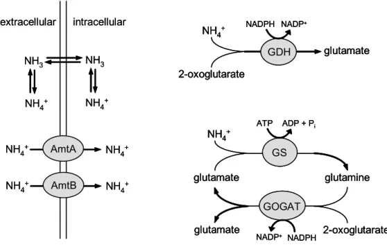
In general, two different uptake routes for ammonium are present (figure 1) and these are used in dependence on the availability of ammonium as described in the following. In aqueous solution, ammonium (NH4+) is in equilibrium with the uncharged and membrane permeable ammonia (NH3). Ammonia can easily enter the cell by passive diffusion across the cytoplasmic membrane. Under ammonium surplus, diffusion of ammonia is sufficient to promote growth, whereas under nitrogen limitation, ammonium transporters are expressed to ensure a proper nitrogen supply of the cell. In C. glutamicum, two ammonium transporter, AmtA and AmtB, are present under nitrogen limitation (Siewe et al., 1996; Jakoby et al., 1999a; Meier-Wagner et al., 2001) to enhance ammonium uptake and to ensure nitrogen supply of the cell.
The mode of transport by these membrane proteins is not clear. Two models are discussed. On one hand, an energy-dependent transport of charged ammonium was observed, indicating that ammonium uptake depends on the membrane
extracellular intracellular
AmtA NH4+ NH4+
NH3 NH3
NH4+
NH4+
AmtB NH4+ NH4+
2-oxoglutarate NH4+ NADPH
glutamate GDH
NADP+
glutamine glutamate
NH4+
2-oxoglutarate glutamate
GS
GOGAT
ATP ADP + Pi
NADPH NADP+
Figure 1: The uptake and assimilation of ammonium in C. glutamicum.The uptake of ammonium from the environment occurs either by diffusion of ammonia or via transport by AmtA and AmtB, respectively. It is not clear whether AmtA and AmtB actively transport ammonium depending on the membrane potential or if they facilitate passive diffusion of ammonia. Two assimilation pathways for ammonium are present in C. glutamicum. One is catalyzed by glutamate dehydrogenase (GDH), the other is a coupled reaction of glutamine synthetase (GS) and glutamate synthase (GOGAT). Both, transport and assimilation are regulated in dependence on the availability of ammonium. In all equations, free protons (H+) are not given. (Burkovski, 2003a; 2003b; 2005)
extracellular intracellular
AmtA NH4+ NH4+
NH3 NH3
NH4+
NH4+
AmtB NH4+ NH4+
2-oxoglutarate NH4+ NADPH
glutamate GDH
NADP+
2-oxoglutarate NH4+ NADPH
glutamate GDH
NADP+
glutamine glutamate
NH4+
2-oxoglutarate glutamate
GS
GOGAT
ATP ADP + Pi
NADPH NADP+
glutamine glutamate
NH4+
2-oxoglutarate glutamate
GS
GOGAT
ATP ADP + Pi
NADPH NADP+
Figure 1: The uptake and assimilation of ammonium in C. glutamicum.The uptake of ammonium from the environment occurs either by diffusion of ammonia or via transport by AmtA and AmtB, respectively. It is not clear whether AmtA and AmtB actively transport ammonium depending on the membrane potential or if they facilitate passive diffusion of ammonia. Two assimilation pathways for ammonium are present in C. glutamicum. One is catalyzed by glutamate dehydrogenase (GDH), the other is a coupled reaction of glutamine synthetase (GS) and glutamate synthase (GOGAT). Both, transport and assimilation are regulated in dependence on the availability of ammonium. In all equations, free protons (H+) are not given. (Burkovski, 2003a; 2003b; 2005)
potential (Kleiner, 1993, Siewe et al., 1996; Meier-Wagner et al., 2001). On the other hand, these transporters were described as gas channels that simply facilitate passive diffusion of uncharged ammonia across the cell membrane (Soupene et al., 1998, 2002; Khademi et al., 2004; Zheng et al., 2004; Javelle et al., 2005).
However, the strict regulation of ammonium transporters was ascribed to prevent the formation of a putative energy-costly futile cycle under ammonium surplus.
Regarding to this theory, ammonium would first be transported into the cell by the use of energy and then it would diffuse passively back out of the cell resulting in a detrimental waste of energy (Castorph et al., 1984; Kleiner, 1985). Sensitivity to ammonium due to futile cycling is suspected to be a universal phenomenon of animals, plant, and bacteria (Wirén et al., 2004). In fact, a number of plant families (Britto et al., 2001; Kronzucker et al., 2001; Britto et al., 2002) as well as animal cells (Martinelle et al., 1993) are known to be sensitive to ammonium. Nevertheless,
ammonium toxicity has not been demonstrated in bacteria and the formation of a putative futile cycle in bacteria is still speculation.
1.3. Assimilation of ammonium
In many bacteria, the primary products of ammonium assimilation are glutamate and glutamine, which are the major intracellular nitrogen donors (Merrick and Edwards, 1995). Glutamate provides nitrogen for most of the transaminases, whereas glutamine donates nitrogen for the synthesis of purines, pyrimidines, arginine, asparagine, tryptophan, histidine, glucosamine, and p-aminobenzoate (Reitzer, 2003). In general, there are two pathways for the assimilation of ammonium forming glutamate or glutamine: the glutamate dehydrogenase (GDH) and the glutamine synthetase/glutamate synthase (GS/GOGAT) pathway (figure 1).
Glutamate dehydrogenases are broadly distributed enzymes which catalyze the reversible reductive amination of 2-oxoglutarate by ammonium to give L-glutamate in an NAD(P)H-dependent reaction (Barker, 1981; Merrick and Edwards, 1995). In contrast to most GDHs identified in higher eukaryotes, which have a dual coenzyme specificity (EC 1.4.1.3) using both, NADH and NADPH, GDHs of prokaryotes and lower eukaryotes act only with one particular coenzyme, NADH or NADPH (Minambres et al., 2000). In general, NADPH-dependent GDHs (EC 1.4.1.4) contribute to anabolism by assimilating ammonium to form glutamate (Consalvi et al., 1991), whereas NADH-dependent GDHs (EC 1.4.1.2) are usually catabolic enzymes for the reverse reaction, the oxidative deamination of glutamate (Duncan et al., 1992). All GDHs described so far are oligomeric enzymes, which consist either of six or four identical subunits. These subunits are either of 50 kDa, 115 kDa, or 180 kDa. According to the oligomeric structure, the subunit size, and the results of hierarchical homology grouping, four different well defined classes of GDHs exist:
α6-50I and α6-50II (small GDHs) as well as α6-180 and α4-115 (large GDHs). The four classes share the same catalytic mechanism, very similar domain structures, and several well-conserved amino acid residues with distinct function. However, the function of the additional amino acids of large GDHs (α6-180 and α4-115 classes) is completely unknown. In bacteria, only hexameric GDHs have been reported yet (Minambres et al., 2000). GDH from Clostridium symbiosum is the most extensively studied GDH with regard to the three-dimensional structure, the catalytic
mechanism, and amino acid residues with distinct function. GDH from C.
symbiosum is NADH-dependent and belongs to the α6-50I class (Minambres et al., 2000). Each subunit of this hexameric GDH consists of two domains separated by a cleft harbouring the active site. One domain binds NADH and the other glutamate and 2-oxoglutarate, respectively (Baker et al., 1992). During the catalytic cycle, a large movement between the two domains occurs, closing the cleft and bringing the substrate and NADH into the correct position for the reaction (Baker et al., 1997).
The distinct functions of several amino acid residues are known (Pasquo et al., 1996; Millevoi et al., 1998; Baker et al., 1992; Baker et al., 1997). One of them is lysine-89 in the active site, which plays a key role for the interactions with the γ- carboxyl group of the substrate (Wang et al., 1994; Stillman et al., 1999). If this residue is altered by site-directed mutagenesis to a leucine residue, the resulting mutant has an almost identical conformation as the wild type GDH, but it is enzymatically inactive (Stillman et al., 1999).
GDH from C. glutamicum belongs to the α6-50I class of small GDHs (Minambres et al., 2000). It is NADPH-dependent, suggesting that it preferentially acts as an anabolic GDH for ammonium assimilation in vivo. The kinetic properties of GDH from C. glutamicum have been investigated by in vitro assays (Shiio et al., 1970).
GDH from C. glutamicum catalyzes the formation as well as the degradation of glutamate in vitro, but the maximum velocity of the formation of glutamate (87.6 µmol/(min*mg)) is 4.6 times higher than that of the degradation (19.2 µmol/(min*mg)). High concentrations of glutamate (400 mM) inhibit the formation of glutamate (75 % inhibition) and the presence of ammonium (10 mM) inhibits the degradation of glutamate (95% inhibition) in vitro. As GDHs from many other organisms, GDH from C. glutamicum has only a low affinity to its substrates ammonium (Km = 3.08 mM) and 2-oxoglutarate (Km = 5.72 mM) and even lower affinity to its product glutamate (Km = 100 mM) (Shiio et al., 1970). In vivo, GDH from C. glutamicum is the major consumer of cellular ammonium and NADPH under nitrogen surplus. Flux measurements revealed that about 72 % of ammonium assimilation is done by GDH (Tesch et al., 1999), which consumes about 50 % of cellular NADPH (Marx et al., 1999). The gdh gene encoding GDH from C.
glutamicum has been identified. It is transcribed monocistronically and transcription starts 284 bp upstream of the start codon of gdh (Börmann et al., 1992).
The second pathway for ammonium assimilation is the glutamine synthetase/glutamate synthase (GS/GOGAT) system (figure 1). In this reaction, ammonium is first attached to glutamate to form glutamine by an ATP-dependent glutamine synthetase (GS), followed by the transfer of the amide nitrogen onto 2- oxoglutarate by an NADPH-dependent glutamate synthase (GOGAT) to form glutamate. In C. glutamicum, two genes encoding glutamine synthetases were identified: glnA coding for a GSI-β subtype enzyme (Jakoby et al., 1997) and glnA2 coding for a GSI-α subtype enzyme (Nolden et al., 2001a). Analysis of a glnA deletion strain revealed that only glnA codes for an active enzyme for glutamine synthesis in C. glutamicum. The glnA mutant was observed to be glutamine- auxotrophic (Jakoby et al., 1996). Hence, the gene product of glnA2 has obviously no glutamine synthetase activity in C. glutamicum and its physiological role is unclear. Glutamate synthase is encoded by the gltBD operon in C. glutamicum (Beckers et al., 2001; Schulz et al., 2001).
The overall reactions of both pathways, GDH and GS/GOGAT, are very similar (figure 1). Both catalyze the NADPH-dependent formation of glutamate from ammonium and 2-oxoglutarate. But in contrast to the GDH-pathway, the GS/GOGAT-system additionally utilizes energy in form of ATP. Consequently, the GS/GOGAT system is strictly regulated to prevent waste of energy. Ammonium is assimilated mainly by GDH if C. glutamicum is growing in ammonium-rich medium.
Under these conditions, GDH activity is on a high level (1.7 U/mg protein), GOGAT activity is not detectable, and GS activity is maintained at a low level (0.1 U/mg protein) exclusively to satisfy the glutamine requirements of the cell (Tesch et al., 1998). As GDH from C. glutamicum has only a low affinity to ammonium (Km = 3.08 mM), it is not able to sufficiently assimilate ammonium under nitrogen limitation.
Glutamine synthetases are known to have a significantly higher affinity to ammonium than glutamate dehydrogenases. Consequently, the GS/GOGAT system is activated in C. glutamicum under nitrogen limitation (1.5 U/mg protein and 35 mU/mg protein, respectively) (Tesch et al., 1998). Thus, utilization of GS/GOGAT allows nitrogen assimilation even under ammonium limitation, but at the expense of energy.
1.4. Nitrogen-dependent regulation
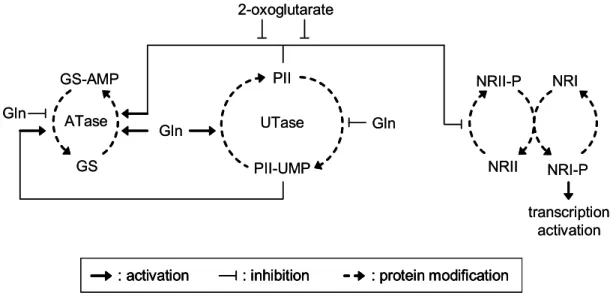
As described above, bacteria regulate nitrogen uptake and assimilation in dependence on the availability of nitrogen sources. For this purpose, bacteria have evolved complex regulatory networks. The most extensive model of nitrogen- dependent regulation has been described for the Gram-negative enteric bacterium E. coli (Merrick and Edwards, 1995; Reitzer, 2003; Ninfa, 2005).
In E. coli, two proteins play a major role in the regulation of nitrogen uptake and metabolism (figure 2). These are uridylyltransferase (UTase) and the signal transduction protein PII. UTase is a sensor for the internal concentration of glutamine. The glutamine pool is low under nitrogen starvation and high under nitrogen surplus. UTase transfers this signal to PII by uridylylation and deuridylylation, respectively. Unmodified PII is present under nitrogen surplus, while PII-UMP is present under nitrogen starvation (Jiang et al., 1998a). In addition, PII itself is a sensor for the internal concentration of 2-oxoglutarate. Under nitrogen starvation, 2-oxoglutarate is accumulated. A high 2-oxoglutarate pool antagonizes the status of unmodified PII, which is present under high glutamine concentrations
PII
PII-UMP
UTase Gln
Gln
2-oxoglutarate
NRII NRII-P
NRI-P transcription
activation NRI ATase
GS GS-AMP Gln
Figure 2: Nitrogen regulation network of E. coli. The nitrogen status is sensed by uridylyltransferase (UTase) in E. coli. UTase reversibly uridylylates PII in response to the internal glutamine pool. PII additionally senses 2-oxoglutarate. These signals are transferred by protein interactions of PII/PII-UMP to the NRII/NRI system for transcriptional control of nitrogen-regulated genes and adenylyltransferase (ATase), which regulates glutamine synthetase (GS) activity. ATase reversible adenylylates GS to regulate its activity, whereas NRII reversibly phosphorylates NRI to influence its ability to activate transcription (Ninfaet al., 2005).
: activation : inhibition : protein modification PII
PII-UMP
UTase Gln
Gln
2-oxoglutarate
NRII NRII-P
NRI-P transcription
activation NRI ATase
GS GS-AMP Gln
Figure 2: Nitrogen regulation network of E. coli. The nitrogen status is sensed by uridylyltransferase (UTase) in E. coli. UTase reversibly uridylylates PII in response to the internal glutamine pool. PII additionally senses 2-oxoglutarate. These signals are transferred by protein interactions of PII/PII-UMP to the NRII/NRI system for transcriptional control of nitrogen-regulated genes and adenylyltransferase (ATase), which regulates glutamine synthetase (GS) activity. ATase reversible adenylylates GS to regulate its activity, whereas NRII reversibly phosphorylates NRI to influence its ability to activate transcription (Ninfaet al., 2005).
: activation : inhibition : protein modification : activation : inhibition : protein modification : activation : inhibition : protein modification
(Kamberov et al., 1995; Jiang et al., 1998a; Jiang et al., 1998b). Thus, PII converts two input signals, the concentrations of glutamine and 2-oxoglutarate, in one output signal (Ninfa et al., 2005). This signal is transferred by PII to other regulatory proteins. On the one hand, PII and glutamine synergistically interact with adenylyltransferase (ATase), which regulates glutamine synthetase by adenylylation and deadenylylation. This results in unmodified and fully active GS under nitrogen starvation, whereas GS-AMP with reduced activity is present under nitrogen surplus (Stadtman, 1990; Jiang et al., 1998b). On the other hand, PII interacts with NRII, which is part of the NRII/NRI two component system. Under nitrogen surplus, PII binds the kinase NRII and thereby represses phosphorylation of the response factor NRI (Jiang et al., 1998b; 1998c). Under nitrogen limitation, NRII is released and phosphorylates NRI, which then activates transcription of σ54-dependent genes.
Among these are genes of nitrogen metabolism, transport, and regulation (Atkinson et al., 2002). For example, expression of nac, coding for the transcriptional regulator Nac, is activated by NRI under nitrogen limitation. Nac itself regulates the transcription of several genes of the glutamate and serine metabolism, e.g. gdh coding for glutamate dehydrogenase. Nac represses gdh transcription under nitrogen limitation and ammonium assimilation is taken over by the GS/GOGAT system (Camarena et al., 1998). Beside that, NRI activates the expression of the glnK gene, coding for GlnK, under nitrogen limitation. GlnK is another PII-type protein. It is also modified by UTase in response to the nitrogen status in the same manner as PII. GlnK is responsible for fine tuning of the nitrogen regulation cascade under nitrogen limitation by the formation of heterotrimers of PII-UMP and GlnK- UMP (Atkinson et al., 1998; 1999; 2002; Forchhammer et al., 1999; van Heeswijk et al., 2000). Additionally, GlnK binds to the ammonium transporter AmtB, if nitrogen- starved cells are exposed to an ammonium rich environment. This deactivates AmtB presumably to prevent the formation of a futile cycle under ammonium surplus.
Additionally, AmtB is discussed to be a sensor for external ammonium and thereby influences the uridylylation state of GlnK (Javelle et al., 2004).
Nitrogen control of C. glutamicum differs substantially from that of the model organism E. coli (Burkovski, 2003a; 2003b). In C. glutamicum, a signal cascade of at least three proteins plays a major role in nitrogen control (figure 3). This cascade
sensor
GlnD
GlnK AMP GlnK
AmtR
transcription
sensor
GlnD
GlnK AMP GlnK
AmtR
transcription
AmtB
Clp FtsH
proteolysis NH4+ AmtB
A B
Figure 3: The nitrogen regulation network of C. glutamicum. A: Under nitrogen limitation, a so far unknown sensor protein induces the adenylylation of GlnK by GlnD. GlnK-AMP binds the transcriptional repressor AmtR to inactivate it. Consequently, AmtR-controlled genes are transcribed. B: After a shift of nitrogen starved cells to an ammonium rich environment, GlnD deadenylylates GlnK-AMP. Unmodified GlnK does not deactivate AmtR anymore. Consequently, AmtR represses the transcription of its target genes. Unmodified GlnK is sequestered to the membrane. It binds AmtB and thereby most probably deactivates AmtB. Beside that, binding to AmtB induces degradation of GlnK by proteolysis, which is depending on the proteases FtsH, ClpCP, and ClpXP. (Stösseret al., 2004)
sensor
GlnD
GlnK AMP GlnK
AmtR
transcription
sensor
GlnD
GlnK AMP GlnK
AmtR
transcription
AmtB
Clp FtsH
proteolysis NH4+ AmtB
A B
Figure 3: The nitrogen regulation network of C. glutamicum. A: Under nitrogen limitation, a so far unknown sensor protein induces the adenylylation of GlnK by GlnD. GlnK-AMP binds the transcriptional repressor AmtR to inactivate it. Consequently, AmtR-controlled genes are transcribed. B: After a shift of nitrogen starved cells to an ammonium rich environment, GlnD deadenylylates GlnK-AMP. Unmodified GlnK does not deactivate AmtR anymore. Consequently, AmtR represses the transcription of its target genes. Unmodified GlnK is sequestered to the membrane. It binds AmtB and thereby most probably deactivates AmtB. Beside that, binding to AmtB induces degradation of GlnK by proteolysis, which is depending on the proteases FtsH, ClpCP, and ClpXP. (Stösseret al., 2004)
consists of the GlnD protein, which is similar to the UTase from E. coli, the GlnK protein, which is the only PII-type protein in C. glutamicum, and AmtR, which is a TetR-type transcriptional repressor (Jakoby et al., 1999; 2000; Nolden et al., 2001b).
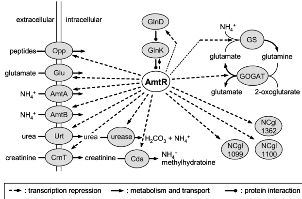
Under nitrogen surplus, AmtR represses transcription of several genes involved in nitrogen metabolism, transport, and regulation (figure 4) (Jakoby et al., 2000;
Nolden et al., 2001b; Beckers, 2004). Under this condition, GlnD and GlnK are present only on a low basal level. In response to nitrogen starvation, GlnD adenylylates GlnK (Strösser et al., 2004), which then binds to AmtR (figure 3A).
AmtR bound by GlnK-AMP does not bind DNA anymore (Beckers et al., 2005).
Consequently, repression of transcription by AmtR is released by GlnK-AMP and the expression of corresponding genes is induced under nitrogen limitation. If nitrogen-starved cells are shifted to an ammonium rich enviroment, GlnD deadenylylates GlnK (Stösser et al., 2004). AmtR cannot be bound by unmodified GlnK, which allows repression of target gene transcription again (figure 3B) (Beckers et al., 2005). Moreover, unmodified GlnK binds to the ammonium
extracellular intracellular
NH4+ AmtA NH4+ AmtB
Glu glutamate
Urt urea
peptides Opp
creatinine CrnT
urease
urea H2CO3+ NH4+ creatinine
methylhydratoine Cda NH4+
NCgl 1099
NCgl 1100 NCgl 1362 GlnD
AmtR
glutamine glutamate
NH4+
2-oxoglutarate glutamate
GS
GOGAT GlnK
: transcription repression : metabolism and transport : protein interaction
Figure 4: The AmtR-regulon of C. glutamicum. The transcriptional repressor AmtR from C. glutamicum regulates the expression of genes coding for transporters, enzymes, and signal transduction proteins as indicated. Repression occurs under nitrogen surplus and is released under nitrogen limitation. (Beckers, 2004)
extracellular intracellular
NH4+ AmtA NH4+ AmtB
Glu glutamate
Urt urea
peptides Opp
creatinine CrnT
urease
urea H2CO3+ NH4+ creatinine
methylhydratoine Cda NH4+
NCgl 1099
NCgl 1100 NCgl 1362 GlnD
AmtR
glutamine glutamate
NH4+
2-oxoglutarate glutamate
GS
GOGAT GlnK
: transcription repression
: transcription repression : metabolism and transport: metabolism and transport : protein interaction: protein interaction
Figure 4: The AmtR-regulon of C. glutamicum. The transcriptional repressor AmtR from C. glutamicum regulates the expression of genes coding for transporters, enzymes, and signal transduction proteins as indicated. Repression occurs under nitrogen surplus and is released under nitrogen limitation. (Beckers, 2004)
transporter AmtB in response to an ammonium upshift. On the one hand, this is suspected to inactivate AmtB, as described for E. coli, to prevent an energy-costly futile cycle in the presence of ammonium. On the other hand, binding of GlnK to AmtB leads to a rapid degradation of GlnK by a process, that involves the proteases FtsH, ClpCP, and ClpXP. Nevertheless, around 5 % of GlnK are protected against proteolysis. The role and the mechanism of protection of GlnK is still unknown (Strösser et al., 2004).
In addition to the transcriptional regulation by the GlnD/GlnK/AmtR cascade, glutamine synthetase is regulated on the level of activity by adenylylation/deadenylylation. In accordance to E. coli, GS-AMP is present under nitrogen surplus and is less active than unmodified GS, which is present under nitrogen starvation (Nolden et al., 2001a). The modification/demodification of GS is catalyzed by ATase. But in contrast to E. coli, ATase works independently of GlnK (Burkovski, 2003b).
While the signal transduction via the GlnD/GlnK/AmtR cascade is well-investigated, the sensor of the nitrogen status in C. glutamicum is still unknown. An overexpression of the corresponding glnD gene leads to a loss of nitrogen control (Nolden et al., 2001b). This observation is inconsistent with a putative role of GlnD as a primary sensor, as described for in E. coli. Consequently, the presence of at least one additional protein was postulated, which functions as a sensor for the nitrogen status of the cell and controls GlnD activity in response to nitrogen supply (figure 3). Beside that, the signal indicating the nitrogen status is unknown as well.
The question if the internal concentrations of glutamate, glutamine, and ammonium, respectively, indicate the nitrogen status in C. glutamicum was investigated (Nolden et al., 2001b). The cellular concentration of glutamate is high under nitrogen surplus as well as under nitrogen starvation. Thus, glutamate can be excluded. For glutamine, Nolden et al. (2001b) observed only a slight drop of the internal concentration from approximately 18 mM to 8 mM 10 minutes after the removal of nitrogen sources. This indicates, that glutamine does not play a major role in sensing the nitrogen status in C. glutamicum. To measure internal ammonium, Nolden et al. (2001) initially used the indophenol method (Jahns et al., 1988). But this method is significantly influenced by high amino acid pools as present in C.
glutamicum and is not reliable under these conditions (L. Nolden, personal communication). Because of that, the values of internal ammonium concentrations presented by Nolden et al. (2001b) might be incorrect.
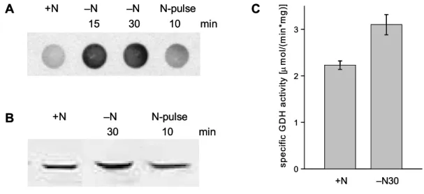
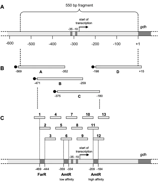
Glutamate dehydrogenase plays an important but so far less investigated role in nitrogen control of C. glutamicum. The presence of the gdh gene, coding for glutamate dehydrogenase, is essential for a functional nitrogen-dependent regulation in C. glutamicum. GS and GOGAT activity are normally downregulated in the C. glutamicum wild type under nitrogen surplus. Deletion of gdh causes a significantly increase of GS activity and deregulation of GOGAT activity (Tesch et al., 1998). In accordance to this, a loss of transcription control by the GlnD/GlnK/AmtR signal cascade was observed upon deletion of gdh. In the C.
glutamicum wild type, transcription of amtA and amtB is repressed by AmtR under nitrogen surplus. Deletion of gdh leads to the loss of repression of amtA and amtB transcription. Consequently, a deletion of gdh abolishes transcription control by the
GlnD/GlnK/AmtR signal cascade (L. Nolden, personal communication). The underlying mechanism for the interaction between gdh and the signal cascade is unknown so far.
In addition to that, GDH seems to have a so far unknown function under nitrogen limitation. In other bacteria, expression of NADPH-dependent GDH is either downregulated under nitrogen starvation or it remains constant (Brenchley et al., 1975; Schwacha et al., 1993; Merick and Edwards, 1995; Camarena et al., 1998). In agreement with this, GDH from C. glutamicum was first reported to be unaffected by nitrogen supply. Tesch and co-workers (1999) could not observe a significant change in GDH activity of C. glutamicum under nitrogen limitation. This observation was supported by transcriptome analyses of C. glutamicum cultivated under nitrogen excess and nitrogen limitation, where gdh transcription was observed to be unaffected (Beckers, 2004; Silberbach, 2004). In contrast to this, L. Nolden observed a significant increase in gdh transcription as well as GDH activity under nitrogen limitation (personal communication). This is remarkable, as an upregulation of NADPH-dependent GDH in bacteria under nitrogen limitation was unknown at that time. Recently published data show, that NADPH-dependent GDH from Ruminococcus flavefaciens is also upregulated on the level of transcription under ammonium limitation (Antonopoulos et al., 2003). The physiological reason for this regulation in R. flavefaciens is still unknown. Interestingly, GDH from R. flavefaciens and GDH from C. glutamicum are closely related. Both are members of the α6-50I class of small GDHs, and with an amino acid identity of 63 %, GDH from R.
flavefaciens is the most similar homolog of C. glutamicum GDH known so far (Antonopoulos et al., 2003). Because of these observations, one can speculate about a putative new and so far unknown physiological role of certain NADPH- dependent GDHs in bacteria under nitrogen limitation. However, the induction of gdh transcription under nitrogen limitation in C. glutamicum seems not to depend on the global nitrogen regulator AmtR. A deletion of the amtR gene did not affect gdh transcription (L. Nolden, personal communication). Consequently, an additional and so far unknown regulatory system for nitrogen-dependent transcription control seems to be present in C. glutamicum, which regulates gdh transcription in response to the nitrogen supply.
1.5. Objectives
Expression of the gdh gene is induced under nitrogen limitation. This regulation does not depend on the global nitrogen regulator AmtR. Consequently, a putative new regulatory system for nitrogen-dependent transcription control is present in C.
glutamicum. The main aim of this work is the identification and characterization of the putative new regulator of gdh transcription.
Glutamate dehydrogenase plays a crucial role for a functional nitrogen control by the GlnD/GlnK/AmtR signal cascade. Deletion of the gdh gene leads to a complete loss of transcription control by AmtR. The underlying mechanism is still unknown.
The characterization of this effect is the second aim of this work.
Ammonium is suspected of being toxic for bacteria due to the formation of a putative energy-wasting transmembrane cycle. An additional aim of this work is to investigate putative ammonium toxicity in C. glutamicum and the presence of a putative futile cycle.
2. Materials and methods
2.1. Bacterial strains, plasmids, and primers
Bacterial strains used in this study are listed in table 1, plasmids are listed in table 2.
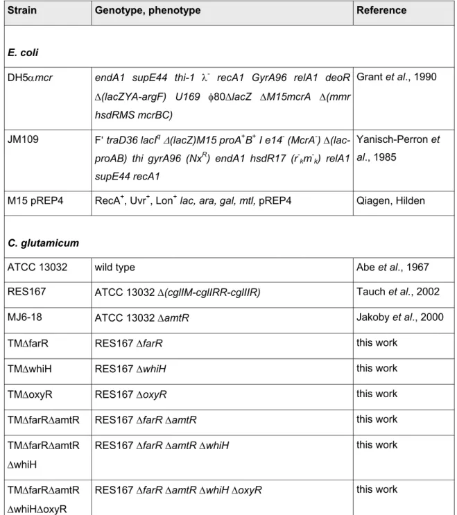
Table 1:E. coliand C. glutamicum strains that were used in this work.For strains that were constructed as part of this work, a detailed description is given in the appendix. NxR:
resistant to nalidixic acid.
Strain Genotype, phenotype Reference
E. coli
DH5αmcr endA1 supE44 thi-1 λ- recA1 GyrA96 relA1 deoR
∆(lacZYA-argF) U169 φ80∆lacZ ∆M15mcrA ∆(mmr hsdRMS mcrBC)
Grant et al., 1990
JM109 F‘ traD36 lacIq ∆(lacZ)M15 proA+B+ I e14- (McrA-) ∆(lac- proAB) thi gyrA96 (NxR) endA1 hsdR17 (r-km-k) relA1 supE44 recA1
Yanisch-Perron et al., 1985
M15 pREP4 RecA+, Uvr+, Lon+ lac, ara, gal, mtl, pREP4 Qiagen, Hilden
C. glutamicum
ATCC 13032 wild type Abe et al., 1967
RES167 ATCC 13032 ∆(cglIM-cglIRR-cglIIR) Tauch et al., 2002
MJ6-18 ATCC 13032 ∆amtR Jakoby et al., 2000
TM∆farR RES167 ∆farR this work
TM∆whiH RES167 ∆whiH this work
TM∆oxyR RES167 ∆oxyR this work
TM∆farR∆amtR RES167 ∆farR ∆amtR this work
TM∆farR∆amtR
∆whiH
RES167 ∆farR ∆amtR ∆whiH this work
TM∆farR∆amtR
∆whiH∆oxyR
RES167 ∆farR ∆amtR ∆whiH ∆oxyR this work
LN∆GDH ATCC 13032 ∆gdh Nolden, unpublished
LN∆GS ATCC 13032 ∆glnA Nolden, unpublished
TM∆gdh∆glnA LN∆GDH ∆glnA this work
JS-1 ATCC 13032 ∆amtA ∆amtB Strösser,
unpublished
Table 2: Plasmids that were used in this work. For plasmids that were constructed as part of this work, a detailed description is given in the appendix. ApR: resistant to ampicilin.
KmR: resistant to kanamycin.
Plasmid Description Reference
pK18mobsacB KmR, ori pUC, mob, sacB Schäfer et al.,
1994 pK18∆amtR Deletion vector for amtR of C. glutamicum, derived from
pK18mobsacB, KmR, ori pUC, mob, sacB
Nolden, 2001
pK18∆farR Deletion vector for farR of C. glutamicum, derived from pK18mobsacB, KmR, ori pUC, mob, sacB
this work
pK18∆whiH Deletion vector for whiH of C. glutamicum, derived from pK18mobsacB, KmR, ori pUC, mob, sacB
this work
pK18∆oxyR Deletion vector for oxyR of C. glutamicum, derived from pK18mobsacB, KmR, ori pUC, mob, sacB
this work
pK18∆glnA Deletion vector for glnA of C. glutamicum, derived from pK18mobsacB, KmR, ori pUC, mob, sacB
Nolden, unpublished
pZ8-1 KmR, ptac, ori C. glutamicum Degussa AG,
Halle
pUC18 ApR, lacZα Viera & Messing,
1982 pUC11-1.8 Expression vector of amtR of C. glutamicum derived from
pUC19, ApR, lacZα
Jakoby et al., 2000
pUCfarR Expression vector of farR of C. glutamicum derived from pUC18, ApR, lacZα
this work
pUCwhiH Expression vector of whiH of C. glutamicum derived from pUC18, ApR, lacZα
this work
pUCoxyR Expression vector of oxyR of C. glutamicum derived from pUC18, ApR, lacZα
this work
pZgdh Expression vector of gdh from C. glutamicum, derived from pZ8-1, KmR, ptac, ori C. glutamicum
this work
pZgdhEC Expression vector of gdh from E. coli, derived from pZ8-1, KmR, ptac, ori C. glutamicum
this work
pZgdh-K92L Expression vector for an enyzmatical inactive mutant of GDH from C. glutamicum, derived from pZ8-1, KmR, ptac, ori C. glutamicum
this work
pQE30Xa ApR, ori ColE1, PT5 Qiagen, Hilden
pQE30Xagdh Expression vector for his-tagged GDH from C. glutamicum, ApR, ori ColE1, PT5
this work
pJC1 KmR, ori C. glutamicum Cremer et al.,
1990 pJCgdhlacZ KmR, ori C. glutamicum, harbouring a fusion of the gdh
promoter of C. glutamicum and lacZ
this work
pK18gdh-lacZ KmR, ori pUC, mob, sacB, harbouring a fusion of the gdh promoter of C. glutamicum and lacZ
Nolden, unpublished PGEM3z-amt ApR, lacZα, harbouring a 0.5 kb fragment of the amtA gene Nolden, 2001 PGEM3z-amtB ApR, lacZα, harbouring a 0.5 kb fragment of the amtB gene Nolden, 2001 PGEM3z-gltB ApR, lacZα, harbouring a 0.5 kb fragment of the gltB gene Nolden, 2001 PGEM3Z-glnA ApR, lacZα, harbouring a 0.5 kb fragment of the glnA gene Nolden, 2001 PGEM4z-gdh ApR, lacZα, harbouring a 0.5 kb fragment of the gdh gene Nolden, 2001 PGEM3z-glnD ApR, lacZα, harbouring a 0.5 kb fragment of the glnD gene Nolden, 2001 pDrive ApR, lacZα, A-T insertion vector Qiagen, Hilden
2.2. Cultivation of bacteria
Culture media used in this study are listed in table 3. If appropriate, antibiotics were added to the media in the following final concentrations: 50 µg/mL carbenicillin, 25 µg/mL kanamycin, or 10 µg/mL kanamycin (latter only for C. glutamicum strains after electroporation). E. coli strains were routinely grown at 37 °C in LB medium or on LB plates. C. glutamicum strains were routinely cultivated at 30 °C in CgC medium, in Brain Heart Infusion (BHI) medium, or on BHI plates. To study various stress conditions under highly reproducible conditions, a standard inoculation schema was applied. C. glutamicum strains were first cultivated in 3 mL BHI medium for 8 hours. This culture was used to inoculate 25 mL CgC medium for overnight growth. This culture, with an overnight OD600 of approximately 25-33, was used to inoculate 100 mL of fresh CgC medium to an OD600 of approximately 1 and cells were grown until the exponential phase was reached (OD600 approximately 4- 5). In case of LN∆GS and TM∆gdh∆glnA, all media were additionally supplemented with 100 mM glutamine. To analyze the effect of various cultivation conditions or medium compositions, the cells were treated in the following as listed in table 4.
28.5 g FeSO4x 7 H2O, 16.5 g MnSO4x H2O, 6.4 g ZnSO4x 7 H2O, 764 mg CuSO4 x 5 H2O, 129 mg CoCl2 x 6 H2O, 44 mg NiCl2 x 6 H2O, 64 mg Na2MoO4x 2 H2O, 48 mg H3BO3, 50 mg SrCl2, 50 mg BaCl2x 2 H2O, 28 mg KAl(SO4)2x 12 H2O, pH (H2SO4) = 1, sterilization by filtration.
Trace element solution
42 g MOPS, 20 g (NH4)2SO4, 5 g urea, 0.5 g KH2PO4, 0.5 g K2HPO4, pH (NaOH) = 7.0, after autoclavation: 10 mL 100 mM CaCl2, 10 mL 1 M MgSO4, 200 mg biotin, 1 mL trace element solution
CgCoC
42 g MOPS, 0.5 g KH2PO4, 0.5 g K2HPO4, pH (NaOH) = 7.0, after autoclavation: 10 mL 100 mM CaCl2, 10 mL 1 M MgSO4, 200 mg biotin, 1 mL trace element solution, 50 mL 50 % glucose
CgCoN
42 g MOPS, 20 g (NH4)2SO4, 5 g urea, 0.5 g KH2PO4, 0.5 g K2HPO4, pH (NaOH) = 7.0, after autoclavation: 10 mL 100 mM CaCl2, 10 mL 1 M MgSO4, 200 mg biotin, 1 mL trace element solution, 50 mL 50 % glucose CgC
15 g Bacto-Agar in BHI medium BHI plates
37 g/L Brain Heart Infusion BHI
15 g Bacto-Agar in LB medium LB plates
10 g Tryptone, 5 g yeast extract, 10 g NaCl LB
Ingredients (per L) Medium
Table 3: Culture media used in this study.
50 mL of a culture was transferred to a 50 mL falcon tube. The falcon tube was sealed. In this airtight container, cells were cultivated at 30 °C.
Oxygen limitation
Cells were harvested by centrifugation and resuspended in 100 mL CgC medium with a temperature of 37 °C. Cells were cultivated at 37 °C.
Heat stress
Cells were cultivated at 30 °C until the stationary phase was reached.
Growth phase
1 % of arginine, citrulline, and ornithine, respectively, was added to the culture medium, and cells were cultivated at 30 °C.
Addition of intermediates
of arginine biosynthesis
Cells were harvested by centrifugation and resuspended in 100 mL pre- warmed CgCoN medium supplemented with either 0.25 M (NH4)2SO4, 0.5 M (NH4)2SO4, or 1 M (NH4)2SO4. As control, cells were resuspended in prewaremd CgCoN medium supplemented with either 0.25 M (NH4)2SO4 + 0.25 M Na2SO4 or 0.25 M (NH4)2SO4 + 0.75 M Na2SO4. After resupending, cells were cultivated at 30 °C. Cultures of TM∆gdh∆glnA were additionally supplemented with 100 mM glutamine.
High ammonium concentrations
Cells were harvested by centrifugation and resuspended in 100 mL CgC medium with a temperature of 15 °C. Cells were cultivated at 15 °C.
Either 100 µM paraquat (superoxide stress) or 58 µM H2O2 (peroxide stress) was added to the culture medium, and cells were cultivated at 30
°C.
1 M NaCl was added to the culture medium, and cells were cultivated at 30 °C.
Cells were harvested by centrifugation and resuspended in 100 mL pre- warmed CgCoC medium supplemented with 1 % of the appropriate carbon source. Cells were cultivated again at 30 °C.
Cells were harvested by centrifugation and resuspended in 100 mL pre- warmed CgCoC medium. Cells were cultivated again at 30 °C. After a certain period of time, 1 % glucose was added to the culture medium to restore carbon surplus conditions.
Cells were harvested by centrifugation and resuspended in 100 mL pre- warmed CgCoN medium supplemented with 1 % of the appropriate nitrogen source. Cells were cultivated again at 30 °C.
Cells were harvested by centrifugation and resuspended in 100 mL pre- warmed CgCoN medium. Cells were cultivated again at 30 °C. After a certain period of time, 200 mM (NH4)2SO4 was added to the culture medium to restore nitrogen surplus conditions. LN∆GS was pulsed with either 200 mM (NH4)2SO4, or 100 mM glutamine, or both, 200 mM (NH4)2SO4and 100 mM glutamine.
Chill stress Oxidative
stress Osmotic
stress Variation of
the carbon source Carbon starvation Variation of the nitrogen
source Nitrogen starvation
Practical procedure Condition
effect of various conditions, C. glutamicumstrains was treated as described in this table.
Table 4: Cultivation protocols for the analysis of various conditions. To study the
2.3. Genetic manipulation of bacteria
2.3.1. Preparation of competent E. coli cells and transformation
To prepare competent E. coli cells, 20 mL LB medium was inoculated with E. coli cells and cultivated for 8 h at 37 °C. 1 mL of this culture was used to inoculate 250 mL SOB medium for overnight growth at 20 °C in a 2 L flask. On the next day, the culture was chilled on ice for 10 min. Cells were harvested by centrifugation (4000 x g, 4 °C, 8 min). The cell pellet was resuspended in 80 mL ice-cold TB buffer and centrifuged again (4000 x g, 4 °C, 10 min). After resuspending in 40 mL TB buffer supplemented with 1.4 mL DMSO, the cells were incubated for 10 min on ice.
Aliquots of 100 µL were transferred to pre-cooled reaction tubes. These were immediately frozen in liquid nitrogen and stored at -80 °C.
For transformation, a 100 µL aliquot of competent E. coli cells was thawed on ice and plasmid DNA was added. The cells were incubated for 30 min on ice. After a heat shock at 42 °C for 30 s, cells were again incubated for 2 min on ice and 900 µL SOC medium was added. The cell suspension was cultivated for 1 h at 37 °C. After that, 200 µL of the cell suspension was plated on LB plates containing the appropriate antibioticum.
TB buffer: 10 mM Pipes, 15 mM CaCl2, 250 mM KCl, pH (KOH) = 6.7. After adjustment of pH, 55 mM MnCl2 were added, sterilization by filtration.
SOB medium: 20 g/L Tryptone, 5 g/L yeast extract, 0.5 g/L NaCl, 2.5 mM KCl. After autoclavation, 5 mL 2 M MgCl2 were added.
SOC medium: 10 g/L Tryptone, 5 g/L yeast extract, 5 g NaCl, 3.6 g/L glucose. After autoclavation, 5 mL 2 M MgCl2 were added.
2.3.2. Preparation of competent C. glutamicum cells and transformation To prepare competent C. glutamicum cells, 20 mL BHI medium was inoculated with C. glutamicum cells and cultivated for 8 h at 30 °C. Subsequently, this culture was used to inoculate 200 mL LB medium with growth inhibitors to an OD600 of 0.35. This was cultivated at 20 °C in a 2 L flask overnight. On the next day, cells were