YggB of Corynebacterium glutamicum - Dual function in osmotic stress response and
glutamate production
Inaugural-Dissertation zur
Erlangung des Doktorgrades
der Mathematisch-Naturwissenschaftlichen Fakultät der Universität zu Köln
vorgelegt von
Kirsten Börngen aus Köln
Köln, September 2009
Berichterstatter:
Prof. Dr. Reinhard Krämer Prof. Dr. Arnd Baumann
Tag der Disputation: 27.11.2009
„Phantasie ist wichtiger als Wissen, denn Wissen ist begrenzt“
Albert Einstein
Abstract
The membrane protein YggB of Corynebacterium glutamicum was previously described to belong to the MscS-type family of mechanosensitive (MS) channels functioning as emergency valves upon osmotic downshift. Bacterial cells respond to rapid water influx by immediate activation of MS channels that allow efflux of compatible solutes to prevent cell lysis. Recently, YggB was also connected to the glutamate export in C. glutamicum under glutamate productive conditions. The mechanism of glutamate export is not fully understood so far although C. glutamicum has been used for the industrial production of amino acids for decades. Deletion of yggB led to a drastic decrease in glutamate excretion while truncation of 110 AA resulted in continuous export of glutamate. In this work YggB was characterized with respect to its dual function as MS channel on the one hand and in the excretion of glutamate on the other. Using the patch clamp technique and additional physiological approaches it was shown that YggB harbors the functions of a pressure- sensitive MS channel similar to the E. coli homolog MscS. However, for the first time also an involvement of a MS channel in bacterial response to hyperosmotic conditions was shown. A so called ‘pump and leak’ model including active betaine uptake (via BetP) and passive betaine efflux (via YggB) to accurately adjust the internal solute concentration, which are accumulated under hyperosmotic conditions to balance the osmotic gradient, is proposed. Concerning the second function of YggB in the glutamate production of C.
glutamicum the integrity of the C-terminal domain was shown to have a strong effect on
the inducibility of glutamate production. However, the exact function of the C-terminal
domain cannot be unequivocally clarified. Additionally, this work provides strong
evidence that glutamate excretion is triggered directly by the C. glutamicum MS channel
YggB.
Kurzzusammenfassung
Das Membranprotein YggB aus Corynebacterium glutamicum gehört zu der MscS- ähnlichen Familie von mechanosensitiven (MS) Kanälen, die im Fall eines osmotischen downshifts als Notfallventil dienen. Bakterielle Zellen antworten auf plötzlichen
Wassereinstrom mit der direkten Aktivierung der MS Kanäle, die den Ausstrom von kompatiblen Soluten aus der Zelle erlauben und so die Zelllyse verhindern. Vor kurzem wurde YggB auch mit dem Export von Glutamat aus C. glutamicum unter Glutamat- produzierenden Bedingungen in Verbindung gebracht. Der Mechanismus des Glutamatexports ist bisher nicht bekannt, obwohl C. glutamicum seit Jahrzehnten zur industriellen Produktion von Aminosäuren genutzt wird. Deletion von yggB hat eine drastische Verringerung der Glutamat Ausscheidung zur Folge während eine Verkürzung um 110 Aminosäuren zu kontinuierlich stattfindender Glutamatproduktion führt. In dieser Arbeit wurde YggB in Bezug auf seine duale Funktion charakterisiert, zum einen als MS Kanal und zum andern bei der Exkretion von Glutamat. Mit Hilfe der Patch clamp Technik und weiteren physiologischen Ansätzen konnte gezeigt werden, dass YggB die Funktionen eines Druck-sensitiven MS Kanals ähnlich wie das E. coli Homolog MscS besitzt.
Allerdings konnte in dieser Arbeit auch zum ersten Mal eine Beteiligung eines MS Kanals
in der bakteriellen Antwort auf hyperosmotische Bedingungen gezeigt werden. Ein
sogenanntes ‚pump and leak‘ Modell wird vorgeschlagen, welches die aktive Aufnahme
(via BetP) und den passiven Ausstrom von Betain (via YggB) beinhaltet, um die interne
Solutkonzentration, welche unter hyperosmotischen Bedingungen akkumuliert werden um
den osmotischen Gradienten auszugleichen, sehr genau einzustellen. Bezogen auf die
zweite Funktion von YggB in der Glutamatproduktion von C. glutamicum, konnte gezeigt
werden, dass die Integrität der C-terminalen Domäne einen starken Effekt auf die
Induzierbarkeit der Glutamatproduktion hat. Allerdings bleibt die genaue Funktion der C-
terminalen Domäne von YggB unklar. Zusätzlich liefert diese Arbeit starke Anhaltspunkte
dafür, dass der Export von Glutamat direkt durch den MS Kanal YggB aus C. glutamicum
vermittelt wird.
Contents
1 Introduction 1
1.1 YggB of Corynebacterium glutamicum 1
1.2 C. glutamicum in general 2
1.3 Osmotic stress response in C. glutamicum 3
1.3.1 Response to hyperosmotic stress conditions 3
1.3.2 Response to hypoosmotic stress conditions 5
1.4 Glutamate production by C. glutamicum 9
1.5 Glutamate export 14
1.6 Thesis objective 16
2 Materials and Methods 17
2.1 Bacterial strains and plasmids 17
2.2 Media and growth conditions 20
2.3 Molecular biological approaches 21
2.3.1 Preparation of competent E. coli cells and transformation 21 2.3.2 Preparation of competent C. glutamicum cells and transformation 22
2.3.3 DNA techniques 23
2.3.3.1 Isolation of plasmid DNA from E. coli and C. glutamicum 23 2.3.3.2 Isolation of genomic DNA from E. coli and C. glutamicum 23 2.3.3.3 Gel electrophoresis and extraction of DNA from agarose gels 23
2.3.3.4 Polymerase chain reaction (PCR) 24
2.3.3.5 Restriction, ligation, and sequencing of DNA 24 2.3.4 Construction of deletion strains and plasmids 25
2.3.5 Site-directed mutagenesis 26
2.4 General analytic methods 26
2.4.1 Cell disruption and membrane preparation 26
2.4.2 Determination of protein concentrations 27
2.4.3 SDS-Polyacrylamide Gel Electrophoresis (PAGE) 27 2.4.4 Staining of SDS-gels - Coomassie Brilliant Blue staining 28
2.4.5 Immunoblot analyses 28
2.4.6 Determination of osmolality 29
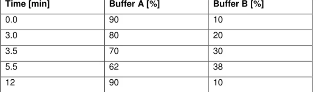
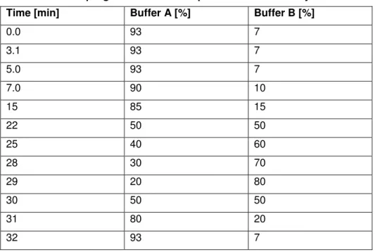
2.4.7 HPLC (high-performance liquid chromatography) – analysis 29
2.5 Biochemical approaches 30
2.5.1 Enzyme assays 30
2.5.2 Analysis of cell viability 31
2.5.3 Efflux of glutamate upon hypoosmotic shock 31
2.5.4 Efflux of glycine betaine upon hypoosmotic shock 32 2.5.5 Betaine uptake and efflux under hyperosmotic conditions 33 2.5.6 Betaine uptake rate during osmotic compensation 33
2.5.7 Determination of membrane potential 33
2.5.8 Determination of the internal pH and the pmf (proton motive force) 34
2.5.9 Test for pyruvate efflux 35
2.5.10 Test for K
+permeability 35
2.5.11 Susceptibility of antibiotics 36
2.5.12 Glutamate production 36
2.5.13 Hypoosmotic stress response under glutamate productive conditions 37
2.6 Electrophysiological approaches 38
2.6.1 Preparation of giant spheroplasts 38
2.6.2 Electrical Recording 38
3 Results 40
3.1 Recombinant YggB proteins 40
3.2 Topology of C. glutamicum YggB 42
3.3 Electrophysiological characterization of YggB 43
3.4 Functionality of C. glutamicum YggB in E. coli 49
3.5 Function of YggB under osmotic stress conditions 50
3.5.1 Glutamate efflux upon osmotic downshift 50
3.5.2 Betaine efflux upon osmotic downshift 52
3.5.3 Betaine accumulation under hyperosmotic conditions 54 3.5.3.1 BetP activity during osmotic compensation 57
3.5.3.2 Energetics during betaine accumulation 59
3.5.4 Growth at different osmolalities 62
3.5.5 Characterization of leakiness mediated by YggB ∆110-His 64
3.5.6 E. coli MscS and MscS/CtYggB in C. glutamicum ∆yggB 65
3.6 Glutamate production 66
3.6.1 Glutamate production upon different treatments 66
3.6.2 Influence of external osmolality, media, and pH 69 3.6.3 Influence of on-going glutamate production on hypoosmotic stress 72 3.6.4 Alternative candidates for the glutamate exporter 73
3.6.5 E. coli MscS and MscS/CtYggB in C. glutamicum ∆yggB 76 3.6.6 LOF- and GOF mutants of E. coli MscS and MscS/CtYggB 78
4 Discussion 82
4.1 Topology of YggB 83
4.2 Electrophysiological characterization of YggB 83
4.3 Significance of YggB under osmotic stress conditions 85
4.4 Involvement of YggB in glutamate production 90
4.5 Function of YggB – current knowledge and future perspectives 96
5 Summary 100
6 Literature 102
7 Supplement 116
Abbreviations
AA amino acids
Am
Rresistance towards ampicillin ATP adenosine triphosphate
BCIP 5-Bromo-4-chloro-3-indolyl phosphate BHI Brain heart infusion
bp base pairs
BSA bovine serum albumin
C carbon
Cm
Rresistance towards chloramphenicol CTAB cetyltrimethylammoniumbromide cpm counts per minute
dm dry mass
DMSO dimethyl sulfoxide DNA deoxyribonucleic acid
EDTA ethylendiaminetetraacetic acid et al . et alii ("and others")
GOF gain of function
HPLC High-performance liquid chromatography IPTG isopropyl-1-thio-β-D-galactosid
kDa kilo Dalton
Km
Rresistance towards kanamycin LB Luria-Bertani
LOF loss of function MM minimal medium
MOPS 3-[N-morpholino]propansufonic acid MS mechanosensitive
OD
600optical density at 600 nm
OPNG o – nitrophenyl β-D-galactopyranoside orf open reading frame
o/n over night
PAGE polyacrylamide gel electrophoresis
PCR polymerase chain reaction
pmf proton motive force
Fmoc fluorenylmethyloxycarbonyl chloride PVDF polyvinylidine difluoride
rpm rounds per minute RT room temperature SDS sodium dodecylsulphate TAE Tris-Acetate-EDTA TCA trichloroacetic acid
TE Tris-EDTA
TEMED N,N,N’,N’-tetramethyl-ethylendiamine Tris 2-amino-hydroxymethylpropane-1,3-diol TPP tetraphenylphosphoniumbromid
(v/v) volume per volume (w/v) weight per volume
wt wild type
∆Ψ membrane potential
∆pH pH gradient
1
1 Introduction
1.1 YggB of Corynebacterium glutamicum
The membrane protein YggB of Corynebacterium glutamicum is proposed to harbor a dual function in the osmotic stress response and in the glutamate production by C. glutamicum.
The combination of these two functions by one protein makes YggB a novel and quite interesting topic.
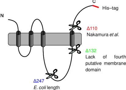
YggB is encoded by the gene NCgl1221 (yggB) and harbors 533 amino acids (AA). Due to structural similarity the protein YggB belongs to the family of MscS-type (Mechanosensitive channel of small conductance) mechanosensitive (MS) channels (Ruffert el al., 1999). These channels function as emergency valves preventing cell lysis under hypoosmotic conditions. Together with yggB another open reading frame (orf), NCgl0843 (mscL), homologous to mscL of E. coli was identified by homology search for MS channel homologs within the genome of C. glutamicum. The related proteins were associated with the two different electrical conductances observed in patch clamp analysis of C. glutamicum membrane fragments fused into liposomes (Ruffert el al., 1999). The MscL homolog (135 AA) has a high sequence similarity to MscL of E. coli (38 % identical and 57 % similar amino acids). On the contrary, YggB is more closely related to MS channel homologs from mycolic acid containing actinomycetes than to the E. coli MscS (only 26 % identical and 46 % similar amino acids) (Nottebrock et al., 2003). The sequence similarity of YggB to MscS of E. coli is restricted to the three N-terminally located transmembrane domains. However, YggB contains 533 AA and is therefore almost double in length compared to the E. coli MscS (286 AA) (Fig. 1.1). The additional C- terminal elongation of C. glutamicum YggB, harboring 247 AA, includes a putative fourth transmembrane domain (www.predictprotein.org) and might therefore also harbor an additional function. However, the putative MS channels of C. glutamicum are barely characterized so far.
Fig. 1.1: Schematic model of MscS and YggB.
Topology of MscS of E. coli (286 AA) and predicted topology of YggB of C.
glutamicum (533 AA).
N
C periplasm
cytoplasm
C N
E. coli MscS C. glutamicum YggB
2
Aside from its suggested function as MS channel, YggB was recently connected to the export of glutamate by C. glutamicum. A truncation mutant, missing 110 AA at the C- terminal end, led to permanent excretion of glutamate (Nakamura et al., 2007).
Additionally, an elevated expression level of the yggB gene was observed upon glutamate triggering treatments (Radmacher et al., 2005, Schiekel, 2005). Since C. glutamicum is the most important organism used in industrial production of L-glutamate, understanding of the mechanisms leading to glutamate production and excretion are of major interest.
Although C. glutamicum has been used in glutamate production for decades the mechanisms leading to glutamate production have not been completely resolved so far.
The possible involvement of YggB in (a) osmotic stress situations functioning as MS channel and (b) glutamate production acting as excretion system makes the protein YggB of C. glutamicum a very interesting study object. For a better understanding, the two fields concerning YggB, osmotic stress and glutamate production, will be described in the following sections.
1.2 C. glutamicum in general
C. glutamicum, a Gram-positive, biotin-auxotrophic bacterium, was isolated about 50 years ago in a screening program for L-glutamate-producing bacteria (Kinoshita et al., 1957, Udeka, 1960). C. glutamicum is a nonmotile, aerobic, and nonsporulating bacterium inhabiting mainly the surface layers of the soil. In its natural environment C. glutamicum has to deal with several stress situations, e.g. osmotic stress caused by sudden changes of the external osmolarity. Like mycobacteria it belongs to the suborder Corynebacterineae which is characterized by very unique cell wall components and a high G+C content of the DNA (Stackebrandt et al., 1997). In addition to a ubiquitous inner plasma membrane, the cell envelope has an outer lipid layer which contains mycolic acids and is probably also organized as a bilayer (Eggeling and Sahm, 2001). Importantly, this hydrophobic layer was shown in related genera to play an important role in drug und substrate transport due to its high impermeability (Jarlier and Nikaido, 1990; 1994). For C. glutamicum the mycolic acids were shown to be essential for the impermeability of the cell wall (Tropis et al., 2005; Gebhardt et al., 2007).
The 3.3 Mb genome of C. glutamicum was completely sequenced by three independent research groups in Europe and Japan (Kalinowski et al., 2003; Ikeda and Nakagawa, 2003;
Yukawa et al., 2007). Due to many genes which are highly conserved within the
Corynebacterineae species and its close relationship to pathogenic organisms, such as C.
3 diphtheria, Mycobacterium tuberculosis, and M. leprae, C. glutamicum is of high interest as non-pathogenic model organism. Furthermore, C. glutamicum is one of the biotechnologically most important bacterial species, because of its intense use for amino acid production. The annual production was more than two million tons of amino acids, mainly L-glutamate and L-lysine in 2005 (Leuchtenberger et al., 2005). Up to now glutamate production alone has increased to an annual production of two million tons (Sano, 2009). Due to this immense industrial importance, glutamate production by C.
glutamicum has been studied intensively.
1.3 Osmotic stress response in C. glutamicum
In the upper layer of the soil, the natural environment of C. glutamicum, changes in external osmolarity are very common as a result of environmental changes, like sunshine and rainfall, respectively. Compensation of these changes is essential for the cell to prevent dehydration or disrupture. Bacterial cells need to maintain an outwardly directed turgor, which pushes the cytoplasmic membrane against the cell wall and appears to be essential for the enlargement of the cell envelope and therefore for growth and cell division (Koch, 1983). To achieve this turgor, solutes are accumulated against their chemical gradient, leading to water flow into the cytoplasm (Epstein, 1986). In high osmolarity environments C. glutamicum actively accumulates these compatible solutes to ensure the continuous flow of water into the cytoplasm. After an osmotic downshift C. glutamicum releases solutes from the cytoplasm via so-called MS channels, thus counterbalancing excessive water influx to prevent cell lysis (Morbach and Krämer, 2002; 2008).
1.3.1 Response to hyperosmotic stress conditions
As first consequence of environmental change to hyperosmotic conditions efflux of water
occurs and cells impend to dehydrate. Thereupon, to maintain cell turgor, C. glutamicum as
other bacteria responds with a fast influx of K
+followed by the accumulation of
compatible solutes, such as glycine betaine, trehalose, and proline, at high concentrations
(Wood, 1999; Morbach and Krämer, 2005a). These compatible solutes harbor two main
characteristics. On the one hand they increase the internal osmolality in order to redirect
water fluxes and maintain the necessary cell turgor. On the other hand compatible solutes
are able to stabilize proteins and protect them against denaturation under hyperosmotic
conditions (Bolen and Rose, 2008). Accumulation of compatible solutes can be
accomplished either by de novo biosynthesis (mainly proline and trehalose) or by uptake
4
LcoP
Compatible solutes
MscL Hypoosmotic stress Hyperosmotic stress
YggB BetP
ProP
EctP Biosynthesis
MtrB
MtrA
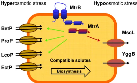
from the surrounding environment (Morbach and Krämer, 2005a). In general bacteria prefer uptake over biosynthesis due to lower energy costs. C. glutamicum harbors four osmoregulated uptake systems for compatible solutes, namely BetP, EctP, ProP, and LcoP (Peter et al., 1998; Morbach and Krämer, 2005a). Among these BetP is the most intensively studied uptake system.
In order to adapt to osmolarity changes of the environment, bacterial cells have to sense these changes. Therefore, C. glutamicum harbors the osmosensory two-component system MtrAB consisting of a membrane-bound histidine kinase MtrB and the soluble response regulator MtrA (Möker et al., 2004). MtrB is able to sense a so far unknown stimulus related to hyperosmotic stress via its cytoplasmatically located phosphorylation domain.
The response regulator MtrA becomes phosphorylated by MtrB and binds to the DNA, regulating the transcription of several genes involved in osmoregulation, e.g. betP, proP, lcoP, and mscL (Krämer 2009).
One of these genes, betP, encodes the transporter BetP which is a specific uptake carrier for glycine betaine. BetP is a secondary transporter which belongs to the BCCT-type family of transporters (Krämer and Morbach, 2004). It consists of 594 AA forming 12 transmembrane segments. Both, the C- and N-terminal, domains of about 50-55 AA are exposed to the cytoplasm. Electron cryo-microscopy of 2D crystals revealed that three BetP proteins form a trimer (Ziegler et al., 2004). Recently, the crystal structure of BetP
Fig. 1.2: Systems involved in the osmotic stress response of C. glutamicum.
Under hyperosmotic conditions compatible solutes are accumulated either via de novo synthesis or via the osmoregulated uptake systems BetP, ProP, EctP, and LcoP. The two-component system MtrBA senses these hyperosmotic conditions and regulates the transcription of several genes which are either up- (green arrow) or down-regulated (red arrow). To prevent cell lysis upon osmotic downshift the MS channels MscL and YggB open within milliseconds to allow the release of compatible solutes.
5 was also solved at a resolution of 3.3 Å (Ressl et al., 2009). The uptake of glycine betaine is energetically coupled to the membrane potential dependent co-transport of two Na
+ions (Peter et al., 1996; Krämer and Morbach 2004; Morbach and Krämer, 2005b). Upon activation BetP transports betaine into the cell in a very specific manner reaching a K
mvalue of 8.9 µM and so is able to build up extremely high betaine gradients of 4 x 10
6(inside/out ratio) (Peter et al., 1996). Besides its transport activity, BetP is also able to function as osmosensor. Three different stimuli are involved in the activation of BetP, (a) the C-terminal, regulatory domain, (b) the cytoplasmic K
+concentration, and (c) negative membrane surface charges (Rübenhagen et al., 2001; Ott et al., 2008). Once the hyperosmotic stress is compensated the cell has to keep the internal betaine concentration at a constant level. Thus, an appropriate fine-tuning mechanism that regulates the internal betaine concentration by balancing uptake and efflux is necessary. In principle, two such mechanisms are possible. One is the gradual downregulation of the transport activity of BetP; the other is the presence of a counteracting efflux system compensating the ongoing betaine uptake. However, a combination of both mechanisms also seems possible. It was already shown that BetP activity is strongly decreased upon adaption to hyperosmotic stress (Botzenhardt et al., 2004). Nevertheless, the knowledge of the exact fine-tuning process of internal betaine concentration is still very limited.
1.3.2 Response to hypoosmotic stress conditions
Mechanosensitive (MS) channels are described as protection system against sudden osmotic downshifts. This rapid decrease of external osmolarity leads immediately to an excessive water influx and consequently to a dramatically increased turgor pressure. To avoid cell lysis, emergency release valves, the MS channels, are activated within milliseconds mediated by the increased membrane tension. As a result, cytoplasmic solutes are released into the environment and the driving force for water entry is reduced (Fig. 1.3) (Morbach and Krämer 2002; Booth et al., 2007).
MS channels
H2O-Influx
compatible solutes MS channel
H2O-Influx
compatible solutes
Fig. 1.3: Response to hypoosmotic stress conditions.
Sudden decrease of the external osmolarity, e. g. caused by rainfall, leads to an excessive water influx. To prevent cell disruption mechanosensitive channels are activated immediately to allow the release of compatible solutes.
6
According to this function, MS channels have to sense lipid deforming forces within the membrane leading to structural rearrangements that lead to channel opening and allow immediate efflux of solutes. However, the way how MS channels respond to mechanical forces along the plane of the cell membrane is not completely clear. Changes in the lipid bilayer caused by increased membrane tension must somehow be sensed and transmitted to the channel (Martinac, 2004).
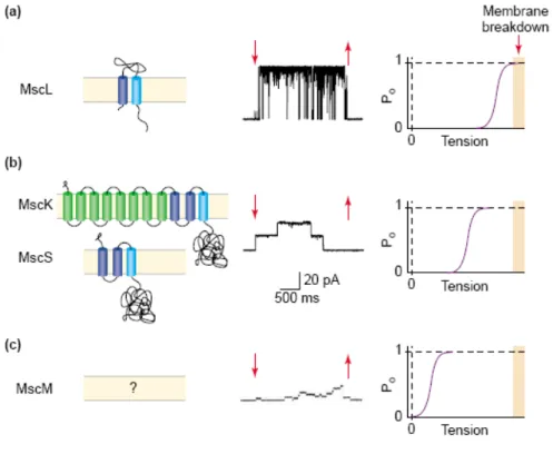
In contrast to C. glutamicum, the MS channels of E. coli have been intensively studied for many years. Using patch clamp techniques, three types of MS channels could be identified in E. coli. Dependent on their conductance the channels are named MscL, MscS and MscM for large, small or very small (mini) conductance (Fig. 1.4) (Berrier et al., 1996; Perozo and Rees, 2003). Corresponding to their conductance the three channels open at different activation thresholds to allow a stepwise response to hypoosmotic conditions (Fig. 1.4) (Martinac et al., 1987; Perozo and Rees, 2003).
While the associated genes of MscL (mscL) and MscS (yggB) have been identified, the gene encoding MscM is still unknown (Levina et al., 1999). MscL and MscS have been
Fig. 1.4: Mechanosensitive channels from E.coli.
Shown are the channels responsible for (a) the MscL (large conductance), (b) two channels of small conductance, MscS (non specific) and MscK (potassium ions), and (c) MscM (mini conductance) mechanosensitive activity as topology model, their single channel activity, and activation threshold.
From: Perozo and Rees, 2003
7 studied most extensively, resulting in the crystal structure of both proteins (Chang et al., 1998; Bass et al., 2002). These two channel proteins are generally not found in animals, but homologs are present in plants and in some fungi and oomycetes. Other structural classes of MS channels are known to exist in higher organisms, from yeast to humans (Kung, 2005). MscL is the largest of the known MS channels. It assembles as homopentamer, each subunit (136 AA) containing two transmembrane domains. For activation a much stronger membrane tension is necessary than it is the case for MscS and MscM. The threshold level of the membrane tension that activates MscL is reached at imminent cell lysis. Accordingly, MscL acts as emergency valve activated at the last moment to allow an extremely fast compensation of the osmotic gradient. Deletion of mscL in E. coli shows no significant differences concerning growth or survivability upon hypoosmotic stress compared to the wild type. Thus, lack of MscL function seems to be compensated by the other MS channels. MscS activity in E. coli is composed of two different channel activities. The gene product of yggB, MscS, constitutes the main part of activity, while kefA encodes a potassium regulated MS channel (MscK). MscS belongs to a family of small membrane proteins of about 300 AA (E. coli MscS: 286 AA). However, MscK is part of a family of larger membrane proteins of more than 700 AA (E. coli MscK:
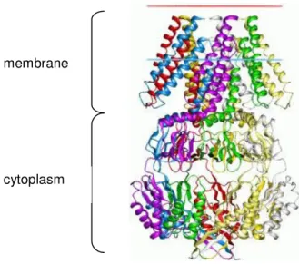
1120 AA). E. coli mutants lacking MscL and MscS are not able to survive severe osmotic downshifts. However, lack of MscK (also KefA) has no influence on the survivability (Levina et al. 1999; Perozo and Rees 2003; Pivetti et al., 2003). Besides sensing membrane stretches (activation threshold is about 50 % of MscL) MscS is able to detect changes of the membrane potential (Sukarev, 2002). The crystal structure revealed that MscS assembles a homoheptamer (Fig. 1.5) (Bass et al., 2002).
Fig. 1.5: E. coli MscS heptamer (side view).
E. coli MscS assembles as homoheptamer. Each subunit is shown in a different colour. The periplasmatic part (N– terminus) is displayed at the top, the cytoplasmic part (C–terminus) at the bottom.
From: Bass et al., 2002 membrane
cytoplasm
8
Each MscS subunit contains 286 AA assembling three transmembrane helices and an additional large, cytoplasmic C–terminal domain. However, some MscS homologs are predicted to have more than three transmembrane (TM) spans which are observed in E.
coli MscS (Pivetti et al., 2003). While the TM1 and TM2 domains act probably as sensor of changes in membrane tension and membrane potential, the TM3 domains of each subunit pack tightly in the heptameric complex to line a pore. In the crystal structure (Fig.
1.5), this pore has a diameter of about 8 – 11 Å at the narrowest point generated by the side chains of Leu105 and Leu109, which form the hydrophobic seal in the closed channel (Bass et al. 2002, Miller et al. 2003). The pore forming TM3 helix contains a conserved glycine- and alanine-rich motif that forms a helix-helix interface. The smooth glycine face allows a sliding of the opposite TM3 helices across each other inducing a conformational change that opens the channel (Edwards et al., 2005). It was proposed that opening and closing of the channel is caused by an iris-like rotation of the helices in the membrane. The current model suggests that under outwardly directed turgor pressure, the density of lipids in the cytoplasmatic membrane is decreased. Thereupon, TM1 and TM2 adjust their conformation to increase their buried volume. This rotation of TM1 and TM2 might lead to a shift of TM3. Movement of TM3 withdraws the side chains of Leu105 and Leu109 from the central pore allowing solvated ion transport (Wang et al., 2008). Single amino acid exchanges within the three TM domains important for channel gating often have a high impact on channel activation. For MscS several loss-of-function (LOF - difficult or no channel opening), and gain-of-function (GOF - easy or flickering channel opening) mutants are described (Nomura et al., 2006; Okada et al., 2002; Miller et al., 2003;
Edwards et al., 2005; Wang et al., 2008). GOF-mutations can especially have severe consequences for the cell as a permanently open channel would cause cell death.
In contrast to the MS channels known for E. coli which release different molecules in a
very unspecific way, C. glutamicum MS channels show much higher substrate specificity
for compatible solutes, mainly betaine and proline (Ruffert et al., 1997). As mentioned
above, two different conductances were observed in patch clamp analysis of C. glutamicum
membrane fragments (Ruffert et al., 1999). Two putative MS channel genes, mscL and
yggB, identified by sequence homologies to the E. coli MS channels, were further
characterized by Nottebrock et al. (2003). While a deletion of mscL showed no phenotype,
the absence of yggB led to a reduced ability to release betaine upon osmotic downshift. A
double mutation had an intermediate phenotype concerning its ability to cope with
hypoosmotic stress indicating the existence of a third efflux channel, not identified yet
9 (Nottebrock et al., 2003). This assumption was further supported by the fact that the double deletion in C. glutamicum was not lethal upon severe hypoosmotic conditions.
However, deletion of yggB also included an essential regulatory sequence, located upstream the genes of isoleucine/valine biosynthesis. This additional deletion resulted in a strain auxotroph for isoleucine, leucine, and valine. Side effects on the phenotype due to this auxotrophy could not be completely excluded (Nottebrock et al., 2003).
As described above, MscS of E. coli has been under intensive investigation by several research groups. The C. glutamicum homolog YggB, on the contrary, has been poorly investigated. Therefore, not much knowledge exists about its function as MS channel.
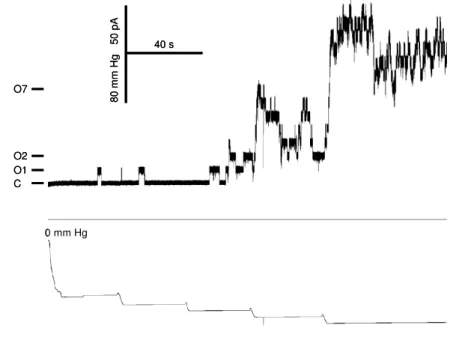
Furthermore, there is still no proof, e.g. using patch clamp analysis, that the YggB protein really harbors the functions of a MS channel.
1.4 Glutamate production by C. glutamicum
Among the amino acids produced by microbial fermentation, L-glutamatic acid in the form of monosodium glutamate (MSG) is the one with the highest production rate. MSG has a unique flavor, which is called umami in Japanese, and is used as flavor-enhancer. About 2.0 million tons of MSG is currently produced worldwide per year by fermentation using coryneform bacteria (Shirai et al., 2005; Sano, 2009). The fermentation process is carried out under strictly controlled conditions (temperature, pH, aeration) using sugar cane syrup as the most common carbon source. The L-glutamic acid excreted by the bacteria into the fermentation solution is then obtained by crystallization (Leuchtenberger et al., 2005).
To induce glutamate productive conditions several treatments are known since C.
glutamicum does not excrete any glutamate under normal growth conditions. As described below all these treatments somehow alter the cell wall. Consequently, the structure of the cell wall and its regulation is supposed to have significant influence on amino acid efflux (Eggeling et al., 2008). A schematic model of the C. glutamicum cell wall is shown in figure 1.6. The cytoplasm is surrounded by the cytoplasmic membrane composed of phospholipids in which the membrane proteins, e.g. importers and exporters, are embedded. The pepdidoglycan–arabinogalactan is arranged on top in outward direction.
The mycolate layer consists of one layer of mycolic acids (mainly dimycolates) which are
esterified with arabinogalactan, and of one layer of non-covalently bound trehalose
mycolates. This upper layer is already part of the outer layer containing also a large
quantity of soluble lipids, such as trehalose di- and monomycolates. The mycolate layer
10
has a highly ordered structure that leads to a cell wall of unusually low permeability (Puech et al., 2001; Eggeling and Sahm, 2001).
The conditions of normal growth of C. glutamicum can be changed towards glutamate productive conditions by several treatments. In general, two major modifications of (a) the cell wall structure and (b) the metabolic flux between tricarboxylic acid (TCA) cycle and glutamate synthesis are involved in glutamate production by C. glutamicum. One possibility to induce glutamate overproduction is limited supply of biotin which is necessary for growth (Shiio et al., 1962). Glutamate excretion triggered by biotin limitation is explained by the inhibition of fatty acid synthesis which leads to a decreased availability of phospholipids and consequently to membrane alterations. In the presence of sufficient biotin, glutamate excretion can be induced by the addition of fatty acid ester surfactants such as Tween 40 (polyoxyethylene sorbitan monopalmitate) or Tween 60 (polyoxyethylene sorbitan monostearate) (Takinami et al., 1965; Duperray et al., 1992).
However, monolaurate or monooleate esters (Tween 20 and 80, respectively) are not effective. Further triggering methods are the addition of (a) the beta-lactam antibiotic penicillin inhibiting cell wall biosynthesis by binding to penicillin-binding proteins, which catalyze the transglycosylation and transpeptidation of peptidoglycan (Nunheimer et al., 1970); (b) the antimycobacterial drug ethambutol targeting a series of arabinosyltransferases, and therefore causing a decreased arabinan deposition in the cell wall (Radmacher et al., 2005); (c) local anesthetics, e. g. chlorpromazine, tetracaine, butacaine, and benzocaine, which change the order of the lipid bilayer by insertion into the membrane (Lambert et al., 1995). It becomes quickly obvious that all these treatments or
Fig. 1.6: Cell wall composition of C.
glutamicum.
On top of the cytoplasmatic membrane the peptidoglycan is located. To the peptidoglycan a layer of arabinogalactan is linked. To the ends of the arabinogalactan mycolic acids (mainly dimycolates) are esterified which together with soluble mycolic acids in the form of trehalose monomycolates and trehalose dimycolates form an outer layer.
Mycolates Phospholipids
Glycolipids
Proteins Porin
11 conditions alter the cell envelope of C. glutamicum. The amount of mycolic acids is reduced by up to approximately 40 % under glutamate production conditions. Additionally, the spectrum of mycolic acids, normally consisting of 30, 32 or 34 carbons is altered to much shorter mycolic acids containing 22 and 24 carbons (Hashimoto et al., 2006).
However, not only treatments directly altering the cell wall induce glutamate production, but also genetic manipulations of the cell wall via fatty acid synthesis can trigger glutamate excretion. Here it has to be mentioned that trehalose becomes covalently linked to fatty acids forming trehalosemono- and dimycolates (Tropis et al., 2005). A mutant lacking the trehalose synthesis genes treS, otsA, and treY is devoid of the mycolic acid layer under distinct cultivation conditions. This lack of mycolates is sufficient to induce continuous glutamate excretion (Gebhardt et al., 2007). Additionally, the overexpression or inactivation of the genes involved in lipid synthesis results in a strong alteration of the phospholipid composition and as a consequence of the modified lipid synthesis in a dramatically changed glutamate efflux (Nampoothiri et al., 2002).
Several genes are up- or downregulated under glutamate productive conditions (Kataoka et al., 2006). One is the gene dtsR1 encoding a homolog of the β-subunit of some biotin- containing enzyme complexes (Kimura et al., 1996). The expression of the dtsR1 gene and therefore the cellular concentration of the protein are decreased under biotin limited conditions and upon Tween 40 addition. A disruption of the dtsR1 gene results in a strain strictly auxotroph for fatty acids (oleic acid). The ∆dtsR mutant produced glutamate efficiently also in excess of biotin (Kimura et al., 1997). Although the related protein DtsR1 has no biotin-binding motif, it seemed very likely that it might form a complex with another subunit that contains biotin. The two carboxylases identified in C. glutamicum which were shown to be essential for fatty acid and mycolic acid synthesis (Gande et al., 2007) were assumed to be counterparts of DtsR1 forming a complex including biotin molecules as co-factors.
As mentioned above, also a change in metabolic flux is involved in glutamate production by C. glutamicum. On the metabolic level a reduced specific activity of the 2-oxoglutarate dehydrogenase complex (ODHC) was observed in the dtsR1 disruptants (Kimura, 2002).
Interestingly, such a decrease in the specific activity of the ODHC was also observed under
biotin limitation inducing glutamate production. Therefore, the ODHC seems to be a key
enzyme in glutamate production, which is located at the metabolic branch point between
the glutamate biosynthesis pathway and the TCA cycle where it catalyzes the oxidative
decarboxylation of 2-oxoglutarate to succinyl-CoA (Kawahara et al., 1997). It was shown
12
that the activity of the ODHC was also decreased under glutamate productive conditions induced by several other treatments than biotin limitation, while the activity of the glutamate dehydrogenase (GDH), catalyzing ammonia assimilation of 2-oxoglutarate to form glutamate, was mainly unchanged. Furthermore, the level of ODHC activity seemed to be inversely correlated to the yield of glutamate (Kawahara et al., 1997). Metabolic flux analysis indicated that attenuation of ODHC activity is the factor with the greatest impact on glutamate formation in the metabolic network (Shirai et al., 2005). ODHC consists of three subunits, one the 2-oxoglutarate dehydrogenase subunit E1o encoded by the gene odhA (Usada et al., 1996). Deletion of odhA results in the elimination of any ODHC activity. The ∆odhA mutant produces glutamate spontaneously under normal growth conditions. In this mutant no alteration in the fatty acid composition of the cells was observed. Glutamate production by this mutant additional to the basal glutamate excretion could be triggered by several treatments (Asakura et al., 2007). However, overexpression of odhA leading to an increased ODHC specific activity results in dramatically reduced glutamate production despite Tween 40 addition (Kim et al., 2009). These results suggest that decrease in ODHC activity is an important factor for glutamate production by C.
glutamicum, leading to increased metabolic flux towards glutamate biosynthesis. Thus, it is very interesting how reduction of the ODHC activity and alterations of the cell envelope are correlated.
Recently, a novel regulation mechanism of ODHC was discovered. The 15 kDa protein
OdhI in its unphosphorylated form is responsible for the inhibition of ODHC by direct
interaction with the E1o subunit (OdhA). OdhI is phosphorylated by the Ser/Thr protein
kinase PknG (Niebisch et al., 2006). Phosphorylated OdhI is inactive and cannot bind to
the ODHC. Dephosphorylation of OdhI is catalyzed by the phosphoprotein phosphatase
Ppp. PknG is a soluble protein which is assumed to be membrane associated whereas Ppp
is a membrane-integral protein. These proteins might function as sensors and their de-
/phosphorylation activity is thought to be dependent on the absence or presence of specific
stimuli. Deletion of the odhI gene nearly abolishes glutamate production. However, the
effect of a pknG deletion varies depending on the inducing conditions but can lead to a
significantly increased glutamate production. The positive influence of the pknG deletion
on glutamate production might be caused by an increased level of unphosphorylated OdhI,
resulting in an increased inhibition of ODHC activity and therefore a higher flux of 2-
oxoglutarate towards glutamate synthesis (Schultz et al., 2007).
13 Taken together glutamate overproduction seems to be the result of a combination of cell wall alteration, possibly mediating the activation of a glutamate excretion system, and metabolic alteration towards efficient glutamate synthesis. A model to combine all effects connected with the induction of glutamate production involves sensing of cell wall alterations, a regulatory cascade up to the central metabolism, and activation of a specific glutamate export system (Fig. 1.7)
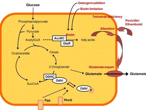
Fig. 1.7: Model for the induction of glutamate production by C. glutamicum.
Treatments triggering glutamate production alter the cell envelope of C. glutamicum.
Biotin limitation and detergent addition alter the membrane by inhibiting fatty acid synthesis. Genetic manipulations resulting in effects like trehalose deficiency or changed fatty acid synthesis also change the composition of the cell wall. Addition of penicillin or ethambutol inhibits cell wall biosynthesis. These cell wall alterations might be sensed by the glutamate export carrier itself or by other membrane bound proteins, like PknG and Ppp. These proteins could regulate the ODHC activity via the phosphorylation status of OdhI.
Modified from Nakamura et al., 2007 and Eggeling et al., 2008 Glutamate Citrate
Glucose
Phosphoenolpyruvate
Pyruvate AcetylCoA
2-Oxoglutarate Oxaloacetate
SucCoA
ODHC
fatty acids DtsR
AccBC Biotin
Biotin limitation Detergent addition
Alteration
Penicillin/
Ethambutol
Glutamate export
Glutamate
OdhI
OdhI P
PknG
Trehalose deficiency
Ppp
14
1.5 Glutamate export
Since C. glutamicum is used for the industrial production of various amino acids, which have to cross the cell membrane on their way into the external medium, a main focus of research laid on the identification of the corresponding export systems. Several exporters have been identified so far, such as LysE exporting the basic amino acids L-lysine and L- arginine (Vrljic et al., 1996), BrnFE exporting branched chain amino acids and L- methionine (Kennerknecht et al., 2002; Trötschel et al., 2005), and ThrE exporting L- threonine (Simic et al., 2001). For the export of glutamate the situation slightly differs.
Because all the triggers known at the time being affected the cell surface of C. glutamicum, it was initially assumed that glutamate passively leaks through the membrane and cell wall (‘leak model’). However, it was shown that membrane permeability does not change during glutamate production, since other amino acids, as well as actetate and ions (H
+, K
+, and Cl
-) do not leak from the cell (Hoischen and Krämer, 1989; 1990). Furthermore, the leak model itself could not explain the accumulation of external glutamate. Therefore, the presence of a specific glutamate export system in the membrane was proposed (Hoischen and Krämer, 1989; Gutmann et al., 1992). However, this export carrier for the industrial important glutamate was not known for decades.
Recently, the MS channel homolog YggB of C. glutamicum was connected to the export of glutamate (Nakamura et al., 2007). As previously mentioned, the activity of the ODHC is significantly decreased under glutamate producing conditions. Therefore, the construction of odhA disruptants was one strategy to increase glutamate productivity. However, these mutants showed a reduced ability to grow on minimal medium and had an extremely unstable phenotype resulting in suppressor mutations. To identify such a mutation in a glutamate producing mutant, a Sau3AI library of wild-type C. glutamicum chromosomal DNA was used. The gene which restored normal growth and abolished glutamate production was identified by sequencing as NCgl1221 (yggB). Several odhA disruptants showing elevated levels of glutamate production revealed mutations within the yggB gene.
One mutant with a yggB (V419::IS1207) gene leading to a protein with a C-terminal truncation of 110 AA showed continuous glutamate production without induction.
Overexpression of yggB from a plasmid resulted in increased glutamate production only
upon induction by various triggers. The deletion of yggB was followed by a dramatically
decrease in glutamate production. However, a small residual excretion of glutamate could
still be observed. Based on the obtained results, Nakamura and co-workers proposed the
following model for the role of YggB in glutamate production by C. glutamicum: (a)
15 several treatments inducing glutamate production cause changes in membrane tension, (b) the structure of YggB is altered by these changes so that YggB becomes activated, and (c) allows glutamate excretion. However, YggB is localized in the cytoplasmic membrane, while most of the treatments which induce glutamate production, alter the cell wall of C.
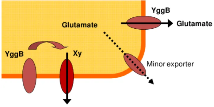
glutamicum. Here the unique C-terminal elongation provides an interesting candidate to sense and transduce these changes of the cell wall under the assumption that it is localized in the periplasmic space. The reduced ODHC activity might be only a consequence of glutamate production and not an activator. However, a decrease in ODHC activity can then further increase glutamate productivity. Besides the proposed major glutamate export system YggB, a second minor glutamate export carrier might exist. However, the existence of another so far unknown glutamate export system is also possible. In this case YggB might just function as regulator of this glutamate exporter Xy (Fig. 1.8) (Nakamura et al., 2007).
YggB
Minor exporter
Glutamate Glutamate
YggB
Glutamate Xy
Fig. 1.8: Possible functions of YggB in the export of glutamate.
YggB might either function as glutamate exporter together with a minor export system responsible for residual glutamate excretion of the yggB deletion strain or as regulator of another unknown export system (shown here as protein Xy).
16
1.6 Thesis objective
The aim of this work was a detailed characterization of the protein YggB of C.
glutamicum. Research on YggB is interesting because a dual function of the protein was proposed as mechanosensitive channel under hypoosmotic conditions on the one hand and an involvement in the export of the industrial important amino acid glutamate on the other.
MS channels can sense alterations of the membrane tension and transduce these alterations into a conformational change allowing solute efflux. As all treatments inducing glutamate production go along with an alteration of the cell envelope, a MS channel harboring the mentioned properties might be a good candidate to be involved in the sensing of these alterations.
However, the actual function of YggB as MS channel has still to be proven. An approved
method to show mechanosensitive properties of a channel are electrophysiological analyses
via the patch clamp technique. Additionally, YggB should be physiologically characterized
under different osmotic stress conditions to gain a comprehensive understanding of its
properties. Regarding the proposed involvement of YggB in the export of glutamate the
main question is if YggB is the glutamate exporter itself or whether it is just a regulator of
another so far unknown glutamate export system. To answer this question, several
physiological and biochemical attempts are made to characterize the role of YggB under
glutamate productive conditions. In addition to a functional characterization of YggB, the
correct topology of the protein in the membrane should be determined. Depending on the
computer program used, the existence of a fourth transmembrane domain was predicted
which would localize the C-terminal elongation in the periplasm.
17
2 Materials and Methods
2.1 Bacterial strains and plasmids
All strains used in this work are listed in table 2.1.
Table 2.1: Bacterial strains
Strain Genotype Reference
E. coli
DH5αmcr endA1 supE44 thi-1 λ- recA1 gyrA96 relA1 deoR
∆(lacZYA-argF) U196 φ80DlacZ ∆M15mcrA ∆(mmr hsdRMS mcrBC)
Grant et al. 1990
BL21 (DE3) F- ompT gal [dcm] [lon] hsdSB (rB –mB -; an E. coli B strain) with DE3, a λ prophage carrying the T7 RNA polymerase gene
Novagen, Darmstadt
Frag 1 F–, rha, thi, gal, lacZ Epstein and Kim,
1971
MJF455 Frag1, ∆mscL::Cm, ∆yggB Levina et al. 1999
MJF465 Frag1, ∆mscL::Cm, ∆yggB, ∆kefA::Km Levina et al. 1999
C. glutamicum
ATCC 13032 wild type Abe et al. 1967
ATCC 13032 ∆mscL Derivative of ATCC 13032 with an in frame-deletion of the mscL gene
Nottebrock et al.
2003 ATCC 13032 ∆yggB Derivative of ATCC 13032 with an in frame-deletion of
the yggB gene
this work
ATCC 13032 ∆mscL
∆yggB
Derivative of ATCC 13032 with in frame-deletions of the mscL and yggB genes
this work
ATCC 13032
∆Cgl0590∆yggB
Derivative of ATCC 13032 with in frame-deletions of the Cgl0590 and yggB genes
this work
ATCC 13032
∆Cgl2211∆yggB
Derivative of ATCC 13032 with in frame-deletions of the Cgl2211 and yggB genes
this work
ATCC 13032
∆Cgl2211∆Cgl0590
Derivative of ATCC 13032 with in frame-deletions of the Cgl2211 and Cgl0590 genes
this work
ATCC 13032
∆Cgl2211∆Cgl0590
∆yggB
Derivative of ATCC 13032 with in frame-deletions of the Cgl2211, Cgl0590, and yggB genes
this work
ATCC 13032 IS::Cgl0063 Derivative of ATCC 13032 with an insertion-deletion of the Cgl0063 gene, Cgl0063::Am
Elena Jolkver
18
ATCC 13032
∆kup ∆CglK
Derivative of ATCC 13032 with in frame-deletions of the kup and CglK genes
Follmann et al., 2009
All plasmids used in this work are listed in table 2.2.
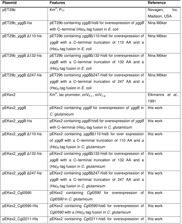
Table 2.2: Plasmids
Plasmid Features Reference
pET29b KmR, PT7 Novagen, Inc.
Madison, USA pET29b_yggB-his pET29b containing yggB-his6 for overexpression of yggB
with C–terminal (His)6-tag fusion in E. coli
Nina Möker
pET29b_yggB ∆110-his pET29b containing yggB∆110-his6 for overexpression of yggB with a C–terminal truncation of 110 AA and a (His)6-tag fusion in E. coli
Nina Möker
pET29b_yggB ∆132-his pET29b containing yggB∆132-his6 for overexpression of yggB with a C–terminal truncation of 132 AA and a (His)6-tag fusion in E. coli
Nina Möker
pET29b_yggB ∆247-his pET29b containing yggB∆247-his6 for overexpression of yggB with a C–terminal truncation of 247 AA and a (His)6-tag fusion in E. coli
Nina Möker
pEKex2 KmR, tac promotor, oriVE.c., oriVC.g. Eikmanns et al., 1991
pEKex2_yggB pEKex2 containing yggB for overexpression of yggB in C. glutamicum
this work
pEKex2_yggB-his pEKex2 containing yggB-his6 for overexpression of yggB with C–terminal (His)6-tag fusion in C. glutamicum
this work
pEKex2_yggB ∆110-his pEKex2 containing yggB∆110-his6 for over expression of yggB with a C–terminal truncation of 110 AA and a (His)6-tag fusion in C. glutamicum
this work
pEKex2_yggB ∆132-his pEKex2 containing yggB∆132-his6 for overexpression of yggB with a C–terminal truncation of 132 AA and a (His)6-tag fusion in C. glutamicum
this work
pEKex2_yggB ∆247-his pEKex2 containing yggB∆247-his6 for overexpression of yggB with a C–terminal truncation of 247 AA and a (His)6-tag fusion in C. glutamicum
this work
pEKex2_Cgl0590 pEKex2 containing Cgl0590 for overexpression of Cgl0590 in C. glutamicum
this work
pEKex2_Cgl0590-His pEKex2 containing Cgl0590-his6 for overexpression of Cgl0590 with a (His)6-tag fusion in C. glutamicum
this work
pEKex2_Cgl2211-His pEKex2 containing Cgl2211-his6 for overexpression of this work
19
Cgl2211 with a (His)6-tag fusion in C. glutamicum
pMS3 pEKex2 fused with a β-galactosidase alkaline phosphatase reporter cassette for topology studies
Seidel et al., 2007
pMS3_yggB pEKex2 containing yggB fused to a β-galactosidase alkaline phosphatase reporter cassette for overexpression in E. coli and in C. glutamicum
this work
pMS3_yggB ∆110 pEKex2 containing yggB ∆110 fused to a β- galactosidase alkaline phosphatase reporter cassette for overexpression in E. coli and in C. glutamicum
this work
pMS3_yggB ∆132 pEKex2 containing yggB ∆110 fused to a β- galactosidase alkaline phosphatase reporter cassette for overexpression in E. coli and in C. glutamicum
this work
pMS3_yggB ∆247 pEKex2 containing yggB ∆110 fused to a β- galactosidase alkaline phosphatase reporter cassette for overexpression in E. coli and in C. glutamicum
this work
pEKex2_mscS pEKex2 containing E. coli mscS for overexpression of mscS in C. glutamicum
this work
pEKex2_mscS-His pEKex2 containing E. coli mscS -his6 for overexpression of mscS with C–terminal (His)6-tag fusion in C.
glutamicum
this work
pEKex2_mscS/CtyggB pEKex2 containing E. coli mscS fused to the C-terminal domain of YggB for overexpression of mscS/CtyggB in C. glutamicum
this work
pEKex2_mscS/CtyggB- His
pEKex2 containing E. coli mscS fused to the C-terminal domain of YggB-his6 for overexpression of mscS/CtyggB with C–terminal (His)6-tag fusion in C. glutamicum
this work
pEKex2_mscS-His I37N/L86N
pEKex2 containing E. coli mscS I37N L86N-his6 for overexpression of mscS I37N L86N with C–terminal (His)6-tag fusion in C. glutamicum
this work
pEKex2_mscS/CtyggB- His I37N/L86N
pEKex2 containing E. coli mscS I37N L86N fused to the C-terminal domain of YggB-his6 for overexpression of mscS/CtyggB I37N L86N with C–terminal (His)6-tag fusion in C. glutamicum
this work
pEKex2_mscS-His A51N/F68N
pEKex2 containing E. coli mscS A51N F68N-his6 for overexpression of mscS A51N F68N with C–terminal (His)6-tag fusion in C. glutamicum
this work
pEKex2_mscS/CtyggB- His A51N/F68N
pEKex2 containing E. coli mscS A51N F68N fused to the C-terminal domain of YggB-his6 for overexpression of mscS/CtyggB A51N F68N with C–terminal (His)6-tag fusion in C. glutamicum
this work
20
pEKex2_mscS-His V40D pEKex2 containing E. coli mscS V40D-his6 for overexpression of mscS V40D with C–terminal (His)6-tag fusion in C. glutamicum
this work
pEKex2_mscS/CtyggB- His V40D
pEKex2 containing E. coli mscS V40D fused to the C- terminal domain of YggB-his6 for overexpression of mscS/CtyggB V40D with C–terminal (His)6-tag fusion in C. glutamicum
this work
pEKex2_mscS-His A106V
pEKex2 containing E. coli mscS A106V-his6 for overexpression of mscS A106V with C–terminal (His)6- tag fusion in C. glutamicum
this work
pEKex2_mscS/CtyggB- His A106V
pEKex2 containing E. coli mscS A106V fused to the C- terminal domain of YggB-his6 for overexpression of mscS/CtyggB A106V with C–terminal (His)6-tag fusion in C. glutamicum
this work
pEKex2_mscS-His L109S pEKex2 containing E. coli mscS L109S-his6 for overexpression of mscS L109S with C–terminal (His)6- tag fusion in C. glutamicum
this work
pQE60 Amr, T5 promotor, Col E1 Qiagen, Hilden
pREP Kmr, lacI Qiagen, Hilden
pQE60-lacI pQE60 with integrated lacI gene this work
pQE60-lacI_yggB pQE60-lacI containing yggB for overexpression of yggB in E. coli
this work
pQE60-lacI_yggB-His pQE60-lacI containing yggB-His for overexpression of yggB with C–terminal (His)6-tag fusion in E. coli
this work
pQE60-lacI_yggB ∆110 pQE60-lacI containing yggB ∆110 for overexpression of yggB ∆110 in E. coli
this work
pQE60-lacI_yggB ∆132 pQE60-lacI containing yggB ∆132 for overexpression of yggB ∆132 in E. coli
this work
pQE60-lacI_yggB ∆247 pQE60-lacI containing yggB ∆247 for overexpression of yggB ∆247 in E. coli
this work