Sensing properties of MtrB-MtrA of Corynebacterium glutamicum:
a two-component system involved in the osmo- and chill stress response
Inaugural-Dissertation zur
Erlangung des Doktorgrades
der Mathematisch-Naturwissenschaftlichen Fakultät der Universität zu Köln
vorgelegt von
Nina Möker aus Berlin
Köln, Mai 2006
Berichterstatter:
Prof. Dr. Reinhard Krämer Prof. Dr. Karin Schnetz
Tag der Disputation: 30.06.2006
Seltsam?
Aber so steht es geschrieben...
(Gespenstergeschichten, Bastei-Verlag)
Sensing properties of MtrB-MtrA of Corynebacterium glutamicum: a two- component system involved in the osmo- and chill stress response
Being an immobile Gram-positive soil bacterium, Corynebacterium glutamicum has to cope with dramatic changes of environmental conditions like an altered osmolarity or temperature.
It was recently shown that the MtrB-MtrA two-component system of this bacterium mediates the expression regulation of osmo- and chill stress-related genes in response to increased medium osmolarity or decreased environmental temperature. The response regulator of this system, MtrA, was shown to act as an activator of the genes proP, betP, and lcoP as well as a repressor of the mscL gene. These results led to the question, which physico-chemical stimuli are recognized by MtrB, in order to mediate the expression regulation of genes in response to changes of the environmental osmolarity and temperature.
To avoid the high complexity of the cellular system, an in vitro system was established for the detailed analyses of the sensory properties of MtrB. Using E. coli liposomes enriched with MtrB, the basic characteristics of the bacterial two-component system could be detected.
These include the autokinase activity of membrane-bound MtrB, the phosphoryl group transfer to soluble MtrA, and the MtrB-catalyzed dephosphorylation of MtrA-P.
By use of the artificial membrane system the influence of a systematic variation of signals related to osmo- and chill stress conditions could be performed. The autokinase activity of MtrB turned out to be stimulated by the presence of numerous osmolytes including sugars, amino acids, compatible solutes, and high molecular weight PEGs. In addition, the histidine kinase was shown to be strongly activated by exposure to low temperature. By fusion of proteoliposomes made from E. coli lipid with synthetic POPG, the sensory properties of MtrB furthermore turned out to significantly depend on the composition of the membrane surrounding.
In order to identify the sensing domain of MtrB, various truncated derivatives of the protein were constructed. In contrast to most bacterial histidine kinases, which are thought to carry their sensory function within the extracytoplasmic loop, MtrB was shown to sense independently of this external domain.
Taken together, the data of this work led to the model that MtrB senses environmental hyperosmotic stress either as an increased osmolarity of the cytoplasm, or as altered membrane properties. Environmental chill conditions are suggested to be sensed as an altered physical state of the membrane.
Sensorische Eigenschaften von MtrB-MtrA von Corynebacterium glutamicum: Vermittlung der Osmo- und Kältestress-Antwort durch ein
Zwei-Komponenten System
Corynebacterium glutamicum ist als immobiles, Gram-positives Bodenbakterium Stress- Situationen durch plötzliche Änderungen seines Habitats, wie zum Beispiel Schwankungen in der externen Osmolarität und Temperatur, ausgesetzt. Es konnte kürzlich gezeigt werden, dass das Zwei-Komponenten System MtrB-MtrA als Reaktion auf erhöhte Osmolarität im Nährmedium, sowie auf erniedrigte Umgebungstemperatur, die Expressionsregulation von Genen vermittelt, die eine wichtige Rolle in der Osmo- und Kältestress-Antwort von C. glutamicum spielen. Dabei agiert der Antwort-Regulator MtrA sowohl als Aktivator der Gene proP, betP und locP, als auch als Repressor des mscL-Gens. Dies führte zu der Frage, welche physico-chemischen Reize als Indikatoren für eine veränderte Osmolarität bzw. Temperatur der Umgebung von MtrB genutzt werden, um als Reaktion die Expressionsregulation spezifischer Gene zu vermitteln. Um die Reizwahrnehmung detailliert zu untersuchen, wurde ein in vitro System aufgebaut. In diesen, mit MtrB angereicherten, E. coli-Liposomen konnten die Basiseigenschaften von Zwei-Komponenten Systemen in vitro imitiert werden. Dies beinhaltete die Autokinase-Aktivität von Membran-gebundenem MtrB, den Transfer der Phosphoryl-Gruppe auf den gereinigten Antwort-Regulator MtrA, sowie die von MtrB katalysierte Dephosphorylierung von MtrA-P.
Mit dem Proteoliposomen-System konnte die Auswirkung vielfältiger, bei osmotischem und Kälte-Stress auftretender Signale analysiert werden. Die Autokinase-Aktivität von MtrB wurde durch die Anwesenheit zahlreicher Osmolyte wie Zucker, Aminosäuren, kompatible Solute und hochmolekulare PEGs deutlich stimuliert. Darüber hinaus konnte eine deutliche Aktivierung von MtrB durch erniedrigte Temperaturen gezeigt werden. Fusion von Proteoliposomen, die aus E. coli-Lipid bestanden, mit synthetischem POPG ergab ferner, dass die Sensoreigenschaften von MtrB von der Membranzusammensetzung deutlich beeinflusst werden.
Des weiteren konnte mit Hilfe von verkürzten Varianten dieses Proteins die periplasmatische Schleife, welche häufig bei den bakteriellen Sensor-Kinasen die Signal-Wahrnehmung vermittelt, als sensorische Domäne für MtrB ausgeschlossen werden.
Zusammengefasst führen diese Daten zu dem Modell, dass MtrB hyperosmotischen externen Stress entweder als erhöhte Osmolarität des Cytoplasmas, oder als veränderte Membraneigenschaft wahrnimmt. Die Wahrnehmung von Kälte wird vermutlich durch die unter diesen Bedingungen veränderte Membranphase vermittelt.
Contents
1. Introduction 1
1.1 Corynebacterium glutamicum 1
1.2 Osmotic stress 2
1.2.1 Osmoregulation in response to hypoosmotic environmental conditions 3
1.2.2 Osmoregulation in response to hyperosmotic environmental conditions 3
1.3 Chill stress 6
1.4 Bacterial two-component systems 7
1.5 The MtrB-MtrA two-component system of Corynebacterium glutamicum 7
1.6 Objectives of this thesis 12 2. Materials and methods 14 2.1 Bacterial strains, plasmids, and oligonucleotides 14 2.2 Growth media and cultivation conditions 17 2.3 Molecular biological approaches 19 2.3.1 Preparation of competent E. coli cells and transformation 19 2.3.2 Preparation of competent C. glutamicum cells and transformation 20 2.3.3 DNA techniques 20 2.3.3.1 Isolation of plasmid DNA from E. coli 20 2.3.3.2 Isolation of genomic DNA from C. glutamicum 20 2.3.3.3 Gel electrophoresis and extraction of DNA from agarose gels 21 2.3.3.4 Polymerase chain reaction (PCR) 21 2.3.3.5 Restriction, ligation, and sequencing of DNA 22 2.3.4 RNA techniques 22 2.3.4.1 Isolation of total RNA from C. glutamicum 22 2.3.4.2 Synthesis of digoxigenin-labelled RNA probes 23 2.2.4.3 RNA hybridization experiments (Dot blots) 24 2.3.4.4 Northern blot analyses 25 2.3.4.5 RT-PCR 26
2.4 Biochemical approaches 27 2.4.1 Determination of protein concentrations 27 2.4.2 SDS-Polyacrylamide Gel Electrophoresis (PAGE) 28
2.4.3 Staining of SDS-gels 29
2.4.3.1 Coomassie Brilliant Blue staining 29
2.4.3.2 Silver staining 29
2.4.4 Immunoblot analyses 29
2.4.5 Identification of MtrB-Strep 30 2.4.6 Purification of MtrA-(His)10 by Ni-NTA affinity chromatography 30 2.4.7 Purification of Strep-MtrB, MtrB-Strep, and truncated derivatives of MtrB-
Strep by Strep-tagII/StrepTactin affinity chromatography 31 2.4.8 Purification of MtrB∆190 by Strep-tagII/StrepTactin affinity chromatography 32 2.4.9 Purification of (His)6-DcuS by Ni-NTA affinity chromatography 33 2.4.10 Preparation of inverted membrane vesicles (IMV) enriched with
Strep-MtrB or MtrB-Strep 33
2.4.11 Reconstitution of Strep-MtrB, MtrB-Strep, truncated derivatives of
MtrB-Strep, or (His)6-DcuS into E. coli lipid liposomes 34
2.4.12 Variation of the lipid composition 35
2.4.13 Determination of the orientation of reconstituted MtrB-Strep by site-
specific proteolysis 35
2.4.14 Phosphorylation assays 36
2.4.15 Fluorescence assays 37
2.4.14 Determination of the osmolality 38
3. Results 39
3.1 Development of in vitro phosphorylation assays for the MtrB-MtrA two-
component system 39
3.1.1 Heterologous synthesis and affinity purification of MtrA-(His)10 40
3.1.2 Heterologous synthesis of MtrB in E. coli 41
3.1.3 Production of MtrB-enriched E. coli inverted membrane vesicles 43 3.1.4 Autophosphorylation activity of MtrB in inverted membrane vesicles 43 3.1.5 Preparation of proteoliposomes enriched with MtrB 44 3.1.5.1 Affinity purification of Strep-MtrB and MtrB-Strep 44 3.1.5.2 Reconstitution of Strep-MtrB and MtrB-Strep into E. coli lipid liposomes 46 3.1.6 Autophosphorylation activity of MtrB in proteoliposomes 46
3.2 In vitro characterization of MtrB and MtrA activity 48
3.2.1 Kinetics of the ATP-dependent MtrB autokinase activity 49
3.2.2 MtrB-MtrA phosphoryl group transfer 50
3.2.3 Dephosphorylation of MtrA-P 51
3.2.4 Orientation of MtrB-Strep in E. coli lipid liposomes 53 3.2.4.1 Influence of Mg2+ on the autophosphorylation activity of MtrB 53 3.2.4.2 Investigation of the effect of Mg2+ on E. coli polar lipid extract liposomes 54 3.2.4.3 Investigation of the orientation of reconstituted MtrB-Strep by
site-specific proteolysis 55
3.2.5 Autokinase activity of MtrB in its solubilized state 57
3.3 Investigation of the stimuli sensed by MtrB with focus on osmostress-
related signals 58
3.3.1 Expression regulation of proP, betP, and mscL after a hyperosmotic
shock with trehalose 58
3.3.2 Influence of various salts on the in vitro autokinase activity of MtrB 59 3.3.3 Development of a control system to discriminate between specific and
non-specific MtrB stimuli by use of the fumarate sensor DcuS 61 3.3.4 Detailed analyses of the osmostress-related in vitro stimulation of the
MtrB-MtrA two-component system 64
3.3.4.1 Influence of various amino acids and compatible solutes on the
in vitro autokinase activity of MtrB 65
3.3.4.2 Influence of various sugars on the in vitro autokinase activity of MtrB 66 3.3.4.3 Influence of various osmolytes on the in vitro MtrB-MtrA phosphoryl
group transfer 68
3.3.4.4 Influence of polyethylene glycols (PEGs) with various molecular
sizes on the in vitro autokinase activity of MtrB 70 3.3.4.5 Investigation of the high osmolarity-induced shrinkage of E. coli
polar lipid extract liposomes 73
3.4 Investigations on the sensing domain of the histidine kinase MtrB 76 3.4.1 Construction, isolation and/or reconstitution of various truncated forms
of MtrB-Strep 77
3.4.2 In vitro autokinase activity and/or sensing properties of various truncated
forms of MtrB-Strep 79
3.4.2.1 In vitro autokinase activity of MtrB∆190 79
3.4.2.2 In vitro autokinase activity of MtrB∆25 and -∆154 80 3.4.2.3 In vitro autokinase activity of MtrB∆L124 and -∆L134 in presence
of various osmolytes 80
3.5 Influence of the membrane composition on the in vitro stimulation of
MtrB activity 82
3.6 Investigation of the stimuli sensed by MtrB with focus on chill stress-
related signals 85
3.6.1 Expression regulation of proP, betP, and lcoP under chill stress conditions 85 3.6.2 Influence of chill on the in vitro autokinase activity of MtrB 87
4. Discussion 91
4.1 Development and characterization of in vitro MtrB and MtrA
phosphorylation assays 92
4.2 Sensing properties of MtrB in dependence of osmotic stress 96
4.3 Sensing properties of MtrB in dependence of the membrane composition 103
4.4 Sensing properties of MtrB in dependence of chill stress 105
4.5 Investigation of the sensing domain of MtrB 106
4.6 Models of MtrB stimulation 109
5. Summary 112
6 References 114
7. Appendix 126
Abbreviations
AHT Anhydrotetracycline ApR Resistance towards ampicilin ADP Adenosine diphosphate ATP Adenosine triphosphate BHI Brain heart infusion
bp Base pairs BSA Bovine serum albumin C Carbon
CSPD Disodium 3-(4-metho xyspiro {1,2-dioxetane-3,2-(5-chloro)triciclo [3.3.1.13,7]decan}-4-yl)phenyl phosphat
DIG Digoxigenin DTT Dithiothreitol
EDTA Ethylendiaminetetraacetic acid et al. et alii ("and others")
g Gravitational acceleration (9.81 m2/s) IPTG Isopropyl-1-thio-β-D-galactosid kb kilo base pairs
kDa kilo Dalton Km Michaelis-Menten constant KmR Resistance towards kanamycin KPi Potassium phosphate buffer LB Luria-Bertani
MM Minimal medium
MOPS 3-[N-morpholino]propansufonic acid OD600 Optical density at 600 nm
orf Open reading frame
PAGE Polyacrylamide gel electrophoresis PCR Polymerase chain reaction
PVDF Polyvinylidine difluoride RT Poom temperature
RT-PCR Reverse transcription polymerase chain reaction SDS Sodium dodecylsulphate
TAE Tris-Acetate-EDTA TCA Trichloroacetic acid
TE Tris-EDTA
TEMED N,N,N’,N’-tetramethyl-ethylendiamine Tris 2-amino-hydroxymethylpropane-1,3-diol
1. Introduction
Living cells, including pro- and eukaryotes, have to cope with stress situations caused by changes of environmental conditions. These include, in addition to variation of the pH, nitrogen limitation, nutrient deprivation, phosphate limitation, and others, changes of the external osmolarity and temperature. Microorganisms have developed various strategies to rapidly adapt to environmental challenges, which is the prerequisite for colonization of different habitats. The cellular response to external stress requires (I) sensing of environmental changes and (II) signal transduction resulting in the regulation of gene expression as well as the activity regulation of specific proteins.
1.1 Corynebacterium glutamicum
Corynebacterium glutamicum is an immobile Gram-positive bacterium which inhabits the surface layers of the soil. It was first described 1957 as glutamate-producing strain Micrococcus glutamicus (Kinoshita et al., 1957). C. glutamicum is an aerobic and non- sporulating bacterium belonging to the irregularly shaped rods (Fig. 1) referred to as coryneform bacteria. Having a complex mycolic acid-containing cell wall and high G+C- content of the DNA, C. glutamicum, like the mycobacteria, belongs to the suborder Corynebacterineae of the mycolic acid-containing actinomycetes. This species is of increasing interest as a non-pathogenic model organism to investigate specific topics that are common to its pathogenic relatives C. diphtheriae, Mycobacterium tuberculosis, and M. leprae. Many of the genes present in the completely sequenced C. glutamicum genome
(3.3 Mb, Ikeda and Nakagawa, 2003; Kalinowski et al., 2003) are highly conserved within the Corynebacterineae species. In addition, C. glutamicum gained high biotechnological relevance as large-scale industrial producer of amino acids. Mainly L-glutamate (1,500,000 tons per year), and L-lysine (550,000 tons per year), as well as smaller amounts of tryptophan, glutamine, alanine, isoleucine, nucleotides, and vitamins are produced by the use of different C. glutamicum strains (Leuchtenberger, 1996;
Hermann, 2003).
Fig. 1: Scanning electron micrograph of C. glutamicum WT (Möker, 2002).
1.2 Osmotic stress
Microorganisms are constantly exposed to environmental challenges. Especially bacteria of the upper soil layers, like C. glutamicum or Bacillus subtilis, must cope with dramatic changes of the osmolarity in their habitat. These can occur for example after rain or periods of sunshine, resulting in either hypo-, or hyperosmotic stress, respectively (Wood, 1999).
Such conditions would cause an increase (hypoosmotic stress) or a decrease (hyperosmotic stress) of the cell turgor and therefore endanger the survival of the bacterium.
In living cells, the cytoplasmic membrane serves as selectively permeable barrier separating the interior from the extralumenal space, which allows water to freely diffuse across the phospholipid bilayer, whereas macromolecules and ionic or polar substances cannot pass by diffusion. The water diffusion, or osmosis, occurs from compartments of high to those of low water potential. In bacterial cells, the water potential (Ψw) and hence the occurrence of osmosis, is affected by the physical pressure of the cell wall as well as the osmotic pressure derived from solutes; it is defined by the osmotic potential (Ψπ) and the turgor potential (Ψp) as follows:
Ψw = Ψπ + Ψp
The osmotic potential Ψπ of a given solution is in proportion with the concentration of solutes.
The elevated concentration of solutes in the microbial cytoplasm generally results in a decreased water potential compared to the surroundings. Consequently, water influx occurs causing a hydrostatic pressure on the cell wall, which increases until an equilibrium with the physical pressure exerted from the cell wall on the cytoplasmic fluid is reached. This hydrostatic pressure, the so-called cell turgor, is exerted from the cytoplasmic membrane on the cell wall and averages in Gram-negative bacteria 3 to 5 atm (Csonka and Epstein, 1996), whereas up to 20 atm are reached in Gram-positive bacteria (Whatmore and Reed, 1990).
For bacterial and plant cells it is highly important to keep the cell volume and the turgor pressure at a constant level. If the turgor pressure is increased, the cell may burst, whereas a decreased turgor pressure can negatively affect cell metabolism and -division (Kempf and Bremer, 1998). The osmotic pressure Π of a solution is defined as follows
Π = - (RT / Vw) ln aw
R = Gas constant, T = Absolute Temperature, aw = water activity, Vw = partial molar volume of water and can be described by the term osmolarity. This indicates the sum of the concentrations of all osmotically active particles (ci) of a solution:
osmolarity = Σ ci ≈ Π / RT
Since the osmolarity of a solution cannot be determined experimentally, the osmotic pressure is described by the osmolality of a solution, i.e. by the concentration of osmotically active particles in relation to one kilogram of solvent (osm/kg).
osmolality = Π / RT
1.2.1 Osmoregulation in response to hypoosmotic environmental conditions If the osmolarity of the environment is suddenly decreased, a microbial cell is exposed to hypoosmotic stress, and the altered gradient between the internal osmolarity of the cytoplasm and the surrounding results in an influx of water. As a consequence, cell turgor is dramatically increased (Booth and Louis, 1999), the cell swells and is faced with rupture.
Most prokaryotes respond to such live-threatening conditions via the rapid release of small osmotically active compounds, like ions, amino acids, or compatible solutes. This fast efflux of cytoplasmic compounds is mediated by mechanosensitive channels (Berrier et al., 1992;
Sukharev et al., 1997), which could be shown to be present in pro- as well as eukaryotic cells (Morris, 1990). In the Gram-negative bacterium Escherichia coli, Berrier et al. (1996) could identify various mechanosensitive channels which were, concerning their conductance, divided into three groups: MscM, MscS and MscL (mechanosensitive channel of mini, small, and large conductance). These are located in the cytoplasmic membrane and were shown to be consecutively activated with an increased membrane strain (Berrier et al., 1996), thus allowing the graded response to osmotic downshifts. In C. glutamicum two mechanosensitive channels could be identified at biochemical and molecular level, which show similar conductances as the E. coli MscL and MscS pores (Ruffert et al., 1999; Nottebrock et al., 2003). However, the analyses of an mscS and mscL deficient mutant pointed on the presence of at least one more efflux system, which seems to be as efficient as the MscS and MscL pores (Nottebrock et al., 2003).
1.2.2 Osmoregulation in response to hyperosmotic environmental conditions In case of hyperosmotic stress, the internal osmolarity of a bacterial cell is lowered compared to the outside. Therefore, water efflux occurs, which results in a loss of turgor; the cell dehydrates and is endangered by plasmolysis. As consequence of the loss of water, cell metabolism and -division is negatively affected (Wood, 1999). To prevent these deleterious effects, microorganisms developed different strategies, which allow adaptation to
hyperosmotic conditions (Galinski and Trüper, 1994; Csonka and Epstein, 1996; Miller and Wood, 1996). One possibility, the co-called salt-in strategy, is used by halophilic archaea and halotolerant bacteria (Galinski and Trüper, 1994). These organisms colonize saline environments and respond to external osmotic upshifts with the internal accumulation of salts as KCl and NaCl to high concentrations (in molar range). Bacteria known to accept only less saline habitats, like E. coli, C. glutamicum or B. subtilis, make use of the salt-out strategy.
These species prevent high intracellular salt concentrations and accumulate organic substances instead, which do not negatively affect cellular metabolism processes, even in case of high (molar) internal concentrations (Galinski and Trüper, 1994; Wood, 1999). These compounds are referred to as compatible solutes and include sugars (trehalose and succrose), polyols (glycerol and glucosylglycerol), amino acids (proline, glutamate, and glutamine), amino acid derivatives (ectoine and taurine), and other zwitterionic substances (glycine betaine and proline betaine). Among bacteria, glycine betaine, ectoine, proline, and trehalose (Fig. 2) have been described to be the most commonly used compatible solutes (Bremer and Krämer, 2000). The intracellular accumulation of these compounds has, next to re-hydration of the cytoplasm, a stabilizing effect on the native conformation of proteins (Yancey, 1994). Compatible solutes were also shown to provide protection of microorganisms and plants against different environmental challenges, for example heat- (Fletcher and Csonka, 1995; Malin and Lapidot, 1996; Caldas et al., 1999; Canovas et al., 2001; Diamant et al., 2001) and cold stress (Ko et al., 1994; Deshnium et al., 1997;
Rajashekar et al., 1999; Bayles and Wilkinson 2000; Xing and Rajashekar, 2001).
Compatible solutes can be internally accumulated in two ways. If present in the environment, the uptake of these substances is favored due to lower energy cost of the cell, however, they can also be gathered independently of the their external availability via de novo synthesis.
Fig. 2: Commonly used compatible solutes in bacteria.
HN
N+ COO- H
N COO-
+
HN2+
-OOC
O
HO HO HO
HOCH2
O O HO
H2COH OH OH
glycine betaine ectoine proline trehalose
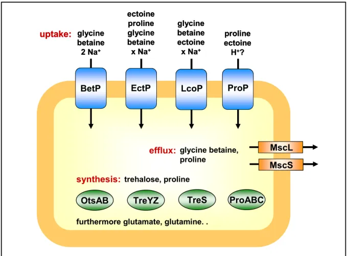
In response to hyperosmotic environmental conditions, C. glutamicum accumulates glutamate, serving as counter-ion for potassium, as well as the compatible solutes proline, trehalose, and glutamine, which can be provided by de novo synthesis (Rönsch et al., 2003, Wolf et al., 2003). If sufficient amounts of osmoprotectants are available in the surrounding, the uptake of glycine betaine, ectoine, and proline into the cytoplasm is favored (Rönsch et al., 2003). Uptake of compatible solutes in C. glutamicum is mediated by five carrier systems, of which four were shown to be osmotically regulated in their activity (Peter et al., 1996;
Peter et al., 1998; Steger et al., 2003). The secondary transport systems BetP, EctP, LcoP, and ProP (Fig. 3) catalyze the symport of the above-mentioned compounds with Na+-ions or protons, each exhibiting different substrate specificities and -affinities (compare Fig. 3), thereby allowing an effective adaptation to osmotic changes of the environment. These carriers are regulated at the level of activity, i.e. the transport rate is adapted to the extend of hypertonicity (Peter et al., 1996; Peter et al., 1997; Steger et al., 2003). Recently, a regulation at the level of expression was shown as well; transcription of the genes proP, betP, ectP and lcoP was found to be induced after an osmotic upshift (Möker, et al., 2004).
Fig. 3: Important osmoregulation systems of C. glutamicum.
trehalose, proline
BetP EctP LcoP ProP
MscS efflux: glycine betaine, MscL
proline
OtsAB TreYZ TreS ProABC glycine
betaine 2 Na+
proline ectoine
H+? ectoine
proline glycine betaine x Na+
glycine betaine ectoine x Na+
synthesis:
furthermore glutamate, glutamine. . uptake:
trehalose, proline
BetP EctP LcoP ProP
MscS efflux: glycine betaine, MscL
proline
OtsAB TreYZ TreS ProABC glycine
betaine 2 Na+
proline ectoine
H+? ectoine
proline glycine betaine x Na+
glycine betaine ectoine x Na+
synthesis:
furthermore glutamate, glutamine. . uptake:
De novo synthesis of proline in C. glutamicum starts with glutamate as substrate and is catalyzed by the enzymes γ-glutamate-kinase, 4-glutamyl-phosphate-reductase, and pyrroline carboxylate-reductase, which are encoded by the proB, proA and proC genes, respectively (Ankri et al., 1996). These genes were shown to be transcriptionally activated after an upshift of the medium osmolarity (Ley, 2001), whereas an activity regulation of proline biosynthesis proteins remains unclear.
C. glutamicum employs three different pathways for the biosynthesis of the disaccharide trehalose: the OtsAB-, the TreYZ-, and the TreS-pathway (Wolf et al., 2003). This sugar can be produced starting from the substrates glucose-6-phosphate and UDP-glucose (OtsAB- pathway), maltooligodextrine (TreYZ-pathway), as well as maltose (TreS-pathway,).
Regarding the TreYZ-pathway, an osmotic regulation at the level of protein activity was observed, however, all three pathways were shown to be osmotically influenced at the level of gene expression (Wolf et al., 2003).
1.3 Chill stress
In addition to osmolarity, temperature is an important physical factor influencing cell physiology of microorganisms. Bacteria can be divided into different classes concerning the temperature ranges providing optimal cell growth and -division. Most bacteria, as e. g. E. coli and C. glutamicum, are referred to as mesophilic, meaning that maximum growth rates are reached at moderate temperatures between 20 and 42 °C. A decrease in temperature causes immediate reduction of molecular dynamics resulting in lowered diffusion rates and conformational alterations of molecular structures. Consequently, three major problems arise from exposing a cell to a sudden decrease in temperature. (I) Chill stress has dramatic effects on enzymatic reactions, thus influencing cell physiology in general. (II) Membrane fluidity decreases, which affects many vital membrane and membrane-associated functions, such as transport, cell division, or energy production. (III) Nucleic acid topology changes causing halts in processes such as transcription, translation, or replication. Bacteria make use of at least three different mechanisms to prevent cease of growth and finally cellular death. Some organisms, as e. g. B. subtilis, respond to a temperature downshift by the production of specific proteins, so-called cold shock proteins (Neuhaus et al., 1999; Derzelle et al., 2003). These are low molecular mass proteins, which can be involved in diverse cellular processes allowing the adaptation to low temperatures. The reduced membrane fluidity in case of low temperatures can be prevented by the synthesis and lipid incorporation of fatty acids with lowered melting points. These include branched-chain and unsaturated fatty acids (Aguilar et al., 2001; Klein et al., 1999; Mansilla et al., 2004; Weber et al., 2002;
Weber et al., 2001). One further possibility to adapt to low temperature is the accumulation of
compatible solutes, which was reported for some mesophilic organisms, as e. g. Listeria monocytogenes (Ko et al., 1994).
In C. glutamicum, the uptake carriers for compatible solutes BetP, LcoP, and EctP, belonging to the betaine-carnitine-choline transporter (BCCT) family, were shown to be osmotically regulated in their activity. Recently, these were also analyzed for their responses to chill stress at the level of activity (Özcan et al., 2005). It could be shown, that LcoP was activated by chill to low extent, whereas BetP was significantly stimulated at low temperature, with maximum glycine betaine uptake activity at 10-15 °C (Özcan et al., 2005). In contrast, EctP was not stimulated by chill. RNA hybridization experiments furthermore revealed that all three genes, betP, lcoP, and ectP, were transcriptionally induced in C. glutamicum after exposure of cells to low temperature (15 °C, Özcan 2003).
1.4 Bacterial two-component systems
The cellular responses to environmental stimuli, such as nutrient starvation, or temperature and osmolarity-induced stress situations, are commonly regulated by a phosphorelay system, which was found in nearly all bacterial species (Nixon et al., 1986) as well as some eukaryotic organisms belonging to yeast, amoeba, fungi, or plants (Ota and Varshavsky, 1993; Maeda et al., 1994; Schuster et al., 1996; Alex et al., 1996; Chang et al., 1993), whereas it is missing in metazoic animals. The standard organization of this phosphorelay system is shown in Figure 4 and comprises a membrane-bound dimeric sensor histidine kinase, generally containing one or two membrane-spanning domains, and a cognate, cytoplasmic response regulator. Because of these two essential compounds this regulation system is commonly known as two-component system. However, there are also examples of soluble sensor kinases, like CheA from E. coli, which is involved in chemotaxis, or multi- component systems, including those related to chemotaxis, which are composed of more than seven proteins (Egger et al., 1997). External stimuli, which are sensed by the histidine kinase, regulate the autokinase activity of this protein (Fig. 4). The sensor catalyses the ATP- dependent trans-autophosphorylation, in which one subunit of the dimer phosphorylates a specific, highly conserved histidine residue within the other subunit, resulting in a phosphoimidazole. Subsequently, the phosphoryl group is transferred to a specific, highly conserved aspartate residue within the regulatory domain of the response regulator (Fig. 4).
This reaction is catalyzed by the response regulator and results in a conformational change of the protein, thereby causing the activation of its effector domain (Klumpp and Krieglstein, 2002). This domain mediates the specific cellular response, in most cases the regulation of gene expression, in which the response regulator acts as transcription factor (Hakenbeck and Stock, 1996). In addition, this protein class can also exhibit distinct functions, as for example the mediation of chemotaxis (Lupas and Stock, 1989). The stability of the
phosphorylated response regulator varies, depending on the system, between few seconds and several hours (Hess et al., 1988; Weis and Magasanik, 1988; Igo et al., 1989; Makino et al., 1989; Wright et al, 1993). The signal transduction is turned off by dephosphorylation of the activated response regulator. Dephosphorylation is catalyzed either by a separate bacterial phosphatase (e. g. CheZ in case of CheA/CheY of E. coli), a phosphatase activity of the sensor kinase (e. g. EnvZ or KdpD of E. coli), or the combined action of the histidine kinase, the response regulator, and an accessory protein (e. g. in case of NtrB/NtrC of E. coli (Comeau et al., 1985; Ninfa et al., 1995; Blat and Eisenbach, 1994).
Fig. 4: Model of the two-component system-mediated signal transduction resulting in the expression regulation of genes in response to environmental stimuli.
1.5 The MtrB-MtrA two-component system of Corynebacterium glutamicum
In C. glutamicum 13 putative two-component systems have been identified: CgtSR1, 2, 4-11, CitAB, MtrBA, and PhoSR (Kocan et al., 2006). Among these systems, the physiological function of only two was proposed: the CitAB system belongs to a family of two-component systems controlling the uptake and metabolism of citrate. Regarding PhoSR, Kocan et al.
P P H
D
Stimulus
ATP ADP
P ATP
ADP
ATP ADP
P
(2006) have recently shown an involvement in the adaptation of C. glutamicum to phosphate- limiting conditions.
In order to examine the involvement of two-component systems in the osmostress response of C. glutamicum, a set of twelve deletion mutants lacking those genes encoding one of the two-component systems each (Kocan et al., 2006), was analyzed. Only one out of twelve examined two-component systems exhibited an involvement in the transcription regulation of osmostress-related genes in this bacterium (Möker, 2002). RNA hybridization experiments, which were carried out with an mtrAB deficient mutant, resulted in an altered expression pattern of four osmoregulated genes (Möker et al., 2004). In C. glutamicum WT exposure to hyperosmotic stress results in the transcriptional induction of genes encoding the secondary uptake carriers for compatible solutes BetP, ProP, LcoP, and EctP, as well as the mechanosensitive channels MscL and MscS. As consequence of the mtrAB deletions, the mRNA levels of betP, proP, and lcoP were strongly decreased in the ∆mtrAB mutant both, before and after an upshift of medium osmolarity (Fig. 5). With regard to these genes, the MtrBA system seems to be essential for the activation of transcription. In contrast, transcription of the mscL gene was increased in the deletion strain before and up to 30 minutes after the hyperosmotic shock (Fig. 5). Thus, the expression of this gene is repressed via MtrBA.
Fig. 5: Expression pattern of the uptake carrier- and mechanosensitive channel encoding genes in response to an increased medium osmolality. Shown are the transcript amounts of the uptake carrier genes proP, betP, lcoP, and ectP, as well as the mechanosensitive channel genes mscL and mscS in C. glutamicum WT (wt) and ∆mtrAB (∆) before (T0) and 5 to 180 minutes after a hyperosmotic shift from 0.3 to 2.2 osm (T5 to T180), Möker et al., 2004.
T0 T5 T15 T30 T45 T60 T120 T180
+NaCl
wt ∆ wt ∆ wt ∆ wt ∆ wt ∆ wt ∆
proP betP lcoP ectP mscL mscS
T0 T5 T15 T30 T45 T60 T120 T180
+NaCl
wt ∆ wt ∆ wt ∆ wt ∆ wt ∆ wt ∆
proP betP lcoP ectP mscL mscS
T0 T5 T15 T30 T45 T60 T120 T180
+NaCl
wt ∆ wt ∆ wt ∆ wt ∆ wt ∆ wt ∆
proP betP lcoP ectP mscL mscS
The plasmid-encoded expression of the mtrAB genes using pEKEx2-mtrAB could complement the changes of the mRNA levels of proP, betP, lcoP, and mscL, therefore proving, that the described effects were indeed a consequence of the deletion, and not of polar effects (Möker et al., 2004).
In contrast to the four above-mentioned genes, the mRNA levels of further genes known to be regulated after an osmotic upshift were unchanged (Möker et al., 2004). They encode a secondary uptake carrier for compatible solutes, EctP, a mechanosensitive channel, MscS, and genes encoding enzymes of each pathway for the biosynthesis of the compatible solutes trehalose and proline, namely OtsA, ProB, TreS, and TreY.
Previous studies using RT-PCR revealed that proP and betP are expressed individually, but also together with the open reading frames (orfs) 3447 and 1449, respectively, which are localized upstream of the two genes (Möker, 2002). The putative product of orf 3447 reveals similarities to membrane protease subunits belonging to the stomatin/prohibitin family, whereas orf 3447 encodes a putative uroporphyrin-III C/tetrapyrrole methyltransferase with unknown function. In order to determine whether these genes are also regulated by the MtrBA two-component system, their expression, and additionally that of the orf located upstream of mscL, was examined in C. glutamicum WT and the mtrAB deletion mutant before and after an increase in medium osmolarity (Fig. 45, section 7.1). It turned out that, similar to betP, proP, and mscL, the transcription of these genes was osmotically induced in C. glutamicum WT. The open reading frames located upstream of proP and betP, orf 3447 and 1449, showed decreased expression levels in the ∆mtrAB mutant, indicating that their expression regulation is also mediated by the two-component system. In contrast, the orf upstream of mscL (1435) was not affected by the deletion of the mtrAB genes. Since the genes located upstream of proP and betP turned out to be regulated by MtrBA, it was necessary to investigate the transcriptional organization of the MtrBA target genes in more detail. Northern Hybridization experiments showed that proP, betP and mscL are mainly transcribed monocistronically (Fig. 46, section 7.2). In case of the two genes encoding uptake carriers weak signals corresponding to the sizes of polycistronic mRNA were also found.
In addition to osmoprotection, the uptake and synthesis of compatible solutes in bacteria has been shown to play a role in the response to thermal stresses, e. g. chill. It was recently shown, that the compatible solute uptake carriers BetP and LcoP are influenced by chill stress in their activity (compare section 1.3). Furthermore, the transcription of their encoding genes was regulated at the level of expression in response to low temperature. RNA hybridization experiments revealed that exposure of C. glutamicum cells to chill stress (15 °C) results in the transcriptional induction of the lcoP and betP genes. In contrast, in the mtrAB deletion mutant, these genes turned out to be transcribed to significantly lower levels,
before and after exposure to low temperature (Özcan, 2003). Consequently, MtrBA seems to mediate, in addition to expression regulation in response to osmotic stress, the chill stress- induced regulation of genes encoding uptake carriers for compatible solutes.
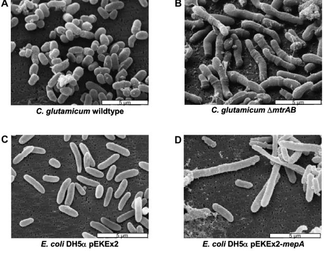
The findings of MtrBA-mediated regulation of gene expression described above were obtained with focus on responses to specific stress conditions. Furthermore, a global analysis of the consequences of the mtrAB deletions on the C. glutamicum genome was performed using DNA microarrays (M. Bott, Forschungszentrum Jülich). These indicated that the two-component system also regulates the expression of a set of genes involved in a different physiological function in C. glutamicum, the cell wall metabolism. In the ∆mtrAB mutant the mRNA levels of genes encoding the lipoproteins MepA and LpqB were significantly increased. The MepA protein shows similarities to a cell wall endopeptidase. A third gene with increased mRNA levels in the deletion mutant was named ppmA, for putative protease modulator (Möker et al., 2004). To verify the hypothesis that these genes are important for cell wall biosynthesis, C. glutamicum wild type (WT) and ∆mtrAB cells were analyzed using scanning electron microscopy. The mutant cells turned out to exhibit a significantly elongated cell morphology (compare Fig. 6). Heterologous expression of the plasmid-encoded mepA gene in E. coli (Fig. 6) resulted in elongated cells with a morphology more or less similar to that of C. glutamicum ∆mtrAB (Möker et al., 2004). These observations led to the suggestion that MepA has a cell wall endopeptidase activity targeting the peptidoglycan layer. These effects, the altered expression of mepA and the elongated cell morphology of the deletion mutant, were complemented by the plasmid-encoded expression of the mtrAB genes in the ∆mtrAB mutant using the vector pEKEx2-mtrAB (Möker et al., 2004).
Taken together, the MtrB-MtrA system seems to be involved in three physiological functions in C. glutamicum, (I) osmoregulation, (II) chill stress response, and (III) cell wall metabolism.
Regarding the sensor kinase MtrB of this system one has to conclude that either a single stimulus is detected, which can act as a measure for the three different purposes, or MtrB is able to detect a set of different stimuli. These signals would likely be connected to the different above-mentioned physiological functions. The MtrBA-mediated expression regulation of osmo- and chill stress-related genes was shown to occur in response to specific conditions connected with osmotic and chill-stress, therefore indicating that MtrB acts as a sensor for both, osmo- and chill-stress. In contrast, the conditions which lead to the repression or induction of the cell wall-related genes remain unknown. The phenomenon, that a sensor is able to detect both, stimuli related to osmo- and chill stress has already been reported for the compatible solute uptake carrier BetP (Peter et al., 1996; Peter et al., 1998, Özcan et al., 2003). A number of two-component systems involved either in chill stress
response, osmoprotection, or cell wall biosynthesis were also described recently (Aguilar et al., 2001; Jung et al., 2000; Mizuno et al., 1988; Raivio et al., 1999; Mascher et al., 2004), but few data are available on systems influencing all three of these processes.
Fig. 6: Scanning electron micrographs showing C. glutamicum WT (A), and ∆mtrAB (B) as well as E. coli DH5α containing pEKEx2 (C) or pEKEx2-mepA (D), Möker et al., 2004.
1.6 Objectives of this thesis
The aim of this work was a detailed characterization of the sensing properties of the MtrB- MtrA two-component system of C. glutamicum. Recent investigations suggested that the histidine kinase MtrB acts as sensor for both, osmo- and chill stress-related stimuli. For a system acting as osmosensor a variety of stimuli are possible. These include changes in external osmolarity, ionic strength and concentration of ions or specific solutes, but also, resulting from the water fluxes across the cytoplasmic membrane, changes in turgor pressure, internal osmolarity, ionic strength, concentration of ions or specific solutes and membrane strain or -shrinkage. For a protein acting as a chill sensor the cellular
A B
C. glutamicumwildtype
5 µm
C. glutamicumwildtype
5 µm 5 µm
C. glutamicum∆mtrAB5 µm C. glutamicum∆mtrAB5 µm5 µm
C D
5 µm
E. coliDH5αpEKEx2
5 µm 5 µm 5 µm
E. coliDH5αpEKEx2
5 µm
E. coliDH5αpEKEx2-mepA
5 µm 5 µm 5 µm
E. coliDH5αpEKEx2-mepA
architectures offer three basic targets which respond almost immediately to a change in temperature, (I) phase condition of membrane-bound lipids, (II) conformation of proteins, or (III) conformation of nucleic acids (Weber and Marahiel, 2002).
Since a set of different parameters changes upon osmotic- and chill stress, a whole cell approach did not seem favorable for the detection of separate stimuli. Instead, for the investigation of the direct signals sensed by MtrB two in vitro membrane systems were chosen. In inverted membrane vesicles enriched with MtrB prepared from E. coli the membrane-spanning sensor kinase is integrated into the cytoplasmic membrane by the E. coli protein insertion machinery, which might provide a correct integration. However, in this system non-specific E. coli membrane proteins are also present. The proteoliposome system provides a more reduced complexity. The isolation of MtrB and the subsequent reconstitution into E. coli lipid liposomes would result in an in vitro membrane system carrying MtrB only. In recent years, proteoliposomes were shown to serve as suitable systems for the analyses of membrane-bound sensor histidine kinases.
Using the above-mentioned MtrB-enriched in vitro membrane systems it was first aimed to define the basic attributes of MtrB and MtrA, including the autokinase activity of membrane- bound MtrB, the phosphoryl group transfer to isolated MtrA, and the dephosphorylation of activated MtrA (MtrA-P). Once the in vitro activities of MtrB and MtrA were defined, the stimuli triggering the MtrB-MtrA signal transduction could be addressed by a systematic variation of possible stimuli related to osmotic and chill stress conditions.
2. Materials and methods
2.1 Bacterial strains, plasmids, and oligonucleotides
2.1.1 Bacterial strains
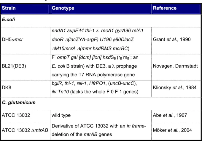
The E. coli and C. glutamicum strains used in these studies are summarized in Table 1.
Tab. 1: E. coli and C. glutamicum strains that were used in this work.
S
Sttraraiinn GeGennoottyyppee RReeffeerreennccee E.coli
DH5αmcr
endA1 supE44 thi-1 λ- recA1 gyrA96 relA1 deoR ∆(lacZYA-argF) U196 φ80DlacZ
∆M15mcrA ∆(mmr hsdRMS mcrBC)
Grant et al., 1990
BL21(DE3)
F- ompT gal [dcm] [lon] hsdSB (rB-mB-; an E. coli B strain) with DE3, a λ prophage carrying the T7 RNA polymerase gene
Novagen, Darmstadt
DK8 bglR, thi-1, rel-1, HfrPO1, (uncB-uncC),
ilv:Tn10 (lacks the whole F 0 F 1 genes) Klionsky et al., 1984 C. glutamicum
ATCC 13032 wild type Abe et al., 1967
ATCC 13032 ∆mtrAB Derivative of ATCC 13032 with an in frame-
deletion of the mtrAB genes Möker et al., 2004
2.1.2 Plasmids
In Table 2, the plasmids used in this work are listed. The construction of the vectors is described in detail in the appendix (7.3).
Tab. 2: Plasmids that were used in this study.
PlPlaassmmiidd DDeessccrriippttiioonn RReeffeerreennccee
pASK-IBA3 ApR, Ptet, expression vector Skerra et al., 1994 pASK-IBA7 ApR, Ptet, expression vector Skerra et al., 1994
PlPlaassmmiidd DDeessccrriippttiioonn RReeffeerreennccee pASK-IBA3-
mtrB-strep
pASK-IBA3 derivative containing mtrB in the PshAI cleavage site fused to the sequence encoding a C-terminal Strep-tag
This work
pASK-IBA7- strep-mtrB
pASK-IBA7 derivative containing mtrB in the PshAI cleavage site fused to the sequence encoding an N-terminal Strep-tag
This work
pASK-IBA3- mtrB∆25
pASK-IBA3-mtrB-strep derivative encoding an MtrB-Strep derivative with a truncation of the N-terminal amino acids 1 to 25
This work
pASK-IBA3- mtrB∆154
pASK-IBA3-mtrB-strep derivative encoding an MtrB-Strep derivative with a truncation of the N-terminal amino acids 1 to 154
This work
pASK-IBA3- mtrB∆163
pASK-IBA3-mtrB-strep derivative encoding an MtrB-Strep derivative with a truncation of the N-terminal amino acids 1 to 163
This work
pASK-IBA3- mtrB∆190
pASK-IBA3-mtrB-strep derivative encoding an MtrB-Strep derivative with a truncation of the N-terminal amino acids 1 to 190
This work
pASK-IBA3- mtrB∆L124
pASK-IBA3-mtrB-strep derivative encoding an MtrB-Strep derivative with a truncation of the N-terminal amino acids 31 to 164
This work
pASK-IBA3- mtrB∆L134
pASK-IBA3-mtrB-strep derivative encoding an MtrB-Strep derivative with a truncation of the N-terminal amino acids 36 to 159
This work
pASK-IBA7- strep-mtrA
pASK-IBA7 derivative containing mtrA in the PshAI cleavage site fused to the sequence encoding an N-terminal Strep-tag
This work
pEKEx2 KmR, lacIq, ptac, E. coli - C. glutamicum shuttle vector
Eikmanns et al., 1991
pEKEx2-mtrAB
pEKEx2 derivative containing the mtrAB genes from C. glutamicum under control of the tac promoter
Möker et al., 2004
pEKEx2-mepA
pEKEx2 derivative containing the mepA gene from C. glutamicum under control of the tac promoter
Möker et al., 2004
PlPlaassmmiidd DDeessccrriippttiioonn RReeffeerreennccee
pET224b-mtrA
KmR, PT7, expression vector derivative containing mtrA fused to the sequence of a C- terminal (His)10-tag
Brocker,
unpublished data
pET28a KmR, PT7, expression vector Novagen, Inc.
Madison, USA
pMW151
pET28a derivative containing dcuS fused to the sequence encoding an N-terminal (His)6- tag.
Janausch et al., 2002
pDrive ApR, KmR, lacZα, A-T insertion vector Qiagen, Hilden
pDrive1435 pDrive derivative containing a 0.51 kb
fragment of the orf 1435 (upstream of mscL) This work pDrive1449 pDrive derivative containing a 0.47 kb
fragment of the orf 1449 (upstream of proP) This work pDrive3447 pDrive derivative containing a 0.48 kb
fragment of the orf 3447 (upstream of betP) This work pGEM-3Z ApR, PT7 and PSP6
Promega, Mannheim
pGEM-4Z ApR, PT7 and PSP6 Promega,
Mannheim p16Sr pGEM-3Z derivative containing a 0.5 kb
fragment of the 16s rRNA gene Nolden, 2001 pbetP pGEM-4Z derivative containing a 0.44 kb
fragment of the betP gene Möker et al., 2004 pectP pGEM-4Z derivative containing a 0.76 kb
fragment of the ectP gene Möker et al., 2004 plcoP pGEM-4Z derivative containing a 0.73 kb
fragment of the lcoP gene Möker et al., 2004 pmscL pGEM-4Z derivative containing a 0.4 kb
fragment of the mscL gene Möker et al., 2004 pproP pGEM-4Z derivative containing a 0.51 kb
fragment of the proP gene Möker et al., 2004 ApR, resistence towards ampicillin. KmR, resistence towards kanamycin


![Fig. 16: MtrB-mediated dephosphorylation of MtrA-P. A - D Phosphorimages of MtrA-P after membrane-bound MtrB and [γ 33 P]ATP and ADP, respectively, were removed by ultracentrifugation and subsequent gel filtration](https://thumb-eu.123doks.com/thumbv2/1library_info/3658118.1503656/62.892.88.786.616.977/mediated-dephosphorylation-phosphorimages-membrane-respectively-ultracentrifugation-subsequent-filtration.webp)
