Intersubunit Cross-talk in the Betaine Permease BetP from
Corynebacterium glutamicum
Inaugural-Dissertation zur Erlangung des Doktorgrades
der Mathematisch-Naturwissenschaftlichen Fakultät der Universität zu Köln
vorgelegt von
Markus Christian Becker aus Daun
Copy-Star Druck und Werbung GmbH
Köln 2013
Diese Arbeit wurde am Institut für Biochemie der Universität zu Köln unter Anleitung von Herrn Professor Dr. R. Krämer durchgeführt.
Berichterstatter: Professor Dr. R. Krämer Professor Dr. U.- I. Flügge Professor Dr. C. Ziegler Tag der mündlichen Prüfung: 23.Mai 2013
„Unsere größte Schwäche liegt im Aufgeben. Der sichere Weg zum Erfolg ist immer es doch noch einmal zu versuchen.“
(Thomas Alva Edison)
I
Contents
Contents ... I Abbreviations ... IV
1 Introduction ... 1
1.1 Osmotic stress ... 1
1.2 Osmosensors ... 2
1.3 The betaine permease BetP ... 3
1.4 The LeuT family of transporters ... 6
1.5 Transport mechanism of LeuT type carriers ... 7
1.6 Transport mechanism of BetP ... 9
1.7 Quarternary structure of BetP and its implications for cross-talk ... 10
1.8 Cross-talk in transport proteins and methods for its analysis ... 13
1.9 The osmo-transportome of C. glutamicum ... 15
2 Experimental procedures ... 17
2.1 Bacterial strains, plasmids and oligonucleotids ... 17
2.1.1 Bacterial strains ... 17
2.1.2 Plasmids ... 17
2.1.3 Oligonucleotides ... 20
2.1.4 Growth media and cultivation conditions ... 23
2.2 Molecular biology ... 24
2.2.1 Preparation of competent E. coli cells and transformation ... 24
2.2.2 Preparation of electrocompetent C. glutamicum cells and transformation ... 24
2.2.3 DNA techniques ... 25
2.2.3.1 Isolation of plasmid DNA from E. coli ... 25
2.2.3.2 Gel electrophoresis and extraction of DNA from agarose gels ... 25
2.2.3.3 Polymerase chain reaction (PCR) ... 25
2.2.3.4 Restriction, ligation, and sequencing of DNA ... 26
2.2.3.5 Site directed mutagenesis ... 26
2.2.3.6 DNA sequencing: ... 27
2.3 Analytical methods ... 27
II
2.3.1 Protein concentration measurements ... 27
2.3.2 Sodium-dodecyl-sulphate-polyacrylamid-gel-elektrophoresis (SDS-PAGE) ... 27
2.3.3 Blue-native-polyacrylamid-gel-elektrophoresis (BN-PAGE) ... 27
2.3.4 Western-Blot ... 28
2.3.5 Gel-Permeation-Chromatography ... 28
2.3.6 Estimation of osmolalities ... 28
2.3.7 Fluorescence microscopy ... 28
2.4 Biochemical Methods ... 29
2.4.1 Heterologous expression ... 29
2.4.1.1 Heterologous expression in E. coli in shaker flasks... 29
2.4.1.2 Heterologous expression in E. coli in bioreactor cultures ... 29
2.4.1.3 Heterologous expression of proteins in C. glutamicum ... 30
2.4.2 Isolation of membrane proteins. ... 30
2.4.2.1 Isolation of membrane proteins via bead mill ... 30
2.4.2.2 Isolation of membrane proteins via french press ... 31
2.4.3 Purification of membrane proteins via affinity tags ... 31
2.4.3.1 Solubilisation of membrane proteins ... 31
2.4.3.2 Purification of Strep tagged proteins: ... 31
2.4.3.3 Purification of His tagged proteins: ... 32
2.4.3.4 Purification of Flag tagged proteins. ... 32
2.4.3.5 Purification of MBP tagged proteins... 32
2.4.4 Chemical protein modifications ... 33
2.4.4.1 Protein labeling with Bodipy dye ... 33
2.4.4.2 Protein labeling with Biotin ... 33
2.4.5 Reconstitution of Proteins ... 33
2.4.5.1 Preparation of liposomes ... 33
2.4.5.2 Reconstitution of proteins ... 34
2.4.6 Radiochemical transport measurements ... 34
2.4.6.1 Betaine uptake measurements of BetP and LcoP in proteoliposomes ... 34
2.4.6.2 Betaine uptake measurements in E. coli MKH13 cells ... 35
2.4.6.3 Betaine uptake measurements in C. glutamicum DHPF cells. ... 36
3 Results ... 37
3.1 Genetic fusion of BetP trimers ... 37
3.1.1 Fusion BetP: a. pFus ... 37
III
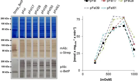
3.1.2 Fusion BetP: b. pFel ... 40
3.2 Individually tagged BetP ... 46
3.2.1 N-terminal affinity tags ... 46
3.3 Stability of BetP trimers ... 54
3.4 C-terminal affinity tags ... 55
3.5 Co-expression and purification of BetP heteromers ... 58
3.6 Cross-talk in BetP heteromers ... 62
3.6.1 Catalytical cross-talk in BetP... 62
3.6.2 Regulatory cross-talk in BetP ... 65
3.7 Re-analysis of monomeric BetP ... 67
3.8 BetP dynamics via single molecule fluorescence ... 72
3.9 Analysis of the osmo-transportome of C. glutamicum ... 75
4 Discussion ... 80
4.1 LcoP is an osmo-sensor ... 80
4.2 Monomeric BetP ? ... 82
4.3 Genetic fusion of BetP trimers ... 84
4.4 The N-terminal domain is sensitive to alterations ... 88
4.5 State of art in the hetero-trimeric BetP production: applications in single molecule approaches and limitations ... 89
4.6 The nature of cross-talk in BetP ... 90
5 Abstract ... 95
6 Zusammenfassung ... 96
7 References ... 97
Danksagung ... 107
Erklärung ... 108
IV
Abbreviations
AHT Anhydrotetracycline
APS Ammoniapersulfat
BCCT Betaine-Choline-Carnitine-Transporter
BHI Brain-Heart-Infusion
Carb Carbenicillin
CBD Chitin-Binding-Domain
CBP Calmodlin-Binding-Pepitde
Cdw cellular dry weight
Cm Chloramphenicol
DDM Dodecylmaltoside
DNA Deoxyribonucleicacid
EDTA Ethylendiaminetetramin
EAAT Excitatory-Amino-Acid-Transporter
GST Glutathione-S-Transferase
IPTG Isopropyl-b-Thiogalactoside
LB Lysogeny-Broth
LDAO Lauroyl-Dimethyl-Amineoxide MBP Maltose-Binding-Protein MFS Major-Faciliator-Superfamily NCS1 Nucleobase-Cation-Symporter
Rpm rounds per minute
SEC Size Exclusion Chromatography
TEV Tobacco Etch Virus
1
1 Introduction
1.1 Osmotic stress
Unicellular organisms face rapid and severe changes in the external osmolality. Due to the permeability of the cell membrane for water, its activity equilibrates fast through the membrane between medium and cytoplasm (Wood 1999). Under physiological conditions the water activity in the cytoplasm is lower than in the surrounding medium so that the turgor can be established. Turgor describes the pressure with which the plasma membrane is pushed against the cell wall. This pressure was shown to be necessary for cell division and growth in bacteria (Koch 2000). Cells may have to encounter generally to different osmotic stress conditions in which water permeates the cell membrane, due to lowered (hyper- osmotic stress) or enlarged (hypo-osmotic stress) external water activity.
Cell integrity under hypo-osmotic stress is achieved by mechanosensitive channels. These proteins can be activated in vitro by pressure gradients indicating that they percept directly raising membrane tension as indicator for cell swelling. Upon opening, these channels release a broad spectrum of low molecular weight compounds thereby increasing the water activity in the cytoplasm (Sukharev et al., 1997).
Upon hyper-osmotic stress the cells take up or synthesise solutes to lower the internal water activity without cytoplasm shrinkage. Not every compound is equally well suited for this purpose. Solutes utilised for lowering the water activity should exhibit no detrimental interference with cytoplasmic compounds e.g. proteins, DNA, etc.
Fig. 1-1: Chemical structure represantation of different compatible solutes.
2
Such compounds are called compatible solutes. The main compatible solutes utilised by bacteria are amino acids and derivatives like proline and betaine, or sugars and polyols like trehalose and sorbitol (Fig. 1-1).The common feature of those compounds is that they form thermodynamically unfavorable interactions with unfolded proteins thereby stabilising the native folded state (Rösgen et al., 2005).
The temporal organisation of adaption to hyper-osmotic stress is very common in all mesophiles for which it was investigated. First the cells take up potassium to maintain turgor. Interestingly it is still not clear how the influx of positive charges is compensated.
Potassium ions are then replaced by compatible solutes taken up from the medium or synthesised in the cytoplasm. In the late state of adaption the gene expression is re-modeled by mainly two-component systems percepting the osmotic stress (Wood 1999).
Solute synthesis is normally much more energy intensive than uptake. Therefore bacteria have evolved a variety of solute uptake systems. The systems for both solute uptake and synthesis are usually redundant. Interestingly the different solute transporters belong to completely different transport protein families suggesting the independent evolution of different uptake systems and thus highlighting the impact of osmotic stress.
1.2 Osmosensors
Within the large group of solute transport proteins only three have been ultimately shown to sense hyper-osmotic stress without any other factor in proteoliposomes:
(i) OpuA is an ABC transporter for betaine and was described in Lactococcus lactis.
Resembling the common bacterial ABC transporter architecture, the protein is composed out of two transmembrane domains fused to a periplasmic binding domain and two nucleotide binding domains (NBD). The NBDs carry a C-terminal extension harbouring a tandem ß-cystathionine-synthase (CBS) domaine followed by a short extension with a cluster of glutamate residues (Van der Heide & Poolman 2000). The transporter was shown to be activated by increasing ionic strength (Van der Heide et al., 2001). The osmo-regulation of this transporter is abolished by deletion of the CBS, whereas deletion of the anionic C-terminal domain leads to regulatory dependence on the head groups of the
3
membrane lipids. Mutations in exposed cationic residues also lead to deregulation of the transporter. It is likely that the C-terminal domain of OpuA senses salt stress via disturbance of ionic interaction with the membrane (Mahmood et al. 2009; Biemans-Oldehinkel et al.
2006).
(ii) ProP from Escherichia coli is a secondary active transporter that mediates import of betaine and prolin together with protons. The transporter belongs to the major faciliator superfamily (MFS) and consists of twelve transmembrane helices. 60 amino acids at the transporters C-terminus form a coiled coil structure in the cytosol and are shown to be important for the activation threshold of the transporter (Hillar et al., 2005).
Osmoregulation of this transporter depends on the lipid environment (Romantsov et al., 2008; Tsatskis et al., 2005). Interestingly the transporter seems to co-localise with cardiolipin at the cell poles suggesting functional inhomogenity of the cell membrane.
(Romantsov et al. 2008; 2010). ProP is activated by all kinds of hyper-osmotic stress including ionic strength, concentration of neutral molecules and macromolecules. This finding suggests that this transporter senses directly its hydration state (Racher et al. 2001;
Culham et al. 2003; Wood 2006).
1.3 The betaine permease BetP
The protein BetP was discovered functionally as osmoregulated betaine/Na+ symport carrier mediating fast (up to 110 nmol x mgcdw-1
x min-1) and high affine (Km(betaine)=8,6 µM) import of this solute in C. glutamicum (Farwick et al., 1995). The carrier encoding gene was identified by functional complementation of an E. coli mutant unable to import or synthesise betaine (Peter et al. 1996). BetP could be purified via an amino terminal StrepII™-tag (Strep-tag) out of E. coli and reconstituted to proteoliposomes where it retained its ability to conduct the import of Na+/betaine driven by the sodium motive force (Rübenhagen et al., 2000). The protein is activated exclusively by raising K+ (Rb+, Cs+) concentration in proteoliposomes suggesting this as being the only relevant stimulus (Schiller et al. 2004; Rübenhagen et al. 2001). However it was shown in vivo, that potassium is not the only stimulus (Ott 2009). BetP belongs to the betaine choline-carnithine transporter (BCCT) family characterised by these substrates. These substrates share a
4
positively charged quarternary nitrogen atom which is coordinated by a highly conserved motif of aromatic amino acids (Ziegler et al. 2010). It possess 12 transmembrane helices and N-, and C-terminal extensions of 59/49 amino acids. These extensions are both located in the cytoplasm (Peter et al. 1996; Rübenhagen et al. 2001). Both extensions were shown to be involved in BetP activation (Peter et al. 1998). The C-terminal domain of BetP was also identified in vitro to be responsible for the perception of rising K+ concentrations (Schiller &
Morbach 2004). Via site directed mutagenesis of the C-domain and functional anlysis of the resultant BetP variants in E. coli and C. glutamicum it was concluded that this domain interacts with the membrane lipids and that not only its charge but also its structure is important for correct signal transduction (Schiller et al. 2006; Ott et al. 2008). The interactions of the C-domain were shown via peptide binding array experiments to occur also with the cytoplasmic loops L2, L4; L8, the N-domain and the C-domain of the adjacent protomers showing that the regulation mechanism is much more complicated (Ott et al.
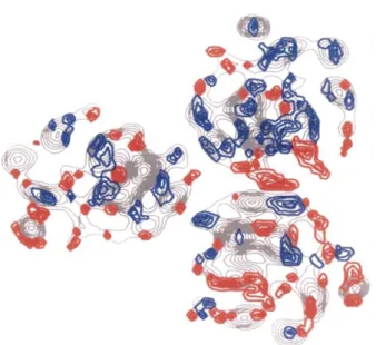
2008). Because of its high stability in detergent solutions, BetP was also choosen as target for structural analysis. A 2D projection map at an resolution of 0,75 nm showed that BetP is a homotrimer where every monomer is most likely to form its own betaine translocation pathway.
Fig. 1-2: Electron density projection map of 2D BetP crystals (Ziegler et al. 2004). Differences from the average densities are highlighted in red (additional densities) and blue (missing densities).
5
Interestingly the subunits were shown to be not symmetrical to each other which is a first hint for catalytical or regulative cross-talk (Fig. 1-2) (Ziegler et al. 2004; Tsai et al. 2011). The protein was also crystallised in detergent solubilised state in typeII 3D crystals (Ressl et al., 2009). Many features of the solved structure are consistent with functional observations and bioinformatic predictions. Each protomer consists of twelve transmembrane helices.
The substrate was found to be bound in an aromatic box build by the side chains of tryptophanes and one tyrosine (Ressl et al., 2009).
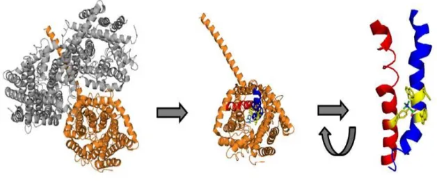
Fig. 1-3: Structure of the BetP trimer (left). One subunit shown with the residues of the betaine binding motif highlighted in yellow (middle). Structural arrangement of the tryptoptophane and tyrosin residues (red) contributing to betaine binding located on TM8 (blue) and TM4 (red) (right). (modified from PDB entry:
2WIT)
The two co-transported sodium ions are bound in remarkably distant sites. The first sodium binding site (Na2) is formed by backbone and sidechain carbonyl oxygens of residues in TM 7 and 3. The second sodium binding site (Na1) is formed by residues of TM 5 and 8.
Mutations in the respective residues abolished sodium binding and sodium coupled transport completely (Khafizov et al., 2012). Whereas the Na2 site was found by structural alignments of BetP with the NSS type transporter LeuT, the Na1 site was established by symmetry investigations of the TM helix arrangement in BetP. The succesful elaboration of the sodium binding sites was only possible, because the BetP structure exhibits a remarkable global fold that was shown in the last years to hold true for many different
6
secondary active transporters and was first discovered in the LeuT structure: the inverted repeat motif (Forrest et al., 2008; Krishnamurthy et al., 2009; Yamashita et al., 2005)
1.4 The LeuT family of transporters
The inverted repeat motif consists of ten transmembrane helices.
tab. 1-1: Different secondary active transport proteins all shown to comprise the inverted repeat architecture first described for the NSS-type transporter LeuT.
transporter substrate/mechanism family source LeuT Leucine/Na+
symport
NSS (Yamashita et al., 2005)
vSglT galactose/Na+
symport
SSS (Faham et al., 2008)
Mhp1 benzylhydantoin/Na+
symport
NCS1 (Weyand et al., 2008;
(Shimamura et al.
2010) CaiT carnithine/y-butyrobateaine
antiport
BCCT (Tang et al., 2010)
AdiC arginin/agmatin antiport
APC (Gao et al., 2009)
ApcT Amino acid/ H+
symport
APC (Shaffer et al., 2009)
GltPH Glutamate/Na+
symport
EAAT (Yernool et. al 2005)
The first five transmembrane helices are structurally related to the following five ones by a two-fold symmetry axis in the membrane plane. This fold contains the transporter core were the transport reaction takes place (Fig. 1-4).
7
Fig. 1-4: The inverted repeat motifs in the structure of a single BetP protomer. First repeat (red) formed by TM3-TM7 and second repeat (blue) formed by TM8-TM12. The c terminal domain as well as the TM1 and 2 not taking part in the formation of the inverted repeat motif are shown in grey. (Modified from PDB entry:
2WIT)
The first two transmembrane helices of each repeat form a tight bundle, whereas the latter two of each repeat form a scaffold which is called a hash domain according to arrangement of the helices (Abramson and Wright, 2009; Krishnamurthy et al., 2009). Interestingly, this architecture was found in transporters of completely distinct families (tab.1-1).
1.5 Transport mechanism of LeuT type carriers
According to the generally accepted model of alternating access (Tanford, 1983) of a single substrate binding site mediating transport in secondary active transporters, at least three different conformations (binding site accessible from the cytoplasm Ci; binding site accessible from the periplasm Ce and binding site not accessible from both compartments Cc) have to be realised during the transport cycle (Jardetzky 1966). The binding site is mainly build by the first helices of each repeat and located halfway across the membrane in
8
LeuT type transporters (Abramson and Wright, 2009). From the different available structures of LeuT type transporters in different conformations, a model for the realisation of the alternating access of the binding site during transport was proposed. This model proposes a relative rocking type movement of the 4 helix bundle against the hash domain as being responsible for the necessary accessibility change of the binding site during the catalytic cycle (Forrest and Rudnick, 2009; Shimamura et al., 2010).
Mhp1 was the first member of the LeuT family that was crystallised in the Ce; Ci and Cc conformation. From this structure the model could convincingly be extended by sub-states occluding the binding site by so-called gating residues prior to the global conformational change in the Ce-Ci transition (Shimamura et al., 2010).
Although good evidence exists for the global conformational transition of LeuT type transporters during transport, the mechanism of coupling the free enthalpy of substrate and co-substrate flux is poorly understood. Molecular dynamic simulations of the sugar transporter vSglT suggested the obligatory order of substrate release as the molecular realisation of free enthalpy coupling (Watanabe et al., 2010). The LeuT transporter itself has recently been crystallised by use of conformation specific antibodies in different conformations. From these structures it was concluded that binding of the substrate to the outward open, sodium bound conformation is the trigger for closure of the extracellular exit leading in addition to the disruption of the two sodium binding sites. It is however not clear how the substrate is released to the cytoplasm as no inward occluded structure of this transporter is availible (Krishnamurthy and Gouaux, 2012).
Several substrate binding sites assigned to the structures of different LeuT type transporters lead to the assumption of regulatory involvement of additional substrate binding sites in the transport pathways of these transporters. In the structure of the BCC-type carnithin/ϒ - butyrobetaine antiporter CaiT a second ϒ-butyrobetaine binding site near the periplasmic end of the substrate pathway was found. Mutations in this site abolished the slight cooperativity observed in binding of both substrates (Schulze et al. 2010). A possible second substrate binding site in the transporter LeuT and its functional relevance is still not clear (Zhao et al. 2011; Quick et al. 2012; Shi et al. 2008; Piscitelli et al. 2010; Krishnamurthy &
Gouaux 2012). BetP comprises an exception of all LeuT-family members found so far because it is the only member of this group where transport activity is regulated on a
9
biochemical level which necessitates the occurrence of more substates resembling active and inactive states of the protein (Ziegler et al. 2010).
1.6 Transport mechanism of BetP
As the structure of BetP has recently been established in different conformational states (Perez et al. 2012; 2011) it was possible to assign the transport cycle according to the generally accepted alternating access model (Jardetzky 1966). The state of the art model of alternating access during the betaine/sodium co-transport in BetP mainly involves the molecular displacements of the first and last helix of each repeat , TM3; 7; 8 and 12 for realisation of the change of accessibility during the transport cycle (Perez et al., 2012).
Binding and dissociation of the substrate betaine is mainly achieved by different rotamer conformations of the aromatic residues W377 and W374 located on TM8 which resembles the first (bundle) helix of the second repeat. The outward open conformation allowing for betaine binding is established by sodium binding to the Na2 site. Binding of betaine to BetP in the outward open conformation leads to the formation of the Na1 site, allowing sodium to bind to the transporter in this site. The periplasmic exit pathway is then closed by a relative movement of the periplasmic halves of TM3 and TM12. Dissociation of sodium from the Na1 site formed by residues of TM5 and TM8 leads to an opening of the intracellular exit by a remarkable displacement of the cytoplasmic half of TM3: The Na2 site and the betaine binding site are consequently disrupted leading to the dissociation of all substrate towards the cytoplasmic exit (Perez et al., 2012).
Although it was possible to assign an alternating access mechanism for BetP by the established crystal structures, the nature of coupling the substrate/co-substrate flux is not clear to date. Furthermore very few conclusions about the regulatory function of the BetP protein could be drawn from the structural analysis for several reasons. First of all it is known that the regulation of the protein strictly depends on the membrane environment (Schiller et al. 2006), a factor which is of course not present in 3D detergent crystals. The crystallisation of BetP neccessitated the truncation of the N-terminus by 29 amino acids, resulting in a protein that lacks the osmoregulation in E. coli membranes completely (Ressl et al., 2009).
10
A second betaine binding site at the periplasmic entry of the protein that was only visible by cooperative betaine binding upon elevated potassium concentration was proposed. The role of this binding site is not clear yet especially as the betaine affinity measured by tryptophan fluorescence exhibited an apperent affinity two orders of magnitude lower than the apperent affinity in transport measurements (Ge et al., 2011).
1.7 Quarternary structure of BetP and its implications for cross-talk
Interestingly BetP and AdiC are the only members of the LeuT family where structural information from membrane embedded proteins in 2D crystals is available. Since the 2D structure of AdiC was solved with the application of 2 fold non-crystallographic-symmetry no asymmetry in this structure could be detected (Casagrande et al., 2008).
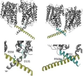
Fig. 1-5: C-terminal inter protomer contacts in the C-terminal helices. Salt-bridge between R558 and D131 in loop2 of the neighbouring protomers (left) and the interaction of R568 and E552 of the same protomers (right). (Structure from PDB entry: 2WIT)
As already mentioned, the protomers in the 2D BetP crystals do not seem to be symmetric to each other (Tsai et al. 2011; Ziegler et al. 2004) (Fig. 1-2). Homo-trimeric BetP prooved to exist in conformationally asymmetric states as well in the recent 3D structures (Perez et al., 2012). Therefore it might be the case that the monomers function in a concerted manner where all three conformations (Ce, Cc and Ci) are present at the same time in the trimer.
This fact would also explain why betaine could be detected in the 3D crystal structure
11
without supplementation of the crystallisation solution with this compound (Ressl et al., 2009). If the substrate would not be trapped in one subunit it could hardly stay bound during crystallisation for kinetic reasons. Cross-talk will most likely involve the residues mediating the monomer contacts. The protein contacts can be nicely seen in the published 3D structures. Only few residues contribute to monomer interactions. Most of the space between the monomers is occupied by non-protein density in the structure (Perez et al., 2012; Ressl et al., 2009). Two salt bridges in the C-terminal domain between R568 and E552 contribute to monomer interaction. Mutations of R568 to proline (charge and structure change) and alanine/aspartate (charge effect) were shown to strongly impair BetP function (Ott et al. 2008). The second salt bridge is formed between R558 in the C-terminal helix of one protomer and D131 of the neighbouring protomer. As this interaction links the regulatory C-terminal domain to the functional relevant first and second bundle helix of the neighbouring protomer it was suggested that this site might be involved in a regulatory crosstalk (Gärtner et al., 2011) although the regulation of the respective R558D BetP mutant is comparable to that of the wild-type protein (Ott et al. 2008). Since the BetP activity is very sensitive to mutations in the C-domain it cannot be immediately concluded that observed effects are due to impaired monomer interaction. Although the quarternary structure of these mutants was not addressed it is unlikely that trimerisation is affected here because it was shown that the deletion of the complete C-domain from Y550 on does not affect the trimerisation (Tsai et al. 2011; Perez et al. 2011). Beside the protomer interactions mediated by the C-domain, few protomer contacts were found at the mebranous interfaces. Van der Waals contacts were found between TM2 and the horizontal amphipathic helix 7 of neighboring protomers (T108 and F112 in TM2, Y340, F344 and F345 in helix 7 and H192 in TM4). The main salt bridges between the neighbouring protomers were found between N331 to D356 and T351 to E425 in TM9 of the neighbouring protomer (Ressl et al., 2009).
12
Fig. 1-6: Side view on one BetP monomer. The positions for the alanine mutations leading to monomerisation of the protein are highlighted in red. (modified from PDB entry: 2WIT)
By in silico alanine scanning approaches several single site mutations could be predicted to impair trimerisation of the protein. The mutant variants W101A/T351A and W101A/T351A/F345A were shown two form complexes that appear much smaller in freeze fracture analysed liposomes in comparison to the wild type protein. These variants were further shown to migrate as monomers upon native PAGE analysis whereas the wild type protein migrates at trimer size. The monomeric vatiants exhibit only a very low but significant uptake activity. These results suggest the functional necessity of cross-talk for the transport function. The monomeric mutant was interpreted to be non-activatable by hyperosmotic stress which also suggests a regulatory relevant cross-talk.
But it has to be taken into account that the conclusions from the monomeric BetP are hardly interpretable in terms of cross-talk because it remains unclear if the functional effects result from missing stabilisation or missing functional cross-talk. Further it is not clear if the observed activity results from monomers or from transient trimers even though strong suggestions for the absence of transient trimers could be found by cysteine crosslinking experiments (Perez et al. 2011).
Nevertheless the nature of the cross-talk remains highly speculative. The current model of BetP activation involves cross-talk as a necessity for the activation of the protein (Perez et al. 2011). In the current model of activation the interaction of the C-terminal domain with loop2 of the adjacent protomer is modulated by potassium. Raising potassium
13
concentration would thereby tighten this interaction and consequently be transduced to the periplasmic side of the protein were the formation of the potassium dependent second betaine binding site is then established (Ge et al., 2011b). The potassium binding at the cytoplasmic face of the membrane would thereby be transduced by TM3 to the periplasmic side of the membrane (Gärtner et al., 2011).
1.8 Cross-talk in transport proteins and methods for its analysis
Cross-talk in transport proteins, meaning that conformational changes in a single subunit affect the function of another subunit, was observed for different systems. For transporters in which the transport pathway is located at the interface between different subunits such a kind of cross-talk is quite obvious. The multidrug exporter EmrE from E. coli is build up by a homo-dimer of two membrane protein subunits. The transport pathway is located at the interface of the two subunits. Consequently it could early be shown by titration studies with inactivated monomers that the inactivation of a single subunit leads to inactivation of the transport function (Yerushalmi et. al 1995). This effect is named negative dominance. The same effects were observed for the phosphate carrier PIC from yeast mitochondria. Again it could be shown in vitro, that the inactivation of a single subunit in the dimer abolishes transport function (Schroers et al., 1998). For those proteins the negative dominance is easy to rationalise as both subunits participate in the formation of a single substrate conduit. In several studies negative dominance was observed for proteins in which one single subunit forms a transport conduit.
For the multidrug efflux pump AcrB as well as for the F1F0-ATPase it is clear that impaired function of a single transporting subunit affects the function of the whole protein as in both examples three substrate binding sites are established for drug binding from the periplasm (AcrB) or the ADP/phosphate binding (F1F0-ATPase) that obviously change their conformation in a concerted manner. Not only cross-talk for catalysis of transporters but also cross-talk in the regulation of a transporter has been described. The ammonium uptake carrier Amt1 from Arabidopsis thaliana is activated by the phosphorylation state of a threonine in the C-terminal soluble domain of this protein. By co-expression of regulation defective mutants together with mutant variants defective in transport but unaffected in
14
the regulatory C-domain, in vivo evidence could be provided for allosteric trans-activation of this domain on the neighbouring subunit (Lanquar et al. 2009; Lalonde et al. 2007).
Two principal different strategies exist to address monomer function in proteins composed of homo-oligomers. First, the protein contacts can be impaired in their flexibility by cross- linking in the interfaces. This approach relies on an in-depth structural knowledge of the protein of interest and it only enables detection of cross-talk that requires large structural rearrangements at the interface. By such an approach it was shown that the trimeric glutamate /sodium symporter GltPH (GltT) of Pyrococcus horikoshi, is not impaired in transport function upon oxidative crosslinking of interface cysteine residues (Groeneveld and Slotboom, 2007).
Another approach is to express artificial heteromers. Via these proteins it can be addressed if the oligomer works in a concerted manner or not. Furthermore it is possible to apply site directed mutagenesis and protein labeling for spectroscopic analysises. To construct such hetero-oligomers generally two strategies can be followed. The protein can be expressed as a fusion construct in which all monomers are located on one polypeptide chain. This strategy was successfully applied to unravel cross-talk in the homo-trimeric multidrug efflux pump AcrB (Takatsuka and Nikaido, 2009) and dimer topology of the multidrug efflux pump EmrE (Steiner-Mordoch et al., 2008). A severe drawback of this strategy is the conformational restriction of the termini in the linked subunits.
Another way to create hetero-oligomers is the sequential affinity purification of oligomers with differently tagged monomers of the protein. This approach was successfully applied to unravel functional cross-talk of the phosphate carrier from yeast mitochondria (Schroers et al., 1998). So far no study published the co-expression and sequential affinity purification of a homo-oligomeric protein by this approach with more than two subunits. The great advantage of this approach for BetP heterotrimer purification would be in that the termini remain their flexibility. The two major drawbacks of this approach are that for stochastic reasons only a maximum of 20% of the protein will comprise correct hetero-trimeric composition and that no in vivo studies with this protein would be possible.
Establishing the heterotrimeric BetP complexes is not only a pre-requisite for the in-depth analysis of protomer cross-talk but also for spectroscopic analysis of this protein via labels attached to engineered cysteine residues. Labeling of engineered cysteines in the
15
homotrimeric protein will always lead to hardly interpretable results, due to the fact that already one cysteine residue labeled in every subunit leads to at least two different distances of the labels. The situation would be even much more difficult if photofluorescent labels are introduced than for the EPR analysis (Nicklisch, 2008).
1.9 The osmo-transportome of C. glutamicum
C. glutamicum is equipped with at least five different secondary active osmolyte transporters (Peter et al. 1998; Steger et al. 2004).
tab. 1-2: Osmo-regulated transport systems in C. glutamicum and their kinetic properties. (Peter et al. 1996;
Peter et al. 1998; Steger et al. 2004; Krämer & Morbach 2004). Classification of these systems according to (Saier, 2000; Saier et al., 1999)
Transporter Substrate co-substrate KM [µM] Vmax
[nmol x mgcdw- 1 x min-1]
Transport protein family
BetP Betaine Na+ 8,6 110 BCCT
EctP Ectoine
Betaine Prolin
Na+ 53
333 1200
27 34 34
BCCT
ProP Prolin
Ectoine
H+ 48
132
8,5 8,6
MFS
LcoP Betaine
Ectoine
Na+ 154
539
71 129
BCCT
Four of those, namely BetP; ProP; EctP and LcoP were shown to be regulated by the external osmolality in vivo. It was shown that the activity of BetP, ProP and LcoP is further controlled on the level of transcription by the MtrBA two component system whereas the EctP coding gene is constitutively transcribed (Möker et al., 2004). BetP as well as LcoP were additionally shown to be activated in vivo by chill stress, whereas the activity of the other osmolyte transporter was not enhanced by low temperatures (Özcan et al., 2005). The only osmolyte transport system of C. glutamicum for which the osmoregulatory function is proven to be an intrinsic protein function in vitro is the betaine permease BetP
16
(Rübenhagen et al., 2000). Any kind of cross-talk in BetP would raise the question about the universality of the underlying mechanism. Most of the recently elucidated structures of secondary active transport proteins from completely distinct families suggested that the underlying mechanisms of transport are remarkably similar (Forrest, 2013; Forrest and Rudnick, 2013). The osmo-regulatory function of secondary osmolyte transporters might also be conserved. For BetP and EctP, both beeing BCC-Transporters from C. glutamicum it was shown that C-terminal soluble domains are crucial for the osmoregulation of the transport activity in vivo (Peter et al. 1998)(Steger 2002). As BetP appeared to be easily accessible for functional studies in vitro as well as for structural analyses it had become the major target for studying the osmoregulation and the transport mechanism of osmolyte transporters. None of the attempts carried out in earlier studies to examine the regulation of EctP in vitro was successful due to the fact that this protein appeared to be not expressed in E. coli cells. None of the other osmolyte transporters of C. glutamicum were subjected to heterologous expression and activity studies so far.
17
2 Experimental procedures
2.1 Bacterial strains, plasmids and oligonucleotids
2.1.1 Bacterial strains
C. glutamicum and E. coli strains used in this study are listed in tab. 2-1
tab. 2-1: Bacterial strains used in this study
Strain Genotype Reference
E. coli DH5 endA1 supE44 thi-1λ- recA1 gyrA96 relA1 eoR (lacZYA- argF) U169 Φ80dlacZ M15 mcrA (mrr hsdRMS mcrBC)
(Grant et al., 1990)
E. coli MKH13 ara39 (argF-lac) U169 relA51 rps150 flbB5301 deoC ptsF25 (putPA)101 V(proP)2 (proU)
(Haardt et al., 1995)
C. glutamicum DHPF ATCC 13032 (betP, proP,
putP, ectP, lcoP)
(Steger et al., 2004)
2.1.2 Plasmids
Plasmids used in this work are listed in tab. 2-2. The listed plasmids were used for the expression of the respective construct in E. coli. For the expression of those in C. glutamicum the coding sequences were cut via Xba1/Nae1 and subcloned to the Xba1/Xma1 cut pXMJ19 vector. pD vectors used for the co-expression of the different constructs were subcloned from the single vectors and are not listed.
18
tab. 2-2: Plasmids used in this study
Plasmid Description/coding sequence (cds) Reference
pASK-IBA5 ColE1;ApR;pTET expression vector (Skerra, 1994)
pXMJ19 CmR,lacI,pTac expression vector (Jakobi et. al 1999)
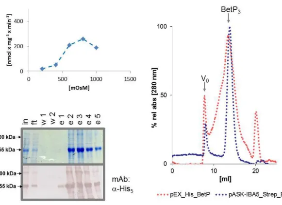
pASK-IBA5_BetP pASK-IB5 drivative encoding for StrepII_tagged BetP_C252T
(Rübenhagen et al., 2000)
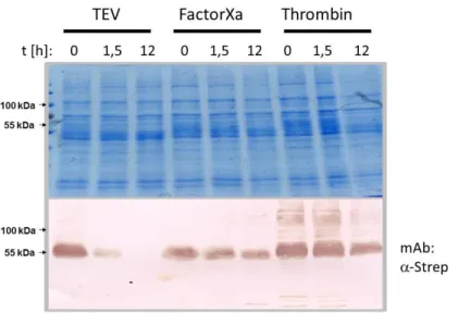
pEX pASK_IBA5 derivative with an TEV recognition
element between Nhe1 and Kas1 site
this study
pEX_His_BetP cds: His10_TEV_BetP this study
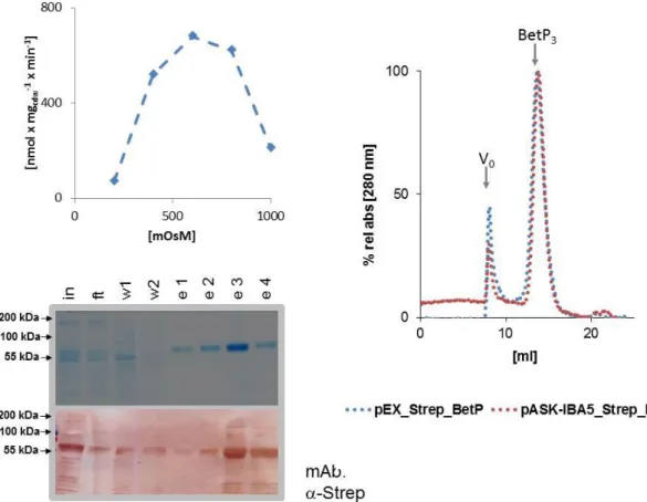
pEX_Strep_BetP cds: StrepII_TEV_BetP this study
pEX_Flag_BetP cds: Flag_TEV_BetP this study
pEX_MBP_BetP cds: maltose-binding-protein_TEV_BetP this study
pEX_CBP_BetP cds: Calmodulin-binding-peptide_TEV_BetP this study
pEX_CBD_BetP cds: Chitin-binding-domain_TEV_BetP this study
pEX_R6_BetP cds: Arginine6_TEV_BetP this study
pEX_D6_BetP cds: Aspartate6_TEV_BetP this study
pEX_GST_BetP cds: Glutathione-S-transferase_TEV_BetP this study
pEX_HIS_SSG6_BetP cds: His10_(SerSerGly)6_TEV_BetP this study
pEX_mVenus_BetP cds: mVenus_TEV_BetP this study
pEX_His_mVenus_BetP cds: His10_mVenus_TEV_BetP this study
pEX_HAT_BetP cds: Lysozyme-chelating-peptide_TEV_BetP this study
pEX_HQ_BetP cds: (HistidineGlutamine)3_TEV_BetP this study
pEX_myc_BetP cds: myc_TEV_BetP this study
pEX_Flag_BetP cds: Flag_TEV_BetP this study
pEX_S_BetP cds: RNAse1-S-fragment_TEV_BetP this study
pEXT_Strep_BetP cds: Strep_Thrombin_BetP this study
pEXF_Strep_BetP cds:Strep_FactorXa_BetP this study
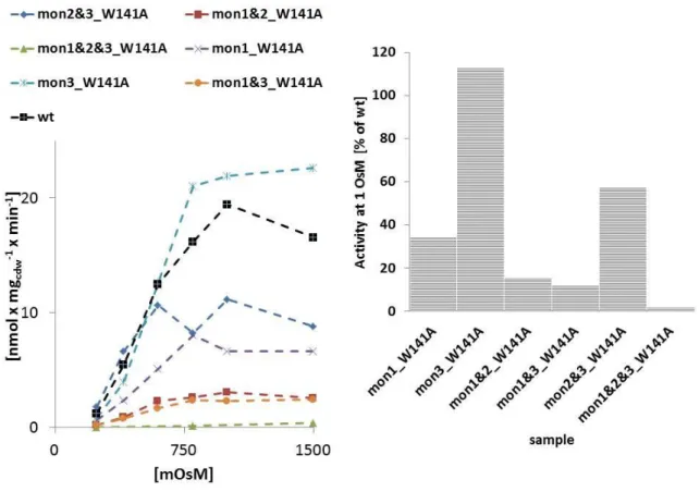
pFus cds:fusion BetP trimers with TEV cleavable linkers this study pFus_mon1_W141A pFus with W141A mutation in the first protomer this study pFus_mon2_W141A pFus with W141A mutation in the second protomer this study pFus_mon3_W141A pFus with W141A mutation in the third protomer this study pFus_mon1&2_W141A pFus with W141A mutation in the first and second
protomer
this study
pFus_mon1&3_W141A pFus with W141A mutation in the first and third protomer
this study
pFus_mon2&3_W141A pFus with W141A mutation in all three protomers this study
pFel cds: fusion BetP with elongatable linkers this study
pFel17 pFel wiht 17 mer SSG linker this study
pFel28 pFel wiht 17 mer SSG linker this study
19
pFel39 pFel wiht 17 mer SSG linker (Salman 2012)
pFel50 pFel wiht 17 mer SSG linker (Salman 2012)
pFel61 pFel wiht 17 mer SSG linker (Salman 2012)
pFel17a pFel wiht 17 mer poly-A linker this study
pFel28a pFel wiht 17 mer poly-A linker (Salman 2012)
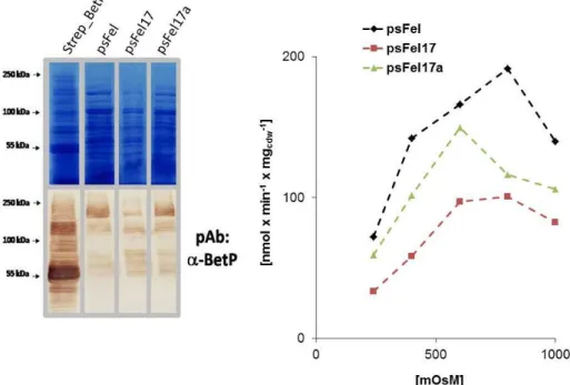
psFel cds: fusion BetP_DC12 with elongatable linkers this study
psFel17 psFel with 17 mer SSG linker (Salman 2012)
psFel17a psFel with 17 mer poly-A linker (Salman 2012)
pFel_mon1_W141A pFel with W141A mutation in the first protomer this study pFel_mon3_W141A pFel with W141A mutation in the first protomer this study pFel_mon1&2_W141A pFel with W141A mutation in the first and second
protomer
this study
pFel_mon1&3_W141A pFel with W141A mutation in the first and second protomer
this study
pFel_mon2&3_W141A pFel with W141A mutation in the first and second protomer
this study
pFel_mon1&2&3_W141A pFel with W141A mutation in all protomers this study
pC17_Strep_BetP _His cds: Strep_BetP_17merSSG_His this study
pC28_Strep_BetP _His cds: Strep_BetP_28merSSG_His this study
pC39_Strep_BetP _His cds: Strep_BetP_39merSSG_His this study
pC9_Strep_BetP_DC12_His cds: Strep_BetP_DC12_17merSSG_TEV_His this study pC9_Strep_BetP_DC12_Flag cds: Strep_BetP_DC12_17merSSG_TEV_Flag this study pC9_Strep_BetP_DC12_MBP cds: Strep_BetP_DC12_17merSSG_TEV_MBP this study pC9_Strep_BetP_Y197L/W377L/T467A
/S468A_DC12_His
pC9_Strep_BetP_DC12_His with indicated mutations in BetP
this study
pC9_Strep_BetP_Y197L/W377L/T467A /S468A_DC12_Flag
pC9_Strep_BetP_DC12_Flag with indicated mutations in BetP
this study
pC9_Strep_BetP_Y197L/W377L/T467A /S468A/A564P_DC12_His
pC9_Strep_BetP_DC12_His with indicated mutations in BetP
this study
pC9_Strep_BetP_Y197L/W377L/T467A /S468A/A564P_DC12_Flag
pC9_Strep_BetP_DC12_Flag with indicated mutations in BetP
this study
pC2_Strep_BetP_Y197L/W377L/T467A /S468A_DC45_His
pC9_Strep_BetP_DC12_His with indicated mutations in BetP
this study
pC2_Strep_BetP_Y197L/W377L/T467A /S468A_DC45_Flag
pC9_Strep_BetP_DC12_Flag with indicated mutations in BetP
this study
pC2_Strep_BetPR129A/I130A/D131S/E 132G/A133G/P134A/DE135/Y197L/W3 77L/T467A/ S468A/A564P__DC45_His
pC2_Strep_BetP with c-terminal His tag and the indicated mutations
this study
pC2_Strep_BetPR129A/I130A/D131S/E 132G/A133G/P134A/DE135/Y197L/W3
pC2_Strep_BetP with c-terminal Flag tag and the indicated mutations
this study
20
77L/T467A/ S468A/A564P__DC45_Flag
pC2_Strep_BetP_eGfP cds: Strep_BetP_6merSSG_eGfP this study
pASK-IBA5_Strep_BetP_A564P cds: Strep_BetP with indicated mutation (Ott et al. 2008) pASK-IBA5_Strep_BetP_Y197L cds: Strep_BetP with indicated mutation (Ressl et al., 2009) pASK-IBA5_Strep_BetP_W377L cds: Strep_BetP with indicated mutation (Ressl et al., 2009) pASK-IBA5_Strep_BetP_Y197L/W377L cds: Strep_BetP with indicated mutation this study pASK-IBA5_Strep_BetP_T467A/S468A cds: Strep_BetP with indicated mutation this study pASK-IBA5_Strep_BetP_Y197L/W377L/
T467A/S468A
cds: Strep_BetP with indicated mutation this study
pASK-IBA5_Strep_BetP_E577C cds: Strep_BetP with indicated mutation this study pASK-IBA5_Strep_BetP_A564P/E577C cds: Strep_BetP with indicated mutation this study pASK-IBA5_Strep_BetP_G510C cds: Strep_BetP with indicated mutation this study pASK-IBA5_Strep_BetP_G511C cds: Strep_BetP with indicated mutation this study pASK-IBA5_Strep_BetP_D512C cds: Strep_BetP with indicated mutation this study pASK-IBA5_Strep_BetP_N513C cds: Strep_BetP with indicated mutation this study pASK-IBA5_Strep_BetP_E175C cds: Strep_BetP with indicated mutation this study pASK-IBA5_Strep_BetP_L447C cds: Strep_BetP with indicated mutation this study pASK-IBA5_Strep_BetP_Q451C cds: Strep_BetP with indicated mutation this study pASK-IBA5_Strep_BetP_W101A/T351A cds: Strep_BetP with indicated mutation (Perez et al. 2011) pASK-IBA5_Strep_BetP_W101A/
F345A/T351A
cds: Strep_BetP with indicated mutation (Perez et al. 2011)
pASK-IBA5_Strep_BetP_D97C/W101A/
S328C/T351A
cds: Strep_BetP with indicated mutation (Perez et al. 2011)
pASK-IBA5_Strep_BetP_D97C/W101A/
S328C/F345A/T351A
cds: Strep_BetP with indicated mutation this study
pC2_Strep_LcoP cds: Strep_LcoP_GT this study
pC2_Strep_LcoP_C483T cds: Strep_LcoP_C483T_GT this study
pEX_Strep_ProP cds: Strep_TEV_ProP this study
pC2_Strep_LcoP_DN20 cds: Strep_DN20_LcoP_C483T_GT this study
pC2_Strep_LcoP_DN40 cds: Strep_DN_40_LcoP_C483T_GT this study
pC2_Strep_LcoP_DC40 cds: Strep_LcoP_C483T_DC40_GT this study
pC2_Strep_LcoP_DC70 cds: Strep_LcoP_C483T_DC70_GT this study
2.1.3 Oligonucleotides
Oligonucleotides used in this study are listed in table 2.3.
tab. 2-3: oligonucleotides used in this study
oligonucleotide sequence
21
EX_TEV_sense CGCGCCGGGCGGTAGCGAAAACCTCTACTTCCAGG EX_TEV_anti GCGCCCTGGAAGTAGAGGTTTTCGCTACCGCCCGG EX_Thrombin_sense CGCGCCGCTGGTTCCGCGTGGTTCTG
EX_Thrombin_anti GCGCCAGAACCACGCGGAACCAGCGG EX_FXa_sense CGCGCCGATCGAAGGCCGTACCG EX_FXa_anti GCGCCGGTACGGCCTTCGATCGG
EX_SSG6_sense CGCGCCGGGCGGTAGCTCTGGTTCTTCCGGCTCCAGCGGTTCTAGCGGCTCCA GCGGTTCTAGCGGTGAAAACCTCTACTTCCAGG
EX_SSG6_anti GCGCCCTGGAAGTAGAGGTTTTCACCGCTAGAACCGCTGGAGCCGCTAGAAC CGCTGGAGCCGGAAGAACCAGAGCTACCGCCCGG
EX_Strep_sense CTAGCGTCGACGGAGATCTTTGGAGCCACCCGCAGTTCGAAAAGGG EX_Strep_anti CGCGCCCTTTTCGAACTGCGGGTGGCTCCAAAGATCTCCGTCGACG EX_His_sense CTAGCCATCACCATCATCACCACCATCACCATCATGG
EX_His_anti CGCGCCATGATGGTGATGGTGGTGATGATGGTGATGG EX_Flag_sense CTAGCGATTATAAAGATGATGATGATAAAGG
EX_Flag_anti CGCGCCTTTATCATCATCATCTTTATAATCG EX_HQ_sense CTAGCCATCAACATCAGCACCAAGG EX_HQ_anti CGCGCCTTGGTGCTGATGTTGATGG
EX_HAT_sense CTAGCAAGGATCATCTCATCCACAATGTCCACAAAGAGGAGCACGCTCATGCC CACAACAAGGG
EX_HAT_anti CGCGCCCTTGTTGTGGGCATGAGCGTGCTCCTCTTTGTGGACATTGTGGATGA GATGATCCTTG
EX_myc_sense CTAGCGAACAGAAACTGATCTCTGAAGAAGATCTGGG EX_myc_anti CGCGCCCAGATCTTCTTCAGAGATCAGTTTCTGTTCG
EX_S_sense CTAGCAAAGAAACCGCTGCTGCGAAATTTGAACGCCAGCACATGGACTCGGG EX_S_anti CGCGCCCGAGTCCATGTGCTGGCGTTCAAATTTCGCAGCAGCGGTTTCTTTG EX_D6_sense CTAGCGATGACGATGACGATGACGG
EX_D6_anti CGCGCCGTCATCGTCATCGTCATCG EX_R6_sense CTAGCCGCCGTCGCCGTCGCCGTGG EX_R6_anti CGCGCCACGGCGACGGCGACGGCGG
EX_CBP_sense CTAGCAAGCGACGATGGAAAAAGAATTTCATAGCCGTCTCAGCAGCCAACCGC TTTAAGAAAATCTCATCCTCCGGGGCACTTGG
EX_CBP_anti CGCGCCAAGTGCCCCGGAGGATGAGATTTTCTTAAAGCGGTTGGCTGCTGAG ACGGCTATGAAATTCTTTTTCCATCGTCGCTTG
EX_mVen_sense ATGAGGCGCCATCGGAGGGCTAGCTGTGAA EX_mVen_anti GCGCGAATTCTAAAACCACCACCTCCGGAACCA EX_CBD_sense GCAGCTAGCACAAATCCTGGTGTATCCGCTTGG EX_CBD_anti ATTGGCGCGCCTTGAAGCTGCCACAAGGCAGGAAC EX_GST_sense GAGCGCTAGCTCCCCTATACTAGGTTATTGG EX_GST_anti GAGCGCTAGCTCCCCTATACTAGGTTATTGG EX_MBP_sense GCAGCTAGCAAAATCGAAGAAGGTAAACTG EX_MBP_anti CCGTTCGAGCTCGAATTAGTCTGCGCGTCT
pC_MBP_sense GATCACCGGTGAAAACCTCTACTTCCAGGGCGGCGGGGCTAGCAAAATCGAA GAAGGTAAA
pC_MBP_anti GATCAAGCTTATTTAGTCTGCGCGTCTTTCAGGGCTTCATCGACAG
pC_Flag_sense CCGGTGAAAACCTCTACTTCCAGGGCGATTATAAAGATGATGATGATAAATAA A
pC_Flag_anti AGCTTTTATTTATCATCATCATCTTTATAATCGCCCTGGAAGTAGAGGTTTTCA pC_His_sense CCGGTGAAAACCTCTACTTCCAGGGCCATCACCATCATCACCACCATCAACATC
22 ATTAAA
pC_His_anti AGCTTTTAATGATGTTGATGGTGGTGATGATGGTGATGGCCCTGGAAGTAGA GGTTTTCA
pC_eGfP_sense GATCACCGGTAGCAAGGGCGAGGAGCTGTT pC_eGfP_anti GCTCAAGCTTCTACTTGTACAGCTCGTCCATGC pFus_mon1_sense ATATGGCGCGGAGACCACTACATCTGACCCAAATC
pFus_mon1_anti GCAAGCTTGCATGGATCCGCCCTGGAAGTAGAGGTTTTCTGAGCCTCCTCGAC GCTTCCCCGCGCCACT
pFus_mon2_sense GCATGGATCCACTACATCTGACCCAAATCCGAAACCG
pFus_mon2_anti GATAAGCTTGCATGGCGCCGCCCTGGAAGTAGAGGTTTTCTGAGCCTCCTCGA CGCTTCCCCGCGCCACTCGC
pFel_mon1_sense ATAAGCTTATTGGATCCACCGCTACCGGTTCGACGCTTCCCCGCGCCACTCGC pFel_mon1_anti GCGGATCCACTACATCTGACCCAAATCCGAAAC
pFel_mon2_sense ATAAGCTTATAATCCGGAACCGCTGGTACCTCGACGCTTCCCCGCGCCACTCGC pFel_mon2_anti GCTCCGGAACTACATCTGACCCAAATCCGAA
psFel_mon1_anti ATAAGCTTATTGGATCCACCGCTACCGGTGCGTCGCTTTGCAGCCAGTTCACGC psFel_mon2_anti ATAAGCTTATAATCCGGAACCGCTGGTACCGCGTCGCTTTGCAGCCAGTTCAC
G
psFel_mon3_anti GAATAAGCTTTTAGCGTCGCTTTGCAGCCAGTTCACGCTTGCGG Fel_L2SSG_sense AAGCTCTGGTAGTTCCGGCAGCTCTGGTGGTACCAGCGGTT
Fel_L2SSG_anti CCGGAACCGCTGGTACCACCAGAGCTGCCGGAACTACCAGAGCTTGTAC Fel_L1SSG_sense CCGGCAGCTCTGGTAGTTCCGGCAGCTCCGGCACCGGTAGCGGTG Fel_L1SSG_anti GATCCACCGCTACCGGTGCCGGAGCTGCCGGAACTACCAGAGCTG Fel_L2A_sense GGCTGCAGCGGCAGCGGCTGCGGCTGCAGGTACCGCTGCAT
Fel_L2A_anti CCGGATGCAGCGGTACCTGCAGCCGCAGCCGCTGCCGCTGCAGCCGTAC Fel_L1A_sense CCGGCGCAGCTGCGGCTGCGGCAGCGGCAGCTACCGGTGCTGCAG Fel_L1A_anti GATCCTGCAGCACCGGTAGCTGCCGCTGCCGCAGCCGCAGCTGCG T467A/S468A_sense GGTACTTTCTTCATTGCCGCTGCTGACTCTGCTTCC
T467A/S468A_anti GGAAGCAGAGTCAGCAGCGGCAATGAAGAAAGTACC E577C_sense CAATGAACACCGCAAGCGTTGCCTGGCTGCAAAGCGACGCAG E577C_anti CTGCGTCGCTTTGCAGCCAGGCAACGCTTGCGGTGTTCATTG E175C_sense GTACCTGGACATGATTGCCACAATGTTGGCG
E175C_anti CGCCAACATTGTGGCAATCATGTCCAGGTAC H176C_sense CTGGACATGATGAATGTAATGTTGGCGTTGC H176C_anti GCAACGCCAACATTACATTCATCATGTCCAG N177C_sense GACATGATGAACACTGTGTTGGCGTTGCTATG N177C_anti CATAGCAACGCCAACACAGTGTTCATCATGTC L447C_sense GGATTGCTTCATGCTTGTCCAGGTGGGCA L447C_anti TGCCCACCTGGACAAGCATGAAGCAATCC Q551C_sense CACTTCCAGGTGGGTGCATCATGGGCATCATCG Q551C_anti CGATGATGCCCATGATGCACCCACCTGGAAGTG G510C_sense GCTATTGCTTTCTTGTGGTGACAATGCCTTG G510C_anti CAAGGCATTGTCACCACAAGAAAGCAATAGC G511C_sense CTATTGCTTTCTGGTTGTGACAATGCCTTG G511C_anti CAAGGCATTGTCACAACCAGAAAGCAATAG D512C_sense CTTTCTGGTGGTTGCAATGCCTTGAGCAAC D512C_anti GTTGCTCAAGGCATTGCAACCACCAGAAAG N513C_sense CTGGTGGTGACTGTGCCTTGAGCAACTTGC N513C_anti GCAAGTTGCTCAAGGCACAGTCACCACCAG
23
pEX&pC_LcoP_sense GAATGGCGCCTCCACCAACTCTGGCAATAA
pEX&pC_LcoP_anti GCGCAAGCTTTTAACCGGTATCCTTTTTCGCGTCCACTTCGCCTTC pEX&pC_ProP_sense GATAGGCGCCAGCCCGATTCGCTCAAAAAAG
pEX&pC_ProP_anti GCGCAAGCTTTTATGCGTTTTGCTTTTCAGCACCTAC LcoP_-Kas1_sense TGCGATGATGTTCGGTGCCGGCATCGGTGT
LcoP_-Kas1_anti ACACCGATGCCGGCACCGAACATCATCGCA LcoP_DN20_sense GACGGCGCCCCTCACGACACCCACCCAGGCCTAG LcoP_DN40_sense GACGGCGCCTTCGGACTCGACAAAACCGTTTT LcoP_DC40_sense GATACCGGTGTCGGTGATGTGATCAGCGGTGGAAT LcoP_DC70_sense GATACCGGTGTGTTCTTCCAAGCCACGAACCACCGCGTTAG Loop2_5´_anti GATTCCGGAGCCGGCGCCTAAGCGAATCGTGCCGAATTTAC Loop2_3´_sense GATTCCGGAGGGGCGTTTCGCACGGTGTCATGGAT
2.1.4 Growth media and cultivation conditions
The growth media used in these studies are listed in tab. 2-4. For agar plates 15 g/L Bacto- Agar (Difco, Detroit, USA) were added. If appropriate, the antibiotics chloramphenicol (Cm) or carbenicillin (Carb) were added to the growth media with a final concentration of 34 μg/mL (Cm) and 100 µg/ml (Carb). E. coli strains were generally cultivated in LB- (Lysogeny Broth) medium (tab. 2-4). During expression in fermenter cultures E. coli was cultivated in LB+ medium. C. glutamicum was grown in BHI complex medium.
tab. 2-4: Cultivation media used in this study.
Medium Ingredients
LB NaCl 10 g/L; yeast extract 5 g/L; tryptone
10 g/L
BHI Brain-Heart-Infusion 37g/L
LB+ NaCl 10 g/L; yeast extract 5 g/L; tryptone
10 g/L; 20 mM KH2PO4/K2HPO4 (pH 7,5);
Glucose 2%
BHIS Brain-Heart-Infusion 37g/L
0,6 M Sorbitol sterilzed by filtration
SOB 20 g/L Tryptone, 5 g/L yeast extract, 0.5 g/L
NaCl, 2.5 mM KCl. After heat sterilization, 5 mL 2 M MgCl2 were added.
24
2.2 Molecular biology
2.2.1 Preparation of competent E. coli cells and transformation
Preparation of chemically competent E. coli DH5 was carried out according to (Inoue et al., 1990). In brief 200 ml SOB medium were inoculated with 1 ml E. coli culture grown in LB to stationary phase. After growth for additional 18 h at 18°C and 200 rpm cells were sored on ice for 10 min. Cells were harvested by centrifugation (4000 g 10 min 4°C) and washed once in 80 ml TB buffer. After resuspension in 20 ml TB the cell suspension was supplemented with 1,4 ml DMSO and incubated for 10 min on ice. After aliquoting to 200 µl cells were flash frozen in liquid N2 and stored at -80°C until use. For transformation one aliquot was supplemented with plasmid DNA and stored on ice for 1h. Hereafter the cells were heat- shocked by insertion of the tube to an incubator set to 42°C for 45 s. The cells were chilled on ice for 5 min and supplemented with 600 µL LB. After incubation at 37°C for 1h at vigorous shaking the cells were streaked out on LB-Agar plates containing the appropriate antibiotics.
TB buffer: 10 mM Pipes, 15 mM CaCl2, 250 mM KCl, pH (KOH) = 6.7. After adjustment of pH, 55 mM MnCl2 were added. The buffer was sterilised by filtration.
E. coli MKH13 cells were transformed according to (Chung et al., 1989). 1 ml E. coli culture was grown to OD600 ~0,4. Cells were harvested and resuspended in 100 µL buffer TSS (LB;
50 mM MgCl2; 2% DMSO). Plasmid DNA was added and the cells were heat-shocked at 42°C for 45 s after incubation for 1 h on ice. The cells were chilled on ice for 5 min and supplemented with 600 µL LB. After incubation at 37°C for 1h at vigorous shaking the cells were streaked out on LB-Agar plates containing the appropriate antibiotics.
2.2.2 Preparation of electrocompetent C. glutamicum cells and transformation
Competent C. glutamicum cells were prepared according to (Liebl et al. 1989).
Electrocompetent C. glutamicum cells were transformed with approx. 400 ng plasmid DNA
25
by a 2,5 kV pulse applied with a half-value-period of typically 5 ms. Subsequently the cells were immediatly supplemented with 1 BHIS medium and streaked out on BHI-agar plates after incubation at 30°C for 2h.
2.2.3 DNA techniques
2.2.3.1 Isolation of plasmid DNA from E. coli
The isolation of plasmid DNA from E. coli cells was performed following the principle of alkaline lysis. For the isolation of plasmid DNA from these cultures, the NucleoSpin®Plasmid DNA Purification kit (Macherey-Nagel, Düren) was used as recommended by the supplier.
2.2.3.2 Gel electrophoresis and extraction of DNA from agarose gels
Gel electrophoresis of DNA was performed using 0.8 to 2 % agarose gels in 1X TAE buffer (40 mM TRIS/Acetate pH7,9; 0,5 mM EDTA) as described by (Sambrook et al. 1989). For this purpose, DNA samples were mixed with 5X Loading Dye (MBI Fermentas, St. Leon-Roth).
After electrophoresis, DNA was stained with ethidium bromide (Biosciences, Freiburg). DNA was isolated from agarose gels using the NucleoSpion® Extract kit (Macherey-Nagel, Düren) as recommended by the supplier.
2.2.3.3 Polymerase chain reaction (PCR)
The in vitro amplification of specific DNA fragments was performed by the polymerase chainreaction using the Phusion polymerase (MBI Fermentas, St. Leon-Roth) as recommended by the supplier. For this purpose, two oligonucleotides were used, flanking the DNA region to be amplified. Oligonucleotides were diluted to a concentration of 10 pmol/μL. The annealing temperature was chosen with respect to the forward and reverse oligonucleotides. For each guanine and cytosine 4 °C, for each adenine and thymine 2 °C are required to separatethe hydrogen bonds. As template, total DNA, plasmid DNA, or a cell suspension, which was diluted in H2Odd and incubated for 10 min at 95 °C, could be used.
The PCR reaction was performed using the thermocycler Mastercycler® personal or Mastercycler® gradient (Eppendorff, Hamburg). A 50 μL PCR reaction mixture was prepared as follows:
10 μL 5x HQ Buffer Phusion
26 2 µl dNTPs 5 µM each
1,5 µl DMSO
2,5 μL oligonucleotides forward [10 μM]
2,5μL oligonucleotides reverse [10 μM]
0,01-1 μL template
0,5 µl Phusion polymerase H2Odd add 50 μL
The amplification reaction was initiated by denaturing the template for 4 min at 95 °C. The following steps were performed in 30 cycles: Denaturing the DNA for 30 seconds at 95°C, hybridisation of the primers for 30 seconds at the specific annealing temperature, andpolymerisation for 1 min per kb at 72 °C. After a final incubation for 10 min at 72 °C, the samples were kept at 4 °C or -20 °C. If appropriate, the PCR product was purified either with the NucleoSpin® Extract Kit (Macherey-Nagel, Düren) as recommended by the supplier, or by gel electrophoresis.
2.2.3.4 Restriction, ligation, and sequencing of DNA
For restriction of DNA, restriction enzymes were used as recommended by the suppliers (MBI Fermentas, St. Leon-Roth). After restriction DNA was purified either with the NucleoSpin® Extract kit (Macherey-Nagel, Düren) following the suppliers recommendations or by gel electrophoresis. Ligations were carried out with the use of T4-DNA-Ligase (MBI Fermentas, St. Leon-Roth) as recommended by the supplier.
For the cloning of small fragments (<80 bp) the respective 3´-5´and 5´-3´DNA fragments were purchased from MWG Operon and ligated immediatly after annealing reaction. The annealing reaction was carried out by mixture of the two oligonucleotides and heating to 95°C with subsequent chilling in steps of 5°C for each 1min.
2.2.3.5 Site directed mutagenesis
For site directed mutagenesis oligonucleotides containing the appropriate mutations flanked by 15 bp of correct template sequence were ordered and the whole plasmid template sequence was amplified in a common PCR reaction. After digest of the parental methylated DNA via Dpn1 (MBI Fermentas, St. Leon-Roth) the amplified vectors were