EPR-based structural and functional
characterization of the C-terminal domain of the osmoregulated glycine betaine
transporter BetP from Corynebacterium glutamicum
Inaugural-Dissertation zur
Erlangung des Doktorgrades
der Mathematisch-Naturwissenschaftlichen Fakultät der Universität zu Köln
vorgelegt von
Sascha Carsten Thomas Nicklisch aus Köln
Köln, Oktober 2008
Berichterstatter:
Prof. Dr. Reinhard Krämer Prof. Dr. Günter Schwarz
Tag der Disputation: 01.12.2008
“Der Mensch hat dreierlei Wege, klug zu handeln: erstens durch Nachdenken, das ist der edelste; zweitens durch Nachahmen, das ist der leichteste und drittens durch Erfahrung, das ist der bitterste.”
(Konfuzius)
Abstract
As a Gram-positive soil bacterium, Corynebacterium glutamicum regularly encounters various kinds of stresses that threaten the cell’s survival. Beside an alternating pH and temperature, the cells are faced with changes in the osmolarity of the external medium.
Recently, it was shown that the secondary glycine betaine uptake system BetP of this bacterium is able to autonomously sense a hyperosmotic stress induced cytoplasmic accumulation of potassium as specific stimulus (osmo-/chemosensor). Subsequently, the carrier is able to regulate its activity in response to the extent of the osmotic stress it is exposed to (osmoregulator). Further studies revealed that changes in the structure and/or orientation of the cytoplasmically exposed C-terminal domain of the carrier seem to be critically involved in stimulus sensing and/or signal transduction. Since the molecular mechanisms related to these processes are barely understood to date, in the present work Site-Directed Spin Labelling-Electron Paramagnetic Resonance (SDSL-EPR) spectroscopy was applied for the first time to probe the molecular dynamics during BetP function. Focus was on the structure and structural changes of the C-terminal domain or adjacent protein and/or lipid domains. Strategically engineered BetP mutants with single cysteine substitutions at the beginning (545), in the centre (572) and close to the end (589) of the C-terminal domain were tested for sustained transport activity as well as for optimal labelling and reconstitution into E. coli lipids to obtain enough highly purified material for EPR analysis. Absorption of about 90% of spin labelled BetP by Bio-Beads was observed when the standard reconstitution procedure was used. We concluded that at least the C-domain is apparently involved in this process. A new procedure for the reconstitution of the carrier into E. coli liposomes was established with about 6-7times higher protein recovery compared to the standard Bio-Bead method. SDSL-EPR revealed that the structure/conformation of the whole C-terminal domain seem to be influenced by the incorporation into E. coli lipids. The activation of BetP mutants by hyperosmotic stress showed at least at the terminal part of the C-domain a certain mobility effect, indicative of a structural/conformational change. However, the extent of this mobility effect was strongly dependent on the nature (osmotic and/or ionic strength) of the used osmolyte.
The preliminary distance measurements of single and double spin labelled BetP variants confirmed an oligomeric state (e.g. trimer) of BetP in detergent as well as reconstituted into liposomes. In addition, a slight effect of a proline substitution in a deregulated triple mutant (BetP-S545C/Y550P/S589C) on the structure of the C-terminal domain was observed.
The data provided in this work are combined in a current model outlining possible
C-terminal structures and dynamic motions upon BetP activation.
Kurzzusammenfassung
Als Gram-positives Bodenbakterium ist Corynebacterium glutamicum regelmäßig diversen Stresssituationen ausgesetzt, die das Überleben der Zelle beeinträchtigen können. Neben wechselnden pH- und Temperatur-Werten, werden die Zellen auch mit Osmolaritätschwankungen des umgebenden Mediums konfrontiert. Es konnte gezeigt werden, dass das sekundäre Glycinbetain Aufnahmesystem BetP aus diesem Bakterium in der Lage ist, den unter hyperosmotischen Bedingungen intrazellulär erhöhten Kaliumgehalt als spezifischen Stimulus autonom zu detektieren (Osmo-/Chemosensor) und seine Aktivität daraufhin an das Ausmaß des Stresses anzupassen (Osmoregulator).
Weiterführende Untersuchungen zeigten, dass Änderungen in der Struktur und/oder der
relativen Orientierung der C-terminalen Domäne des carrier einen starken Einfluss auf die
Stimulusdetektion und/oder die nachgeschaltete Signaltransduktion haben. Da die
molekularen Mechanismen solcher sensorischen Eigenschaften von Transportproteinen
bislang nur unzureichend untersucht und verstanden sind, wurde in der vorliegenden
Arbeit eine Kombination aus ortspezifischer Spinmarkierung (site-directed spin labelling,
SDSL) und Elektronenspinresonanzspektroskopie (ESR) angewendet, um die molekulare
Dynamik während des Aktivierungsprozesses von BetP zu untersuchen. Im Fokus lagen
dabei zum einen die tertiäre Struktur des Transporters als auch die Aufklärung von
strukturellen Änderungen der C-Domäne sowie deren mögliche Interaktion mit
angrenzenden Protein- und/oder Lipid-Bereichen im aktivierten Protein. Strategisch
eingeführte Cystein-Reste zu Beginn (545), in der Mitte (572) und nahe dem Ende (589)
der C-Domäne wurden auf ihre jeweilige Aktivitätsregulation hin überprüft. Darüber hinaus
wurden die Markierungs- und Rekonstitutionsschritte für jede Mutante optimiert, um eine
hohe Ausbeute an spinmarkiertem, gereinigtem Material für die EPR-basierten Analysen
zu erhalten. Es konnte eine hochgradige Absorption von BetP durch den direkten Kontakt
zu den Bio-Beads (bis zu 90%) während des herkömmlichen Rekonstitutions-Assay
identifiziert werden. Daraufhin wurde eine neue Rekonstitutionsmethode für den
verlustfreien Einbau von spinmarkiertem BetP Protein in E. coli-Liposomen etabliert,
deren Effizienz um etwa 60% höher lag als mit der herkömmlichen Methode. Die SDSL-
ESR-Studien zeigten zum einen, dass der Einbau von solubilisiertem BetP in E. coli-
Liposomen einen ausgeprägten Einfluss auf die Konformation und/oder räumliche
Orientierung der C-Domäne hat. Eine hyperosmotisch induzierte Aktivierung des
Transportproteins zeigte zudem eine erhöhte spinlabel-Mobilität am Ende der C-Domäne
auf, die auf strukturelle Änderungen während des Aktivierungsprozesses hinwies. Das
Ausmaß der Mobilisierung war dabei maßgeblich von der Art (Ionenstärke, Osmolalität)
des benutzten Osmolytes abhängig. Die vorläufigen Ergebnisse der Abstandsmessungen
von Einzel- und Doppelcystein-Mutanten bestätigten einen oligomeren Zustand (z.B.
Trimer) von solubilisiertem und in E. coli-Liposomen eigebautem BetP. Darüber hinaus konnte gezeigt werden, dass der zusätzliche Einbau eines Prolins in einer deregulierten Dreifachmutante BetP-S545C/Y550P/S589C einen Einfluss auf die Struktur der C-terminalen Domäne im nicht aktiven Zustand des Proteins hat.
Die in der vorliegenden Arbeit gewonnenen Daten konnten in einem aktuellen Topologie-
Modell kombiniert werden, das die möglichen Konformationen und die Dynamik der
C-terminalen Domäne während des Aktivierungsprozesses von BetP zusammenfasst.
Contents
1. INTRODUCTION... 1
1.1. Corynebacterium glutamicum ... 1
1.2. Osmosis and osmotic properties of a cell ... 3
1.3. Measurement of osmotic pressure... 4
1.4. Hypoosmotic stress in microorganisms ... 5
1.5. Hyperosmotic stress and response in microorganisms ... 6
1.6. Osmosensing and osmoregulation ... 9
1.7. Osmoregulated uptake systems ... 9
1.8. Solute uptake systems in C. glutamicum ... 11
1.9. Glycine betaine transporter BetP from C. glutamicum ... 13
1.10. EPR (Electron Paramagnetic Resonance) ... 19
1.11. Objectives of this thesis ... 21
2. MATERIALS AND METHODS ...23
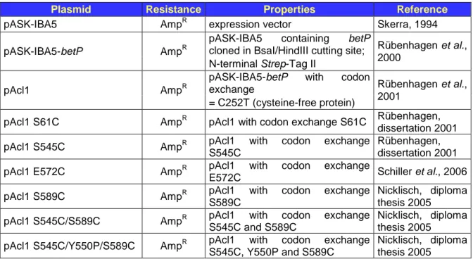
2.1. Bacterial strains and plasmids ... 24
2.2. Growth media and cultivation conditions ... 24
2.3. Molecular biological approaches ... 26
2.4. General analytical approaches ... 28
2.5. Biochemical approaches ... 32
2.6. EPR measurements ... 39
3. RESULTS ...41
3.1. Substitution mutants and strategic labelling ... 43
3.2. Activity regulation of BetP variants in E. coli lipids ... 45
3.3. Optimization of the reconstitution process ... 48
3.4. Mobility profiles of C-terminal BetP variants ... 52
3.5. Distance measurements with BetP variants ... 65
4. DISCUSSION ...74
4.1. Activity regulation of BetP variants in E. coli lipids ... 75
4.2. Optimization of the reconstitution process ... 77
4.3. Mobility profiles of C-terminal BetP variants ... 79
4.4. Distance measurements with BetP variants ... 88
4.5. Future aspects ... 95
5. SUMMARY ...97
6. REFERENCES ...99
7. APPENDIX... 113
Abbreviations
ABC ATP-binding cassette
AHT Anhydrotetracycline
Amp
RResistance towards ampicillin
AP Antarctic phosphatase
BCCT family Betaine-choline-carnitine-transporter family BCIP 5-bromo-4-chloro-3-indoyl phosphate
bp Base pairs
BSA Bovine serum albumin
CAPS N-cyclohexyl-3-aminopropanesulfonic acid CrOx Chromium-(III)-oxalate (Cr(C
2O
4)
3−)
cysless = cysteine-less (mutant protein lacking any native cysteine) DDM n-dodecyl-β-D-maltopyranoside
DEER Double electron-electron resonance
DMSO Dimethylsulfoxide
DNA Desoxyribonucleic acid
dNTP Desoxynukleosidtriphosphate
DPG Diphosphytidylglycerol
DOPC 1,2-Dioleoyl-sn-Glycero-3-Phosphocholine DOPG 1,2-Dioleoyl-sn-Glycerol-3-Phosphoglycerol
DTB Desthiobiotin
DTT Dithiothreitol
DW (cell) Dry weight
EDTA Ethylendiaminetetraacetic acid EPR Electron paramagnetic resonance
ESE Electron spin echo
ESR Elektronenspinresonanz
et al. et alii (latin = “and others”)
FRET Fluorescence resonance energy transfer g Gravitational acceleration (9.81 m/s
2)
GB Glycine betaine
H
2O
ddDouble-distilled water, “bidest. water”
HABA 4-Hydroxy-azobenzene-2-carboxyl acid
HFS Hyperfine splitting
kb Kilo base pairs
kDa Kilo Dalton
K
MMichaelis-Menten constant
KP
iPotassium phosphate buffer
LB medium Luria Bertani medium
LPR Lipid-protein ratio
MSC Mechanosensitive channel
MFS Major facilitator superfamily
MTSSL (1-oxyl-2,2,5,5-tetramethylpyrroline-3-methyl)-methanethiosulfonate NBT Nitro-blue tetrazolium chloride
NaP
iSodium phosphate buffer NiEDDA Ni(II) ethylenediaminediacetate OD
600Optical density at 600nm
PAGE Polyacrylamide gel electrophoresis
PCR Polymerase chain reaction
PE Phosphatidylethanolamin
PEG Polyethyleneglycol
PG Phosphatidylglycerol
PVDF Polyvinylidine diflouride
RT Room temperature
SDS Sodium dodecyl sulphate
SDSL Site-directed spin labelling SHFS Superhyperfine splitting
SSS Sodium/Solute Symporter
tetA Tetracycline resistance gene
TCA Trichloroacetic acid
TEMED N,N,N’,N’-tetramethyl-ethylendiamine
TM Transmembrane
TRIS 2-amino-hydroxymethylpropane-1,3-diol o/n culture Overnight culture
RPM Rounds per minute
V
maxMaximal uptake rate
1. Introduction
Beside fluctuations in temperature, pH or nutrient depletion, most microorganisms have to deal with alternating osmolalities (water scarcity or surplus) – so called osmotic stress - in their natural habitat (Gross et al., 1996, Desmond et al., 2002). A stress emerges, when a changing environmental factor influence the growth or even survival of an affected organism. To overcome or bypass these deleterious circumstances, various survival strategies have been employed by bacteria (Hecker et al., 1996, Hecker et al., 2001). For this purpose, microorganisms have to sense outside stimuli and subsequently respond to sudden environmental changes with appropriately regulated gene expression and protein activity to ensure survival and cell proliferation.
1.1. Corynebacterium glutamicum
As an immobile, topsoil (= mineral horizon A;
Allaby, 1994) bacterium Corynebacterium glutamicum is regularly exposed to an ever- changing environment. It was first described 1957 as glutamate-producing strain Micrococcus glutamicus (Kinoshita et al., 1957, Udaka, 1960).
C. glutamicum is a Gram-positive, non- pathogenic, facultative-anaerobic and non- sporulating bacterium that possesses a club- (greek „coryne“= club) or rod-shaped cell morphology referred to as coryneform bacteria (Figure 1). With a G/C content of 46-74mol-% Corynebacteria belong to the big group of high-G/C-containing bacteria forming the class Actinobacteria (Figure 3; Funke et al., 1995; Abe et al., 1967). Among these, the heterogeneous order of Actinomycetales - which includes the Mycobacteria, Nocardia and the Corynebacteria - is characterized by a unique cell wall structure (Stackebrandt et al., 1997; Daffé et al., 1998; Daffé, 2005). This so-called mycolic acid layer (mycolate layer) is bearing on the top of the normal peptidoglycan (murein) and arabinogalactan layer (Figure 2). In addition to the plasma membrane it provides a second permeability barrier, containing channel-forming proteins, the porins, which facilitate the diffusion of hydrophilic substances (Nikaido, 1994, 2003;
Lichtinger et al., 1998; Puech et al., 2001). Due to its close phylogenetic relationship to pathogenic bacteria like Mycobacterium tuberculosis, Mycobacterium leprae or Corynebacterium diphtheria, as common germs for severe illnesses and diseases, C. glutamicum is still in the focus of medical investigations as a non-pathogenic model
Figure 1: Scanning electron micrograph of C. glutamicum ATCC13032 WT (N. Möker, 2002).
organism. Today, particular attention is paid on the bacterium due to its utmost importance for large-scale production of amino acids.
Among these are
L-glutamate and L-lysine with current production rates of 1.500.000 tons/year and 550.000 tons/year, respectively (Kataoka et al., 2006, Hermann, 2003).
However, during fermentation cells are subjected to various kinds of stress. One of the stresses concerned is a hyperosmotic environment due to the accumulation of high substrate or product concentrations in the external medium (Kawahara et al., 1997; Varela et al., 2004). Thus, the investigation of the physical response of cells to encounter these unfavourable situations is of great importance for the economic sense of industrial production.
oBacteria (eubacteria) oActinobacteria (class)
oActinomycetales (order)
oCorynebacterineae (suborder) oCorynebacteriaceae
oMycobacteriaceae CMN-group
oNocardiaceae oGordoniaceae oTsukamurellaceae oWilliamsiaceae
Figure 3: Taxonomic lineage for Corynebacteriaceae drawn up with NCBI Taxonomy Browser (http://www.ncbi.nlm.nih.gov/Taxonomy/). The main representatives within the suborder of Corynebacterineae are summarized to the so-called CMN-group (Corynebacteria, Mycobacteria, Nocardia).
Peptidoglycan (murein) Arabinogalactan Mycolate layer
Cytoplasmic membrane Outer layer
Mycolic acids
Glycolipids
Phospholipids
Porins
Integral membrane proteins
Peripheral membrane proteins
Figure 2: Schematic illustration of the cell wall composition of the taxonomic order Actinomycetales (Figure 3).
1.2. Osmosis and osmotic properties of a cell
Osmotic (greek “osmos” = penetration, push, drive) stress is
a common challenge
encountered by eukaryotic and prokaryotic cells. The semipermeability of the cytoplasmic membrane is responsible for the influence of the external osmolarity on the bacterial cell, because it retains macromolecules, ions and polar substances but allows free diffusion of water in both directions, known as the fundamental principle of osmosis (Figure 4; Bovell, 1963).
The chemical potential of water – also referred to as water activity
ψW– is a sum of both, the solute potential
ψS(osmotic potential) and the pressure potential (
ψP, turgor pressure) of a given solution:
(1)
ψW=
ψS+
ψP(water activity)
The water activity
ψwquantifies the tendency of water to move from one area to another due to osmosis: The lower the overall water activity, the higher the potential to attract water and vice versa. The turgor pressure
ψPis a mechanical, outward-directed pressure exerted from the plasma membrane on the cell wall of plants and bacteria and can be positive (tension) or negative (suction power). In most bacteria the cell turgor is essential for growth and cell division and reaches values up to 15-25atm in Gram-positive bacteria, whereas in Gram-negative organisms it ranges from 1-5atm (Koch, 1983; Poolman and Glaasker, 1998). The solute potential (
ψS, osmotic potential), however, is always negative and can be described by Van’t Hoff’s equation for diluted solutions (<100mM):
(2) ψS
= - (R * T * c) (solute potential)
With R as the ideal gas constant, T as the absolute temperature in °K and c as the total solute concentration.
Hence,
ψSis directly proportional to the absolute temperature and the concentration of dissolved particles (colligative property) in a given solution and decreases with a rising amount of solutes. In reference to equation (1), water flows through a cytoplasmic
Hydrostatic pressureψP
Osmosis
Pure water Solution
Semipermeable membrane
∆t
Figure 4: Schematic illustration of osmosis. Pure water (high water activity, ψW) diffuses through a semi permeable membrane to a solution with lower water activity generating a hydrostatic pressure (ψP). ∆t = time difference.
membrane towards a lower chemical potential until both, the solute potential
ψSand the turgor pressure
ψPare equal (Cosgrove, 2000):
(3)
ψW=
ψS+
ψP= 0, if
ψS=
ψP(full turgescence)
Due to the fact, that the cytoplasm is an aqueous solution supplied with salts, sugars, amino acids and other chemicals compounds, the water activity
ψWof living cells under physiological conditions is more negative than that of the surrounding medium, leading to an inward directed water flux that maintains the crucial cell turgor. Fluctuations in the external osmolarity, on the other hand, may reduce the cell’s water activity and hence increase its turgor pressure (hypoosmotic stress) or on the contrary decrease the cell’s
ψP(hyperosmotic stress). According to equation (1) and the colligative property of
ψS(2), the overall water activity of a cell is mainly determined and can be adjusted by the internal solute pool (osmoregulation). Bacteria and plants thereby have to be able to tightly regulate their internal solute concentrations to ensure viable cell functions and reproduction (Kempf and Bremer, 1998; Chater and Nikaido, 1999).
1.3. Measurement of osmotic pressure
As a prerequisite for studies on bacterial osmoregulation and osmosensing it is important to be able to measure the osmotic pressure imposed by growth media and supplements.
For this purpose, the water activity can be linked to the osmotic pressure
Πby the relation:
(4)
Π= - (R * T / V
W) * ln
ψW(osmotic pressure)
Here, V
Wis the partial molar volume of water and measured in [m
3* mol
-1] = [J * m
-3] = [N * m
-2], so in pressure units.
From equation (4) the osmolarity of a given solution can be derived as the sum of the molar concentrations of all osmotically effective particles (
Σc):
(5) Osmolarity [mol/L] =
Σc ~
Π/ R * T
The osmolarity can be calculated but not measured. That’s why in practical approaches the osmotic pressure per weight of solvent, the osmolality, is used:
(6) Osmolality [osmol/kg] =
Π/ R * T
The osmolality in turn can be measured, but not calculated because the different properties of a particular solute (e.g. charge, size, form) are included into the total water potential as single-potentials and affect it to different extents. So, the osmolality is merely an approximation for the osmolarity of a given solution, however simply quantifiable for investigations on microbial adaptation to osmotic stress situations.
1.4. Hypoosmotic stress in microorganisms
A hypoosmotic (greek “hypo“= below, under) stress occurs for living cells when the external osmolality is decreased, e.g. the water activity of the external medium is higher in relation to the cytoplasm of the cell. This causes an instant influx of water and an increase in cell turgor. Although the cell walls of Gram-negative bacteria can sustain pressures up to 100atm, the permanent increase of
ψPwould inevitably lead to cell burst (Csonka, 1989; Carpita et al., 1985). To overcome this unfavourable situation, microorganisms have to dispose internal solutes to even out the differences in chemical potential and thus leading to a decrease in turgor.
For this purpose, specific transporters and mechanosensitive channels (Mscs) as release
valves are rapidly activated to reduce the driving force for water entry (Anishkin and Kung,
2005; Hamill and Martinac, 2001; Csonka and Epstein, 1996). Mscs are ubiquitous among
prokaryotes, unspecific and have different pore sizes as well as different sensitivities
towards the applied mechanical stress that activates them (Berrier et al., 1992). They all
have in common, that their activity is directly controlled via changes in the lateral tension
of the membrane as a response to changes in the external osmolality (downshift). Upon a
change from high to low osmolalities, the open probability of these channels increases by
several orders of magnitude as a function of the coupling mechanism between protein
conformation and membrane stretch (pressure sensitivity). Thereby, the respective
channels work cooperatively: upon hypoosmotic stress the channel with the lowest
conductance opens first, sequentially followed by the channels with higher conductance to
provide a graduation of efflux response (Martinac 2001; Bezanilla and Perozo, 2002). In
this context, the best studied efflux channels so far are the two species MscL
(MechanoSensitive Channel of Large conductance) and MscS (Small) from E. coli, with
conductances of 3nS and 1nS, respectively (Levina et al., 1999; Sukharev et al., 1993,
1999). In addition, a third and yet barely studied MscM (Mini) channel (~0.1nS) opens as
first response under hypoosmotic conditions and - with rising pressures - additionally the
MscS channel (Berrier et al., 1996). At turgor pressures causing a tension sufficient to
rupture the membrane, also the MscL channel opens. Whereas single deletion mutants
lacking the gene for MscS (yggB) or MscL (mscL) remain fully functional after applying a
hypoosmotic shock, the respective double mutants die, indicating the physiological
importance of both channels and implying that MscM alone is not able to protect E. coli during an osmotic downshift (Sukharev et al., 1994; Levina et al., 1999; Booth and Louis, 1999).
Homologues of MscL and MscS could also be identified in Mycobacterium tuberculosis (Chang et al., 1998) and in Corynebacterium glutamicum (Nottebrock et al., 2003). In case of C. glutamicum, the MscL- and MscS-channel seem to be responsible for the specific efflux of glycine betaine, proline and – to a less extent – of some cations (Ruffert et al., 1997, 1999). However, the respective double deletion strain lacking the genes for both channels still showed a hypoosmotically induced glycine betaine efflux, arguing for at least one additional MS channel (Nottebrock et al., 2003).
1.5. Hyperosmotic stress and response in microorganisms
During a hyperosmotic (greek “hyper“ = above, excess) stress the osmolality of the external medium is increased and water flows out of the cell, thereby changing the cell’s hydration, volume and/or turgor pressure. The consecutive water efflux inevitably leads to plasmolysis and growth slowdown or even a stop of growth. As a response, cells have developed a variety of mechanisms to restore cell turgor and balance water stress.
According to Wood (1999) this response can be at least divided in three overlapping phases: (1) an instant but passive dehydration of the cytoplasm (seconds-minutes), (2) an active process of rehydrating the cytoplasm by accumulation of ions or osmolytes (up to an hour) and (3) a remodelling of the cell by changes of gene expression profiles (up to one or more hours).
As an initial step during the rehydration phase, many bacteria transiently and rapidly accumulate potassium (Dinnbier et al., 1988; Whatmore and Reed, 1990; Whatmore et al., 1990). Potassium is a widespread ion in nature and the dominant cation in the cytoplasm of microorganisms with amounts of 100-600mM (Ballal et al., 2007; McLaggan et al., 1994). The increase in internal potassium can be achieved by both, passively via hyperosmotic-induced water efflux out of the cells and actively via specific potassium uptake systems. In E. coli active accumulation is mediated by the low affinity and low capacity Kup system as well as the high efficient transporters Kdp and TrK as the main transport systems for potassium (Bakker, 1993; Schlösser et al., 1995). Recently, two potassium uptake systems were identified in C. glutamicum: a Kup system with similarities to the cation/proton symporter Kup from E. coli and a potassium channel (CglK) with striking similarity to MthK from Methanobacterium thermoautotrophicum (Becker, 2007).
Preliminary results with knock-out deletion strains indicated, that CglK is the major
constituent for potassium uptake in C. glutamicum (Becker, 2007; personal
communication, M. Becker). However, both systems could not yet be further characterized biochemically or kinetically. Also, there is no evidence for a specific activation of potassium uptake systems in C. glutamicum upon hyperosmotic stress. To maintain electroneutrality and to prevent alkalisation in E. coli and C. glutamicum cells, the potassium accumulation is accompanied by the synthesis of glutamate as a counterion, whereas in B. subtilis the nature of the counter ion still remains elusive (Caylay et al., 1991; McLaggan et al., 1994; Morbach and Krämer, 2002; Whatmore and Reed, 1990). In addition, the internal nucleic acid counterion putrescine or other protons are exchanged by potassium and exported (Csonka, 1989; Munro et al., 1972).
In contrast to (extreme) halophiles with their common “salt-in” strategy, moderate osmotolerant bacteria use a “organic- solute-in” strategy in a second phase of the hyperosmotic response to prevent chaotropic effects during the adaptation phase (Oren 2006, 2008). In this regard, the internal charge accumulation (potassium, glutamate) leads to aggregation of cellular macromolecules and thus may interfere with the metabolism (Wood, 1999). To counteract this effect, the ions are exchanged against neutral, so- called compatible solutes either by uptake or biosynthesis (Csonka, 1989; Galinski and Trüper, 1994). These osmoprotectants are characterized by being compatible with normal physiological functions of the cell while accumulated to very high intracellular levels up to several moles per litre (Braun, 1997; Arakawa and Timasheff, 1985; Timasheff, 1991). In addition, they are neutral or zwitterionic at physiological pH and soluble up to molar concentrations (Csonka, 1989). Depending on their molecular structure, these compounds can be subdivided into (i) amino acids and derivatives (e.g. glutamate, proline, ectoine, and glycine betaine), (ii) polyols (e.g. glycerol, glycosylglycerol), (iii) sugars (e.g.
trehalose, sucrose) and (iv) others (e.g. carnitine, choline-O-sulphate). Among these, glycine betaine, ectoine, proline and trehalose are the most abundant and thus most effective osmoprotectants in the domain Bacteria (Oren, 2008; Figure 5).
N H
+ C
H CH2 C H2 N H
C H3
O
O Ectoine
NH2+ CH C H2
C H2 C H2
O
O N+
C H2
O O CH3 C
H3 CH3
O O CH2
OH O H
OH
OH O OH
OH OH
C H2 OH Glycine betaine
Trehalose Proline
Figure 5: Chemical structure of selected compatible solutes.
Beside their function as osmoprotectants, these organic solutes have the intrinsic property to stabilize the native conformation of proteins. This attribute can be explained by the model of
“preferential exclusion” proposed by Arakawa and Timasheff (1985). The model indicates that compatible solutes are preferentially excluded from the hydration shell of a protein, leading to stronger interactions of the water molecules with the protein surface (preferential hydration, Figure 6). The stabilizing effect of the organic solutes thereby derives from the different affinities towards the native and denatured status of the protein: in a denatured protein the lipophilic peptide backbone is exposed, leading to thermodynamically unfavourable interactions with the predominantly hydrophilic compatible solutes which in turn promote the native protein conformation. One mechanism for preferential exclusion is the “sterical exclusion” that occurs, when the radius of the compatible solute is considerably bigger than that of the water molecule leading to a tight solvation shell around the protein (Arakawa and Timasheff, 1985).
A second mechanism deals with the increased surface tension of the respective solvation shell due to the inhomogeneous distribution of the solvents within the cytoplasm (according to Gibbs isotherm). The denaturation of a protein thereby leads to an increased free surface energy - which is the product of surface tension multiplied by the overall surface – leading to an unfavourable energy imbalance compared to the native state. This energy imbalance is reduced, when the protein (and its hydration shell) adopts a small volume or overall surface, so to say the native conformation (Timasheff, 1998).
Since the uptake of compatible solutes is more favourable in terms of energy and carbon cost as compared to biosynthesis, the activation of specific uptake systems for compatible solutes is in general the first response (short-term response) to hyperosmotic conditions.
For the sake of long-term adaptation, enzymes for de novo synthesis of compatible solutes and for transporters get additionally regulated at the level of gene expression.
However, if compatible solutes are not present in the environment, the bacterium is forced to exclusively synthesize its osmoprotectants. C. glutamicum thereby uses several pathways to synthesize glutamine (Frings et al., 1993; Rönsch et al., 2003), proline (Ankri et al., 1996; Ley, 2001) and trehalose (Wolf et al., 2003) after an osmotic upshift.
S S S
S S
S S S S
S
S
S
S S
S S
S
S
S S
S
S
S S
S
S S
S
S S
S
S
S
S S
S
S S
S
S
S S
S
S
S
S S
S S
H2O
H2O H2O
Figure 6: Schematic model of the
“preferential exclusion” of compatible solutes leading to a preferential hydration of the protein (according to the basic idea from Timasheff, 1998 and Lee, 2000). Blue = hydration shell of H2O, pink = solute shell.
1.6. Osmosensing and osmoregulation
Most bacteria possess uptake systems in their membrane which mediate the immediate accumulation of compatible solutes upon an osmotic upshift of the external medium. These systems have to be tightly-regulated on the level of protein activity as well as at the level of gene expression to ensure a proper adaptation to varying hyperosmotic stress (osmoregulation). Thus, as a basic prerequisite for an efficient osmoregulation, cells have to exhibit sensitive receptor systems which either directly or indirectly perceive the encountered stress (osmosensors). The detected stimulus has then to be converted and transmitted to a cell intrinsic osmoregulatory network that finally regulates the catalytic activity of its transport systems and/or biosynthesis pathways depending on the extent of the emerged stress. According to this, the elucidation of bacterial osmosensory and osmoregulatory mechanisms relies on the identification and characterization of osmosensory transporters, their encoding genes as well as the solutes and ions that serve as substrates and cosubstrates, respectively. For this purpose, current investigations focus on the measurement of the respective transport activity under (hyper-) osmotic conditions in vivo with intact cells and in vitro with inverted membrane vesicles (IMV) or artificial lipid vesicles reconstituted with purified transporters (proteoliposomes, Figure 7). Referring to the latter, this artificial membrane system provides the major opportunity to manipulate a set of conditions (e.g. the internal and external buffer, lipid and protein composition/ratio, liposome size, etc.) that facilitates the discrimination of activating stimuli (e.g. turgor pressure, membrane strain, internal and external osmolarity/ionic strength/ion concentration, macromolecular crowding) or substrate and cosubstrate specificity (Wood, 2007; Papahadjopoulos, 1978; Olsen et al., 1979).
1.7. Osmoregulated uptake systems
Since the import of solutes against their electrochemical gradient requires metabolic energy, primary (ATP-Binding Cassette, ABC) and secondary (ion-linked) transporters are commonly used as uptake systems for osmoprotectants (Bremer and Krämer, 2000;
Wood, 1999; Wood et al., 2001). Among these, ProP of Escherichia coli and BetP of Corynebacterium glutamicum are the best studied secondary uptake systems in which the solute transport is coupled to the electrochemical gradient of a cosubstrate. One of the
lumen
Figure 7: Schematic illustration of a closed lipid vesicle system reconstituted with purified transport proteins (proteoliposomes).The internal compartment (= lumen, blue) is separated via the lipid bilayer with its incorporated membrane proteins (green).
best studied ABC transporter in terms of activity regulation upon hyperosmotic stress is OpuA from Lactococcus lactis in which the transport is directly coupled to ATP hydrolysis as the driving force. These prototypical osmosensory transporters are characterized by detecting changes in osmotic pressures and respond by mediating the uptake of compatible solutes without the assistance of other proteins. In this context, great efforts are currently made on revealing the detailed molecular mechanism of osmostress detection and signal transduction within these autonomously working carrier proteins.
The ABC transporter OpuA from L. lactis facilitates the unidirectional transport of glycine betaine at the expense of two molecules ATP (Van der Heide et al., 2000; Patzlaff et al., 2003). The protein consists of 573 amino acid residues forming eight
α-helical transmembrane domains and a binding protein domain oriented to the periplasm. Its initial uptake rates increase with a rising medium osmolality (osmoregulator), whereas the osmolality threshold for the transporter activation increases with increasing amount of anionic lipids in the membrane fraction of proteoliposomes (Van der Heide et al., 2001). In addition, the rapid activation of OpuA seems to be indifferent to the nature of the triggering ion, although divalent ions are more effective than monovalent ions. To this respect and due to the fact that the ionic strength of a solution varies as the square of ion charge (equation (7); Wood, 2007), OpuA was proposed to be a ionic strength sensor (Biemans- Oldehinkel et al., 2006; Van der Heide et al., 2001).
(7) I = 1 / 2 *
Σ(m
i* z
i2) (ionic strength of a solution)
With i = amount of ions, m
i= ion molalities (moles/kg solvent) and z
i= ion charges.
Accordingly, the proposed model for the osmosensing mechanism of OpuA is based on electrostatics: under physiological conditions intracellular tandem CBS (Cystathionine-
β- Synthase) domains of OpuA are interacting with the surrounding membrane surface.
Upon hyperosmotic shock, the internal ionic strength rises and the competing ions release the CBS domains from the membrane thereby activating the transporter (electrostatic switch). In addition, the C-terminal stretch of anionic residues serves as a modulator reducing the ionic strength threshold for activation via electrostatic repulsion with the predominantly anionic membrane surface (Biemans-Oldehinkel et al., 2006).
In E. coli a member of the Major Facilitator Superfamily (MFS), ProP, mainly catalyzes the uptake of proline, glycine betaine and ectoine in symport with H
+(Culham et al., 1993;
Cairney et al., 1985a). It is a 500-residue integral membrane protein with 12
transmembrane helices as well as N- and C-terminal hydrophilic domains, both facing the
cytoplasm. The C-terminal domain contains specific heptad repeats: a peptide that can form characteristic, homodimeric, antiparallel,
α-helical coiled-coil structures stabilised by electrostatic interactions and putatively leading to a ProP dimerisation in vivo (Hillar et al., 2005; Culham et al., 1993). It was assumed, that the sensory function of ProP is at least partially located in this C-domain (Culham et al., 2000). In this regard, the C-terminal coiled coil structure seemed to tune the osmotic activation threshold (modulator function), because a higher osmotic pressure was required to activate ProP derivatives with disrupted coiled-coils (Tsatskis et al., 2005). Although ProP is fully functional in the absence of any other protein, its maximal activity is reduced to 20% in strains lacking a certain accessory protein, called ProQ. ProQ is a soluble cytoplasmic protein of 232 amino acids that is proposed to fine tune the osmotic response of ProP in terms of activity (Wood, 1999; Kunte et al., 1999). In contrast to OpuA, ProP seems to respond to osmotically induced changes in cytoplasmic ions (predominantly potassium) and also non- ionic solutes and macromolecules (Culham et al., 2003). Recent considerations suggest that ProP senses the osmotic pressure by its hydration state: as the osmotic pressure increases, water molecules are subtracted from the protein leading to a dehydration- induced structural change that activates ProP (Wood, 2006). Due to the fact that both OpuA and ProP are able to mediate the accumulation of compatible solutes in cells and in artificial lipid vesicles (proteoliposomes), they unify the properties of a transporter, an osmosensor and an osmoregulator.
Analogous investigations were carried out on the osmoregulated carrier BetP in C. glutamicum. However, as a prerequisite for a detailed analysis of a single uptake system in vivo, accessory osmoregulated transporters had to be identified and characterized to discriminate specific and general contributions of each in terms of compatible solute uptake.
1.8. Solute uptake systems in C. glutamicum
Corynebacterium glutamicum possesses five secondary carriers for the uptake of
compatible solutes (Peter et al., 1998a; Steger et al., 2004; Figure 8). Among these, the
constitutively expressed ectoine/betaine/proline transporter EctP is only regulated at the
level of activity after an osmotic upshift whereas the high affinity proline uptake system
PutP is independent from the external osmolality and supposed to take up proline in
symport with sodium for the anabolic cell metabolism (Weinand et al., 2007). The activity
of the proline/ectoine permease ProP, the betaine/ectoine carrier LcoP and the betaine
transporter BetP is regulated by the external osmolality at the level of activity and
expression. Together with the carnitine transporter CaiT (Eichler et al., 1994) from E. coli,
and the two betaine uptake systems BetL (Sleator et al., 1999) from Listeria
monocytogenes and OpuD (Kappes et al., 1996) from Bacillus subtilis, the three secondary carriers BetP, LcoP and EctP belong to the BCCT (Betaine-, Carnitine-, Choline-Transporter) family which is involved in the uptake of quaternary ammonium compounds (Saier, 2000). ProP from C. glutamicum belongs to the MFS (Major Facilitator Superfamily) transporters like ProP from E. coli (Culham et al., 1993), whereas PutP is assigned to the SSS (Sodium:Solute Symporter) family like the proline carrier OpuE from B. subtilis (Von Blohn et al., 1997).
The four osmoregulated secondary transporters in C. glutamicum (BetP, EctP, LcoP, and ProP) catalyze the symport of their respective compatible solute with Na
+-ions or protons, each exhibiting different substrate specificities and -affinities (Table 1). The overlapping substrate spectra thus allow an effective adaptation to osmotic changes of the environment if a particular solute is not available.
To this regard, RNA dot blot experiments and appropriate complementation studies showed, that the hyperosmotic stress-induced expression of the genes betP, proP and lcoP is supposed to be mediated via an osmosensitive two-component signal transduction system composed of the sensor kinase MtrB and its cognate response regulator MtrA (Möker et al., 2004).
GB*
proline
MscL MscS GB*
BetP 2 Na+
ectoine GB*
LcoP Na+ ectoine
proline GB*
EctP Na+
ProP H+
PutP proline
Na+ ectoine
proline
Kup K+
Synthesis: - proline - trehalose - glutamate - glutamine
Msc ?
* GB = glycine betaine
CglK K+
Figure 8: Transport systems for compatible solutes in C. glutamicum. Depicted are the five secondary transporters for compatible solutes with their specific substrates and cosubstrates, the two potassium uptake systems Kup and CglK as well as a putative (Msc?) and two already characterized (MscL, MscS) efflux channels for glycine betaine and proline export (Peter et al., 1998b; Becker, 2007; Nottebrook et al., 2003).
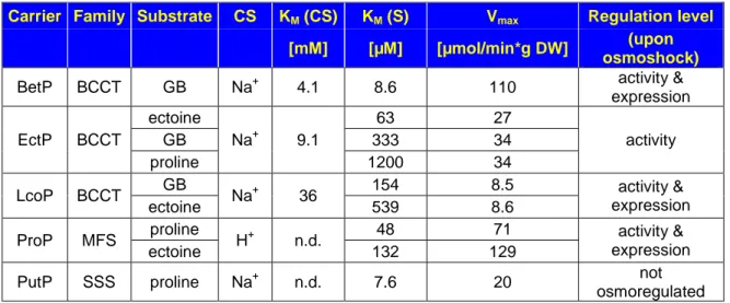
Table 1: Osmoregulated uptake systems in C. glutamicum and respective kinetic parameters. According to the kinetic characterization by Farwick et al. (1995), Peter et al. (1998b) and Steger et al. (2004).
Carrier Family Substrate CS KM (CS) KM (S) Vmax Regulation level [mM] [µM] [µmol/min*g DW] (upon
osmoshock)
BetP BCCT GB Na+ 4.1 8.6 110 activity &
expression EctP BCCT
ectoine
Na+ 9.1
63 27
activity
GB 333 34
proline 1200 34
LcoP BCCT GB
Na+ 36 154 8.5 activity &
expression
ectoine 539 8.6
ProP MFS proline
H+ n.d. 48 71 activity &
expression
ectoine 132 129
PutP SSS proline Na+ n.d. 7.6 20 not
osmoregulated Note: BCCT=Betaine-Choline-Carnitine-Transporter; MFS=Major Facilitator Superfamily; SSS=Sodium:Solute
Symporter; GB = glycine betaine; S = substrate; CS = cosubstrate; DW = dry weight (of cells).
1.9. Glycine betaine transporter BetP from C. glutamicum
As mentioned above, one of the best-studied carriers involved in osmoregulation of C. glutamicum is the secondary glycine betaine transporter BetP that catalyzes the symport of its sole substrate with two sodium ions (Farwick et al., 1995; Peter et al., 1996). BetP is a 595- residue integral membrane protein that comprises 12 transmembrane segments as well as a highly negatively charged hydrophilic N-terminal domain of approximately 62 amino acids and a highly positively charged hydrophilic C-terminal domain of 55 amino acids. Both of these terminal domains are cytoplasmically exposed and important for osmostress-dependent activity regulation (Figure 9; Peter et al., 1997; Rübenhagen et al., 2000). Driven by the electrochemical sodium potential
∆µ
Na+, BetP is able to build up extremely high glycine betaine gradients of 4 x 10
6(inside:outside). The carrier exhibits a moderate affinity of 8.6µM for its substrate glycine betaine and a high K
M(= lower affinity)
Figure 9: Predicted topology model of the secondary glycine betaine carrier BetP-C252T from Corynebacterium glutamicum (TMHMM 2.0,
“http://www.cbs.dtu.dk/services/TMHMM/”). The transmembrane segments are displayed as green cylinders. Marked in yellow is a conservative region of the proteins in the BCCT family that is possibly involved in substrate binding (Vinothkumar et al., 2006). The predominant charges of each cytoplasmic extension at physiological pH are depicted as “–“ (negative) and “+” (positive). The suffix “-C252T” represents the substitution of the sole native cysteine at amino acid position 252 for a threonine to obtain a cysless (cysteine-less) BetP variant.
of 4.1mM for its cosubstrate sodium (Farwick et al., 1995; Table 1). With V
maxvalues of 110 [µmol/min*g cell dry weight], BetP is together with the acetate and glucose transporters one of the fastest uptake systems in C. glutamicum (Ebbighausen et al., 1991; Marx et al., 1996).
1.9.1. BetP - osmosensing and activity regulation
In the absence of any osmotic stress BetP is almost inactive. Once the external osmolality rises and the activation threshold is reached (0.3-0.4osmol/kg), BetP in C. glutamicum cells gets activated in less than one second (activated state) with its activation optimum at 1.2osmol/kg (Peter et al., 1996; Peter et al., 1998b). As soon as the hyperosmotic stress has been compensated by the appropriate uptake of compatible solutes, BetP activity is reduced (activity adaptation) to prevent excessive solute accumulation (Morbach and Krämer, 2000). In this adaptation phase, the net uptake of glycine betaine is reduced to 66% of the overall transport rate due to both, a reduced import activity and a specific counter-exchange activity of the transporter (Botzenhardt et al., 2004). The heterologous expression of BetP in E. coli cells as well as the functional reconstitution of purified BetP protein in proteoliposomes led to the conclusion that this transporter operates autonomously (e.g. independent of accessory proteins or macromolecules) and thus harbours altogether three functions: (i) the catalytic activity of glycine betaine transport, (ii) sensing of hyperosmotic stress, and (iii) osmoregulation, i.e. adjustment of the transport rate to the actual extent of osmotic stress (Rübenhagen et al., 2000; Morbach and Krämer, 2004b).
The search for the activating stimulus took advantage of proteoliposomes that cannot build up a turgor pressure due to the lack of a cell wall-like structure that may sustain a certain hydrostatic pressure. Reconstituted BetP was shown to become fully activated while retaining the characteristic regulation pattern found in cells after an osmotic upshift.
Thus, the (high) turgor pressure in the Gram-positive Corynebacterium glutamicum could be ruled out as an activating stimulus. Other possible stimuli, like membrane tension, internal or external osmolarity as well as ionic strength, showed no significant influence on BetP activation (Rübenhagen et al., 2001). The effect of molecular crowding (Minton, 2005; Wood, 2007), e.g. the enrichment of cytoplasmic molecules due to cell dehydration, could not be investigated up to now. Nevertheless, using the hydration state of a protein as an indicator for hyperosmotic stress is still a challenging suggestion and was already proposed by Wood (2006) for the activation of ProP from E. coli. It could be figured out, that an increase in the luminal K
+concentration alone, i.e. at the side where the hydrophilic domains are located, is sufficient to activate BetP (Rübenhagen et al., 2001).
In addition, cations with similar physical properties as K
+, such as Rb
+and Cs
+, also
induced the activation of BetP, whereas bigger ions (NH
4+) or macromolecules (choline) did not. These results necessitated a reclassification of the so far entitled BetP- osmosensor to a chemosensor or - in particular - a potassium sensor (Rübenhagen et al., 2001).
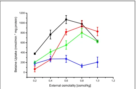
Unexpectedly, when heterologously expressed in E. coli cells, the activation profile of BetP upon hyperosmotic stress still remained similar, while the optimum of the transport activity was shifted to lower values of 0.6-0.8osmol/kg, e.g. a lower external osmolality was required to reach optimal activation of the transporter (Figure 10; Peter et al., 1996). Further investigations in the proteoliposomal system proved a strong influence of the membrane phospholipid composition on the activation profile of BetP: the higher the fraction of negatively charged phospholipids, the higher was the threshold concentration of K
+necessary for activation (Krämer and Morbach, 2004b).
In particular, the regulation pattern of the reconstituted transporter resembles that of native BetP in C. glutamicum cells, when the liposomes comprised more phosphatidylglycerol (Schiller et al., 2006). Although, the internal potassium concentration was shown to be a specific stimulus for BetP activation, the sensitivity towards K
+seemed to be at least partly dependent on the net (negative) charge of the surrounding phospholipid headgroups in the corresponding membrane system (Table 2).
Table 2: Phospholipid headgroup composition of the inner and outer membrane extracts from E. coli K12 and C. glutamicum ATCC13032 according to Morein et al. (1996) and Hoischen and Krämer (1990).
Phospholipid net charge
E. coli (grown at 37°C) C. glutamicum (grown at 30°C)
IOM IOM
[% of total phospholipids]
PE neutral (0) 79 ± 3 ~ 0
PG negative (-1) 17 ± 3 87 ± 1.9
DPG neutral (0) 4 ± 2 1 ± 0.4
Note: PE=phosphatidylethanolamine; PG= phosphatidylglycerol; DPG=diphosphatidylglycerol; IOM=inner and outer membrane fraction.
0,2 0,4 0,6 0,8 1,0 1,2 1,4
0 20 40 60 80 100 120
External osmolality [osmol/kg]
E. coli C. glutamicum
Betaine uptake [nmol/min*mg DW]
Figure 10: Activation profile of BetP-C252T expressed in C. glutamicum (black curve) and E. coli MKH13 cells (red curve) under hyperosmotic stimulation (Ott, 2008). The external osmolality was adjusted by the addition of NaCl.
1.9.2. BetP – Putative sensory domains and binding sites
The potassium-specific activation of BetP requires a binding site for the ion. Except for a sequence homology to a potassium binding site of pyruvate kinases in loop 2 (Jurica et al., 1998; Schiller et al., 2004a), the primary sequence of the transporter does not include a motif known to be involved in K
+recognition. Due to the fact, that the half-maximal activation of BetP is reached at an internal potassium concentration of 220mM (Rübenhagen et al., 2000), the putative sensory domain of the protein is supposed to need high amounts of the stimulating ion to detect and transmit the signal for activation.
In search of the defined localization of the sensor region within the protein, former investigations in intact C. glutamicum cells demonstrated that both hydrophilic domains of BetP strongly influence the activation profile. Truncation of the N-terminal domain of BetP led to a decrease in osmosensitivity, e.g. a higher osmotic stress was necessary to activate an N-terminally truncated BetP (Peter et al., 1998a). However, the truncation of the C-terminal domain by 25 and 45 amino acids led to deregulation of the carrier protein.
As a consequence, these BetP mutants were permanently active in betaine transport, independent of the applied hyperosmotic stress. On the other hand, truncation of only 12 terminal amino acids led to partial deregulation, e.g. the activity optimum of this
∆12 mutant was shifted to lower osmolalities, but the protein was still able to sense K
+(Peter et al., 1998a; Schiller et al., 2004b). These preliminary experiments revealed, that the C-terminal domain (i) was crucial for sensing the stimulus (K
+) and furthermore (ii) acted as an inhibitory element for BetP activation, because terminal deletions with more than 25 amino acids led to a permanent activation even in the absence of osmotic stress.
By series of constructs, it was intended to narrow down the putative sensory region within
the last 25 amino acids of the C-terminal extension. For this purpose, a set of C-terminal
mutants were generated via site-directed mutagenesis and analyzed with regard to an
involvement in signal perception and/or activity regulation of BetP. It turned out, that a
specific glutamate residue at position 572 seemed to be critically involved in potassium
sensing: substitutions with glutamine (E572Q), aspartate (E572D), lysine (E572K) and
proline (E572P) led to a deregulated activation profile of the respective BetP variant,
either heterologously expressed in E. coli or reconstituted in proteoliposomes made from
E. coli lipids (Schiller, 2004; Schiller et al., 2004b). However, in C. glutamicum cells all
mutants - except the proline variant E572P - regained their ability to detect osmostress
(Schiller et al., 2006). As a result, it was stated that (i) the higher amount of negatively
charged PG (phosphatidylglycerol) in the membrane of C. glutamicum stabilizes the
inactive conformation of the sensory domain (Table 2) and (ii) the correct conformation
and/or orientation of the C-terminal domain is important for correct stimulus sensing.
The latter conclusion was drawn based on the fact that - according to former in silico and CD-spectroscopy analysis – the central part of the C-terminal domain (amino acid positions 553-586) was thought to form the secondary structure of an
α-helix (Figure 11;
Burger, 2002). The introduction of proline as a common “helix-breaker” was supposed to alter the conformational properties of the helix. This in turn might impair crucial protein- potassium-membrane interactions and render BetP permanently active (Figure 11, insert upper left). Advanced efforts have been spent on a proline-scan within the C-terminal domain of BetP that confirmed this hypothesis. In addition, it could be shown that not only a retained integrity of the central part of the
α-helix, but also the primary structure of this protein domain seemed to be essential to ensure a proper osmoregulated activity response of BetP (Ott, 2008).
To summarize the results of former and current investigations spent on BetP, Figure 12 displays a preliminary model for the osmotically induced activation of the glycine betaine transporter. It focuses on the presumably different conformations of the C-terminal extension during the activation process as a prerequisite for correct stimulus sensing and signal transduction.
E572P Y550P
0,2 0,4 0,6 0,8 1,0
0 25 50 75 100
BetP-Cysless BetP-Y550P BetP-E572P
Betaine uptake rate [nmol/min* mg dw]
External osmolality [osmol/kg]
540 550 560 580 590
L V K D L S N D V I Y L E Y R E Q Q R F N A R L A R E R R V H NEH R K R E L A A K R R R E R K A S G A G K R R C C C C C C C C E E E E E H H H H H H H H H H H H H H H H H H HHH H H H H H H H H H H H H H C C C C C C C C C 9 4 2 1 3 6 7 8 7 9 9 8 5 0 3 4 4 5 6 7 8 8 8 7 6 6 6 5 4 4 3 266 6 7 7 7 8 8 7 7 6 5 4 3 1 0 2 7 7 7 8 8 7 9
Figure 11: Schematic illustration of the BetP carrier and a secondary structure prediction of the C-terminal domain (3D-PSSM, http://www.sbg.bio.ic.ac.uk/~3dpssm/index2.html). Yellow-highlighted is the predicted α-helical structure within the C-domain. Blue-highlighted are two engineered single proline mutants ahead (BetP- Y550P) and in the central part of the putative α-helical stretch (BetP-E572P). The respective, deregulated activation profiles of both proline mutants (heterologously expressed in E.coli MKH13 cells) are depicted in the upper left. As comparison, the wildtype-like cysless protein (BetP-C252T) with its respective uptake rates is plotted in the same graph (Schiller et al., 2006). H = α-helix; C =coil; E = extended (e.g. β-form).