Structural transitions in full-length human prion protein detected by xenon as probe and spin labeling of the N-terminal domain
Sunilkumar Puthenpurackal Narayanan1, Divya Gopalakrishnan Nair1, Daniel Schaal2, Marisa Barbosa de Aguiar1, Sabine Wenzel2, Werner Kremer1, Stephan Schwarzinger2 &
Hans Robert Kalbitzer1
Fatal neurodegenerative disorders termed transmissible spongiform encephalopathies (TSEs) are associated with the accumulation of fibrils of misfolded prion protein PrP. The noble gas xenon accommodates into four transiently enlarged hydrophobic cavities located in the well-folded core of human PrP(23–230) as detected by [1H, 15N]-HSQC spectroscopy. In thermal equilibrium a fifth xenon binding site is formed transiently by amino acids A120 to L125 of the presumably disordered N-terminal domain and by amino acids K185 to T193 of the well-folded domain. Xenon bound PrP was modelled by restraint molecular dynamics. The individual microscopic and macroscopic dissociation constants could be derived by fitting the data to a model including a dynamic opening and closing of the cavities.
As observed earlier by high pressure NMR spectroscopy xenon binding influences also other amino acids all over the N-terminal domain including residues of the AGAAAAGA motif indicating a structural coupling between the N-terminal domain and the core domain. This is in agreement with spin labelling experiments at positions 93 or 107 that show a transient interaction between the N-terminus and the start of helix 2 and the end of helix 3 of the core domain similar to that observed earlier by Zn2+-binding to the octarepeat motif.
Prion diseases or transmissible spongiform encephalopathies (TSEs) are a family of rare progressive neurode- generative disorders that affect both humans and animals alike. Examples of prion-causing diseases are bovine spongiform encephalopathy (BSE) in cattle and Creutzfeldt-Jakob disease (CJD) in humans1–5. These conditions are associated with the accumulation of an oligomeric conformational scrapie isomer PrPScr of the host-encoded monomeric prion protein PrPc. According to the “protein-only” hypothesis6, it is suggested that the conforma- tional isomer of PrPScr is able to convert other isoforms to the infectious isomer in an autocatalytic process. A detailed knowledge of this conformational transition is mandatory for explaining the molecular basis for prion diseases7.
Mammalian PrPc consists of two domains, a flexibly disordered N-terminal segment and a globular C-terminal domain containing three-helices and two short antiparallel-strands8. In addition, it contains a single disulfide bond linking cysteine residues at positions 179 and 214 between α 2 and α 3, which stabilize the folded structure of the normal protein. It has been suggested that the conformational state of the prion protein disulfide bond may have implications for correct maturation and function of this protein9,10. Prionic diseases were identified in many species of mammals, that also show a transmission specificity and infectivity of prions inter species11. Species bar- riers seem to occur in the majority of cases, but there are indications that species barrier crossing to infect another species requires specific changes of the amino acid sequence. Many groups have been focusing their research in discovery of these critical regions of the primary sequence of the protein, using modern techniques12–18. A region especially important for species differences in infectivity is the loop between β -strand 2 and α -helix 2 from
1Institute of Biophysics and Physical Biochemistry and Centre of Magnetic Resonance in Chemistry and Biomedicine (CMRCB), University of Regensburg, 93040 Regensburg, Germany. 2Research Center for Bio-Macromolecules and Department of Biopolymers, NW1/BGI, University of Bayreuth, 95447 Bayreuth, Germany. Correspondence and requests for materials should be addressed to S.S. (email: stephan.schwarzinger@uni-bayreuth.de) or H.R.K. (email:
hans-robert.kalbitzer@biologie.uni-regensburg.de) Received: 16 February 2016
accepted: 26 May 2016 Published: 24 June 2016
OPEN
www.nature.com/scientificreports/
residue 166 to 172 that occurs in two conformational states, in one state it contains a 310-helix, in the other state a type-I β -turn. The population of the two states in different species mainly depends on the existence of a tyrosine or phenylalanine residue in position 16918. For oligomerisation and fibril formation presence of the character- istic sequence motif AGAAAAGA (residues 113–120) is necessary19 which is part of the unfolded N-terminus of the prion protein and is located just in front of the well-folded core. After oligomer formation the residues of the AGAAAAGA motif are immobilized whereas all seven tryptophan residues located in the N-terminal range from amino acid 31 to 99 including the four tryptophan residues of the copper binding octarepeat remain freely mobile20. There are also neuroprotective mutations such as G127V, which leads to a complete protection for Kuru and CJD21.
Proteins are not rigid entities. For proper function they have to exist in several conformational states (“excited states”) with higher free energies than the ground state. This has been shown exemplarily for proto oncogene Ras involved in cellular signal transduction22. A pivotal role for the corresponding structural transitions play fluctu- ating low density zones and cavities in the protein23. These regions often can be identified by their typical strong, non-linear pressure response in high-pressure NMR spectroscopy24; the corresponding structural transition can be characterized by thermodynamic analysis of the data. Such a study has been reported for the human and the Syrian hamster prion protein15,25,26 where four different states of the folded core with differences of Gibbs free energies Δ G0 of approximately 3, 11, and 19 kJ/mol could be defined. The first transition would describe a transi- tion N1 to N2 in the natively folded ensemble with a relative population of 0.326.
An alternative method to detect structural fluctuations is based on the interaction of a protein with xenon detected by NMR-spectroscopy. Xenon is a noble gas that specifically recognizes hydrophobic cavities in macromolecules27–30. The interaction between xenon and the atoms of the protein are established uniquely through London dispersion forces. It can also bind to cavities that in the ground state are too small to host a xenon atom, but open up transiently. Thus, the corresponding rare conformational states can be detected by xenon binding. This has been shown earlier for the mutant HPr(I14A) of the histidine containing protein that contains a hydrophobic cavity that is filled by a side chain of a leucine residue in the ground state31. Here, xenon recognizes and stabilizes the lowly populated conformation of the wildtype protein that opens up to encompass a xenon atom32. Correspondingly, in the present study xenon is used to probe structural transitions coupled to fluctuations of hydrophobic cavities in huPrP(23–230) and correlate them with the transitions detected by high pressure NMR-spectroscopy.
In a first approximation, typical NMR parameters such as chemical shifts, relaxation times, and missing NOEs indicate that the N-terminal segment is mobile and disordered. However, already in the early paper by Zahn et al.8 the authors realized from an analysis of their NMR data that between the unfolded part of the protein and the well-folded part at least transient interactions have to exist since in the folded part chemical shift changes were observed when the N-terminal residues were removed. These chemical shift changes comprise residues 187 to 193 in the C-terminal part of helix 2 and the eight successive residues from position 219 to 226 in the C-terminal part of helix 3. These regions, defined by chemical shift differences between full length PrP(21–230) and truncated PrP(120–230), are part of a region from amino acids 186 to 226 with an exceptionally large pres- sure coefficients. These pressure coefficients change significantly when the N-terminal part is truncated, indicat- ing (transient) interactions between the N-terminal domain and the core domain. Especially G127, E168, H187, T192, E207, E211, and Y226 show significant differences in the NMR detected pressure response after removal of the N-terminus26. There is a number of additional evidence that there is some functional and conformational cou- pling between the N-terminal domain and the core domain, for instance, elimination of a segment between the N- and C-terminal domains in transgenic mice results in an embryonic lethal phenotype33,34. A most interesting region in the N-terminal domain is the octarepeat region (PHGGGWGQ)4 from amino acid 60 to 91 that is able to bind Cu2+- and Zn2+-ions35–37. Binding of these ions induces PrP endocytosis38, inhibits in vitro fibril formation39 and suppresses PrPScr amplification40. Recently, double electron-electron resonance (DEER) experiments showed multiple equilibria of the N-terminus relative to the core domain of mouse PrP as detected by spin labels at position of Q61 and T200. Zn2+-binding to the octarepeat induces an almost complete shift of conformational equilibrium to a state where the octarepeat region is close to T200 at the start of helix 3 suggesting that this closed conformation protects the PrP from fibril formation41. Zn2+-binding to the octarepeat itself leads to chemical shift and line widths changes in the region encompassing F140 to Y145 in loop 1, Q171 to T182 at the start of helix 2, and K203 to Y217 in helix 341.
For getting more experimental information about such an conformational equilibrium, we introduced spin labels just after the octarepeat in position 93 and in front of the AGAAAAGA motif in position 107 and observed their relaxation enhancement effects on the [1H, 15N]-HSQC spectra of human prion protein as presented in the following.
Results
Identification of xenon binding sites. When Xenon binds to hydrophobic cavities its binding can be detected by chemical shift perturbations and/or cross peak volume changes in [1H-15N] heteronuclear single quantum coherence (HSQC) spectra of a protein. [1H-15N]-HSQC spectra of 15N-enriched huPrP(23–231) were used to localize xenon binding sites on the protein structure at different xenon concentrations defined by slight xenon overpressure28. A reference [1H-15N]-HSQC spectrum was measured and assigned based on the data deposited in the biological magnetic resonance data bank (BMRB#4402) as well as in-house assignments26. Upon subjecting the samples to xenon pressures of 0.2, 0.4, 0.8 and 1.4 MPa (see Materials and Methods), correspond- ing to xenon concentrations of 8.8, 17.6, 35.2, and 61.6 mM in the protein solution respectively28, for a number of amide resonances changes of their chemical shift positions as well as their signal intensities/volumes were observed (Table 1). This demonstrates that the xenon atoms dissolved are interacting with the prion protein in a site-specific, ligand-like fashion in solution.
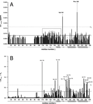
Figure 1A summarizes the combined chemical shift differences Δδcomb of each observable residue j as a func- tion of the protein primary structure. The secondary structure of the prion protein is indicated at the bottom of the figure. Many residues show a significant chemical shift response by xenon binding above the standard deviation σ0 indicative for a generalised conformational change of the protein upon xenon binding (Table 1).
This also includes residues such as K24 and R25 located at the start of the unstructured N-terminus and residues in or close to the AGAAAAGA (113–120) motif required for fibril formation. In the folded core structure of the prion protein xenon effects are observed in all secondary structure elements of PrP (Table 1). The region showing
Residues Location
K24, R25, N47, M109, M112 unfolded N-terminus
A113, G114, A117 AGAAAAGA motif
V122, G123, G126 G127, Y128 loop 0
G131 β -strand 1
S132, M134, S135, R136, I138, I139, H140, F141, G142 1oop 1
Y145, Y146, D147, Y149,Y150, R151, E152 α -helix 1
N153 loop 2
H155, R156 3/10 helix 1
Y157 loop 3
Y162 β -strand 2
M166, E168, S170 loop 4
N173, N174, V176, D178, C179, V180, N181, T183, I184, T188, α -helix 2
K194 3/10 helix 2
G195, F198 loop 5
E200, M205, M206, R208, V209, E211, Q212, I215, T216, Q217, E219 α -helix 3
E221 loop 6
S222, A224, G229 α -helix 4
Table 1. Residues influenced by xenon bindinga. aResidues showing significant (≥σ0) chemical shift or cross peak volume changes with xenon binding are depicted in normal letters and italics, respectively. Residues that have at least one pressure coefficient B1,2> 2 σ015,22 are depicted in bold letters.
Figure 1. Chemical shift and cross peak volume perturbation by xenon binding. Maximum combined chemical shift changes Δ δcomb (A) and relative changes of the cross peak volumes − Δ Vmax/V0= (V0 − V(cmax))/V0 (B) observed in the [1H, 15N]-HSQC spectra of 15N enriched huPrP(23–230) are plotted as a function the residue number j at 293 K. V0 is the cross peak volume in the absence of xenon and V(cmax) the volume at the maximum xenon concentration cmax of 61.6 mM. Solid line, standard deviation σ 0 to zero, dotted line, 2σ 0. P marks prolines, X other residues that are not visible or are not assigned in the spectra, 0 residues where satisfactory values could not be obtained. The error bars correspond to the standard errors. White bars represent residues that do not show saturation behaviour at the highest xenon concentration.
www.nature.com/scientificreports/
an almost continuous combined chemical shift difference above σ 0 is highlighted in Fig. 1 by a grey rectangle. This region starts with M112 and ends with H155 including the amino acids of the preceding flexible N-terminus as well as the first β -strand and α -helix of the folded core. Chemical shift responses above 2σ0 are observed for V161 and F198 upon xenon binding (Fig. 1).
In addition to the chemical shift perturbations observed upon xenon binding a reduction of the signal vol- umes of the cross-peaks in the 1H-15N HSQC spectra was detected. Figure 1B displays the maximum relative vol- ume changes induced by the highest xenon pressure used in this study as a function of the position in the amino acid sequence. The solid line again represents the standard deviation σ 0, the dotted line 2σ 0. Residues with a volume change larger than σ 0 are summarized in Table 1. Values above σ 0 can be considered as significant, values above 2σ 0 as highly significant. Volume changes above 2σ are seen for M109 located in the unfolded N-terminal region as well as for residues G142 located in loop 1 and Y149 in α -helix 1 of the well-folded core. There are two clusters of strongly affected amino acids, one at the start of helix 2 (S170, N173, V176, D178), one at the end of helix 3 (I215, Q217, S222, A224) (Fig. 1). In addition, a few new signals in the [1H-15N]-HSQC spectra appear that in the absence of xenon have not been observed and are therefore not assigned.
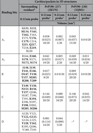
NMR structures published of the human prion protein exhibit a number of hydrophobic cavities in the well-folded core (amino acids 125 to 231) but the full length prion protein may also transiently form new cavities when the presumably disordered N-terminus gets in contact with the compactly folded part of the protein. The cavities were analysed with the program CASTp in two sets of 20 NMR structures each published by Zahn et al.8 (pdb accession code 1QM1) and Ilc et al.42 (pdb accession code 2KUN), respectively. The size and shape of the cavities calculated by the program depends on the size of the probe that should fit into the cavity. We used two dif- ferent probes, one with the size of a water molecule (radius 0.12 nm) and one with the size of a xenon atom (radius 0.22 nm). While the first NMR data set only contains coordinates of the folded core part of the structure (amino acid 125 to 228), the second data set provides additional information about the N-terminal amino acids (amino acid 90 to 227). It also has a point mutation at position 212 (Q to P), a mutation that is not expected to change the number or the properties of the cavities compared to the wild type. Using a water molecule as probe, it was found that cavities are not conserved among and within structural bundles as evidenced by cavity B shown in Fig. 2. An exception is cavity C that is present in all cases. Using a xenon atom as a probe the number of cavities that can accept a xenon atom without steric clashes is significantly reduced in the structural bundles. Cavity B is not large enough to host a xenon atom in any of the structures of the two data sets. Nevertheless, our experimental data support its (transient) existence in solution, since strong local effects induced by xenon binding are observable. A fifth cavity D is formed by the N-terminal part of the protein in some structures deposited in the data set 2KUN.
This part of the structure is not included in data set 1QM1 and can therefore not be analyzed (Fig. 2). However, as a rule, the total volumes of most of the cavities found to fit a water molecule are larger than the corresponding volume of a sphere with the radius of a xenon atom with a volume of 0.045 nm3 (Table 2), indicating that these cavities could adopt xenon after a suitable change of their shapes.
In first approximation the largest chemical shift or cross peak volume changes are to be expected close to xenon binding sites where xenon binding induces local conformational changes. These residues listed in Table 2 correlate well with the residues forming the surface of the cavities detectable in the two bundles of NMR struc- tures (1QM1 and 2KUN). A good correlation is observed but not all residues delineating a cavity experience chemical shift changes above σ0. This is to be expected since not all residues will be in direct contact with the xenon atom and/or involved in the required shape change of the cavity under consideration. More than 50% of Figure 2. Cavities and pockets in human prion protein structures. Cavities were calculated with a probe radius of 0.12 nm in the huPrP structure deposited as (a) 2KUN (20th structure of the bundle) and (b) 1QM2 in the protein data base, respectively. The surface of the cavities is represented in violet. For more details see Table 2.
the 65 residues showing significant effects > σ0 after xenon binding are part of a cavity surface (Tables 1 and 2), most of the other residues contribute to the second order shell of a cavity. From the twelve residues with xenon effects > 2σ0 nine are located close to a cavity, eight of them directly contribute atoms to the surface of the cavities.
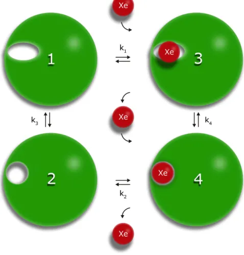
Affinity of individual cavities for xenon. Since we experimentally demonstrated xenon binding to all cavities, either (1) the NMR structures do not represent the cavities with sufficient reliability and all cavities are always large enough to accept a xenon atom, or (2) the cavities exist in a second transient state of the protein, where a xenon atom can be incorporated. A special case is cavity D that is only formed when the N-terminus is in contact with the main folded structure. This interaction has to be transient, since it is only found in a small number of the structures. A satisfactory fit of the experimental data was only possible assuming an equilibrium between an open and a closed state of the cavities. However, the quality of the data does not allow a stable fit including the equilibrium constant for the open and closed state as additional free parameter. In a first approxi- mation for this equilibrium constant the statistical distribution of the states, where the cavities are large enough to accommodate a xenon atom in the solution NMR structural bundles obtained by restraint molecular dynamics, can be used as an estimate of the occupancy of the states where a xenon atom can be incorporated (open state 1) and where not (closed state 2). The minimum scheme for such a dynamical equilibrium is depicted in Fig. 3.
Cavity D is only formed by the interaction of the core structure with the N-terminus (corresponding in the model of Fig. 3 to the open state) and disappears when the contact is lost (“closed” state). For reasons of simplicity the model assumes that xenon binding to different cavities is independent. When xenon cannot bind to the closed state since it does not fit into the corresponding cavity or the xenon binding state does not exist in one state, the xenon binding constant k1 can be set to zero.
The response of the chemical shifts and the cross peak volume changes on xenon binding depends on the time scale of the transitions between the four different states. It is assumed that in the closed state xenon cannot bind to the cavities. When the exchange is fast on the NMR time scale for all four possible transitions depicted
Binding Site
Cavities/pockets in 3D structures Surrounding
residuesb PrP(90–237)
(2KUN) PrP(90–230) (1QM1) 0.12 nm probe 0.12 nm
probec 0.22 nm probec 0.12 nm
probec 0.22 nm probec Volume [nm3]
A1
G131, S132, M134, V161, Y162, Y163, F175, V176, C179, C214, I215, Q217, Y218, E219,
R220
0.058 (0.023)
18/20 0.002 (0.007)
1/20 0.045 (0.037)
15/20 0.0 0/20
A2
R164, E168, S170, N171, N173, N174
0.041 (0.025)
19/20 0.005 (0.017)
2/20 0.049 (0.039)
16/20 0.011 (0.024)
4/20 B
I138, I139, F141, E146, D147, Y150, Y157, M205, R208, Y209
0.017 (0.025)
8/20 0.0 0/20 0.069 (0.019)
20/20 0.0 0/20
C
Y149, Y150, N153, R156, Y157, Q186, H187, T190, T191, K194, E196, N197, F198, D202, M205, M206
0.144 (0.049)
20/20 0.080 (0.040)
18/20 0.146 (0.075)
20/20 0.042 (0.035)
14/20
D
A120, V121, V122, G123, G124, L125, Y128, Y162, K185, Q186, V189, T193
0.081 (0.114)
14/20 0.044 (0.090)
5/20 –d –d
Table 2. Cavities and xenon binding sites in the human PrPa. aCavities were calculated with CASTp using two different probe radii (water, 0.12 nm; Xe, 0.22 nm). The structural ensembles of the PDB entries 2KUN and 1QM1 have been used for the analysis. bResidues contributing at least one atom to the surface of the cavities determined with a sphere of 0.12 nm radius in the 20th structure of the ensembles of the PDB entry 2KUN and the 1st structure of the PDB ensemble 1QM1, respectively. Residues in bold letters show significant xenon effects above σ0. cThe cavities were calculated for all 40 structures of the two PDB data sets deposited. The mean volume and the standard deviation of the mean value (values in brackets) are listed for the cavities defined by a probe of the size of a water molecule and a xenon atom, respectively. In addition, the number of structures is listed where a cavity can be detected that is large enough for accepting a water molecule or a xenon atom.
dN-terminal part is not contained in the PDB entry 1QM1.
www.nature.com/scientificreports/
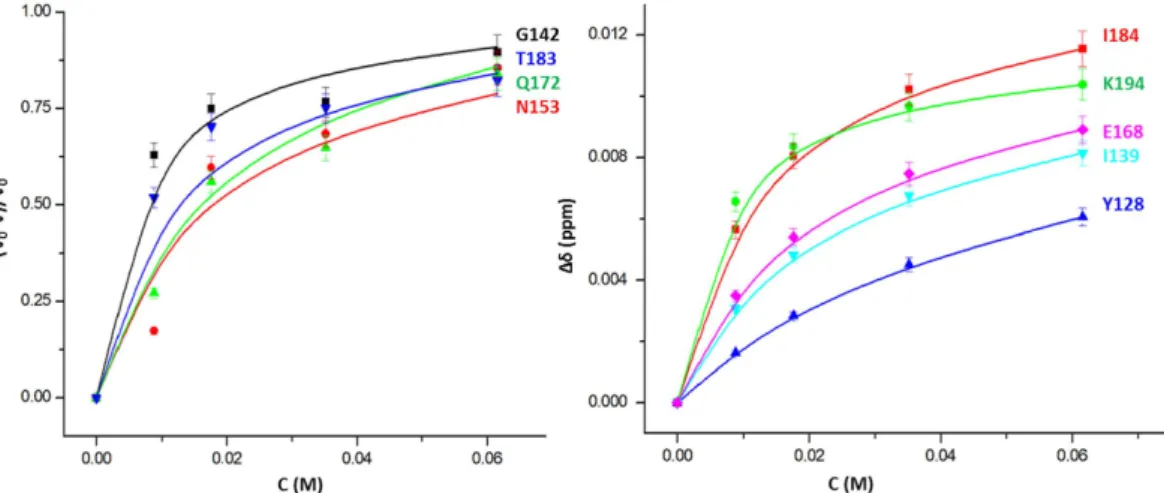
in Fig. 3 (1/τij ≫ |Δωij|, with τij being the exchange correlation times for the exchange between states i and j and Δωij corresponding to the associated chemical shift differences), the dependence of the observed chemical shift changes on the xenon concentration can be fitted with eq. 16. They are dependent on the association constant k2i of xenon to the open state of a given binding site i and the equilibrium constant k3i for the equilibrium between open and closed state. From the largest chemical shift change, a lower limit for the exchange correlation time can be estimated as 35 ms for the equilibrium between the open and closed state. Figure 4 shows examples for chemical shift changes of residues typical for the different cavities. The curves were fitted with the association constants given in Table 3 that describe the thermodynamic equilibrium for all residues assignable to a given cavity (Table 2). Only the chemical shifts in the different states had to be fitted individually since they depend on the environment of an individual spin in the absence and presence of xenon, respectively.
For some residues no chemical shift changes but only a decrease of their cross peak volumes V(c) is observed with increasing xenon concentrations c. Such behaviour could be due to an increased T2-relaxation causing a reduction of the INEPT-transfer and/or by a two-site slow exchange process. Since overall only small changes of the cross peak line widths were observed in the HSQC spectra, the comparatively large volume reductions can only be explained by the second process. When the exchange correlation time is slow in the NMR time scale for a given residue between the open and closed state and fast for the xenon interaction itself, eq. 9 can be applied.
A common fit of the relative volume changes of residues close to a given cavity gives association constants that within the limits of error are identical to those obtained from the fit of the chemical shift changes.
The obtained microscopic association constants k2 for xenon binding to the open state vary between 0.1 and 4.5 mM−1 (Table 3). However, the exact values depend on the probability of a cavity to be open and closed described by k3. As stated above the experimental data is not sufficient to simultaneously determine the equilib- rium constant k3 with sufficient precision, but the fit of the data is consistent with the assumption, that the NMR structural bundles represent the occupancies sufficiently well. The apparent xenon affinity can be approximated by k2/(1 + k3) and is dependent on the opening probability of a xenon binding site. Small opening probabilities will decrease the apparent xenon affinities accordingly. The apparent xenon affinities calculated from the data given in Table 3 are of the order of 0.1 mM−1 corresponding to apparent dissociation constants of 10 mM and vary from cavity to cavity. However, since they are obtained from a fit of many different residues enclosing a given cavity their fit error is rather small and indicates that their differences are significant.
Figure 3. Xenon binding in a two state model. In state 1 the cavity is too small to accommodate a xenon atom, in state 2 it has opened up. The equilibrium constants ki are defined as k1 = [3]/([Xe][1]), k2 = [4]/([Xe][2]), k3 = [1]/[2], k4 = [3]/[4].
Structure of the prion protein with xenon bound. The structural model of the prion protein with xenon bound at the four cavities was calculated by placing xenon atoms in the four cavities, optimizing the struc- ture by a simulated annealing approach on the basis of pseudo restraints obtained from the chemical shift and cross peak volume changes after binding of xenon. The obtained structures were refined in explicit water (for details see Materials and Methods). The lowest energy structure is shown in Fig. 5 together with the residues showing significant effects after binding of xenon. The structure with five xenon atoms bound is compared with the initial structure where some of the cavities are too small to accept a xenon atom. Xenon binding induces some structural rearrangements in the xenon binding cavities but only small global changes are required for accommo- dating the xenon atoms. The xenon atoms mainly interact with hydrophobic amino acid side chains and aromatic rings, in cavity A1 with V161 and F175, in cavity B with F141 and Y150, in cavity C with Y157 and F198, and in cavity D with L125, Y128, and Y162. Only cavity A2 does not show an interaction with such typical hydro- phobic residues. Here the methylene groups of E168, S170, and N174 constitute the main interaction partners (see supplementary Table S1). Note that the positions of the first N-terminal residues in this figure and especially the location of G93 is quite arbitrary, since it is the result of the molecular dynamics refinement but is not sup- ported by sufficient experimental restraints.
Figure 5 depicts those residues where significant pressure effects were reported in the absence of xenon15,26. As to be expected (see Introduction), they also cluster around the cavities and largely overlap with the residues that are sensitive to xenon binding.
Detection of transient structural transitions by paramagnetic enhancement. For obtaining information about transient interactions of the N-terminus with the globular domain, which also lead to for- mation of the transient cavity D, full-length prion protein variants with amino acid substitutions G93C and Figure 4. Dependence of chemical shift and cross peak volumes on the xenon concentration. The sample contained 0.25 mM of 15N-enriched huPrP(23–230) in 5 mM acetate buffer, 0.1 mM DSS, pH 4.5 in 90%
1H2O/10% 2H2O. Temperature 293 K. For residues typical for the various cavities (left) the relative intensity changes −ΔV/V0 and (right) the combined chemical shift changes Δδcomb are plotted as a function of the free xenon concentration c. The curves shown were calculated with the parameters of Table 3, only the chemical shifts in the different states are free fit parameters.
Binding Site k2 [mM−1] Δ G24 [kJ M−1] popenb Xe k3b Xe Kapp Xe [mM] Δ G12 [kJ M−1] popenb H2O Δ GH2O [kJ M−1]
A1 3.2 ± 0.4 − 19.7 ± 0.3 0.025 39 12.5 − 8.9 0.83 3.8
A2 0.46 ± 0.01 − 14.91 ± 0.03 0.15 5.7 14.6 − 4.2 0.88 4.7
B 4.5 ± 0.4 − 20.5 ± 0.2 < 0.024 > 39c 9.1 < − 9.0 0.7 2.1
C 0.11 ± 0.03 − 11.5 ± 0.7 0.80 0.25 11.4 3.4 > 0.98 > 9.0
D 0.34 ± 0.02 − 14.24 ± 0.02 0.25 3.0 11.8 − 2.7 0.7 2.7
Table 3. Equilibrium constants and free energies for xenon binding to huPrP (23–230)a. aMean association constants kji for binding sites i were calculated with eqs 9 and 16 assuming k1 = 0. Temperature 293 K. For definition of the constants see Fig. 3. bpopen, probability of the open state in absence of xenon calculated from the NMR structures 2KUN and 1QM1 (Table 1). Site D is only transiently formed by N-terminus and thus the state, where the cavity is formed corresponds to the open (binding) state. k3 has been calculated from popen as k3 = (1 − popen)/popen. The apparent xenon dissociation constant Kapp is defined by Kapp = 1/k2+ k3/k2. cIn none of the NMR structures cavity B is large enough for accommodating a sphere of the size of the xenon atom.
However, a smaller population than 0.025 is not excluded by statistics. For the calculations the constant k2 was set to 39.
www.nature.com/scientificreports/
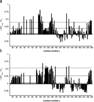
T107C were labelled with compounds containing unpaired electrons. In particular, the two different spin labels (1-oxyl-2,2,5,5-tetramethyl-∆ 3-pyrroline-3-methyl methanethiosulfonate and 1-oxyl-3-(maleimidomethyl)-2, 2,5,5-tetramethyl-1-pyrrolidine) were applied in this study. Residues spatially close to the active spin label should show a reduction of their cross peak volumes due to enhanced T2- relaxation caused by strong electron-nucleus dipole-dipole coupling when compared to the reduced diamagnetic sample. Since the labeling reaction itself was not quantitative and since for the N-terminal residues no unique structure is to be expected, the analysis of the paramagnetic enhancement (PRE) was only done in a qualitatively manner by comparing differences in cross peak volumes of the paramagnetic and diamagnetic sample (after reduction of the spin label). The relative cross peak volume changes −ΔV/V0= (V0 − V)/V0 were plotted as a function of the sequence position with the cross peak volumes V and V0 of active and reduced spin label, respectively. Solid and broken lines represent stand- ard deviation σ0 and 2σ0 to zero of −ΔV/V0 the V/V0 (Fig. 6). The residues with significant cross peak volume changes are summarized in Table S2. They are assumed to be spatially close to the spin label in the time average but the averaging is non-uniform because of the r−6-dependence of the PRE effect.
Besides direct effects in the neighborhood of the spin label attached, both labeled variants show similar rel- ative effects in the sequence positions 30–50, 120–135, ~165 and C-terminal region around residue 220–231. A number of residues show an increase of the cross peak volume in the labeled sample relative to the volume after reduction of the spin labels indicating that residual unattached spin label in solution enhances selectively the longitudinal relaxation of water protons and thus resulting in increased signal to noise ratio in the paramagnetic sample43.
Most of the structures published by Ilc et al.42 deposited in PDB 2KUN ensemble cannot provide an explana- tion for the observed paramagnetic effects since the N-terminal domain is too distant from the globular domain.
As an example we show the observed effects in the first structure of the bundle deposited in PDB 2KUN, where the residues that are significantly affected by the presence of the spin labels are given in a color code (Fig. 7).
However, the structure of the prion protein selected for calculating the xenon containing structure (Figs 2 and 5) qualitatively explains many paramagnetic effects that cannot be explained by other structures of the flexible N-terminal domain of the same structural bundle, including e.g. the first structure of the same bundle, since the N-terminal domain apparently is too distant from the core domain (Fig. 7).
Discussion
Interactions of xenon in hydrophobic cavities. We were able to locate five binding sites for the xenon atoms by their NMR response to xenon binding. They correspond to cavities that are mostly large enough to accommodate water molecules and that are present in part of the NMR structures deposited in 2KUN and 1QM1, respectively. However, cavity D is only visible in some members of the structural bundle of 2KUN, since 1QM1 does not provide structural information about the corresponding flexible part of the N-terminal tail.
Site A1 shows a response of xenon binding at residues G131, S132, M134, V161, Y162, Y163, F175, V176, C179, C214, I215, Q127, Y218, E219, and R220 (Table 2). It is open for accepting a water molecule in almost Figure 5. NMR-Structure of huPrP with bound xenon. The NMR structure of huPrP with xenon bound was calculated from the NMR data starting with the 20th structure of 2KUN. (a) Overlay of the structure in the presence (red) and absence of xenon (green). (b) Lowest energy structure with residues showing significant effects induced by binding of xenon and/or by applying high pressure15,22. Residues showing only xenon effects > σ0 (red) and in addition high pressure effects > 2σ0 (orange). Residues showing high pressure effects > σ0 only are depicted in blue, residues not detectable (e.g. prolines) or with smaller effects are depicted in grey.
all structures of the bundles deposited. Assuming that NMR data are sufficiently representative for the solution conformation of the proteins (what we will do in the following) a probability to be open popen of 0.83 for water would follow resulting in a corresponding ΔGH2O of 3.8 kJ mol−1 (Table 3). However, in most cases a xenon atom Figure 6. Paramagnetic effects in spin labelled full-length prion protein. The relative change − Δ Vmax/V0 = (V0 − Vmax)/V0 of cross peak volumes as a function of the sequence position j in [1H, 15N]-HSQC spectra of
15N-enriched huPrP(23–231) variants G93C (a) and T107C (b) labeled with methanesulfonothioate and maleimide based nitroxyl spin label, respectively. Solid and broken lines represent standard deviations σ0 and 2σ0. Proline residues are marked with P. Additionally invisible and unassignable residues in the spectra are marked with X. V0 corresponds to the cross peak volume after reduction of the spin label and Vmax the cross peak volume after labeling (see Materials and Methods).
Figure 7. Mapping of paramagnetic effects on spin labeled full-length prion protein. Paramagnetic effects of MTSL and maleimide-linked nitroxyl spin label on huPrP variants G93C (a) and T107C (b), respectively.
Very strong paramagnetic effects > 2 σ0 are colored in orange, strong effects > σ0 in red, small and negative effects < σ0 are colored in blue and unassigned residues in grey. The labeling position is displayed as spheres and the alanine-rich region in sticks. Mapping of the paramagnetic effects and in silico mutagenesis was done with Pymol (Schrödinger LLC) and the first structure of PDB entry 2KUN.
www.nature.com/scientificreports/
cannot enter the cavity reflected by an apparent xenon dissociation constant Kapp of 12.5 mM and a Δ G12-value of − 8.9 kJ mol−1. Site A2 is adjacent to this region and reacts on xenon binding by chemical shift and cross peak volume changes at R164, E168, S170, N171, N173, and N174. It can accept a water molecule in most of the struc- tures (popen, 0.88; Δ GH2O of 4.7 kJ mol−1). However, only in a few cases it is large enough to accept a xenon atom (Kapp 14.6 mM; Δ G12 − 4.2 kJ mol−1).
Of note, residues close to these two cavities are involved in prion diseases: E129M and D178N are characteris- tic mutations in Fatal Familiar Insomnia (FFI) while for Creutzfeldt-Jakob disease (CJD) mutations of the residues D178 and V180 are known to be involved in the disease development. The xenon interaction site A1 covers the polypeptide segment from 165–175, the β 2-α 2 loop, which was identified by the Wüthrich group in a whole number of prion structures to occur in different conformations in different species8,18,44,45 and which seems to be of crucial importance for disease progression in a mouse model46.
Xenon binding site B comprises residues I138, I139, F141, E146, D147, Y150, Y157, M205, R208, and Y209.
It is open for a water molecule in all structures of the structural data set 1QM1 but only for 8 of the 20 structures of the 2KUN ensemble. Here, the two structural bundles differ significantly. The average Δ GH2O is 2.1 kJ mol−1. In none of the structures a xenon atom can fit into the cavity (Kapp 9.1 mM; Δ G12 < − 9.0 kJ mol−1). None of the residues forming cavity B is known to be involved in any prionosis. However, the residues preceeding helix 1 have been shown to be involved in the species barrier.
Xenon binding site C has a large surface and comprises residues Y149, Y150, N153, R156, Y157, Q186, H187, T190, T191, K194, E196, N197, F198, D202, M205, M206. The cavity is large enough for accepting a water mol- ecule in all structures of the two data sets. It is also large enough to accept a xenon atom in the majority of the structures given, resulting in an apparent xenon dissociation constant Kapp, of 11.4 mM and a corresponding free energy Δ G12 of < 3.4 kJ mol−1. Mutations of F198, whose aromatic ring interacts directly with xenon, are impli- cated in the Gerstmann-Sträussler-Scheinker (GSS) syndrome.
The xenon binding cavity D is characterized by chemical shift changes of residues preceding the first β -strand of the prion protein (V122, G123, G126, G127, and Y128) but it is also formed by K185, Q186, V189, and T193.
Structural models of the part preceeding the first β -strand are only deposited in the PDB-file 2KUN and have been omitted in 1QM1 since it does not adopt a single well defined rigid structure. However, it shows a number of intra residual NOEs as well as 15N-T1, T2, and 1H-15N-NOEs that are typical for regions with residual local structure42. Using a water molecule as probe, a cavity D is observed in 14 out of 20 structures deposited. These observations confirm the validity of the NMR structures in this region, at least as transient conformations in thermodynamic equilibrium. Peptides encompassing the region from amino acids 106 to 127 that form the xenon binding site D are also known to reproduce the main neuropathological features of prion-related transmissible spongiform encephalopathies13. The chemical shifts of residues inside this region (M109, M112, A113, G114) and thus their local environments are also influenced by xenon binding to cavity D. As revealed by NMR-spectroscopy in oligomers formed by reduced prion protein the AGAAAAGA motif (amino acid 113 to 120) is strongly immo- bilized20. This palindromic motif is required for oligomerisation and the fibril formation of PrP19,47,48.
Correlation between high pressure response and xenon binding sites. It is known that large high-pressure effects are often observable close to cavities in proteins indicating local conformational transitions.
Since xenon binding to cavities is also accompanied by local conformational transitions the two effects should be correlated. Table 1 and Fig. 5 show that indeed the two effects correlate quite well supporting the idea that xenon binding as well as pressure response is due to structural transitions.
Structure of the N-terminal region. In monomeric prion protein the N-terminal part appears to be mainly unstructured and from the NMR point of view rather mobile although high pressure NMR reveals some residual structure26. Even after oligomerisation formed by reduced prion protein the NMR spectra of seven tryp- tophan residues contained in the N-terminal part of the prion protein (amino acid 31 to 99) indicate a high peptide-like mobility20. An exception seems to be the supposed copper binding octarepeat (amino acids 60 to 91) in the presence of Cu2+- or Zn2+-ions that might have a physiological role in neuroprotection35–37. In the absence of metal-ions DEER-experiments with spin labels at position of Q61 and T200 of mouse PrP show that the distance distribution of the spin labels is characteristic for a conformational equilibrium where the octarepeat region is close to the core domain in about 20% of the structures. Zn2+-binding to the octarepeat induces an almost complete shift of conformational equilibrium to a state where the octarepeat region is close to T200 at the start of helix 3. It leads to chemical shift and line widths changes at M128 and in the regions encompassing F140 to Y145 in loop 1, Q171 to T182 at the start of helix 2, and K203 to Y217 in helix 341. These chemical shift changes differ from the chemical shift changes observed upon removal of the N-terminal segment, where residues 187 to 193 located in the C-terminal part of helix 2 and residues 219 to 226 in the C-terminal part of helix are affected8. A somewhat better correlation is found for the residues identified by high pressure NMR spectroscopy: significant differences in the NMR detected pressure response after removal of the N-terminus26 were observed for G127, E168, H187, T192, E207, E211, and Y226.
Unfortunately, only a few residues of the octarepeat region itself could be resolved with sufficient accuracy in the xenon experiments. They do not show a specific xenon response.
The deimination of R27, R40 and R51 in recombinant PrP caused an alteration of the copper-binding affinity of the octarepeats49 although it is rather far away from the metal binding site. In this region K24, R25, and N47 show a significant effect upon xenon binding, probably by perturbing the transient interactions with the core domain by e.g. stabilizing cavity D.
For further analysis of interdomain contacts nitroxide spin labels based on disulphide and maleimide link- ing chemistry were applied and results were compared to previously published data as well as xenon binding
experiments reported here. In principle, a reduction of the cross peak volumes should occur when the spin label is close to a nucleus under consideration. Such a cross peak volume reduction is observed for many residues of PrP (Fig. 6). However, some residues show negative relative changes of signal volumes most likely arising from remaining paramagnetic label in solution, resulting from incomplete washing after the coupling reaction. Low concentrations of paramagnetic agent have shown to enhance relaxation properties43. Since quantitative analysis of paramagnetic relaxation experiments in a multistate equilibrium predicted for the N-terminal segment are very error-prone we only qualitatively account stretches of residues showing continuous paramagnetic effects.
In general, more or less pronounced paramagnetic effects of different strength are observed all over the protein sequence (23–231) in both variants, meaning that the flexible spin-labeled N-terminal tail transiently contacts the globular domain in different locations as predicted by the DEER experiments41. The two spin labels were placed at two strategically interesting positions, position 93 is close to the octarepeat region extending from amino acid 60 to 91 and position 107 is located in front of the palindromic motif AGAAAAGA (residues 113–120). Effects of the spin labels in positions 93 and 107 on distant N-terminal amino acids as well as non-uniformly expressed paramagnetic effects suggest a transiently ordered conformation of this part of the sequence. Due to the lack of experimental NOE distance restraints in the flexible tail no structural data is available. The high flexibility and the repetitive nature of the octarepeat region prevented resonance assignments and through-space structural infor- mation. Few remaining signals, however, show strong paramagnetic influence especially in G93C, meaning that residue positions 50–81 are in close spatial proximity to the spin label at position 93 (Fig. 6, Table S2). G93, W99, K101, S103, K106, M109, K110, M112 show a significant non-random pressure response26. In this region also significant effects downstream from the positions of the spin labels are observed. Surprisingly, the N-terminal AGAAAAGA (113–120) motif is affected several times weaker by the spin label at position 93 than preceding or successive parts of the sequence.
In spin labelled G93C and T107C many residues in helix 2 and 3 of the globular domain show significant par- amagnetic influence with the strongest effects on the C-terminal end of helix 3 (Fig. 6) that cannot be explained with the structure shown in Fig. 7. The spin label at position 107 shows strong effects (> 2σ0) for M166 and Q172 at the start of helix 2, in the preceding loop 4, and at Y226, Q227, R228, G229 at the end of helix 3. A structure such as shown in Fig. 5 could qualitatively explain this effect. Strong paramagnetic effects in the same regions are also found for the spin label in position 93 that should also come close to these regions. Zahn et al.8 predicted as one of the possible interaction sites amino acids 219 to 226. Among others E168 and Y226 were predicted by high pressure NMR spectroscopy as possible interaction sites with the N-terminal domain. In the presence of Zn2+-ions the octarepeat region located close to the spin label at position 93 is predicted to also be in contact with amino acids Q171 to T182 at the start of helix 2, and K203 to Y217 in helix 341. Mapping these effects on a struc- ture of the prion protein shows that in the time-average the spin labels at position 93 and 107 and probably the whole N-terminus preferentially interacts only with one side of the protein, whereas on the opposite side there are only small effects (Fig. 7). A similar but not identical arrangement was also proposed by41. This is also reflected in the minimal paramagnetic effects observed in the helix 2-loop-helix 3 region on the opposite site of the globular domain. This is in agreement with the fact that in the fully matured cellular form of the prion protein, two glycans in position Asn 181 and Asn 197 are blocking any interdomain interaction.
Addition of phosphatidylglycine (POPG) and RNA leads to a conversion of recombinant PrP into a highly pathogenic protease resistant form rPrP-res similar to the pathogenic PrPSc isoform50. Again, the interaction affecting the properties of PrP occur to a large degree in the flexible amino terminus. Specifically, the POPG inter- action takes place through the N-terminal positively charged domain and a hydrophobic domain encompassing residues 100–13451, which are indicated to be at least transiently close in space by the spin label experiments.
Other residues strongly influenced by the spin labels, as e. g., K186 cannot be explained by a structure such as that shown in Fig. 5. This is also true for many residues predicted as possible interaction sites of the N-terminus.
However, the position of the first stretch of amino acids is not defined by NOEs in the original structure. A struc- ture where the N-terminal domain is directly in contact with the globular domain (as e.g. shown in Fig. 5) can only represent a weakly populated structural state in the equilibrium, since otherwise homonuclear NOE data could be observed leading to a defined structure of the N-terminal domain. When this state is associated with the formation of the xenon binding cavity D, an estimate of its relative population would be 0.25 (Table 2), which agrees well with the DEER-data predicting similar populations in the absence of Zn2+-ions. In agreement with the assumption of multiple states of the N-terminal region molecular dynamics calculations using the PRE effects as restraints could not find a unique structure explaining all restraints.
In the set of NMR structures given of huPrP(Q212P) by Ilc et al.42 the orientations of the N-terminal region (90–110) relatively to the folded core are not unique. However, the set contains several structures that could explain our experimental data (existence of cavity D as well as paramagnetic effects between the N-terminal domain and the globular domain) and that are shown in Figs 2 and 5. In this model the loop containing the spin label at amino acid 107 is fixed by two interactions to the core: K104 could interact with Y225 and K110 could form a salt bridge with E168 or a hydrogen bond with S170 (Fig. 5). These relations are shown in more detail in Fig. S1 and are in qualitative agreement with the paramagnetic data (Table S2). K104, E168 and E211 (in blue) display significant high pressure effects as well as effects from xenon binding. The positively charged side chain of K110 exhibits a distance of 0.47 nm to the hydroxyl group of S170 and of 0.56 nm to the carboxyl group of E168.
The amino group of K104 is separated by 5.9 Å from the hydroxyl group of Y225.
Conformational states of PrP and cavity fluctuations. Four different states of Syrian hamster PrPC 15 as well as human PrPC 26 were stabilized by high pressure and characterized by high resolution multidimensional NMR at atomic detail. Moreover, species-specific differences in the response upon hydrostatic pressure were observed by high-resolution multidimensional NMR spectroscopy for the core domain (121–230) of Syrian hamster and human PrPC 15. Both proteins fluctuate between two well-folded conformations N1 and N2 and two
www.nature.com/scientificreports/
excited states I1 and I215. Although the amino acid sequence between both prion proteins is nearly identical (only 14 amino acids out of 110 residues are different between both core domains) Kremer et al.15 observed significant differences of the pressure response for the individual residues. This indicates that the secondary structure of the complete folded core, which is nearly identical for all mammalian PrP proteins, is affected differently by high hydrostatic pressure implying structurally different excited intermediates. Oligomers formed by reduced human prion huPrPC(23–231) dissociate under pressure into monomers that occur in rare, metastable excited states of PrPC stabilized at high pressure20.
In the full-length huPrPC(23–230) residues K101, S103, K104, K106, N108, M109, and M112 of the unfolded amino-terminal region showed significant chemical shift changes upon application of high pressure. In the struc- tured core domain of huPrPC significant pressure shifts of residues M129, G131, I139, H140, F141, C179, T183, Q186, H187, T188, V189, T191, K194, G195, E196, N197, F198, T199, D202, M205, E207, V210, E211, E219, and R220 are observed. Most of them are close to the five cavities (xenon binding sites) described here (Table 1, Figs 2 and 5).
The NMR structures as well as the xenon binding data indicate that these cavities are in a dynamic equilibrium between open and closed states. Such an equilibrium between conformational states where the cavities are open and contain water or where they are closed and too small to accept a water molecule could explain the observed pressure effects: The free energy difference Δ G012 between the two native states N1 and N2 is 3.0 ± 1.4 kJ mol−1 and the volume difference Δ V012 is − 86 ± 17 mL mol−1 (0.14 ± 0.03 nm3)15. A positive Δ G combined with negative Δ V value means that the state dominating at ambient pressure gets rare at high pressure and vice versa. Cavities A1, A2, B, C, and D are characterized by free energy differences between the water containing and the closed state of 3.8, 4.7, 2.1, > 9.0, and 2.7 kJ mol−1, respectively. For all cavities except cavity C these free energy values satisfactorily agree with the energy difference between state N1 and N2. However, according to the definition of Δ GH2O given in Table 2 all cavities preferentially contain at least one water molecule at ambient pressure. If the transition observed by high pressure NMR would describe the equilibrium between an open state containing a water molecule and a closed state excluding water, then at low pressure the water containing state N1 should pre- vail but would be replaced by closed state N2 at high pressures. It is usually observed that high pressure favours an increase of the size of hydrated cavities, thus the structural states derived from the pressure response do probably not describe the hydration of the cavity. In the present case it would lead to a further hydration (opening) of the cavities, as it is required for xenon binding. If the process described by the high-pressure experiments would describe the opening of a cavity to a size large enough to accommodate a xenon atom then cavity A2 and cavity D would be appropriate candidates with Δ G120 values of − 4.2 and − 3.7 kJ mol−1, respectively. Both are located close to an area probably involved in the fibril formation process. The observed volume difference would correspond to the volume of approximately 5 water molecules with a molar volume of 18 mL mol−1. The van-der-Waals volume of a xenon atom calculated from a radius of 0.22 nm would be 27 mL mol−1, which is the total volume difference that would correspond to approximately three xenon atoms bound.
The free energy differences for the transition from the native state to intermediate states I1 and I2 are 10.8 ± 1.9 kJ mol−1 and 18.6 ± 2.9 kJ mol−1, respectively15,26, the corresponding volume differences are − 66 ± 26 mL mol−1 and − 125 ± 32 mL mol−1. Since the Δ G0-values were mainly determined from cross peak vol- ume changes, the sign of the free energy difference is uniquely defined. The large Δ G0 values indicate that the states assumed at high pressure are dominant at ambient pressure. This means that the enlargement of the cavities cannot be correlated with the transition to the intermediate states but has to describe another process on the pathway to PrPScr.
Conclusion
We have shown here that by use of xenon as a probe it is possible to detect cavities in proteins that in the deposited structures are too small to accept a xenon atom (e.g. cavity B) but have to be transiently enlarged. We have derived here the corresponding equations (see Materials and Methods) for a two state model with an open and a closed state of cavities in equilibrium. The functional dependence is different from a simple one state binding model, since the data can only be satisfactorily fitted by a model allowing more than one state. Qualitatively, such an effect is experimentally observed here (Fig. 4). However, for a quantitative description the number of parameters to fit was too high, thus we assumed that the NMR data reflect the ratio of opening and closing as a first approx- imation. In principle, using more data points (which are not easy to obtain) it should be possible to completely extract all parameters with sufficient accuracy.
The N-terminal region is clearly not in a uniquely folded structure but nevertheless xenon effects are observa- ble in a well-defined region, which is populated for at least a fraction of time. Here, xenon is able to detect a tran- sient binding site (cavity D) that is reported in some of the NMR structures of the structural ensemble if 2KUN where the partly folded N-terminal region is in contact with the core region.
High pressure NMR is also known to detect fluctuations close to cavities and thus indirectly cavities. In fact, we can observe a good correspondence between our xenon data set and the high pressure data published before.
In general, transient structures in a multistate equilibrium as derived from NMR-parameters of the N-terminal domain typical for a disordered and highly mobile protein segment and partly quantified by DEER experiments41 cannot be determined exactly by experiments only, but experimental data can be used to select or discard models as we have done here. Another supplementary method is the use of spin labels that can detect transient interac- tion sites but again cannot be used for a direct structure determination when no unique structure exists (as in our case).
From the biological point of view fluctuating cavities are structurally weak points where conformational tran- sitions are often initiated and which we have now characterized for the prion protein. Cavities A2 is formed in part by the loop between β -strand 2 and α -helix 2 from residue 166 to 172, a region that is highly important for species differences in infectivity and that is involved in a conformational transition between a 310-helix in one