Cytoskeleton proteins involved in
chromosome segregation and cell division in Corynebacterium glutamicum
Inaugural-Dissertation zur
Erlangung des Doktorgrades
der Mathematisch-Naturwissenschaftlichen Fakultät der Universität zu Köln
vorgelegt von
Catriona Donovan
aus Tralee, Irland
Köln, March 2012
Berichterstatter: Prof. Dr. Reinhard Krämer
Institut für Biochemie der Universität zu Köln
Prof. Dr. Marc Bramkamp
Institut für Biochemie der Universität zu Köln und
Biozentrum, Department Biologie I, Ludwig-Maximilians Universität, München
Tag der mündlichen Prüfung: 11-06-2012
This dissertation is dedicated to my parents.
Index
Index
I. Abstract V
ΙΙ. Zusammenfassung VII
III. Abbreviations IX
1. Introduction 1
Chapter I
1.1 DNA replication initiation 1
1.2 DNA partitioning systems 1
Plasmid partitioning systems 2
Chromosome encoded partitioning systems 3
Chromosome segregation in Bacillus subtilis 4 Chromosome segregation in Caulobacter crescentus 5 Chromosome segregation in Vibrio cholerae 6 1.3 Chromosome condensation - SMC and organization of chromosomes 7
1.4 Cell division 9
Spatial regulation of cytokinesis – negative regulation 9 Novel spatial and temporal regulation of cytokinesis 10 Spatial regulation of cytokinesis – positive regulators 10 1.5 Cell polarity – integrated temporal and spatial regulation 12 1.6 Corynebacterium glutamicum – cell division and chromosome segregation 13 Chapter II
1.7 The bacterial cytoskeleton 17
Bacterial actin homologues – ParM 18
Divergent bacterial actin families – AlfA and Alf7A 18 1.8 Identification of a C. glutamicum actin-like protein 20
1.9 C. glutamicum prophages 21
1.10 Aim of the research 22
2. Results 24
Chapter I
2.1 The Par chromosome segregation system of C. glutamicum 24
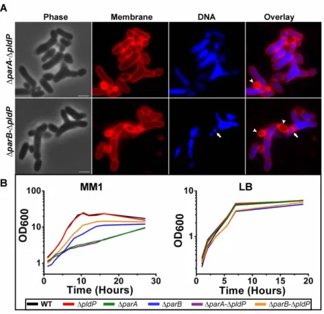
Phenotypes of par null deletion mutants 24
In vivo localization of ParA and PldP 27
Phenotypes of double par deletion mutants 28
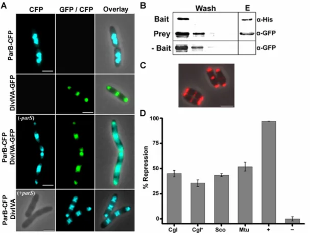
ParA, unlike PldP, is necessary for polar localization of ParB 29 2.2 Polar tethering of the chromosome origins in C. glutamicum 30 DivIVA physically interacts with ParB, in vivo 31
DivIVA interacts with ParB, in vitro 33
Index
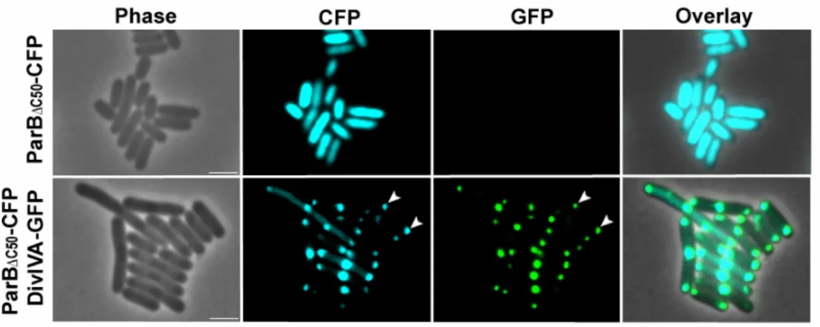
The N-terminal domain of ParB interacts with DivIVA 33 In C. glutamicum, mutation of the conserved arginine (R21) of
ParB significantly alters interaction with DivIVA 35 A central region of DivIVA is necessary for ParB interaction 37 In the synthetic E. coli system, ParB interacts with DivIVA
via a diffusion-capture mechanism 39
The ParB-DivIVA interaction is conserved in Actinobacteria 39
ParA interacts with both ParB and DivIVA 42
2.3 Chromosome organization influences cell growth and division site selection 44 2.4 Characterization of the orphan ParA-like protein, PldP 49
PldP exists as a monomer in solution 49
PldP binds membranes via a putative C-terminal amphipathic helix 50
In vivo, PldP binds to the membrane 51
Identification of potential PldP interaction partners 52 Chapter II
2.5 Does AlpC exhibit actin-like properties? 55
AlpC assembles into filaments, in vivo 55
AlpC filament dynamics 57
AlpC exists as a monodisperse species in solution 57
AlpC hydrolyzes both ATP and GTP 58
AlpC polymerization is nucleotide dependent, in vitro 58 2.6 What is the molecular role of AlpC in C. glutamicum? 60 AlpC filament assembly requires a host specific factor 60
Attempts to identify an AlpC adaptor protein 60
Filament assembly of AlpC at physiological concentration 61 In the absence of alpC, prophage induction rate is reduced 61 In vivo co-visualization of the CGP3 prophage and AlpC 62
3. Discussion 64
Chapter I
3.1 ParA and ParB: role in chromosome segregation 64
ParA has a possible additional role in regulation
of chromosome replication initiation 67
Pleiotropic nature of parA and parB mutation – possible additional roles 68 3.2 Polar anchoring of the oriC is mediated by ParB-DivIVA interaction 70
The N-terminal of ParB interacts with DivIVA 73
ParA might support polar oriC anchoring 73
ParB interacts with a central region of DivIVA 74
In C. glutamicum, DivIVA truncation mutants are
viable but acquire suppressor mutations 75
Index
3.3 Regulation of cell division in C. glutamicum 76
PldP: role in cell division 77
PldP: possible role in regulation of ParA 78
Simultaneous deletion of PldP and ParB or ParA
increases growth from ectopic sites 88
Orphan ParA-like proteins organize the bacterial subcellular environment 81 3.4 Proposed model for chromosome segregation in C. glutamicum 82 3.5 The ParB-DivIVA interaction is conserved in Actinobacteria 84 Chapter II
3.6 AlpC is an actin-like protein 85
3.7 AlpC is necessary for efficient prophage induction 86 3.8 Host actin related proteins organize phage replication 87
4. Experimental Procedures 89
Table 4.1: Plasmids used in this study 89
Table 4.2: Oligonucleotides used in this study 92
Table 4.3: Bacterial strains used in this study 96
Table 4.4: Overview of C. glutamicum strains constructed in this study 97
4.1 Strain construction 98
4.2 Plasmid construction 98
4.3 Media and cultivation conditions 103
4.4 Molecular biological techniques 104
General cloning techniques 104
DNA amplification 104
Agarose gel electrophoresis and gel extraction 104
Sequencing 104
Site-directed mutagenesis 104
DNA quantification 105
Overlap extension PCR 105
Preparation of competent cells and transformation 105
Fluorescence microscopy 105
Time-lapse microscopy with microfluidic chamber 105
Bacterial Two-Hybrid and β–galactosidase-assay 106 Determination of circular phage DNA using quantitative PCR 106
4.5 Biochemical methods 106
Polyacrylamid gel electrophoresis 106
Immunoblotting 106
Heterologous overexpression and protein purification 107
ParB purification 107
PldP purification 107
AlpC purification 108
Concentration by ultrafiltration 108
Nucleotide hydrolysis assay 108
Index
Co-elution Assay 108
Pull Down Assay 109
PldP membrane binding 109
Sodium carbonate extraction 110
5. Literature 111
6. Supplementary Figures 125
7. Scientific Activities 131
8. Acknowledgements 132
9. Erklärung 133
10. Curriculum Vitae 134
Abstract
I. Abstract
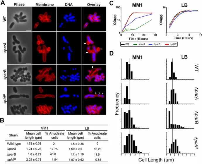
The chromosome partitioning system of the rod-shaped actinomycete, Corynebacterium glutamicum consists of the Walker-type ATPase (ParA), a DNA binding protein (ParB) and centromere-like parS sites, located at the origin-proximal region of the chromosome. ParB binds parS sites, specifically.
ParA is recruited to the ParB-parS nucleoprotein complex, likely providing the driving force needed to relocalize replicated oriC’s to the opposite cell pole. The ParB-oriC complex is then stably attached to the cell pole, where it remains and the cell divides in-between the segregated chromosomes. The phenotypic consequences of mutation of parA or parB include reduced growth rates, a high frequency of anucleate cells and altered cell lengths. Also, in the absence of parA, the oriC is mislocalized.
To date, polar origin tethering factors have been identified in only few bacteria. Thus, we wanted to identify and analyze the Actinobacteria chromosome polar targeting factor. A synthetic in vivo approach was employed to analyze the anchoring of the ParB-oriC nucleoprotein complex to the cell poles via interaction with DivIVA. It was shown that DivIVA is necessary and sufficient to recruit ParB, therefore also tether the oriC at the cell poles. With this synthetic system, in combination with mutational analysis, the interaction sites between ParB and DivIVA were mapped. In Corynebacterium glutamicum, mutation of the N-terminal of ParB showed reduced polar oriC localization. In addition, the interaction between ParB and DivIVA was demonstrated for other members of the Actinobacteria phylum, including the notorious pathogen Mycobacterium tuberculosis and Streptomyces coelicolor.
Corynebacterium glutamicum, which undergoes cell division between the segregated nucleoids but not necessarily precisely at midcell, does not possesses the conventional positive or negative FtsZ regulators found in other rod-shaped bacteria. However, Corynebacterium glutamicum encodes an orphan parA-like gene (pldP, for ParA-like Division Protein). In this thesis a number of subtle differences between ParA and PldP were highlighted, showing that PldP is not involved in chromosome segregation, but probably in regulation of cytokinesis. Similar to the MinD protein of B.
subtilis, PldP contains a putative C-terminal amphipathic helix. In vivo, PldP localizes, probably early, to the division septum and the localization pattern is highly reminiscent of MinD. Further in vitro analysis showed that PldP can bind membranes.
Contrary to the long-standing assumption, some Corynebacterium glutamicum strains encode an actin homologue. The cg1890 (designated AlpC, for Actin-Like Protein Corynebacterium) gene was recently identified as a putative actin-like protein. At the sequence level, AlpC shares little homology with actin and other actin-like proteins, however, it does contain the actin signature motive involved in nucleotide binding and hydrolysis. In vivo, AlpC forms dynamic structures, which assemble into long straight filaments and dissemble into foci. In vitro, AlpC can hydrolyze both ATP and GTP and exhibits nucleotide dependent polymerization. Mutation of the actin signature motif (AlpCD301A) abolishes nucleotide hydrolysis but not polymerization. Thus, AlpC is a genuine member of the actin-like superfamily.
On the genome, alpC is one of the first on the CGP3 prophage region. This prophage no longer forms infectious phage particles and most of the coding regions show little homology to known
Abstract
bacterial genes. The CGP3 prophage can excise from the bacterial chromosome and exist as multiple copies of circular DNA. In vivo co-visualization of induced prophage and AlpC filaments suggests that the Corynebacterium glutamicum actin-like protein is not involved in segregation of the prophage particles. However, in the absence of alpC, the frequency of prophage induction is drastically reduced.
Thus, we speculate that AlpC plays a role in replication the CGP3 prophage.
Zusammenfassung
II. Zusammenfassung
Das Chromosom-Partitionierungs-System des keulenförmigen Aktinomycets Corynebacterium glutamicum besteht aus einer „Walker-type“ ATPase (ParA), einem DNA-Bindeprotein (ParB) sowie zentromeren parS-Sequenzen, die sich an einer ursprungsnahen Region auf dem Chromosom befinden. ParB bindet spezifisch an parS-Sequenzen. ParA wird an den ParB-parS Nukleoprotein- Komplex rekrutiert und stellt dabei die treibende Kraft zur Verlagerung der replizierten oriC’s zum gegenüberliegenden Zellpol. Der ParB-oriC Komplex wird dann stabil an den Zellpol gebunden sodass die Zellteilung zwischen den getrennten Chromosomen stattfinden kann. Phänotypische Konsequenzen nach Mutationen von parA und parB sind reduziertes Wachstum, eine hohe Anzahl DNA-freier Zellen sowie veränderte Zellängen. Des Weiteren ist die oriC in Abwesenheit von ParA misslokalisiert.
Bis heute wurden Mechanismen zur polaren Chromosomenbindung nur in wenigen Bakterien identifiziert. Ein Ziel dieser Arbeit ist die Identifizierung und Charakterisierung des Chromosomsegregations-Mechanismus in Aktinobakterien. Zur Untersuchung der polaren Verankerung des ParB-oriC Nukleoprotein-Komplexes als Folge einer Interaktion mit DivIVA wurde ein synthetischer in vivo Ansatz etabliert. Es konnte gezeigt werden, dass DivIVA nötig und ausreichend ist um ParB, und somit den oriC, an die Zellpole zu rekrutieren. Des Weiteren konnte in dem in vivo Assay, zusammen mit Mutationsanalysen, die Bindestellen von ParB und DivIVA identifiziert werden. In Corynebacterium glutamicum führte eine Mutation des N-Terminus von ParB zu einer reduzierten polaren oriC-Lokalisation. Darüber hinaus konnte die Interaktion zwischen ParB und DivIVA für weitere Mitglieder der Abteilung der Aktinobakterien gezeigt werden, inklusive dem Krankheitserreger Mykobakterium tuberculosis sowie Streptomyces coelicolor.
Corynebacterium glutamicum, dessen Zellteilung zwischen den getrennten Chromosomen aber nicht zwangsläufig an der exakten Zellmitte stattfindet, besitzt keine konventionellen positiven oder negativen FtsZ-Regulatoren, die man in anderen keulenförmigen Bakterien findet. Jedoch kodiert Corynebacterium glutamicum ein parA-ähnliches Gen (pldP, engl.: ParA-like Division Protein). In der vorliegenden Arbeit werden einige subtile Unterschiede zwischen ParA und PldP beschrieben, die zeigen, dass PldP keine Rolle in der Chromosomensegregation spielt, vermutlich aber in der Zellteilung. Ähnlich dem MinD Protein aus Bacillus subtilis enthält PldP eine mögliche C-terminale amphipatische Helix. In vivo lokalisiert PldP, wahrscheinlich zu einem frühen Zeitpunkt des Zellzyklus, an das Zellteilungsseptum und gleicht somit dem Lokalisationsverhalten von MinD. Außerdem haben in vitro Studien gezeigt, dass PldP an Membranen bindet.
Gegensätzlich langjähriger Annahmen kodieren einige Corynebacterium glutamicum Stämme ein Homolog zum eukaryotischen Aktin. Das Gen cg1890 (umbenannt zu AlpC, engl.: Actin-like Protein Corynebacterium) wurde kürzlich als mögliches Aktin-ähnliches Protein identifiziert. Obwohl AlpC nur wenige Sequenzhomologien zum Aktin sowie anderen Aktin-ähnlichen Proteinen hat, enthält es das typische Aktinmotiv, dass in Nukleotidbindung und Hydrolyse involviert ist. In vivo bildet AlpC dynamische Strukturen die zu langen Filamenten assemblieren bzw. Foki disassemblieren können. In
Zusammenfassung
vitro kann AlpC sowohl ATP als auch GTP hydrolysieren und nukleotidabhängig polymerisieren.
Mutationen im Aktin-typischen Motiv verhindert Nukleotidhydrolyse jedoch nicht die Polymerisation.
Somit ist AlpC ein Mitglied der Aktin-ähnlichen Superfamilie.
Im Genom ist alpC eines der ersten Gene der CGP3 Prophagenregion. Dieser Prophage ist nicht mehr in der Lage infektiöse Phagenpartikel zu formen und die meisten Kodierungsregionen zeigen wenig Homologie zu bekannten bakteriellen Genen. Der CGP3 Prophage kann sich vom bakteriellen Chromosom trennen und als vielfache Kopie zirkulärer DNA existieren. In vivo Co- Visualisierungen induzierter Prophagen- und AlpC-Filamente lässt vermuten, dass das Aktin-ähnliche Protein aus Corynebacterium glutamicum in der Segregation der Prophagenpartikel keine Rolle spielt.
Jedoch ist in Abwesenheit von alpC die Häufigkeit der Prophageninduktion drastisch reduziert. Daher wird vermutet, dass AlpC eine Rolle in der CGP3 Prophagenreplikation spielt.
Abbrevations
III Abbreviations
ADP adenosine-5'-diphosphate
ATP adenosine-5'-triphosphate
Bp base pairs
DAPI 4'-6-Diamidino-2-phenylindole
BCIP 5-Bromo-4-chloro-3-indolyl phosphate CFP cyan fluorescent protein
CGXII minimal medium
DMSO dimethyl sulfoxide
DNA deoxyribonucleic acid
dNTP deoxyribonucleotide
eCFP enhanced cyan fluorescent protein EDTA ethylenediaminetetraacetic acid GFP green fluorescent protein
H2Od deionized water
H2Odd double deionized water IPTG isopropyl β-D-1-thiogalactopyranoside
kanR kanamycin resistance
Kav elution constant
kb kilo base pairs
kDa kilodalton
LB Luria Bertani medium
mCHERRY red fluorescent protein
MM1 minimal medium
MWCO molecular weight cut-off
n sample size
NBT nitro blue tetrazolium chloride OD600 optical Density at 600nm
PAGE polyacrylamid-gel electrophoresis
PCR polymerase chain reaction
PVDF polyvinylidene fluoride
SDS sodium dodecylsulfate
TAE tris-Acetat / EDTA-buffer
Tris tris-(hydroxymethyl)-aminomethane
V volt
YFP yellowfluorescent protein
Introduction
1. Introduction
Chapter I
Cytoskeleton elements involved in chromosome segregation and cell division
Bacteria exhibit a high degree of intracellular organization, both in the timing of essential processes and in the placement of the chromosome, division site and regulatory proteins. The high accuracy of spatial and temporal regulators ensures that chromosome replication initiation along with chromosome segregation and condensation are coordinated with cell growth and division, ensuring that each daughter cell receives a complete copy of the genetic material. Recent advances in microscopy and cytological techniques along with structural and biochemical analysis have greatly advanced our knowledge on the organization and regulation of the bacterial cell.
1.1 DNA replication initiation
Initiated from a single origin of replication (oriC), the bacterial chromosome is replicated bidirectionally and terminates at the opposite end of the chromosome (terC), where termination proteins disengage the replication machinery from the DNA (Sherratt, 2003). Replication initiation is mediated by a single initiator protein, DnaA, which specifically interacts with conserved motifs at the oriC (Messer, 2002;
Kaguni, 2006). In the ATP bound state, DnaA forms high-ordered multimeric complexes and cooperatively binds to oriC (Nishida et al., 2002). Binding of ATP-DnaA promotes local unwinding of the DNA duplex at AT-rich sequences, facilitating loading of the DNA helicase, and recruitment of other necessary initiator proteins (Sutton et al., 1998).
Replication initiation is strictly coordinated with cell cycle progression. Depending on the organism, a variety of mechanisms regulate the activity of DnaA, including mechanisms that affect the cellular concentration of DnaA, nucleotide bound state and accessibility to binding sites. Recent evidence shows that replication initiation and elongation is directly linked with nutrient availability in Escherichia coli and Bacillus subtilis (Janniere et al., 2007; Maciag et al., 2011). However, in some organisms (for example, E. coli) replication can be initiated more than once per cell cycle, giving rise to daughter cells that inherit a semi-replicated chromosome (Cooper and Helmstetter, 1968; Niki and Hiraga, 1998; Nielsen et al., 2007). Although this normally only occurs when there is an abundance of nutrients, regulation of multiple initiation events per cell cycle remains largely obscure.
1.2 DNA partitioning systems
The organization and dynamic behavior of the bacterial chromosome is particularly evident during chromosome replication and segregation. In E. coli and vegetative B. subtilis cells the newly
Introduction
duplicated origins rapidly move from the midcell to the cell quarter positions, sites which represent the predivisional site in each daughter cell (Niki and Hiraga, 1998; Webb et al., 1998; Niki et al., 2000). In Caulobacter crescentus, the oriC is positioned at the cell pole and after replication the sister origin is moved to the opposite cell pole (Mohl and Gober, 1997; Viollier et al., 2004). More interestingly, in the gamma-proteobacterium Vibrio cholerae, which contains two circular chromosomes, the oriC of chromosome I (chrI) is segregated unidirectionally similar to C. crescentus, while the oriC of chromosome II (chrII) is found at the cell centre and is segregated bidirectionally similar to the chromosomes of E. coli and B. subtilis (Fogel and Waldor, 2005; Fiebig et al., 2006). Irrespective of cellular localization and track of movement, common to all is the rapid and directed translocation of the oriC which occurs prior to completion of chromosome replication.
Thus, it appeared that, similar to the eukaryotic chromosome segregation apparatus, a mitotic- like apparatus could also exist in bacteria. As early as 1977, an active partitioning system was suggested for the low copy plasmid P1 prophage from E. coli due to its extremely efficient maintenance, despite often being present in as few as two copies prior to division (Prentki et al., 1977). Indeed, the parABS locus was later identified and shown to be necessary and sufficient for stable maintenance of P1 prophage (Abeles et al., 1985). Homologues of the par locus were subsequently identified on the bacterial chromosome, and found to be highly conserved, found across the bacterial domain and in many archaea, as well as in some chloroplasts and mitochondria (Gerdes et al., 2000; Livny et al., 2007). However, the chromosome of E. coli and Mycoplasma species are examples of the few exceptions.
Plasmid partitioning systems
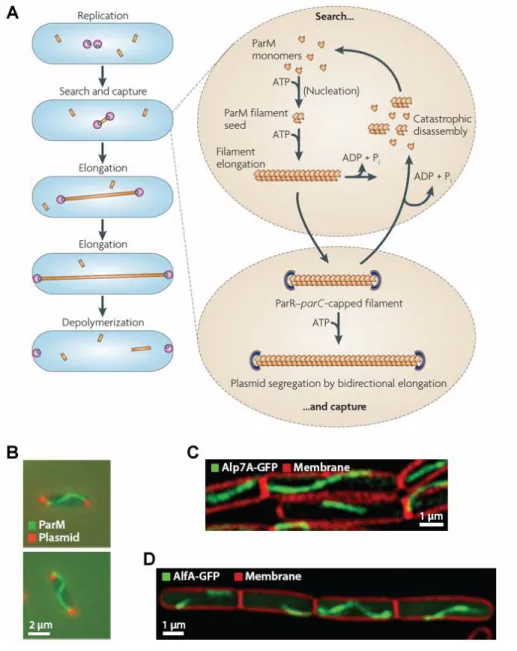
The genetic organization of par loci is similar for both chromosome and plasmid encoded systems. In general, the par locus entails two trans-acting proteins encoded in an operon (parA and parB, respectively) and cis-acting “centromere-like” elements. The centromere-binding protein (ParB) binds the centromere-like element (parS) forming a nucleoprotein complex (Funnell, 1987). The segrosomes are recognized and subsequently segregated by the action of dynamic cytoskeleton filaments composed of ParA protein, which, depending on the plasmid partitioning system, is either a Walker-A P loop ATPase (ParA, Type I), an actin-like ATPase (ParM, Type II) or a tubulin-like GTPase (TubZ, Type III) (Gerdes et el., 2000, Larsen et al., 2007). (Refer to chapter II for more details on type II plasmid partitioning systems).
The par loci of Type I plasmid partitioning systems are among the best characterized, including E. coli P1 and F plasmids (Ogura and Hiraga, 1983; Austin and Abele, 1983a; Austin and Abele, 1983b; Abeles et al., 1985). Type I systems can be divided further into type Ia and Ib which differ in genetic organization and homology of the Par proteins encoded (Davis et al., 1992; Hayes et al., 1994; Radnedge et al., 1998; Libante et al., 2001). Both systems encode a Walker-type ATPase located upstream of the centromere-binding protein. In type Ia the centromere-like element is located downstream of the par operon, while in type Ib this element is found upstream, often in the promoter
Introduction
region of the par operon. Type Ia ParA proteins contain a putative N-terminal helix-turn-helix (HTH) DNA binding motive and a C-terminal Walker box domain. The ParA protein can associate with the operator sequence of the par promoter to down-regulate transcription (Davis et al., 1992; Davey and Funnell, 1994; Davey and Funnell, 1997). The putative DNA binding motive is absent in type Ib ParA proteins. However, the centromere-binding protein functions as a transcription regulator upon binding the centromere-like element, which is situated in the promoter region of the par operon. Thus, in both systems autoregulation maintains the correct stoichiometry of Par proteins which is essential for plasmid partitioning (Hirano et al., 1998; Friedman and Austin, 1998).
The P1 and F plasmids exhibit very specific subcellular localizations. When only a single plasmid is present, midcell localization is observed, while in cells that contain two plasmids each plasmid is positioned at the cell quarter positions (Kim and Wang, 1999; Erdmann et al., 1999). This rapid segregation is highly dependent on the Par system (Gordon et al., 1997). Cooperative binding of ParB to the centromere-like element leads to the formation of higher ordered partition complexes.
Although controversial, this interaction is thought to mediate plasmid pairing (Jensen et al., 1998;
Edgar et al., 2001). Plasmid pairing ensures that plasmids that use similar replication or segregation mechanisms are incompatible and cannot coexist in the same cell (Austin and Nordstroem, 1990).
Subsequent to nucleoprotein complex formation, a fully active segrosome is formed only when bound by ParA. The ParA protein forms dynamic gradients, oscillating over the bacterial nucleoid, directing the movement of the plasmid (Ringgaard et al., 2009). The activity of ParA is modulated by ParB, regulating the nucleotide bound state of ParA and its affinity for the nucleoid or polymerization into filaments. Numerous models have been proposed in an attempt to explain the dynamic oscillating activities of ParA and the directed movement of replicated plasmids. A recent model proposed by Ringgaard and co-workers suggests that ATP hydrolysis and depolymerization of the ParA-ATP polymers is stimulated by interaction with ParB (Ringgaard et al., 2009). However, the nucleoprotein complex remains attached to and follows the end of the retracting ParA filament. Released ParA monomers rebind ATP and polymerize in regions furthest away from the nucleoprotein complex.
Another model suggests that ParA forms filaments that extend from pole to pole and again upon encountering the nucleoprotein complex, the depolymerizing ParA filament pulls the replicated plasmid to their correct subcellular position. Although the molecular mechanism has not been completely unraveled, it is clear that in the absence of one component of the Par system, plasmids are not stably maintained.
Chromosome encoded partitioning systems
Chromosome encoded par loci are classified as Type I, encoding ParA ATPases which contain Walker A / B motifs that have low ATPase activity. The parAB locus and centromere-like elements are generally located in the origin-proximal region (Gerdes et al., 2000; Livny et al., 2007), suggesting a functional preservation among different bacterial species. Structural and biochemical analysis revealed that ParB (Spo0J) of Thermus thermophilus has four domains, a flexible N-terminal domain,
Introduction
a central HTH domain followed by a linker domain and a C-terminal dimerization domain (Leonard et al., 2004). It has been suggested that the C-terminal domain is the primary dimerization domain, while the N-terminal and HTH domains compose a secondary dimerization domain that is necessary for DNA binding. In vitro, Spo0J can stimulate the ATP hydrolysis of Soj (ParA), which is further enhanced in the presence of parS DNA (Leonard et al., 2005). In some plasmid and chromosome encoded Par systems, the ParA interaction site has been mapped to a short but highly conserved part of the N- terminal of ParB / Spo0J (Surtees and Funnell, 1999; Figge et al., 2003; Ravin et al., 2003). In vitro, the hydrolysis rate of ParA can be specifically stimulated by the N-terminal 20 amino acid of ParB (Leonard et al., 2005). Structural and biochemical analysis of Thermus thermophilus Soj revealed that upon binding ATP Soj dimerizes forming a “nucleotide sandwich dimer”. Only as an ATP bound dimer can Soj bind DNA, both of which are prerequisites for polymerization (Leonard et al., 2005).
B. subtilis, C. crescentus, E. coli and V. cholerae have emerged as model organisms for studying the molecular mechanisms of chromosome segregation, to date. Although E. coli does not encode a Par system, segregation has been studied in detail in this organism (Reyes-Lamothe et al., 2008; Wang and Sherratt, 2010). Despite the high degree of conservation at the levels of sequence and genetic organization, studies on chromosomal ParA and ParB revealed a lack of uniformity in their action in different organisms. New insights into the molecular role(s) of Par systems show an involvement in a number of overlapping systems and subsequent mutation of the Par system lead to pleiotropic phenotypes, making it difficult to decipher their precise role. However, a role in segregation is underlined by the fact that chromosomally encoded Par systems can stabilize unstable plasmids as long as the plasmid contains the cognate parS sequence (Lin and Grossman, 1998; Yamaichi and Niki, 2000; Godfrin-Estevenon et al., 2007).
Chromosome segregation in Bacillus subtilis
In B. subtilis, the ParB homologue (Spo0J) binds at least 8 parS sites found in the origin-proximal region of the chromosome and spreads latterly along the DNA to form foci that localize at the cell quarter positions, coinciding with the oriC region during vegetative growth (Lin et al., 1997; Glaser et al., 1997; Lin and Grossman, 1998; Murray et al., 2006). The ParA homologue (Soj) exhibits two distinct localization patterns; as cytoplasmic foci which are dependent on Spo0J and septal localization that is dependent on the division site selection protein MinD. (Autret and Errington, 2003;
Murray and Errington, 2008). In the absence of spo0J, Soj remains statically associated with the nucleoid (Marston and Errington, 1999; Quisel et al., 1999; Murray and Errington, 2008).
On a molecular level, the interplay between Soj and Spo0J influences multiple processes.
Upon binding ATP, Soj dimerizes and subsequently exhibit non-specific, cooperative DNA binding activity (Murray and Errington, 2008). Two positively charged residues of the N-terminal of Spo0J (amino acid 3 and 7, both lysine) can stimulate the ATPase activity of Soj, driving the Soj-ATP dimer to a monomer and subsequent release from DNA (Gruber and Errington, 2009). This cycle of DNA binding and ATP-hydrolysis is important for the regulation of replication initiation (Murray and
Introduction
Errington, 2008; Scholefield et al., 2011). In the monomeric form, Soj directly interacts with DnaA and inhibits DNA replication (Scholefield et al., 2012). Positive regulation of DnaA by Soj requires its DNA binding activity. Although dimeric Soj also directly interacts with DnaA, the molecular mechanism of activation has not been elucidated, yet.
In vegetative cells, soj mutants do not exhibit any gross segregation defects (Ireton et al., 1994; Webb et al., 1998), while deletion of sop0J does not affect origin localization and dynamics, but does however result in mild segregation defects (approximately 2 % anucleate cells) (Lin and Grossman, 1998). In addition, contrary to other ParA homologues, assembly of Soj into filaments, either in the presence or absence of DNA, has not been observed (Scholefield et al., 2011). However, Soj and Spo0J are involved in chromosome organization during sporulation. Under nutrient limitation B. subtilis cells can undergo a complex morphological developmental route leading to the production of endospores. Sporulation specific genes are transcribed, the chromosome adopts an elongated structure (called an axial filament) extended from pole to pole and an asymmetric septum forms close to one cell pole, forming the prespore compartment. Due to the relaxed configuration some of the axial filament becomes trapped in the prespore. Prior to closure of the septum, the remainder of the chromosome is pumped into the prespore compartment by the DNA translocase, SpoIIIE. Analysis of the organization of the axial filament revealed that the oriC, representing about one quarter of the chromosome, is positioned at the cell pole in the prespore compartment while the terC is in the mother cell (Wu and Errington, 1998; Sullivan et al., 2009). In the absence of soj and spo0J the organization of the axial filament is dramatically altered, frequently resulting in prespores lacking the oriC (Wu and Errington, 1998; Lee et al., 2003; Sullivan et al., 2009).
Soj was suspected to act as a transcription regulator that negatively regulates sporulation specific genes (Ireton et al., 1994). More recently, this block in sporulation was shown to be indirect, through the activation of the Sda replication checkpoint (Murray and Errington, 2008). In the absence of spo0J entry into the sporulation pathway is blocked as the nucleotide bound state of Soj is not regulated, which subsequently effects DnaA mediated replication initiation leading to activation of Sda.
However, what remains to be understood is the consequence of the interaction between Soj and the cell division protein MinD (Autret and Errington, 2003; Murray and Errington, 2008). ATP bound Soj interacts with and is recruited by MinD to the division septa, and in the absence of minD Soj is diffused throughout the cytoplasm (Scholefield et al., 2011). One could imagine that MinD fulfills a role similar to Spo0J, regulating the nucleotide bound state of Soj, perhaps to synchronize initiation of replication and cell division. However, in the absence of minD chromosome replication and organization is not affected (Marston et al., 1998).
Chromosome segregation in Caulobacter crescentus
C. crescentus is an α-proteobacterium that divides unequally into a swarmer cell and a sessile stalked cell, which, aside from distinct morphological features, have different cell fates. DNA replication is initiated only when the swarmer cell differentiates into a stalked cell (Jensen and Shapiro, 1999;
Introduction
Jensen et al., 2001). After origin duplication, the sister origin is translocated from the flagellated pole (old pole) to the opposite cell pole. Using high-resolution microscopy, retraction of ParA filaments was shown to coincide with the directed movement of the ParB bound origins (Ptacin et al., 2010). Similar to the situation in B. subtilis, ParB regulates ParA activity by modulating the nucleotide bound state of ParA (Easter and Gober, 2002). ParA monomers bind ATP and dimerize, which subsequently result in DNA binding or polymerization (Ptacin et al., 2010; Schofield et al., 2010). ParA forms filaments that pull the replicated chromosome to the opposite cell pole. Upon interaction with ParB the ATPase activity of ParA is stimulated, hydrolyzing ATP and thereby releasing ParA units from the filament, resulting in shortening of the filament and subsequent pulling of the oriC to the opposite cell pole (Fig.
1.1A). Released ParA is recruited to the cell pole by interaction with TipN, preventing further interaction with ParB (Lam et al., 2006). Together, TipN and PopZ hold ParA and the partitioning complex at the cell poles preventing additional rounds of replication initiation until completion of division. After completion of cell division, TipN relocates to the new cell pole, releasing ParA which can again polymerize restarting the cycle.
Although, the parA and parB genes are indispensable in C. crescentus and severe chromosome segregation defects result from overexpression of ParA or ParB, depletion of ParB or mutations in the ParA ATPase active site (Mohl et al., 1997; Mohl et al., 2001), ParA is not involved in regulation of replication. Instead, rapid degradation of DnaA at the end of every cell cycle regulates initiation of replication (Gorbatyuk and Marczynski, 2005). However, mutation of parAB additionally results in a block in cytokinesis (Mohl et al., 2001; Figge et al., 2003). Thus, the parAB locus of C.
crescentus has multiple additional roles outside of chromosome segregation and the act of chromosome segregation itself serves as a check point for initiation of cell division.
Chromosome segregation in Vibrio cholerae
V. cholerae is a Gram-negative, rod-shaped gamma-proteobacterium that contains two unequally sized circular chromosomes, chrI (2.96 Mb) and chrII (1.07 Mb). Although each chromosome encodes parAB genes, phylogenetic analysis suggests that ParAB from chrI (ParA1 and ParB1) cluster with chromosomal ParAB proteins, while the Par proteins encoded on chrII (ParA2 and ParB2) cluster with plasmid and phage Par proteins (Gerdes et al., 2000; Heidelberg et al., 2000; Yamaichi and Niki, 2000). The product of each encoded par system is necessary for segregation of the parental chromosome and is thought not to influence segregation of the other chromosome. Indeed, the nucleotide sequence of the centromere-like elements on chrI and chrII differs significantly. The nucleotide sequence of parS2 is restricted to Vibrio and Photobacteria species and are only bound by their cognate ParB protein (Yamaichi et al., 2007; Livny et al., 2007). The origin of chrI exhibits a polar localization and sister origins are segregated to the opposite cell pole. ParA1 interacts with the ParB1- parS1 nucleoprotein complex and functions to anchor the nucleoprotein at the cell pole and segregate the sister origin via a “pulling” mechanism (Fig. 1.1B) (Fogel and Waldor, 2006). Lack of parBI leads to reduced polar localization of the origins and also DNA replication overinitiation (Kadoya et al., 2011).
Introduction
Although overinitiation of replication was not observed for a parA1 mutant, increased DNA content was seen upon overexpression of ParA1 in a parAB1 mutant strain suggesting that ParB1 mediates it action through ParA1.
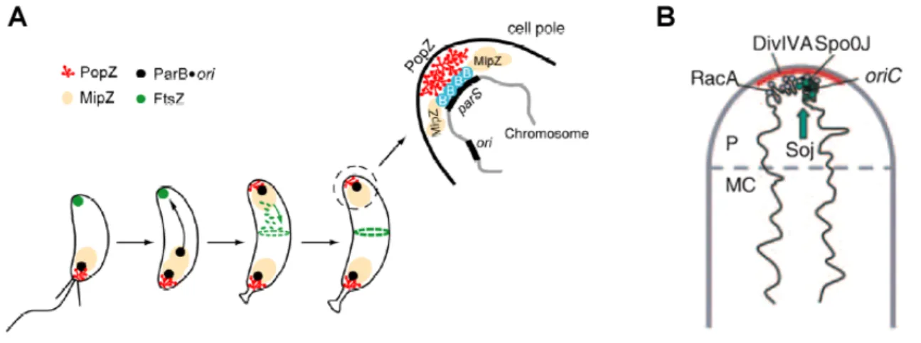
Figure 1.1: Model of chromosome segregation in C. crescentus and V. cholerae. In both organisms ParA polymerizes into filaments which depolymerize upon interaction with the ParB bound origin. A “pulling”
mechanism has been proposed, whereby the retracting ParA filament pulls the replicated origin to the opposite cell pole. (A) In C. crescentus, the origin is positioned at the old pole in a newborn cell. The duplicated origin is bound by ParB and upon interaction with the ParA filament, which extends almost the entire length of the cell, ParB stimulates the hydrolysis of ParA-ATP. ParA subunits are released from the depolymerizing filament and held at the new cell pole through interactions with TipN, preventing further polymerization and / or interaction with ParB. After completion of cell division, ParA is released from the pole as TipN moves to the site of most recent division. (B) In the predivisional cell, V. cholerae ParA holds the oriC at the cell poles through interaction with an as-of-yet unidentified factor (I). After duplication of the origins ParA polymerization is initiated, which is possibly linked to assembly of the cell division apparatus or the inward growing septum (II). When the cell has completed division, one end of the ParA filament is attached to the cell pole and the other end interacts with the ParB-oriC.
Again, ParB stimulates hydrolyses of the ParA bound ATP, causing the ParA filament to retract, pulling the sister origin to the opposite cell pole (III, IV). Models adopted from Kirkpatrick and Viollier, 2010; Fogel and Waldor, 2006, respectively.
Less is known about segregation of chrII. In contrast to chrI, chrII undergoes a symmetric division moving bidirectionally to the cell quarter positions. Null mutants of parAB2 exhibit altered growth and localization of chrII, often leading to cells that lack chrII (Yamaichi et al., 2007). In the absence of chrII, cells undergo one round of division before cell death.
1.3 Chromosome condensation - SMC and organization of chromosomes
In parallel to replication and also probably segregation of the oriC, the newly replicated DNA must be reorganized. One of the main players in chromosome organization and condensation is the highly conserved structural maintenance of chromosomes (SMC) protein, found across all three kingdoms of life. Bacterial cells normally encode a single SMC protein, which forms a coiled-coil protein with a
Introduction
hinge domain at one end and an ABC-type ATPase domain at the other end (Saitoh et al., 1994; Löwe et al., 2001). Upon homodimerization, contacts of the hinge regions lead to a V-shaped dimer which can subsequently form a ring-like structure through ATP dependent interactions between the ATPase domains and / or associations with the kleisin family protein, ScpA and ScpB (Soppa et al., 2002;
Volkov et al., 2003; Hirano and Hirano, 2004; Schwartz and Shapiro, 2011). These complexes are thought to interconnect different regions of the chromosome, stabilizing them in a twisted, condensed conformation (Hirano and Hirano, 2006). In B. subtilis, null mutant of smc display pleiotropic phenotypes, including growth defects, anucleate cells and unorganized chromosome with aberrant oriC and terC localization, less condensed and often guillotined chromosomes that have been chopped by the division septum (Britton et al., 1998; Mascarenhas et al., 2002; Soppa et al., 2002).
Simultaneous deletion of smc and spo0J exaggerates the phenotype of the single smc mutant (Autret et al., 2001). Similarly, deletion of the origin-proximal parS sites led to a phenotype intermediate of the single mutants (Sullivan et al., 2009).
In B. subtilis, SMC localization is dependent on complex formation with ScpA and ScpB (Lindow et al., 2002). The finding that the SMC complex accumulates as foci around the oriC region gave the first hint that SMC proteins could be directly involved in chromosome segregation (Britton et al., 1998). Indeed, recruitment of SMC to the oriC is dependent on Spo0J-parS nucleoprotein complex formation and subsequent lateral spreading of Spo0J on the chromosome (Sullivan et al., 2009;
Gruber and Errington, 2009). Thus, another role of Spo0J is as a chromosome loading factor for site specific recruitment of the SMC complex. Once in position at the oriC, SMC acts as an “organizing center” interacting with large regions of the chromosome, functioning in chromosome reorganization, condensation and also aiding segregation. However, in B. subtilis other recruitment factors must exist as deletion of spo0J does not lead to gross segregation defects (Lin and Grossman, 1998). Non- specific association of SMC with the chromosome is speculated to be sufficient for chromosome organization in the absence of spo0J, at least in vegetative cells. Such functionally redundant mechanisms ensure faithful chromosome segregation and can, at least partly, explain the mild spo0J mutant phenotype.
Spo0J plays a role in regulating the activities of Soj, which in turn regulate replication initiation, segregation of the oriC and transcription of early sporulation specific genes. Spo0J additionally marks the oriC priming it for loading of SMC, which functions in segregation of the bulk of the chromosome.
Interestingly, mutant forms of Spo0J were isolated that are either defective in regulation of Soj or in the SMC pathway demonstrating that the different functions of Spo0J are independent from each other (Gruber and Errington, 2009).
E. coli does not encode homologues of SMC, ScpA or ScpB, however they encode the structurally and functionally related MukB, MukF and MukE proteins (Niki et al., 1991; Yamanaka et al., 1996). The characteristic architecture of SMC proteins is shared by MukB and MukF is classified as a kleisin (Fennell-Fezzie et al., 2005). Mutation of either component causes atypical nucleoid localization, anucleate cells and temperature sensitive growth defects, all phenotypes that are indicative of a role in chromosome organization and segregation (Yamanaka et al., 1996). Akin to B.
Introduction
subtilis SMC, MukBEF complexes are targeted to the oriC regions (Danilova et al., 2007). How this targeting occurs is not well understood. However, chromosome partitioning appears to be mediated through interaction with DNA topoisomerases IV, which is involved in DNA decatenation (Hayama and Marians, 2010; Li et al., 2010). A direct physical interaction is necessary for MukB function, but the molecular mechanisms have not been elucidated, yet.
1.4 Cell division
Once the chromosomes have been replicated and (partially) segregated, the bacterial cell will divide to produce two genetically identical daughter cells. Temporal control of cytokinesis ensures that cell division does not precede chromosome duplication and partitioning. The cell division apparatus is made up of a large number of proteins. The foundation for assembly of the division complex is laid down by the tubulin homologue FtsZ (Bi and Lutkenhaus, 1991; Wang and Lutkenhaus, 1993).
Polymerization of FtsZ at the midcell region results in the formation of a ring-like structure. These Z- rings act as a scaffold for the assembly of additional proteins into the contractile Z-ring, forming a multisubunit complex, known as the divisome (Adams and Errington, 2009).
Spatial regulation of cytokinesis – negative regulation
Many organisms employ a dual mechanism to spatially regulate divisome positioning and assembly, including the Min system and nucleoid occlusion (Woldringh et al., 1990; Harry, 2001; Wu and Errington, 2004). The Min system of E. coli consists of MinC, MinD and MinE, all of which work collectively to prevent division sites from occurring at the cell poles (de Boer et al., 1988; de Boer et al., 1989). Upon binding ATP, MinD associates with the cell membrane and recruits MinC (de Boer et al., 1991). MinC is a division inhibitor that prevents FtsZ from forming a stable cytokinetic ring (Hu et al., 1999; Hu and Lutkenhaus, 2000). The third component, MinE, binds MinD stimulating ATP hydrolysis (Fu et al., 2001). MinD-ADP disassociates from the membrane and as a consequence also MinC and MinE (Hu and Lutkenhaus, 1991). In the cytoplasm, nucleotide exchange results in reassembly of MinD-ATP at the cell membrane where MinE concentration is lowest, namely the opposite cell pole. This cycle of events results in a pole to pole oscillation of MinCD, leading to a MinCD gradient which is lowest at midcell, where FtsZ polymerizes.
The Min system of B. subtilis does not oscillate and consists of four proteins. The topological determinant, DivIVA, localizes to the curved membranes of the cell poles and is also recruited late to the assembling divisome (Cha and Stewart, 1997; Edwards and Errington, 1997). DivIVA recruits MinJ to the cell poles and division septa (Patrick and Kearns, 2008; Bramkamp et al., 2008). MinJ is an adaptor protein necessary for recruitment of MinD and also indirectly for MinC. Contrary to the role of the Min system in E. coli, recent evidence suggests that the B. subtilis Min system is important for disassembly of the divisome machinery (van Baarle and Bramkamp, 2010). In the absence of MinCD or MinJ, division proteins remain associated to the divisome at the cell pole instead of localizing at
Introduction
midcell. This subsequently leads to the initiation of a new round of cytokinesis close to the original cell division site, leading to rows of minicells.
Spatial regulation by the nucleoid occlusion system employs DNA binding proteins that bind to the nucleoid, inhibiting FtsZ polymerization over areas that contain DNA (Wu and Errington, 2004).
Nucleoid occlusion may also provide a checkpoint-like mechanism for preventing division before replication and segregation of the sister chromosome has been completed. In B. subtilis, the Noc protein protects the nucleoid and in E. coli the unrelated SlmA carries out a similar role. In vitro, SlmA directly binds FtsZ, promoting polymer assembly (Bernhardt and de Boer, 2005; Cho et al., 2011). It has been proposed that recruitment of FtsZ away from the membrane may compete with other FtsZ assembly proteins, thus inhibiting the formation of a functional cytokinetic ring. Interestingly, Noc is highly homologous to the partitioning protein ParB and in a similar mechanism Noc binds to a palindromic sequence found through out the chromosome (Wu et al., 2009).
Novel spatial and temporal regulation of cytokinesis
Not all bacteria that divide in a medial fashion have a Min or Noc system, for example C. crescentus.
Thus, alternative spatial regulators must exist. In C. crescentus, a novel cell division protein, MipZ, spatially regulates FtsZ polymerization (Thanbichler and Shapiro, 2006). MipZ is a homologue of ParA and a member of the Walker A ATPase family. MipZ is highly conserved among α-proteobacteria. In C. crescentus, MipZ is an essential protein. It forms a complex with the polar ParB-oriC nucleoprotein complex, but does not localize as tight foci. Instead, MipZ forms a gradient that peaks at the cell poles, where the origins are localized, and decreases towards midcell. In vitro, MipZ directly interacts with FtsZ stimulating its GTPase activity, inhibiting FtsZ polymerization. Interestingly, MipZ is distantly related to MinD, however the underlying mechanism of spatial regulation is different (see below).
Subsequent to cell division, the ParB-oriC nucleoprotein complex is found at the old cell pole, while FtsZ forms foci at the new cell pole (the site of most recent cell division) (Fig. 1.2 A). After initiation of chromosome replication and segregation, MipZ binds the origins through direct interaction with ParB. Depolymerizing ParA filaments translocate the sister origin is to the opposite cell pole.
Upon reaching the cell pole, MipZ stimulates the GTPase activity of FtsZ, displacing it from the cell pole. FtsZ subsequently moves to a region in the cell where MipZ concentration is lowest, namely midcell. Here, the divisome assembles and the cell divides. Depletion of MipZ leads to the formation of minicells and sporadic division events. Additionally, depletion of ParB or overexpression of ParA leads to a block in cell division resulting in long filamentous cells (Mohl et al., 2001). Alterations in the ratio of ParA to ParB might lead to increase ParA-ADP, which binds DNA, possibly leading to repression of cell division genes (Mohl et al., 2001). Thus, MipZ, together with ParA and ParB, spatially and temporally synchronizes cytokinesis with chromosome segregation.
Introduction
Spatial regulation of cytokinesis – positive regulators
Aside from negative regulators of division site placement, positive regulators of FtsZ polymerization have also been recently been described for Streptomyces coelicolor. S. coelicolor is a member of the Actinobacteria family, which grows by tip extension and hyphal branching forming the mycelia of vegetative hyphae (Flärdh and Buttner, 2009). On occasion crosswalls form in vegetative hyphae separating the mulitgenomic compartments. Morphological differentiation into aerial hyphae is accompanied by increased chromosome replication resulting in numerous copies of non-segregated chromosomes filling the aerial hyphae. Cessation of growth is closely followed by development into endospore chains. During this development phase, chromosome segregation and condensation is closely followed by the assembly of regularly spaced FtsZ rings, marking the localization of sporulation septa (Schwedock et al., 1997).
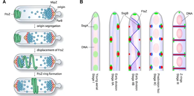
Figure 1.2: Spatial and temporal regulation of FtsZ in C. crescentus and S. coelicolor. (A) In Caulobacter crescentus MipZ is an inhibitor of FtsZ polymerization. MipZ forms a complex with the ParB bound origin at the old cell pole. After replication of the origin, ParA mediates translocation of the sister origin to the opposite cell pole, which is closely followed by MipZ. Upon reaching the opposite cell pole, MipZ displaces FtsZ. FtsZ reassembles where MipZ concentration is lowest. Thus, MipZ exploits origin segregation for its subcellular positioning and additionally spatially and temporally regulates cell division. (B) In S. coelicolor FtsZ and SsgB are diffused in young aerial hyphae, while SsgA forms foci (Stage I). SsgA dependent recruitment of SsgB in the pre- sporulation hyphae marks the predivisional sites. FtsZ uses SsgAB as landmarks for localization into long spiral- like filaments throughout the hyphae. SsgB interacts with the membrane and FtsZ, tethering FtsZ in position. Z- rings are formed and the divisome matures in coordination with chromosome segregation and condensation resulting in equally sized prespore compartments. As the prespore matures, SsgA remains localized at alternating sides of the division septum. Adopted from Thanbichler and Shapiro, 2006; Willemse et al., 2010.
Interestingly, in S. coelicolor deletion of ftsZ does not perturb growth in vegetative and aerial hyphae, which has not been reported for any other organism to date, however spore development defects are observed (McCormick et al., 1994; McCormick and Losick, 1996). Similar to C. crescentus,
Introduction
the canonical nucleoid occlusion and Min system are absent in S. coelicolor. Two novel positive regulators of FtsZ, SsgA and SsgB, were recently identified in S. coelicolor (Willemse et al., 2010).
Null mutants of ssgA and ssgB are blocked at stages preceding the onset of sporulation specific cell division (van Wezel et al., 2000; Keijser et al., 2003). In the absence of ftsZ, SsgA and SsgB still localize, however no septa are produced (Willemse et al., 2010). In young aerial hyphae, SsgA expression is upregulated and, as development proceeds, SsgB is recruited by SsgA to alternating sides of the aerial hyphae membrane (Fig. 1.2 B). FtsZ begins to localize as spiraling filaments that are speculated to attach to the membrane bound SsgB. Subsequently, Z-rings form and sporulation septa develop. In light of these data, it is obvious that bacteria have developed multiple mechanisms to spatially and temporally organize cell division.
1.5 Cell polarity – integrated temporal and spatial regulation
Bacterial cells exhibit a high level of intercellular organization. Proteins or protein complexes are distinctly and selectively localized to specific subcellular regions, spatially organizing the cytoplasmic environment and in turn influencing temporal regulation. In rod-shaped bacteria, the cell poles have emerged as important sites for the intracellular localization of proteins, lipids and nucleic acids, affecting numerous cellular processes including chemotaxis, pole morphogenesis, symmetric and asymmetric cell division and chromosome segregation (Edwards and Errington, 1997; Goley et al., 2007; Lam et al., 2006; Ringgaard et al., 2011). Polarity can be generated as a result of cell division.
Cytokinesis in rod-shaped bacteria gives rise to offspring with differently organized cell poles (Lawler and Brun, 2007). Specific proteins are inherited at the site of most recent division (new pole) historically distinguishing the two poles, setting up spatial cues in the progeny cells that once again guide the cell through the cell cycle. In some bacteria, the oriC is a molecularmarker of cell polarity, which is firmly localized to one cell pole, the old pole immediately after division (Webb et al., 1997).
Following chromosome replication, the newly replicated origin is translocated to the opposite cell pole and is subsequently attached to the opposite cell pole.
Although polar localization of the oriC is not common for all bacteria, Caulobacter crescentus and sporulating Bacillus subtilis exhibit polar attachment of the origin. In C. crescentus, a multimeric self-organizing protein (PopZ) was recently identified as a chromosome origin tethering factor (Fig. 1.3 A) (Bowman et al., 2008; Ebersbach et al., 2008). Polar localized PopZ sets up a polar epicenter that not only anchors the chromosome origins, but also stabilizes bipolar gradients of the cell division inhibitor MipZ (Thanbichler and Shapiro, 2006) and mediates the localization of morphogenic and cell cycle regulating proteins (Bowman et al., 2010). However, PopZ is not highly conserved and many bacterial species exhibit polar localization of chromosome origins. Thus, the mechanism of polar origin anchoring must differ in unrelated bacteria.
In B. subtilis, upon entry into sporulation rearrangements of the chromosome lead to anchoring of the oriC regions at the cell pole where asymmetric division will occur. Tethering the