Impact of trehalose and mycolate biosynthesis on the cell envelope of
a Corynebacterium glutamicum L-lysine production strain
Inaugural-Dissertation zur Erlangung des Doktorgrades
der Mathematisch-Naturwissenschaftlichen Fakultät der Universität zu Köln
vorgelegt von Henrike Gebhardt
aus Köln
Köln, Oktober 2005
1. Referent: Herr Prof. Dr. Reinhard Krämer 2. Referent: Herr Prof. Dr. Thomas Langer
Tag der Disputation: 28. November 2005
Kurzzusammenfassung
Einfluß von Trehalose – und Mycolatbiosynthese auf die Zellwand eines Corynebacterium glutamicum L-Lysinproduktionsstamms
Corynebacterium glutamicum besitzt, im Gegensatz zu anderen Gram-positiven Bakterien, in der Zellwand eine äußere Lipiddoppelschicht, die Mycolatschicht, die eine Permeabilitätsbarriere darstellt. Trehalose ist ein wichtiger Bestandteil der Mycolatschicht und an der Biosynthese von Mycolat beteiligt. Es konnte gezeigt werden, dass der erste Schritt der Mycolatbiosynthese, die Kondensation zu Trehalosemonomycolat, in der Zellwand stattfindet. Um die Bedeutung von Trehalose für die Mycolatschicht zu untersuchen, wurde die Zusammensetzung der Mycolatschicht gezielt durch die Kultivierung eines trehalosedefizienten Stammes mit verschiedenen Kohlenstoffquellen, in An- und Abwesenheit von Trehalose manipuliert. Ein trehalosedefizienter Stamm, der auf einem C. glutamicum L-Lysinproduzenten basierte, wurde gewählt, um zu überprüfen, ob die Veränderung der Mycolatzusammensetzung die Exkretion von L-Lysin steigern könnte.
Weiterhin sollte die Lysinexkretion mit der Permeabilität der Zellwand korreliert werden. Die Analysen zeigten, dass Trehalose essentiell für die Mycolatbiosynthese war, wenn Saccharose oder Fruktose als Kohlenstoffquelle dienten, während Glukose Trehalose als Akzeptor und Überträger von Mycolsäuren ersetzen konnte. Externe Trehalose konnte cytoplasmatische Trehalose nur teilweise für die Mycolatsynthese ersetzen, so dass die Supplementierung des Mediums mit Trehalose im trehalosedefizienten Stamm nicht vollständig die Eigenschaften der Mycolatschicht des Ausgangsstammes wiederherstellen konnte. Eine unvollständige Mycolatschicht erhöhte die Permeabilität der Zellwand, und gleichzeitig steigerte sie die Exkretion von Lysin und regte die Exkretion von Glutamat an.
Die Permeabilitätsbarriere kann scheinbar nur dann aufgebaut werden, wenn alle Bestandteile in der Menge vorhanden sind, die eine korrekte Anordnung der Mycolatschicht ermöglicht.
Die physiologische Funktion von einem der drei Trehalosestoffwechselwege in C. glutamicum, dem OtsAB-Weg, war vor Beginn der Arbeit noch unbekannt. Die Analyse von Stämmen mit nur einem funktionellen Trehalosestoffwechselweg zeigte, dass der OtsAB-Weg der wichtigste Trehalosesyntheseweg unter Kohlenstofflimitierung war. Da Trehalose das wichtigste kompatible Solut unter Stickstofflimitierung ist, könnte der OtsAB- Weg für die Synthese von Trehalose als Schutzsubstanz gegen osmotischen Stress unter Kohlenstoff- und Stickstofflimitierung notwendig sein, eine Mangelsituation, die im Boden, dem natürlichen Lebensraum von C. glutamicum, häufig auftritt.
Abstract
Impact of trehalose and mycolate biosynthesis on the cell envelope of a Corynebacterium glutamicum L-lysine production strain
In contrast to other Gram-positive bacteria all members of the suborder of Corynebacterineae, including Corynebacterium glutamicum, contain a cell envelope that comprises an outer lipid bilayer, the mycolate layer, which is considered as permeability barrier. Trehalose is an important component of the mycolate layer and involved in the biosynthesis of mycolate. The first step of mycolate biosynthesis, the condensation of trehalose monomycolate was proven to be located in the cell envelope. The composition of the mycolate layer was specifically manipulated by growing a trehalose deficient strain on different carbon sources in the absence and presence of trehalose to investigate the importance of trehalose for the corynebacterial mycolate layer. A strain deficient in trehalose biosynthesis deriving from a C. glutamicum L-lysine production strain was chosen to examine whether the alteration of the cell envelope could improve lysine production and to test whether lysine excretion was correlated with the permeability of the cell envelope. Trehalose was shown to be essential for mycolate synthesis, when sucrose or fructose were the carbon source, whereas glucose could replace trehalose as acceptor and translocator of mycolic acids. External trehalose substituted cytoplasmic trehalose only partially for mycolate synthesis so that supplementation of the medium with trehalose could not completely restore the properties of the mycolate layer of the trehalose deficient strain to those of the parental strain. An imperfect mycolate layer increased the permeability of the cell envelope, and at the same time enhanced the excretion of lysine and triggered the excretion of glutamate. Since synthesis alone of the native components of the mycolate layer was not sufficient to restore its native properties, the packing of the mycolate layer seemed to be crucial for its low permeability.
The physiological function of one of the three different trehalose metabolic pathways of C. glutamicum, of the OtsAB-pathway, was unknown. Analysis of trehalose synthesis of strains defective in individual trehalose synthesis pathways showed that the OtsAB-pathway was the predominant trehalose synthesis pathway under carbon limiting conditions. Since trehalose is the predominant compatible solute under nitrogen limitation, the OtsAB-pathway might be necessary to synthesise trehalose as protectant against osmotic stress, when C. glutamicum is exposed to the coincidental limitation of carbon and nitrogen, which occurs frequently in its natural soil habitat.
Contents
1 INTRODUCTION 1
1.1 Functions of Trehalose 1
1.1.1 Trehalose as protectant against environmental stress 1
1.1.2 Trehalose as carbon source 2
1.1.3 Trehalose as component of the bacterial cell envelope 2
1.2 Trehalose synthesis pathways in C. glutamicum 3
1.3 Corynebacterial cell envelope 6
1.3.1 Plasma membrane 7
1.3.2 Cell wall skeleton 7
1.3.3 Porins 8
1.3.4 Outer layer 9
1.4 Biosynthesis of mycolic acids and of mycolate 10
1.5 Amino acid production with C. glutamicum 11
1.5.1 L-lysine production with C. glutamicum 11
1.5.2 L-glutamate production with C. glutamicum 13
1.6 Objectives of the PhD-Thesis 14
2 MATERIALS AND METHODS 16
2.1 Strains, plasmids and oligonucleotides 16
2.2 Media and culture conditions 17
2.3 Molecular biology methods 18
2.3.1 DNA digestion, ligation and purification 18
2.3.2 Competent cells and transformation 18
2.3.3 Polymerase chain reaction 18
2.3.4 Agarose gel electrophoresis 18
2.3.5 Construction of a strain defective in trehalose synthesis 18 2.3.6 Construction of strains inactivated in glycogen synthesis 19
2.4 Biochemical methods 19
2.4.1 Determination of protein concentration 19
2.4.2 Cell disruption 20
2.4.2.1 Cell disruption by permeabilisation with CTAB 20
2.4.2.2 Cell disruption by methanolysis 20
2.4.2.3 Mechanical cell disruption 20
Contents
2.4.3 Quantification of amino acids by HPLC 20
2.4.4 Quantification of trehalose by GC 21
2.4.5 Determination of the cytoplasmic glycogen concentration 21 2.4.6 Quantification of carbon sources in bacterial cultures 21
2.4.7 Isolation, fractionation and analysis of lipids 22
2.4.7.1 Extraction of glycolipids 22
2.4.7.2 Extraction of arabinogalactan bound mycolic acids 22
2.4.7.3 Quantification of mycolic acids by GC 22
2.4.8 Determination of the uptake of glucose, trehalose or betaine into C. glutamicum cells 23 2.4.9 Determination of the permeability of the cell envelope 24
2.4.9.1 Determination of glycerol uptake rates 24
2.4.9.2 Assay for glycerol kinase activity 24
2.4.9.3 Determination of the susceptibility of C. glutamicum strains to antibiotics 24 2.4.9.4 Measurement of the outer membrane permeability by using β-lactamases 25
3 RESULTS 26
3.1 Function of the trehalose synthesis pathway OtsAB 26
3.1.1 Carbon source triggered glycogen limitation 26
3.1.1.1 Significance of single trehalose synthesis pathways under carbon limitation 28 3.1.1.2 Significance of single trehalose synthesis pathways under carbon limitation after
hyperosmotic shock 29
3.1.2 Inactivation of glycogen biosynthesis 31
3.1.2.1 Construction of C. glutamicum strains inactivated in glycogen synthesis 31 3.1.2.2 Significance of single trehalose synthesis pathways in C. glutamicum mutants
inactivated in glycogen biosynthesis after hyperosmotic shock 31
3.2 Localisation of the synthesis of trehalose monomycolate 35
3.3 Importance of trehalose for the cell envelope of a C. glutamicum L-lysine production
strain 38
3.3.1 Characterisation of C. glutamicum L-lysine production strain ATCC 21527 39 3.3.2 Construction of a C. glutamicum lysine production strain defective in trehalose biosynthesis
42 3.3.3 Effect of trehalose deficiency on the growth behaviour of C. glutamicum lysine producer
ATCC 21527 42
3.3.4 Effect of trehalose availability and carbon source on the composition of the mycolate layer 44 3.3.5 Quantitative effect of trehalose supplementation on the amount of mycolate 48 3.3.6 Influence of trehalose on the permeability of the cell envelope 51 3.3.6.1 Inspection of the Zimmermann - Rosselet assay for C. glutamicum 51
Contents
3.3.6.3 Diffusion of glycerol as an indicator for the permeability of the cell envelope 55 3.3.7 Impact of the availability of trehalose and of the carbon source on the excretion of amino
acids 57
3.3.8
Effect of the carbon source on the metabolism of LP∆treS∆otsA∆treY and of ATCC 21527 63
4 DISCUSSION 65
4.1 Function of the trehalose synthesis pathway OtsAB 65
4.2 Localisation of the synthesis of trehalose monomycolate 68
4.3 Importance of trehalose for the cell envelope of a C. glutamicum L-lysine production
strain 70
4.3.1 Impact of the availability of trehalose and of the carbon source on the composition of the
mycolate layer 70
4.3.2 Impact of the composition of the mycolate layer on growth of a C. glutamicum L-lysine
production strain 73
4.3.3 Impact of the composition of the mycolate layer on the permeability of the cell envelope 74 4.3.4 Impact of the availability of trehalose and of the carbon source on the excretion of amino
acids 77
5 SUMMARY 81
6 REFERENCES 83
Abbreviations
Abbreviations
AGM Arabinogalactan mycolate ATP Adenosine 5’-triphosphate BHI Brain heart infusion BSA Bovine serum albumin
C carbon
cdw Cell dry weight
CTAB N-cetyl-N,N,N-trimethylammoniumbromide EDTA Ethylenediamine-tetraacetic acid
FBP Fibronectin binding protein GC Gas chromatography
GMM Glucose monomycolate
HPLC High performance liquid chromotography kb Kilo basepares
KPi Potassium phosphate buffer LB Luria-Bertani
LDH Lactate dehydrogenase
MIC Minimum inhibitory concentration MM Minimal medium
MSTFA N-methyl-N-trimethylsilyltrifluoracetamide N Nitrogen
OD Optical density
ODHC 2-oxoglutarate dehydrogenase complex OPA Ortho-phthaldialdehyde
ORF Open reading frame
osM osmolal, dimension of osmolality, definied as number of osmotically active particles per kg solution
PBS Phosphate buffered saline PCR Polymerase chain reaction
PIPES Piperazine-1,4-bis(2-ethanesulfonic acid) PK Pyruvate kinase
PPP Pentose phosphate pathway PTS Phosphotransferase System
PTV Programmed temperature vaporizer TCA Trichloro acetic acid
TDM Trehalose dimycolate
Abbreviations THF Tetrahydrofurane
TLC Thin-layer chromatography TMM Trehalose monomycolate
Introduction
1 Introduction
1.1 Functions of Trehalose
Trehalose (α-1,1-glucopyranosyl-glucopyranose) is a non-reducing disaccharide present in a large variety of both prokaryotes and eukaryotes, such as bacteria yeast, fungi, insects, invertebrates and plants, but not in mammals (Argüelles et al., 2000; Elbein et al., 2003). Its unique physical properties, including high hydrophilicity, chemical stability, non-hygroscopic glass formation and the absence of internal hydrogen bonding, make it an ideal protectant against different stresses such as heat, cold, dessication or osmotic stress. Furthermore, trehalose serves as carbon and energy source or as storage carbohydrate and controls metabolic pathways as a signalling molecule in yeast and in plants. In Corynebacterineae, a suborder within the Actinomycetales, trehalose is a crucial building block of the cell envelope.
Exceptionally for Gram-positive bacteria, the cell envelope of Corynebacterineae comprises an outer lipid bilayer, named mycolate layer, distinct from the plasma membrane, which is composed of trehalose and mycolic acids. (Argüelles et al., 2000; Reinders et al., 1997;
Hounsa et al., 1998; Fillinger et al., 2001; Elbein et al., 2003).
In Corynebacterium glutamicum trehalose is utilized as protectant against osmotic stress, but it cannot serve as a carbon source. Since C. glutamicum belongs to the suborder of Corynebacterineae, its cell envelope comprises trehalose as a building block of the mycolate layer. The redundancy of three different pathways for trehalose synthesis emphasises the importance of trehalose for C. glutamicum (Wolf et al., 2003; Wolf, 2002).
1.1.1 Trehalose as protectant against environmental stress
Unique chemical and physical properties make trehalose a good protectant against various stresses. Since trehalose is a non-reducing disaccharide which does not form internal hydrogen bonds, it is an inert substance which is stabile over a wide range of temperature and pH. Analogous to other compatible solutes, trehalose stabilizes the native configuration of proteins by preferential exclusion to protect them against osmotic stress (Arakawa &
Timasheff, 1985). Furthermore, trehalose protects proteins during extreme drying in the complete absence of water. Trehalose forms hydrogen bonds between its hydroxyl groups and the polar residues of the protein to maintain the native conformation of the protein (Carpenter & Crowe, 1989).
One option for C. glutamicum to cope with osmotic changes is the de novo synthesis of trehalose (Wolf et al. 2003). The level of trehalose accumulation depends on the nutrient supply, especially the availability of nitrogen and the nature of the carbon source. Under
Introduction
nitrogen limiting conditions trehalose is the predominant compatible solute, while in the presence of excess nitrogen trehalose synthesis is lower in favour of proline synthesis.
C. glutamicum accumulates more trehalose grown on maltose than on sucrose.
1.1.2 Trehalose as carbon source
Microorganisms utilizing trehalose as carbon and energy source require an uptake system for trehalose and a possibility to degrade the disaccharide to glucose. Escherichia coli takes up trehalose via a phosphotransferase (PTS). The resulting trehalose-6-phosphate is cleaved by a phosphotrehalase into glucose and glucose-6-phosphate (Boos et al., 1987;
Boos et al., 1990). At leasttwo trehalose transport systems are present in Saccharomyces cerevisiae: the high-affinityH+-trehalose symporter Agt1p and a low-affinity uptake system that could be a facilitated diffusion process (Han et al., 1995; Stambuck et al., 1996).
Trehalose can be hydrolysed to glucose either by the neutral trehalase Nth1 or by the acid trehalase Ath1 (Jules et al., 2004).
C. glutamicum cannot utilize trehalose as carbon source indicating that this bacterium has either no uptake system for trehalose or no possibility to degrade it into a consumable form.
Analysis of the genome sequence provided no information which of the two systems might not function in C. glutamicum since similarities neither to genes encoding uptake systems for trehalose nor to genes encoding trehalases were identified (Wolf, 2002). Nevertheless, a degradation system for trehalose was detected in C. glutamicum. The trehalose synthase TreS which catalyses in vitro the transglycosylation of maltose to trehalose and the reverse reaction in equilibrium, was found to degrade rather than synthesise trehalose under physiological conditions (Wolf et al., 2003). The presence of a degradation possibility indicates that the inability of C. glutamicum to utilize trehalose as carbon source might be caused by lack of an uptake system for trehalose.
1.1.3 Trehalose as component of the bacterial cell envelope
The cell envelope of Gram-negative bacteria contains a membrane composed of phospholipids and lipopoysaccharides which is distinct from the plasma membrane, whereas cell walls of most Gram-positive bacteria lack an outer membrane and are largely composed of peptidoglycan (Schlegel & Zaborosch, 1992). Although bacteria such as corynebacteria or mycobacteria which are members of the suborder of the Corynebacterineae are Gram- positive bacteria harbouring a peptidoglycan based cell envelope, their cell envelope comprises an outer membrane similar to the cell envelope of Gram-negative bacteria. This additional membrane, named mycolate layer, is composed of the glycolipids trehalose
Introduction monomyclolate (TMM) and trehalose dimycolate (TDM), which consist of a molecule trehalose esterified by one or two mycolic acids, and mycolic acids covalently linked to the cell wall compound arabinogalactan (Brennan & Nikaido, 1995). Trehalose is not only a component of the mycolate layer as building block of TMM and TDM, but also involved in the synthesis of arabinogalactan bound mycolate because TMM is believed to serve as mycolyl- donor for the synthesis of arabinogalactan mycolate (AGM) as well as of TDM (Shimakata &
Minatogawa, 2000). Consequently, trehalose should be essential for the synthesis of TDM, TMM and arabinogalactan mycolate, the three main components of the mycolate layer.
In mycobacteria the mycolate layer is responsible for the extremely low permeability of the cell envelope and consequently for the resistance of the human pathogen Mycobacterium tuberculosis to most common antibiotics (Brennan & Nikaido, 1995). Furthermore, trehalose dimycolate, also known as cord factor, plays a role in the persistence of this pathogenic bacterium in the host cell, presumably by inhibiting the fusion between lysosomes and phagosomes containing the bacteria (Spargo et al., 1991). The function of a variety of other trehalose containing glycolipids such as acetylated trehalose or sulphate containing glycolipids remains to be clarified.
In C. glutamicum trehalose seemed indeed to be essential for the synthesis of the mycolate layer since the trehalose deficient C. glutamicum ATCC 13032 strains Cgl∆otsA∆treY∆treS and Cgl∆otsA∆treY were devoid of mycolate when they were cultured on sucrose as carbon source (Wolf et al., 2003). However, when the trehalose deficient Cgl∆otsA∆treY was supplemented with maltose, this strain synthesised arabinogalactan mycolate and a glycolipid which was neither TMM nor TDM. This glycolipid was identified as maltose monomycolate (Wolf, 2002; M. Daffé, personal communication). The synthesis of maltose monomycolate suggests that apart from trehalose other sugars may be involved in mycolate synthesis. This suggestion was supported by the observation that glucose monomycolate was synthesised by a C. glutamicum strain deleted in the mycolyltransferase PS1 (Puech et al., 2000). The type of carbon source could provide a tool to manipulate the mycolate composition in a trehalose deficient C. glutamicum strain to characterize the properties of the mycolate layer.
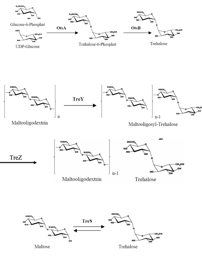
1.2 Trehalose synthesis pathways in C. glutamicum
At least five different trehalose synthesis pathways are identified in bacteria. Three pathways utilize activated glucose as substrate, while the others convert maltose or maltodextrin into trehalose. The most common synthesis route is the OtsAB-pathway. It catalyses the condensation of UDP-glucose and glucose-6-P to trehalose-6-P and the following
Introduction
dephosphorylation to trehalose (Argüelles et al., 2000). The trehalose glycosyltransferring synthase TreT in the hyperthermophilic Archaeon Thermococcus litoralis forms trehalose and ADP from ADP-glucose and glucose (Qu et al., 2004). Pyrococcus horikoshii possesses a similar glycosyltransferase utilizing UDP-glucose and glucose as substrate (Ryu et al., 2005).
In contrast, the bacterial species Rhizobium and Arthrobacter, the Archaeum Sulfolobus and Brevibacterium helvolobum, a close relative of Corynebacterium glutamicum, and mycobacteria utilize the storage carbohydrate maltodextrin as substrate for trehalose synthesis by the TreYZ-pathway (Maruta et al., 1996a/b/c; Kim et al., 2000; De Smet et al., 2000). In the first step, the terminal maltosyl-residue of maltodextrin is transglycosylated by TreY to a trehalosyl-unit which is subsequently cleaved off by TreZ. A further way to synthesise trehalose is the transglycosylation of maltose to trehalose catalysed by the trehalose synthase TreS in a single step reaction. Not in all organisms is the synthesis of trehalose the prevalent direction of the reaction catalysed by TreS. Whereas in mycobacteria TreS catalyses predominantly the synthesis of trehalose (Pan et al., 2004), in Rhodobacter sphaeroides f. sp. denitrificans IL106 TreS catalyses the synthesis of maltose (Makihara et al., 2005).
In contrast to other bacteria, C. glutamicum possesses not only one trehalose synthesis pathway, but three of the described pathways - OtsAB, TreYZ and TreS - indicating an important function of trehalose (Fig. 1). Whereas an in vitro enzymatic assay showed that the trehalose synthase TreS of C. glutamicum catalysed the transglycosylation of trehalose to maltose and the reverse reaction in equilibrium, under in vivo conditions TreS catalyses in C. glutamicum the degradation rather than the synthesis of trehalose (Wolf et al., 2003).
Since no trehalases were identified in C. glutamicum, the TreS-pathway could be an alternative for the degradation of trehalose. The TreYZ-pathway seems to be the main trehalose synthesis pathway in C. glutamicum. It is responsible for the accumulation of trehalose as a compatible solute after a hyperosmotic shock. RNA-hybridisation experiments indicated that also the OtsAB-pathway is involved in the response of C. glutamicum to an osmotic shock, since the otsA gene was upregulated five fold after an osmotic upshift (Wolf et al., 2003). In contrast, data of Shimakata and Minatogawa (2000) suggest a function of the OtsAB-pathway in the synthesis of the cell envelope component trehalose monomycolate.
These authors concluded from an in vitro assay that trehalose-6-phosphate, the product of OtsA, the first enzyme of the OtsAB-pathway, is essential for the biosynthesis of trehalose monomycolate. This conclusion was contradicted by the fact that a C. glutamicum strain deleted in the otsA gene synthesised trehalose mycolate (Wolf et al., 2003). Hence, the function of the OtsAB-pathway remained unclear. Only the simultaneous deletion of all three trehalose synthesis pathways in C. glutamicum resulted in a mutant devoid of mycolate suggesting that not OtsA alone, but both the enzymes of the OtsAB- and of the TreYZ-
Introduction pathway are involved in the synthesis of the components of the mycolate layer (Wolf et al., 2003).
TreZ TreZ
Fig. 1: Trehalose biosynthesis pathways in C. glutamicum
Introduction
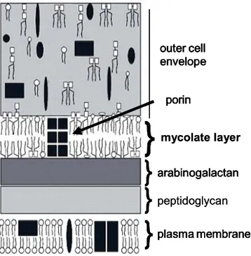
1.3 Corynebacterial cell envelope
C. glutamicum shares the structure of the cell envelope with all members of the suborder of Corynebacterineae which includes also nocardia, rhodococci and mycobacteria (CNM-group) (Fig. 2). The plasma membrane is the innermost layer of the cell envelope which is protected by the cell wall skeleton. Peptidoglycan covalently linked to arabinogalactan which in turn is esterified by mycolic acids forms the cell well skeleton. The mycolic acids pair with trehalose mycolates to a lipid bilayer called mycolate layer. This lipid bilayer in addition to the plasma membrane is a phylogenetic trait of the Corynebacterineae which distinguishes their cell envelope from a usual cell envelope of Gram-positive bacteria. The outer layer of the cell envelope consists to 90 % of polysaccharides. In some corynebacterial strains the S-layer protein is attached to the outermost surface (Puech et al., 2001).
plasma membrane arabinogalactan
} }
}
porin outer cell envelope
}
mycolate layer plasma membrane arabinogalactan} }
}
porin outer cell envelope
}
plasma membrane peptidoglycan arabinogalactan
} }
}
porin outer cell envelope
}
plasma membrane arabinogalactan
} }
}
porin outer cell envelope
}
mycolate layer plasma membrane arabinogalactan} }
}
porin outer cell envelope
}
plasma membrane peptidoglycan arabinogalactan
} }
}
porin outer cell envelope
}
Fig. 2: Model of the cell envelope of C. glutamicum according to Puech et al. (2001), modified. From the cytoplasmic to the external side of the bacteria the cell envelope is composed of the plasma membrane, a cell wall skeleton and an outer layer. The plasma membrane is a lipid bilayer of phospholipids (empty oval symbols) and proteins (dark rectangles and elypses). The cell wall skeleton consists of peptidoglycan covalently linked to arabinogalactan which in turn is esterified by corynomycolic acids (thin parallel bars). The covalently bound mycolic acids arrange to the inner leaflet of a lipid layer with non-covalently linked lipids such as trehalose dimycolate (a pair of empty squares with two pairs of thin parallel bars) and trehalose monomycolate (a pair of empty squares with one pair of thin parallel bars). The outer leaflet of the lipid layer which is called mycolate layer is formed by non-covalently linked trehalose mycolate. Porins (dark squares) span the mycolate layer.
The outer layer consists to 90% of polysaccharides, but contains also non-covalently linked lipids and proteins.
Introduction
1.3.1 Plasma membrane
The plasma membrane of C. glutamicum is mainly composed of polar phospholipids possessing palmitic (C16:0) and octadecenoic (C18:1) acids as fatty acid chains.
Phosphatidylglycerol is the main head-group of phospholipids found in C. glutamicum (80 %) complemented by diphosphatidylglycerol (cardiolipin), phosphatidylinositol and phosphatidylinositol dimannosides (PIM2).
1.3.2 Cell wall skeleton
Three different layers build the cell wall skeleton in C. glutamicum. As common in all Gram- positive bacteria a thick peptidoglycan layer neighbours the plasma membrane. In Corynebacterineae as in most other bacteria the peptidoglycan layer is composed of β-1,4- linked N-acetylglucosamine and N-acetyl muramic acid residues linked to tri- or tetrapeptides such as L-Ala-D-Glu-meso-diaminopimelic acid or L-Ala-D-Glu-meso-diaminopimelic acid-D- Ala (Schleifer & Kandler, 1972).
The second layer consists of the heteropolysaccharide arabinogalactan which is composed of a linear alternating β-D-galactofuranosyl backbone connecting to a 3,5-branched α-D- arabinofuranosyl structure. The galactofuranosyl backbone is attached by a phosphodiester link to the peptidoglycan. The terminal β-arabinofuranosyl residues are esterifyed on position 5 by mycolic acids (Puech et al., 2001; Alderwick et al., 2005).
Mycolic acids are long chain α-alkyl-β-hydroxy fatty acids and characteristic components of the mycolate layer, the third layer of the cell wall skeleton. In mycobacteria mycolic acids possess a very long chain (C60-90) and may be oxygenized or hydroxylated, whereas in nocardia and corynebacteria the alkyl-chain is shorter and the mixture of saturated and unsaturated fatty acids homologous (C40-50 nocardomycolic acids; C22-36 corynomycolic acids) (Qureshi et al., 1984). Mycolic acids linked to arabinogalactan constitute the arabinogalactan mycolate (AGM) or cell wall-bound mycolate. The esterification of one or two mycolic acids with a molecule trehalose yields trehalose monomycolate (TMM) and trehalose dimycolate (TDM), respectively, which are part of the extractable mycolate. TMM is the mycolyl-donor for the synthesis of arabinogalactan mycolate and TDM (Shimakata & Minatogawa, 2000).
Electron microscopy of freeze-fractured preparations of whole cells of C. glutamicum, a method revealing lipid bilayers by characteristic fracture planes, indicated that the cell envelope of the C. glutamicum wild type contained, additionally to the plasma membrane, a second lipid bilayer closer to the cell surface. Since this lipid bilayer was missing in Corynebacterium amycolatum, a naturally mycolate deficient strain, it was considered to be composed of mycolate (Puech et al., 2001). Whereas the inner lipid layer of the mycolate layer is formed mainly by arabinogalactan mycolate and minor amounts of extractable mycolate, the outer leaflet consists exclusively of TDM and TMM (Daffé & Draper, 1998;
Introduction
Puech et al., 2001). Thus, the cell envelope of the Gram-positive Corynebacterineae possesses an outer membrane similar to the cell envelope of Gram-negative bacteria. In Gram-negative bacteria phospholipids form the inner and lipopolysaccharides the outer leaflet of the outer membrane. This outer membrane exhibits a permeability barrier for hydrophilic as well as hydrophobic solutes. The low permeability of the outer membrane explains why Gram-negative bacteria are more resistant to some antibiotics than most Gram- positive bacteria (Schlegel & Zarborosch, 1992). In contrast, Jalier and Nikaido (1994) showed that the permeability of the cell envelope to some hydrophilic antibiotics of the Gram- positive Mycobacterium chelonea, harbouring a mycolate layer, was 3 times lower than that of Gram-negative E. coli and 10 times lower than that of Gram-negative Pseudomonas aeruginosa. Similar to the outer membrane in Gram-negative bacteria, in mycobacteria the mycolate layer is considered to be responsible for the low permeability of the cell envelope and consequently it is supposed to be crucial for the low susceptibility to most antibiotics (Brennan & Nikaido, 1995). A correlation of mycolate layer and permeability of the cell envelope in C. glutamicum was revealed by the inactivation of a mycolyltransferase in C. glutamicum. The inactivation decreased the cell wall linked-mycolate of the mutant to 50 % and caused increased uptake rates for glycerol and acetate (Puech et al., 2000). These results indicated that the nature of the mycolate layer may influence also the permeability of the cell envelope in C. glutamicum.
1.3.3 Porins
Similar to the outer membrane in Gram-negative bacteria, the mycolate layer of the Gram- positive Corynebacterineae may constitute a permeability barrier impeding the uptake of hydrophilic nutrients. The outer membrane of Gram-negative bacteria contains channel- forming proteins named porins facilitating the permeation of small hydrophilic molecules across the outer membrane. These porins are trimers of identical subunits. Each monomer contains one channel of 4 nm length (Cowan et al., 1992). Also the mycolate layer of Corynebacterineae comprises porins which passage hydrophilic solutes through the lipid bilayer, but the structure of these porins differs from that of the Gram-negative porins. In Corynebacterineae a porin is an oligomeric protein forming a single channel (Kartmann et al., 1999; Lichtinger et al., 1998; Riess et al., 1998). The best studied porin of this type is MspA, the main porin of Mycobacterium smegmatis. It is a tetrameric-protein forming a cone-like structure with a single pore of 10 nm length. A further difference between Corynebacterineae and Gram-negative bacteria is the number of porins. M. smegmatis contains 15-fold less porins per µm2 cell wall than a Gram-negative bacterium which results in 45 less pores per µm2 since one porin of a Gram-negative bacterium contains three pores. Longer channels
Introduction and lower pore numbers may cause lower permeability of the cell envelope of M. smegmatis compared to Gram-negative bacteria (Niederweis, 2003). Expression of MspA in the pathogenic and slow growing Mycobacterium tuberculosis and Mycobacterium bovis BCG accelerated the uptake of glucose and enhanced the susceptibility to β-lactam antibiotics and to isoniazid, ethambutol and streptomycin, indicating that porins are responsible for nutrient supply as well as the uptake of drugs currently used in tuberculosis chemotherapy (Mailaender et al., 2004). Furthermore, deletion of the porin gene mspA in the non- pathogenic M. smegmatis rendered it multidrug resistant (Stefan et al., 2004) and enhanced its intracellular persistence in macrophages, demonstrating that persistence depends on the permeability of the mycolate layer (Sharbati-Tehrani et al., 2005).
In C. glutamicum three different porins are identified which exhibit an oligomeric structure similar to MspA. The major porin is PorA, a cation-selective cell wall channel formed by an oligomer of a 45-amino-acid polypeptide (Lichtinger et al., 1998 and 2001). The characterisation of a porA deletion mutant indicated the presence of an alternative anion- selective cell wall channel with low channel conductivity. An anion-selective channel protein was identified with a conductance of 700 pS in 1M KCL and called PorB. Analysis of the genome sequence of C. glutamicum suggested the presence of a further anion-selective porin similar to PorB which was named PorC (Costa-Riu et al., 2003a/b). Additionally to PorA, the cation-selective channel PorH was identified (Hunten et al., 2005). Since the pore diameter of PorA is broader and the channel conductivity higher than that of the other porins, PorA was considered as the predominant hydrophilic channel in C. glutamicum. This assumption was confirmed by the fact that the deletion of the porA gene in the C. glutamicum wild type ATCC 13032 rendered the mutant less susceptible to the antibiotics ampicillin, kanamycin, streptomycin and tetracycline (Costa-Riu et al., 2003a). Furthermore, higher resistance to the antibiotics of the porA deletion mutant indicated that similar to mycobacteria, also the mycolate layer in C. glutamicum constitutes a permeability barrier for the uptake of antibiotics. Whereas in mycobacteria the impact of the mycolate layer on the uptake of antibiotics is of special interest, in C. glutamicum the influence of the cell envelope on the efflux of solutes is a crucial issue, since it is one of the most important industrial producers of amino acids.
1.3.4 Outer layer
The outer layer of the corynebacterial cell envelope consists to 90% of polysaccharides which are composed of glucose, mannose and arabinose (Puech et al., 2001). Some corynebacterial strains, but not the strains used in this study, have a protein attached to the cell surface – the S-layer protein – PS2 (Chami et al., 1997). Furthermore, the outer layer
Introduction
contains various lipids, mostly TDM and TMM, but also phospholipids like PIM2 and phosphatidyl glycerol (Puech et al., 2001).
1.4 Biosynthesis of mycolic acids and of mycolate
Mycolic acids are α-alkyl-β-hydroxy fatty acids and a phylogenetic trait of the Corynebacterineae suborder within the Actinomycetales. As the α-alkyl-β-hydroxy structure, the so called mycolic motif, is common to the whole suborder, the enzymatic set for mycolic acid biosynthesis is supposed to be shared by the Corynebacterineae, but the mechanism of mycolic acid biosynthesis is not known in detail. Use of cell-free extracts showed that two palmitic acids serve as precursors for the condensation to a C32 mycolic acid (Shimakata et al., 1984; Walker et al., 1973). Recently, the enzyme catalysing the condensation of two fatty acids to a mycolic acid was identified as polyketide synthase Pks13 in C. glutamicum (Portevin et al., 2004).
Also the following mycolate synthesis is still a matter of discussion. The first step is the transport of a newly synthesised mycolic acid and its condensation with trehalose to yield trehalose monomycolate (TMM). Based on results of an in vitro assay in Corynebacterium matruchotii Shimakata and Minatogawa (2000) suggest a role of trehalose-6-phosphate as an intermediate acceptor of mycolic acids. Contradicting these results, Wolf et al. (2003) demonstrated that trehalose-6-phosphate is not essential for mycolate syntheses in C. glutamicum in vivo. A mutant devoid of trehalose-6-phosphate due to deletion of otsA, the gene encoding trehalose-6-phosphate synthase, synthesised trehalose mycolate. In contrast, in Cgl ∆otsA∆treS∆treY, which is devoid of both trehalose and trehalose-6-P due to inactivation of all trehalose synthesis pathways, no mycolate was detected. These results emphasize the essentiality of trehalose rather than the importance of trehalose-6-phosphate for mycolate synthesis. Regardless of the acceptor, a mycolyltransferase is predicted for synthesis of TMM. The identity of this mycolyltransferase is unclear.
The last steps of mycolate synthesis are the least controversial. Mycolyltransferases transfer the mycolyl-residue from TMM either to the free position 6 of the trehalose-residue of another TMM to yield trehalose dimycolate or to a terminal arabinofuranosyl of the arabinogalactan layer to yield arabinogalactan mycolate. In Mycobacterium tuberculosis the three antigen 85 proteins were identified as mycolyltransferases by site directed mutagenesis and in vitro enzymatic assays. The antigen 85 proteins, which are also known as fibronectin binding enzymes (FBP), comprise a fibronectin-binding site which enables interaction with macrophages of the host immune system. The fibronectin-binding-site demonstrates that these mycolyltransferases are exported from the cell. The presence of a signal peptide which
Introduction of these enzymes. Since the mycolyltransferases are exported from the cytoplasm, the synthesis of TDM and AGM is supposed to be localised in the cell envelope (Belisle et al., 1997; Ronning et al., 2000; Ronning et al., 2004).
In C. glutamicum the first mycolyltransferase identified due to sequence similarity to the antigen 85 proteins was encoded by the csp-1 or cop-1 gene and called PS1 (Joliff et al., 1992). A csp-1 mutant accumulated TMM and synthesised less TDM than the wild type proving the mycolyltransferase activity of PS1 and indicating the presence of further mycolyltransferases in C. glutamicum. In the genome of C. glutamicum six putative mycolyltransferase genes were identified. One gene was the already known, cop-1. The other five were named cmt1-5 (Brand et al., 2003) or cmytA-E (Sousa-D’Auria et al., 2003).
Since a C. glutamicum ATCC 13032 mutant deleted in cop1, cmt1 and cmt2 could not synthesise TDM, but the single and double mutants produced TDM, Brand et al. (2003) concluded that cop1, cmt1 and cmt2 code for mycolyltransferases synthesising TDM. Cross- complementation experiments in C. glutamicum CGL 2005 suggested two different classes of mycolyltransferases. CMytA (PS1) and cMytB (Cmt2) could replace each other in TDM as well as in arabinoglactan-mycolate synthesis, whereas a cmytA deletion mutant was only complemented in TDM production by the overexpression of cmytC (cmt1) or cmytD (cmt5).
Sousa-D’Auria et al. (2003) concluded that the mycolyltransferases cMytA (PS1) and cMytB (Cmt2) transfer a mycolyl-residue to TMM as a well as to arabinogalactan, whereas cMytC (Cmt1) and cMytD (Cmt5) synthesise TDM, but no arabinogalactan mycolate.
1.5 Amino acid production with C. glutamicum
1.5.1 L-lysine production with C. glutamicum
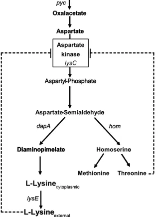
L-lysine is an essential amino acid for mammals which has to be provided in sufficient amounts in animal feed to meet the nutritional requirements. Since plant based feedstuff like corn, wheat or barley is poor in lysine, supplementation with lysine provides a possibility to increase the nutrient value. C. glutamicum is the sole organism utilized for industrial production of L-lysine which exceeds 600,000 tons per year of lysine. Lysine belongs to the aspartate familiy of amino acids as well as homoserine, methionine, threonine and leucin, which are all synthesised starting from oxalacetate, a component of the tricarboxylic acid cycle (Fig. 3).
Lysine biosynthesis is regulated by the aspartate-kinase which is feedback inhibited by lysine and threonine. Since this feedback inhibition limits lysine synthesis, an aspartate-kinase resistant to feedback inhibition provides a tool to optimize lysine production. A further option
Introduction
to increase lysine synthesis is the restriction of the synthesis of the by-products homoserine, methionine and threonine. Conventional lysine production strains are based on the mutagenesis of aspartate-kinase and of enzymes of by-product synthesis, since they were obtained by random mutagenesis followed by screening for resistance to lysine or threonine analogues and/or nutritional requirement for by-products (Nakayama et al., 1973).
Oxalacetate Aspartate
Aspartyl-Phosphate
Threonine Methionine
Diaminopimelate Homoserine Aspartyl-Phosphate
Threonine Methionine
Diaminopimelate
Aspartate-Semialdehyde Aspartate-Semialdehyde
Homoserine dapA
cytoplasmic
lysE
hom pyc
L-Lysine L-Lysine
external
Aspartate kinase
lysC Oxalacetate
Aspartate
Aspartyl-Phosphate
Threonine Methionine
Diaminopimelate Homoserine Aspartyl-Phosphate
Threonine Methionine
Diaminopimelate
Aspartate-Semialdehyde Aspartate-Semialdehyde Aspartate-Semialdehyde Aspartate-Semialdehyde
Homoserine dapA
cytoplasmic
lysE
hom pyc
L-Lysine L-Lysine
external
Aspartate kinase
lysC
Fig. 3: Biosynthesis of the amino acids of the aspartate-family in C. glutamicum. Dashed arrows, regulation at enzyme level (feedback inhibition).
A further approach to improve lysine production uses genetic engineering. Defined mutagenesis avoids secondary mutations which affect growth or stability. A crucial target also of genetic engineering is the feedback inhibition of the aspartate-kinase which is encoded by lysC. Overexpression of lysC alleles encoding aspartate-kinases, which were not feedback inhibited, increased lysine synthesis (Thierbach et al., 1990). A genetic approach to reduce the formation of by-products is the inactivation of the homoserine dehydrogenase (hom) the first enzyme of the metabolic branch leading to homoserine, methionine and threonine synthesis (Eikmanns et al., 1991). Consequently, overexpression of dapA encoding dihydrodipicolinate synthase, an enzyme of the lysine biosynthesis pathway competing with the homoserine dehydrogenase for their common substrate L-aspartate semialdehyde, increased lysine synthesis (Eggeling et al., 1998). A further bottleneck for lysine production are anaplerotic reactions as shown by overexpression of pyc encoding
Introduction pyruvate carboxylase which synthesises oxalacetate, the precursor of all amino acids of the aspartate familiy (Peters-Wendisch et al., 2001).
A further obstacle for the production of lysine is the export of the amino acid. The first permeability barrier for lysine is the plasma membrane. Transport across the plasma membrane is mediated by the exporter LysE (Broer et al., 1991a/b; Vrljic et al., 1995; Vrljic et al., 1996; Bellmann et al., 2001). Product excretion seems to constitute a further bottleneck for lysine production, since overexpression of lysE increased lysine excretion (Pfefferle et al., 2003). Apart from the plasma membrane the cell envelope of C. glutamicum comprises a second lipid bilayer, the mycolate layer, which is supposed to impede the permeation of solutes. However, the exact impact of the mycolate layer on the excretion of amino acids is unknown (Eggeling & Sahm, 2001).
1.5.2 L-glutamate production with C. glutamicum
Although more than 1,000,000 tons per year of L-glutamate are produced by C. glutamicum, little is known about the mechanism causing efflux of glutamate from the cell. Special treatment of the cell is always required such as biotin limitation, temperature upshift or addition of penicillin (Kimura, 2003). Some of these treatments interfere with the fatty acid composition of the plasma membrane e. g. the biotin containing acyl-CoA carboxylase, the first enzyme of fatty acid biosynthesis, might be the target of biotin limitation. The “leak model” suggested that increased permeability of the plasma membrane facilitated the passive diffusion of glutamate, but evidence for an L-glutamate exporter ruled out this model (Hoischen & Krämer, 1990; Gutmann et al., 1992).
Currently, two different models for glutamate efflux are discussed. Eggeling & Sahm (2001) propose that the excretion of glutamate is determined by the structure of the corynebacterial cell envelope. Treatments inducing glutamate excretion may facilitate the passage through the mycolate layer by increasing its permeability. Since the components of the cell wall skeleton peptidoglycan, arabinogalactan and mycolic acids are linked by covalent bonding, alteration of any of these components influences the mycolate layer. Thus, treatment with penicillin, which inhibits the synthesis of peptidoglycan, as well as addition of ethambutol, which interferes with the synthesis of arabinogalactan, may increase the permeability of the mycolate layer (Nunheimer et al., 1970; Radmacher et al., 2005b).
Treatments which manipulate fatty acid synthesis, e. g. biotin limitation, influence the fatty acid composition of the plasma membrane and they may similarly alter the composition of mycolate layer since fatty acids are essential for mycolate synthesis. This assumption was supported by the inactivation of the two fatty acid synthases FAS-IA and FAS-IB in C. glutamicum. The inactivation mutants fasA and fasB excreted glutamate without special
Introduction
treatment and they exhibited an altered plasma membrane as well as a reduced amount of mycolic acids (Radmacher et al., 2005a).
Systematic genetic engineering of fatty acid biosynthesis indicated that also the alteration of the plasma membrane may influence glutamate excretion since the appropriate lipid composition and the suitable membrane tension activate the L-glutamate carrier (Nampoothiri et al., 2002).
Kimura (2003) explains glutamate overproduction by the “metabolic flux change model”.
Already 30 years ago the central role of the activity of the 2-oxoglutarate dehydrogenase complex (ODHC) in glutamate production had been discovered (Shingu & Terui, 1971).
ODHC belongs to the tricarboxylic acid cycle (TCA cycle) and catalyses the oxidative decarboxylation of 2-oxoglutarate to succinyl-CoA. Thus, ODHC competes with glutamate dehydrogenase, the first enzyme of the glutamate synthesis pathway, for their common substrate 2-oxoglutarate. Low ODHC activity may shift metabolic fluxes from the tricarboxylic acid cycle to glutamate production. The significance of ODHC for glutamate excretion was confirmed by the observation that activity of ODHC was reduced under conditions which cause glutamate overproduction such as biotin limitation or treatment with Tween 40 or penicillin (Kawahara et al., 1997), and that inactivation of ODHC by disrupting the odhA gene triggered glutamate excretion (Kimura, 2005). Analysis of metabolic fluxes proved that decreased ODHC activity directed carbon fluxes towards glutamate synthesis (Shirai et al., 2005).
How the special treatments triggering glutamate efflux influence the change of metabolic fluxes is unknown. Kimura (2005) suggests that fatty acid synthesis, the target of most treatments, and metabolic fluxes in TCA cycle and glutamate synthesis are controlled on the transcriptional level by the same global metabolic regulator.
1.6 Objectives of the PhD-Thesis
The first part of this project assessed the role of trehalose as protectant against osmotic stress. Since an involvement of the OtsAB trehalose synthesis pathway in mycolate synthesis, as suggested by Shimakata and Minatogawa (2000) could be ruled out (Wolf et al., 2003), the function of the OtsAB pathway remained unknown. RNA-hybridisation experiments indicated a role of this pathway in the response to osmotic stress in C. glutamicum (Wolf et al., 2003). Since the TreYZ-pathway is regarded as the predominant trehalose synthesis pathway after a hyperosmotic shock, conditions were searched which required the OtsAB-pathway for the synthesis of trehalose under osmotic stress.
Introduction The major part of this project aimed to investigate the function of trehalose as an important component of the corynebacterial cell envelope. In contrast to other Gram-positive bacteria all members of the suborder of Corynebacterineae, including C. glutamicum, contain a cell envelope that comprises a second lipid bilayer apart from the plasma membrane, the mycolate layer, which is considered as permeability barrier (Puech et al., 2001). An important building block for the biosynthesis of the mycolate layer is trehalose, since trehalose monomycolate (TMM) serves as a precursor for trehalose dimycolate (TDM) and arabinogalactan mycolate (AGM), which are, together with TMM, the main components of the mycolate layer. Whereas TDM and AGM are known to be synthesised in the cell envelope, it is unclear where the condensation to TMM is located. One objective of this project was to find out whether TMM is synthesised in the cell envelope like TDM and AGM or in the cytoplasm, where its building blocks trehalose and mycolic acids are synthesised.
Since the trehalose deficient C. glutamicum strains Cgl∆otsA∆treY∆treS and Cgl∆otsA∆treY were devoid of mycolate when they were cultured on sucrose, trehalose seemed to be essential for mycolate synthesis (Wolf et al., 2003). However, Cgl∆otsA∆treY cultivated in medium supplemented with maltose synthesised AGM and a glycolipid identified as maltose monomycolate (Wolf, 2002; M. Daffé, personal communication) indicating that trehalose was not the sole sugar acceptor for mycolic acids. This assumption was supported by the detection of glucose monomyolcate in a C. glutamicum strain deleted in the mycolyltransferase PS1 (Puech et al., 2000). A further objective of this project was to analyse how the carbon source specifically influences the composition of the mycolate layer of a C. glutamicum strain inactivated in trehalose synthesis. Supplementation of the medium with trehalose was tested as an additional tool to influence the synthesis of the mycolate layer (Tzvetkov et al., 2003). The comparison of strains harbouring differently composed mycolate layers should reveal how the composition determines the properties of the mycolate layer, especially its permeability. A C. glutamicum L-lysine production strain inactivated in trehalose biosynthesis was chosen for these experiments to examine whether excreted lysine as a measure for the efflux of solutes could be correlated to the permeability of the cell envelope. Moreover, analysis of this strain should reveal whether the alteration of the cell envelope improves lysine production.
Materials and Methods
2 Materials and Methods
2.1 Strains, plasmids and oligonucleotides
this study L-Lysine production strain carrying
chromosomal deletions in the otsA, treYand treSORFs
LP∆treS∆otsA∆treY
this study L-Lysine production strain carrying
chromosomal deletions in the otsAand treSORFs LP∆treS∆otsA
this study L-Lysine production strain carrying a
chromosomal deletion in the treSORF LP∆treS
Nakayamaet al.
(1972) L-Lysine production strain, lysCFBR, leuCD, hseCD
ATCC 21527
this study Wild type carrying chromosomal deletions in the
glgCORF Cgl∆glgC
this study Wild type carrying chromosomal deletions in the
treY, treS andglgCORFs Cgl∆treY∆treS∆glgC
this study Wild type carrying chromosomal deletions in the
otsA, treSandglgCORFs Cgl∆otsA∆treS∆glgC
Wolf et al.(2003) Wild type carrying chromosomal deletions in the
otsA, treYand treSORFs Cgl∆otsA∆treS∆treY
Wolf et al. (2003) Wild type carrying chromosomal deletions in the
treYand treSORFs Cgl∆treY∆treS
Wolf et al. (2003) Wild type carrying chromosomal deletions in the
otsAand treSORFs Cgl∆otsA∆treS
Abeet al. (1967) Wild type
ATCC 13032 C. glutamicum
Reference Description
Strain
this study L-Lysine production strain carrying
chromosomal deletions in the otsA, treYand treSORFs
LP∆treS∆otsA∆treY
this study L-Lysine production strain carrying
chromosomal deletions in the otsAand treSORFs LP∆treS∆otsA
this study L-Lysine production strain carrying a
chromosomal deletion in the treSORF LP∆treS
Nakayamaet al.
(1972) L-Lysine production strain, lysCFBR, leuCD, hseCD
ATCC 21527
this study Wild type carrying chromosomal deletions in the
glgCORF Cgl∆glgC
this study Wild type carrying chromosomal deletions in the
treY, treS andglgCORFs Cgl∆treY∆treS∆glgC
this study Wild type carrying chromosomal deletions in the
otsA, treSandglgCORFs Cgl∆otsA∆treS∆glgC
Wolf et al.(2003) Wild type carrying chromosomal deletions in the
otsA, treYand treSORFs Cgl∆otsA∆treS∆treY
Wolf et al. (2003) Wild type carrying chromosomal deletions in the
treYand treSORFs Cgl∆treY∆treS
Wolf et al. (2003) Wild type carrying chromosomal deletions in the
otsAand treSORFs Cgl∆otsA∆treS
Abeet al. (1967) Wild type
ATCC 13032 C. glutamicum
Reference Description
Strain
Tab. 1: Bacterial strains used in this study
ab. 2: Oligonucleotides used in this study.
T
5'-tca ttc cat atc gtc ctt ttc-3' treSGa
1.800 5'-atg act gat acc tct ccg ttg-3'
treSgs2
5'-tca aaa ctc act atc ggg tac-3' treYa
2.739 5'-gca cgt cca att tcc gca ac-3'
treYs
5'-cgt tga cgt cgt ggg tat aga cc-3' otsAa
1.808 5'-tct gcc agt gga tat gac tgt cc-3'
otsAs
Size of amplified fragment (kb) Oligonucleotide sequence (5'-3')
Designation
Materials and Methods Tab. 3: Plasmids used in this study
Eikmanns, Ulm pK19mobsacBcarrying ∆glgC
pK19IMC
Wolf et al. (2003) pK19mobsacBcarrying a 0.71 kb deletion
in thetreSORF pK19mobsacB∆treS
Wolf et al. (2003) pK19mobsacBcarrying a 0.34 kb deletion
in thetreYORF pK19mobsacB∆treY
Wolf et al. (2003) pK19mobsacBcarrying a 0.21 kb deletion
in theotsA ORF pK19mobsacB∆otsA
Schäfer et al. (1994) oripUC, KmR, mob sacB
pK19mobsacB
Wolf, 2002 pUC19 carrying a 2.5 kb fragment containing
the treSORF pUC19treS
Wolf, 2002 pUC18 carrying a 2.7 kb fragment containing
the treYORF pUC18treY
Wolf, 2002 pUC18 carrying a 1.8 kb fragment containing
the otsAORF pUC18otsA
Yanisch-Perronet al.(1985) plac, ApR
pUC18/19
Reference Description
Plasmids
2.2 Media and culture conditions
All cultivations were performed in shake flasks under aerobic conditions. Escherichia coli strains were kept at 37 °C and C. glutamicum strains at 30 °C. In both cases, LB medium was used as complex medium. CgXII medium (Keilhauer et al., 1993) was used as minimal medium for C. glutamicum. As carbon source 4 % sucrose, 4 % glucose or 4 % fructose were used. When indicated 2 % or 0.5 % of trehalose were added to the medium. The lysine producer strain ATCC 21527 and its derivatives were supplemented with 0.2 g/L L-leucin and 0.4-0.8 g/L D/L-homoserine. High osmolality was adjusted by the addition of 750 mM NaCl (equivalent to an osmotic upshift of approx. 1.5 osM). For all strain characterizations, cells from LB precultures were washed once in CgXII medium and used to inoculate CgXII precultures, which were grown to exponential or early stationary phase. Subsequently, from those cultures, the CgXII main cultures were inoculated to an initial optical density (OD600) of 1. If the main culture differed from the preculture in any parameter, e.g. carbon source or NaCl content, a washing step between pre- and main culture was included.
Materials and Methods
2.3 Molecular biology methods
2.3.1 DNA digestion, ligation and purification
Standard techniques like DNA digestion (enzymes purchased from New England Biolabs;
Schwalbach), ligation using T4 ligase (New England Biolabs, Schwalbach) and plasmid DNA purification (NucleoSpin Plasmid; Macherey Nagel, Düren) were performed according to the manufacturer’s protocol.
2.3.2 Competent cells and transformation
Competent E. coli cells were prepared and transformed according to Inoue et al. (1990).
Competent C. glutamicum cells were prepared as described by van der Rest et al. (1999) and transformed by electroporation (2.5 kV, 600 Ω, 2.5 µF) with a Gene-Pulser (Bio-Rad, München).
2.3.3 Polymerase chain reaction
The knock-out of genes in the genome in C. glutamicum mutants was examined by the amplification of DNA fragments containing internal deletions in the case of allelic replacement. Amplification of DNA fragments was carried out using Taq PCR Master Mix Kit (Qiagen, Hilden) according to the manufacturer’s protocol. Per reaction 2.5 mM MgCl2 were added. Oligonucleotides were obtained from MWG Biotech (Ebersberg, Germany).
Sequence information on the C. glutamicum genome was generously provided by the Degussa AG (Hanau, Germany).
2.3.4 Agarose gel electrophoresis
To test C. glutamicum mutants for allelic replacement DNA fragments were amplified by PCR and analyzed by agarose gel electrophoresis (1 x TAE: 0.04 M Tris, 0.5 mM EDTA, pH adjusted to 7.5 with acetic acid) according to Sambrook et al. (1989). DNA extraction from the agarose gels was done with the NucleoSpin Extract II-Kit (Macherey Nagel, Düren) according to the manufacturer’s protocol.
2.3.5 Construction of a strain defective in trehalose synthesis
The knock-out of genes in the genome of C. glutamicum ATCC 21527 was performed by the method of allelic replacement described by Schäfer et al. (1994) to construct a C. glutamicum lysine producer strain defective in all three trehalose synthesis pathways.
Derivatives of the vector pK19mobsacB containing the coding regions of otsA, treY and treS with internal deletions were kindly provided by A. Wolf and described recently (Wolf et al.,
Materials and Methods with pK19mobsacB derivatives by means of electroporation (van der Rest et al., 1999).
Plasmid integration into the genome was verified by selecting kanamycin-resistant and sucrose-sensitive colonies (the expression of the sacB gene encoding the levan sucrase is toxic in sucrose-containing media). To promote re-excision of the plasmid DNA, positive clones were grown overnight in LB broth containing 25 µg/ml kanamycin, washed and cultivated in CgXII without phosphate and ammonium source containing 0.5 % glucose for 6 h. Since the knock-out mutants were supposed to have a slower growth rate than the wild type, the starvation step should prevent that the knock-out mutants were overgrown by clones comprising the wild type allele. The starved cells were plated on LB agar supplemented with 15 % sucrose in different dilutions, usually between 10 -3 and 10 -5. Positive colonies obtained from the subsequent selection (KmS, SucR) were tested for allelic replacement by PCR (oligonucleotides are listed in Tab. 2). Single or double deletion strains were used instead of C. glutamicum ATCC 21527 to generate strains carrying multiple gene deletions. LP∆treS∆otsA was constructed from LP∆treS and LP∆treS∆otsA∆treY from LP∆treS∆otsA (Tab. 1).
2.3.6 Construction of strains inactivated in glycogen synthesis
Glycogen biosynthesis was inactivated by inserting the vector pK19IMC (kindly provided by B. Eikmanns) via homologous recombination into the glgC gene in the genomes of C. glutamicum ATCC 13032, Cgl∆otsA∆treS and Cgl∆treY∆treS (Schäfer et al. 1994). In short, competent wild type cells were transformed with pK19IMC by means of electroporation (van der Rest et al., 1999). Plasmid integration into the genome was verified by selecting kanamycin-resistant colonies. The ∆glgC insertion strains Cgl∆glgC Cgl∆otsA∆treS∆glgC and Cgl∆treY∆treS∆glgC were constructed (Tab. 1). These insertion strains were examined for cytoplasmic glycogen accumulation.
2.4 Biochemical methods
2.4.1 Determination of protein concentration
Protein concentration was determined according to the method of Bradford (1976). The solution was calibrated using bovine serum albumin.
Materials and Methods
2.4.2 Cell disruption
2.4.2.1 Cell disruption by permeabilisation with CTAB
For the analysis of cytoplasmic amino acids by HPLC, 1 ml cell culture was separated from the medium by rapid filtration using 0.45 µm glass fibre filters (GF, Schleicher & Schuell GmbH, Dassel, Germany). The cells were washed twice with 2 ml of CgXII medium and incubated in 1 ml 0.1 % N-Cetyl-N,N,N-trimethyl-ammonium bromide (CTAB) for 10 min to release the cytoplasmic solutes. After a centrifugation step (20,000 g; 7 min, 4 °C) the supernatant was collected and stored at -20 °C until use.
2.4.2.2 Cell disruption by methanolysis
Cells were disrupted by methanolysis for the quantitative analysis of cytoplasmic trehalose.
In short, 2 ml samples were taken from cell cultures. Cells were separated from the growth medium by centrifugation. The cells were washed once with 1 ml of fresh, isoosmotic CgXII medium and frozen with liquid nitrogen. Cells were permeabilized by incubation in methanol at 70 °C. Cell debris was removed by centrifugation and the supernatant was used for the preparation of GC-samples.
2.4.2.3 Mechanical cell disruption
2 ml samples were taken from cell cultures. The cells were washed with 50 mM Tris, 50 mM NaCl, pH 6.3 and 300 mg glass beads were added. The cells were broken up mechanically by means of a FastPrep FP120 (QBiogene, Heidelberg, D) (2 x 4.5 s, 6.5 m/s). The cell debris was removed by centrifugation (20,000 g; 10 min, 4 °C).
2.4.3 Quantification of amino acids by HPLC
For the quantitative analysis of external amino acids, especially lysine and glutamate, 1 ml samples were taken from cell cultures. Cells were separated from the growth medium by centrifugation and analysis was performed from the diluted supernatant. Cytoplasmic fractions were analysed for the quantification of cytoplasmic amino acids (cf. 1.4.2.1). The concentration of the amino acids was determined using a reversed phase high-performance liquid chromatography (HPLC) system (HP 1090 Liquid chromatograph, HP1046A fluorescence detector; Hewlett Packard) with automated fluorescent precolumn derivatization by ortho-phthaldialdehyd/mercaptopropionic acid (OPA). Solutes were separated by means of a reversed phase column (Multospher, CS Chromatographie Service) at 40 °C with a flow rate of 0.5 ml/min. The mobile phase was a mixture of solvent A (20 mM sodium actetate, pH 7.2, 0.3 % tetrahydrofurane, 0.04 % triethylamine) and 100% methanol (solvent B). A


![Tab. 4: Uptake rates of [ 14 C]trehalose and of [ 14 C]glucose of ATCC 13032 and Cgl∆treY∆treS∆otsA after 10 min (trehalose) or 5 min (glucose) incubation](https://thumb-eu.123doks.com/thumbv2/1library_info/3660924.1503744/48.892.108.747.161.287/uptake-rates-trehalose-glucose-atcc-trehalose-glucose-incubation.webp)
