Trehalose Transport in Corynebacterium glutamicum and its Significance for Cell Envelope Synthesis
I n a u g u r a l – D i s s e r t a t i o n zur
Erlangung des Doktorgrades
der Mathematisch-Naturwissenschaftlichen Fakultät der Universität zu Köln
vorgelegt von Alexander Werner Eck
aus Speyer
Köln, 2014
Berichterstatter/in: Prof. Dr. Reinhard Krämer Prof. Dr. Karin Schnetz
Tag der mündlichen Prüfung: 21.05.2014
Corynebacterium glutamicum ist ein Gram-positives, apathogenes Bodenbakterium. Es ist ein wichtiger industriell genutzter Aminosäureproduzent und dient als Modellorganismus für die Synthese der Zellhülle in verwandten pathogenen Arten wie Mycobacterium tuberculosis, dem Tuberkuloseerreger. Ein auffälliges Merkmal der Zellhülle dieser Bakterien ist das Vor- handensein einer zweiten Permeabilitätsbarriere ähnlich der äußeren Membran Gram- negativer Bakterien. Wichtige Bestandteile sind Trehalosemono- (TMM) und dimycolat (TDM), langkettige Fettsäuren, die mit einem Molekül des Disaccharids Trehalose verestert sind. Frühere Versuche mit C. glutamicum zeigten, dass die Verknüpfung im Periplasma erfolgt, wofür der Export beider Moleküle notwendig ist. Dieses Modell wurde durch neue Studien in Frage gestellt, welche MmpL-Transporter mit dem Export von TMM aus dem Cytosol in Verbindung brachten. Um das beschriebene Modell der TMM-Synthese zu überprüfen, wurde daher in dieser Arbeit der Export von Trehalose untersucht.
In einem C. glutamicum Teststamm konnten die cytosolische Umwandlung zuvor aufgenom- mener Maltose in Trehalose und dessen Exkretion nachgewiesen werden, wobei eine extrazelluläre Umwandlung ausgeschlossen wurde. Die bestimmte Exportrate von 0.19 nmol
× mg
-1cdw × min
-1ist für die TMM-Synthese während des Wachstums ausreichend und war unabhängig von der Aktivität mechanosensitiver Kanäle, welche Trehalose in anderen Organismen freisetzen, und von der TMM-Synthese. Zusammen mit mechanistischen Analysen der Exkretion deutet dies auf das Vorhandensein eines Trehalosetransporters hin, der diese für die TMM-Synthese im Periplasma bereitstellt. TMM und TDM wurde nur in Zellen nachgewiesen, in denen einer von zwei redundanten MmpL-Transportern aktiv war.
Es wird daher ein Modell vorgeschlagen, das eine periplasmatische Synthese von TMM aus zuvor exportierten Substraten und einem MmpL-vermittelten Transport von TMM aus der äußeren Schicht der Plasmamembran zur Mycolatschicht vereint.
Da ein rationaler Ansatz nicht zur Identifizierung eines Trehaloseexporters führte, wurde ein
genetisch kodierter Trehalosesensor entwickelt und optimiert, welcher für das Screening
einer Mutantenbibliothek eingesetzt werden sollte. Die Affinität dieses Sensors konnte
jedoch durch gerichtete Mutagenese nicht ausreichend reduziert werden, um eine in vivo-
Applikation zu ermöglichen. Der Trehaloseexporter von C. glutamicum ist daher weiterhin
unbekannt.
Corynebacterium glutamicum is a Gram-positive, non-pathogenic soil bacterium. It is one of the main industrial producers for amino acids and also serves as a model organism for cell envelope synthesis in related pathogenic species like Mycobacterium tuberculosis, the causative agent of tuberculosis. A common feature of their cell envelope is the presence of a second permeability barrier similar to the outer membrane of Gram-negative bacteria.
Important constituents of this lipid bilayer are the glycolipids trehalose monomycolate (TMM) and trehalose dimycolate (TDM), long chain fatty acids esterified to the disaccharide trehalose. Previous experiments with C. glutamicum indicated that the linkage of trehalose and a mycolic acid precursor takes place in the periplasm, necessitating the export of both substrates. This model was challenged by the recently described connection of MmpL transporters to TMM transport from the cytosol to the periplasm. To validate the current model of TMM synthesis, the export of trehalose was investigated in this work.
In a C. glutamicum test strain, the cytosolic conversion of imported maltose to trehalose and the excretion of the latter could be shown. The extracellular conversion of substrate to trehalose could be excluded. Trehalose accumulation in the supernatant occurred with a specific rate of 0.19 nmol × mg
-1cdw × min
-1, which is sufficient to maintain mycolic acid synthesis during growth. The rate of trehalose excretion was independent of mechanosensitive channels, which mediate trehalose excretion in other bacteria, and of TMM synthesis. Together with mechanistic analyses, this indicates the presence of a carrier dedicated to provide trehalose for TMM synthesis in the periplasm.
Confirming the results published by other groups, the detection of TMM and TDM in whole cell extracts was dependent on the activity of either of two MmpL proteins. Nevertheless, a model is presented in this work that allows the periplasmic synthesis of TMM after precursor export and that assumes the MmpL-catalysed transport of TMM from the outer leaflet of the plasma membrane across the periplasm to the outer membrane.
A trehalose export carrier could not be identified in a rational approach. Thus, a genetically
encoded trehalose sensor was constructed and optimised to allow the screening of a mutant
library. Although functional in vitro, the affinity of this sensor for trehalose is still too high
and could not be reduced sufficiently by site-directed mutagenesis to allow its application in
C. glutamicum. Thus, the trehalose export system of C. glutamicum remains unknown.
1.1 CORYNEBACTERIUM GLUTAMICUM IS AN INDUSTRIAL PRODUCER AND A MODEL ORGANISM ... 1
1.2 CELL ENVELOPE ARCHITECTURE IN C. GLUTAMICUM ... 2
1.3 TREHALOSE SYNTHESIS IN C. GLUTAMICUM ... 4
1.4 TREHALOSE CATABOLISM IN C. GLUTAMICUM ... 6
1.5 TREHALOSE AS PRECURSOR FOR TMM SYNTHESIS ... 8
1.6 AIMS OF THIS PROJECT ... 11
2 MATERIAL AND METHODS ... 12
2.1 BACTERIAL STRAINS, PLASMIDS, AND OLIGONUCLEOTIDES ... 12
2.2 MEDIA AND CULTIVATION OF E. COLI AND C. GLUTAMICUM ... 18
2.2.1 MEDIA ... 18
2.2.2 CULTIVATION OF C. GLUTAMICUM IN SHAKE FLASKS ... 19
2.2.3 BIOREACTOR CULTIVATION OF C. GLUTAMICUM ... 19
2.3 MOLECULAR BIOLOGY METHODS ... 20
2.3.1 DNA PURIFICATION, DIGESTION, AND LIGATION ... 20
2.3.2 POLYMERASE CHAIN REACTION ... 20
2.3.3 SITE-DIRECTED DNA MUTAGENESIS ... 20
2.3.4 AGAROSE GEL ELECTROPHORESIS ... 21
2.3.5 COMPETENT E. COLI AND C. GLUTAMICUM CELLS AND TRANSFORMATION ... 21
2.3.6 INTEGRATION MUTAGENESIS AND GENE DELETION IN C. GLUTAMICUM ... 22
2.3.7 RNA HYBRIDISATION EXPERIMENTS ... 22
2.4 BIOCHEMICAL METHODS ... 23
2.4.1 HETEROLOGOUS EXPRESSION IN E. COLI BL21(DE3) ... 23
2.4.2 CHROMATOGRAPHIC PURIFICATION OF PROTEINS ... 23
2.4.3 INVESTIGATION OF TREHALOSE BINDING VIA INTRINSIC PROTEIN FLUORESCENCE ... 23
2.4.4 FLUORESCENCE MEASUREMENTS WITH METABOLITE NANOSENSORS ... 24
2.4.5 IN SITU CALIBRATION OF METABOLITE NANOSENSORS ... 24
2.4.6 DETERMINATION OF MALTOSE AND TREHALOSE TRANSPORT RATES ... 25
2.4.7 INVESTIGATION OF SOLUTE RELEASE AFTER HYPOOSMOTIC SHOCKS ... 25
2.5 ANALYTICAL METHODS ... 25
2.5.1 DETERMINATION OF PROTEIN CONCENTRATIONS ... 25
2.5.2 SDS-PAGE AND WESTERN-BLOT ANALYSIS... 26
2.5.3 CARBOHYDRATE ANALYSIS VIA TLC ... 26
2.5.6 QUANTITATIVE ANALYSIS OF AMINO ACIDS VIA HPLC ... 28
2.5.7 GLYCOLIPID EXTRACTION AND ANALYSIS BY TLC ... 28
3 RESULTS ... 29
3.1 THE TREHALOSE IMPORT SYSTEM AS A TARGET FOR OPTIMISATION OF C. GLUTAMICUM STRAINS ... 29
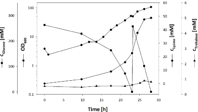
3.1.1 TREHALOSE RECYCLING IMPROVES L-LYSINE PRODUCTION ... 29
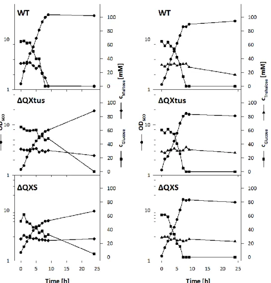
3.1.2 DELETION OF THE TUS-GENES INCREASES TREHALOSE PRODUCTION ... 32
3.2 INVESTIGATION OF TREHALOSE EXPORT IN C. GLUTAMICUM ... 35
3.2.1 CONSTRUCTION OF A TEST STRAIN FOR THE INVESTIGATION OF TREHALOSE EXCRETION ... 35
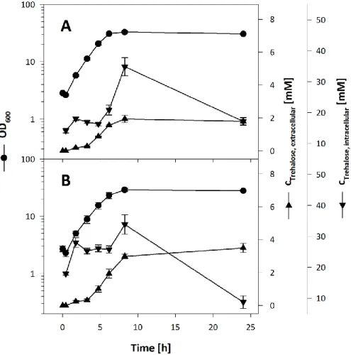
3.2.2 RADIOCHEMICAL ANALYSIS OF TREHALOSE EXCRETION BY C. GLUTAMICUM ΔMALQΔTREXΔTUS ... 37
3.2.3 ENZYMATIC ASSAY FOR QUANTITATIVE TREHALOSE DETECTION ... 40
3.2.4 QUANTITATIVE ANALYSIS OF TREHALOSE EXCRETION ... 42
3.2.5 CONTRIBUTION OF MECHANOSENSITIVE CHANNELS TO THE EXCRETION OF TREHALOSE ... 43
3.2.6 CONTRIBUTION OF PUTATIVE SUGAR EXPORT SYSTEMS TO THE EXCRETION OF TREHALOSE... 45
3.2.7 INVESTIGATION OF TREHALOSE EXCRETION IN C. GLUTAMICUM ΔMALQΔTREXΔTUS IMCG2893 ... 46
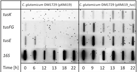
3.2.8 TRANSCRIPTIONAL REGULATION OF THE PUTATIVE TREHALOSE EXPORT SYSTEM... 49
3.3 CONSTRUCTION AND APPLICATION OF A GENETICALLY ENCODED TREHALOSE NANOSENSOR ... 51
3.3.1 ESTABLISHING METABOLITE NANOSENSORS IN C. GLUTAMICUM ... 52
3.3.2 THE GENE CG0834 ENCODES THE BINDING PROTEIN OF THE TREHALOSE ABC UPTAKE SYSTEM ... 55
3.3.3 DEVELOPMENT OF A TREHALOSE NANOSENSOR ... 57
3.3.4 IMPROVING THE SENSOR RESPONSE BY LINKER MODIFICATIONS ... 59
3.3.5 TREHALOSE QUANTIFICATION WITH FRET-SENSORS ... 61
3.3.6 DEVELOPMENT OF AFFINITY MUTANTS ... 62
3.4 SYNTHESIS AND TRANSPORT OF MYCOLIC ACIDS IN C. GLUTAMICUM ... 65
3.4.1 TREHALOSE UPTAKE IS NOT REQUIRED TO RESTORE MYCOLIC ACID SYNTHESIS ... 66
3.4.2 TREHALOSE EXPORT IS INDEPENDENT OF MYCOLIC ACID SYNTHESIS ... 68
3.4.3 TWO RND-TYPE PROTEINS ARE INVOLVED IN MYCOLIC ACID METABOLISM OF C. GLUTAMICUM ... 69
3.4.4 INACTIVATION OF CG2893 LEADS TO ALTERED CELL ENVELOPE COMPOSITION ... 71
4 DISCUSSION ... 73
4.1 TREHALOSE EXPORT IN C. GLUTAMICUM ... 73
4.1.1 QUALITATIVE AND QUANTITATIVE ANALYSIS OF TREHALOSE EXPORT ... 73
4.3 CHARACTERISATION OF TUSE AND TREHALOSE NANOSENSOR CONSTRUCTION ... 83
4.3.1 THE GENE CG0834 ENCODES THE BINDING PROTEIN OF THE ABC TREHALOSE UPTAKE SYSTEM ... 84
4.3.2 CONSTRUCTION OF A GENETICALLY ENCODED TREHALOSE NANOSENSOR ... 85
4.3.3 ENGINEERING THE TREHALOSE BINDING SITE OF TUSE ... 86
4.4 MANIPULATION OF TREHALOSE UPTAKE LEADS TO OPTIMISED PRODUCTION STRAINS ... 88
5 REFERENCES ... 91
ABC ATP Binding Cassette
APS Ammonium Persulfate
ATCC American Type Culture Collection
ATP Adenosine Triphosphate
BCIP 5-Bromo-4-chloro-3-indolyl Phosphate
BHI Brain Heart Infusion
BLAST Basic Local Alignment Search Tool
bp Base Pairs
CAPS N-cyclohexyl-3-aminopropanesulfonic Acid
cDNA Copy Desoxyribonucleic Acid
cdw Cellular Dry Weight
CTAB Cetyltrimethyl Ammoniumbromide
DIG-11-UTP Digoxigenin Deoxyuridine Triphosphate
DNA Desoxyribonucleic Acid
EDTA Ethylenediaminetetraacetic Acid
FRET Förster Resonance Energy Transfer
g Gravitational Force
HPLC High Pressure Liquid Chromatography
IM Integration Mutant
IMAC Ion Metal Affinity Chromatography
IPTG Isopropyl-β-D-thiogalactoside
LB Luria Bertani
MES 2-(N-Morpholino)ethanesulfonic Acid
MFS Major Facilitator Superfamily
MOPS 3-Morpholinopropane-1-sulfonic Acid
NBT Nitro Blue Tetrazolium Chloride
OD600 Optical Density at 600 nm
PAGE Polyacrylamide Gel Electrophoresis
PBP Periplasmic Binding Protein
PCR Polymerase Chain Reaction
PIPES 1,4-Piperazinediethanesulfonic Acid
RNA Ribonucleic Acid
RND Restriction, Nodulation, Cell Division
rpm Rounds per Minute
SDS Sodiumdodecyl Sulfate
SEC Size Exclusion Chromatography
SOB Super Optimal Broth
TAE Tris-Acetate-EDTA Buffer
TB Terrific Broth
TDM Trehalose Dimycolate
TE Tris EDTA Buffer
TEMED Tetramethylethylenediamine
TLC Thin Layer Chromatography
TMM Trehalose Monomycolate
Tris Tris(hydroxymethyl)aminomethane
2TY Tryptone Yeast Extract Medium
WT Wild Type
YP/s Yield Coefficient Product/Substrate
1 Introduction
1.1 Corynebacterium glutamicum is an industrial producer and a model organism
Corynebacterium glutamicum is a Gram-positive soil bacterium belonging to the Corynebacterineae suborder of the Actinobacteria phylum (Liebl, 2005). C. glutamicum has been known since the 1950s for its ability to excrete L-glutamate into the culture super- natant (Kinoshita et al., 1957) and today it is the main industrially applied producer of L-glutamate and other amino acids. 2.5 million tons of L-glutamate, which is mainly used as a flavour enhancer, and 1.5 million tons of L-lysine, which is mainly used for animal nutrition, are produced in fermentation processes with C. glutamicum per year (Becker & Wittmann, 2012). Besides amino acids, nucleoside monophosphates, which also serve as flavour enhancers, are produced with C. glutamicum (Demain et al., 1966; Kazarinova et al., 2002).
The continuous optimisation of production strains demands a profound understanding of metabolic and regulatory pathways to increase product yields, to reduce the formation of byproducts, or to allow the use of alternative carbon sources. Substrate import and product export are also important targets to further boost production (Burkovski & Krämer, 2002).
C. glutamicum has been studied for several decades, which has led to the development of a variety of techniques to construct genetically engineered C. glutamicum strains. The production of multiple bulk and high-value compounds like vitamins, solvents, diamines, ethanol, and organic acids in genetically modified C. glutamicum strains has been achieved in the laboratory scale, showing potential future applications for this bacterium (Sahm &
Eggeling, 1999; Smith et al., 2010; Blombach et al., 2011; Mimitsuka et al., 2007; Kind et al., 2011; Inui et al., 2004; Okino et al., 2005).
The Corynebacterineae suborder also comprises pathogenic species like Mycobacterium
leprae, Corynebacterium diphtheriae, and Mycobacterium tuberculosis, the causative agents
of leprosy, diphtheria, and tuberculosis, respectively. The latter causes about 2 million
deaths per year, which is more than any other infectious disease (Dye, 2006). C. glutamicum
serves as a model organism for cell envelope synthesis within the Corynebacterineae
suborder. The cell envelope of Corynebacterineae shows a complex structure compared to
other Gram-positive bacteria. While the envelope of C. glutamicum resembles that of myco-
bacteria and other related bacteria in structure and function, its composition is
comparatively simple (Puech et al., 2001). In addition, C. glutamicum is non-pathogenic, grows rapidly on different carbon sources, its genome has been sequenced completely (Kalinowski et al., 2003), and it has been used successfully for the expression of mycobacterial genes (Puech et al., 2001).
1.2 Cell envelope architecture in C. glutamicum
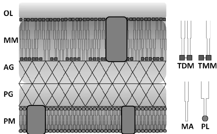
A common feature of the cell envelope of Corynebacterineae (Figure 1) is the presence of a second lipid membrane similar to the outer membrane of Gram-negative bacteria, which can be seen as a fracture plane in freeze-fractured cells (Puech et al., 2001). This bilayer mainly consists of mycolic acids, long chain α-alkyl, β-hydroxy fatty acids with 22 to 36 carbon atoms in C. glutamicum, that are covalently linked to the disaccharide trehalose as trehalose monomycolate (TMM) and trehalose dimycolate (TDM) as well as to the arabinogalactan layer, forming an approximately 42 Å thick hydrophobic permeability barrier in C. glutamicum (Bansal-Mutalik & Nikaido, 2011; Rath et al., 2013). The arabinogalactan layer is covalently attached to both the peptidoglycan layer and the mycolic acid layer, constituting a mycolyl-arabinogalactan-peptidoglycan complex on top of the plasma membrane. The cell envelope is completed by an outer layer mainly consisting of carbohydrates and a minor share of free fatty acids (Puech et al., 2001). In some but not all species, an additional surface layer consisting of proteins can be found (Chami et al., 1995;
Soual-Hoebeke et al., 1999). Mycolic acids found in corynebacteria have a simple structure in comparison to their mycobacterial equivalents. In general, the latter are longer (C
70– C
90) and carry modifications like desaturations, cyclopropane rings, methoxy groups, and keto groups. Besides TMM and TDM, a multitude of other lipid compounds is present in the mycolic acid layer of mycobacteria like phthiocerol dimycocerosates, glycopeptidolipids, polyacyltrehaloses, and sulfolipids (Brennan & Nikaido, 1995).
The mycolic acid layer is a major determinant of the low permeability of the cell envelope for antibiotics and other noxious compounds (Liu et al., 1996; Jackson et al., 1999). Mycolic acid synthesis is essential in mycobacteria and thus of high medical interest as a drug target for the treatment of tuberculosis. This extends to the synthesis of trehalose, a non-reducing disaccharide, which forms the polar head groups of TMM and TDM (De Smet et al., 2000;
Kalscheuer et al., 2010).
Figure 1: Cell envelope structure of C. glutamicum. PM: plasma membrane; PG: peptidoglycan layer;
AG: arabinogalactan layer; MM: mycolic acid membrane; OL: outer layer; TDM: trehalose dimycolate; TMM: trehalose monomycolate; MA: mycolic acids; PL: phospholipids; Boxes:
membrane proteins. Structure and composition are adapted from Puech et al. (2001).
In C. glutamicum, the mycolic acid layer has also been investigated for its contribution to amino acid excretion. L-glutamate excretion can be triggered under different conditions like biotin limitation, the addition of surfactants, antibiotics, or by temperature upshifts (Shiio et al., 1962; Nara et al., 1964; Takinami et al., 1964; Delaunay et al., 1999; Radmacher et al., 2005b). The mechanosensitive channel MscCG has been shown to be involved in L-glutamate transport across the plasma membrane (Nakamura et al., 2007). Restricted by the hydrophobicity of the mycolic acid layer, porin proteins located in the mycolic acid layer probably allow the efflux of amino acids to the surrounding. Four different porins were identified in C. glutamicum and mycolylation of two of them has been shown recently (Huc et al., 2013). Notably, mycolic acid synthesis is not essential in C. glutamicum. Strains unable to synthesise trehalose constantly excreted L-glutamate to the medium, an effect caused by the lack of trehalose mycolates in these mutants (Gebhardt et al., 2007).
The investigation of trehalose metabolism in C. glutamicum is thus of interest for the
optimisation of C. glutamicum production strains as well as for the identification of new drug
targets for the treatment of tuberculosis.
1.3 Trehalose synthesis in C. glutamicum
Trehalose plays an important role in many organisms. It protects proteins and membranes during anhydrobiosis, serves as transport sugar in the haemolymph of arthropods, acts as compatible solute under conditions of hyperosmotic stress, and it is an important precursor for the mycolic acid membrane in Corynebacterineae (Strom & Kaasen, 1993; Crowe et al., 1998; Arguelles, 2000; Luzardo et al., 2000; Tropis et al., 2005).
In C. glutamicum, changes of the external osmolality trigger a sequence of adaptation mechanisms to adjust the osmotic strength of the cytosol to the external conditions to maintain the turgor pressure of the cell. An immediate response to hyperosmotic conditions is the uptake of K
+to prevent the loss of water from the cell (Wolf et al., 2003). For long- term adaptation, K
+is then replaced by the accumulation of compatible solutes. These can either be taken up from the medium like glycine betaine, or they can be synthesised endogenously, like L-glutamate, L-proline, and trehalose. Trehalose synthesis becomes the most important adaptation mechanism under conditions of nitrogen starvation in the absence of compatible solutes in the medium (Wolf et al., 2003).
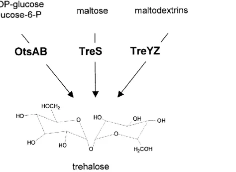
In C. glutamicum, M. tuberculosis, and M. smegmatis three pathways are present for the synthesis of trehalose (Figure 2) (De Smet et al., 2000; Wolf et al., 2003). In contrast to many other bacteria, the most important pathway for trehalose synthesis under hyperosmotic conditions in C. glutamicum is the TreYZ-pathway. Two enzymes, maltooligosyltrehalose synthase (TreY) and maltooligosyltrehalose trehalohydrolase (TreZ), convert maltodextrines, which are intermediates of the glycogen metabolism in C. glutamicum, to trehalose.
Expression of treY and treZ is constitutive to enable a fast response to sudden osmotic
upshifts and is only slightly induced under hyperosmotic conditions (Wolf et al., 2003). The
OtsAB-pathway converts the activated precursors UDP-glucose and glucose-6-phosphate to
trehalose by the sequential action of trehalose-6-phosphate synthase (OtsA) and trehalose-
6-phosphate phosphatase (OtsB). Although otsA expression is slightly enhanced after
osmotic upshifts, its activity is dispensable for trehalose synthesis under these conditions
and thus the physiological function of this pathway in C. glutamicum remains unclear (Wolf
et al., 2003). In the third pathway, trehalose synthase (TreS) catalyses the conversion of
maltose to trehalose, but in C. glutamicum this pathway is only relevant for trehalose
synthesis during growth with maltose as substrate (Tzvetkov et al., 2003). Although this
reaction is reversible, flux from trehalose to maltose seems to prevail in the cell. TreS could
thus be important for trehalose degradation after conditions of hyperosmotic stress (Wolf et al., 2003; Kim et al., 2010a; Miah et al., 2013).
TreS catalyses the interconversion of maltose and trehalose by a double displacement mechanism. A nucleophilic attack of the glucosidic bond by the catalytic L-aspartate residue leads to the intramolecular release of glucose, which reorients without leaving the catalytic site of the enzyme, and attacks the enzyme-bound intermediate (Zhang et al., 2011). Besides the transglucosylating activity of TreS, the release of small amounts (<10%) of glucose as a byproduct was also observed in vitro (Koh et al., 1998; Pan et al., 2004; Kim et al., 2010a).
Two additional pathways for trehalose synthesis are known in other bacteria. Trehalose glycosyltransferring synthase (TreT) catalyses the formation of trehalose from ADP-glucose and glucose (Qu et al., 2004) and trehalose phoshphorylase (TreP) uses glucose-1-phosphate and glucose as substrates for trehalose synthesis (Maruta et al., 2002). However, both pathways are not present in corynebacteria.
Figure 2: Trehalose synthesis in C. glutamicum. OtsA: trehalose-6-phosphate synthase; OtsB:
trehalose-6-phosphate phosphatase; TreS: trehalose synthase; TreY: maltooligosyltrehalose syn- thase; TreZ: maltooligosyltrehalose trehalohydrolase. Adapted from Wolf et al. (2003) with modifications.
M. tuberculosis and M. smegmatis possess the same pathways for trehalose synthesis as
C. glutamicum. While viable C. glutamicum mutants lacking all three pathways for trehalose
synthesis have been successfully created (Wolf et al., 2003; Tropis et al., 2005), this was not
possible in mycobacteria and trehalose synthesis is thus thought to be essential in these bacteria (Woodruff et al., 2004; Murphy et al., 2005). Trehalose synthesis and its metabolism are thus interesting drug targets for the treatment of mycobacterial infections.
1.4 Trehalose catabolism in C. glutamicum
C. glutamicum can use different substrates like sugars, sugar alcohols, organic acids, or peptides as carbon and energy sources. In mixed-substrate cultivations, the co-utilisation of most carbon sources is observed for this bacterium (Arndt & Eikmanns, 2008). Besides its role as osmoprotectant and precursor for the synthesis of mycolic acids, trehalose also serves as substrate for growth for C. glutamicum (Henrich, 2011).
The transport system responsible for trehalose uptake was recently identified (Henrich, 2011). The gene cluster cg0830 – cg0835 (tus-genes) encodes an ABC trehalose uptake system with high affinity (K
M= 0.16 µM) but low transport capacity (v
max= 2.5 nmol × mg
-1cdw × min
-1). Genes in this cluster encode two putative membrane proteins, cg0831 (tusF) and cg0832 (tusG), a putative periplasmic solute binding protein, cg0834 (tusE), and an ATPase protein, cg0835 (tusK) (Henrich, 2011). Further, two genes encoding proteins with unknown function are also encoded in the tus-cluster. The gene cg0830 encodes a predicted membrane protein with two transmembrane domains, cg0833 putatively encodes a soluble cytosolic protein (Schulte, 2011). Both are not required but beneficial for trehalose uptake (Rehorst, 2013). The genes cg0830 and cg0833 form a transcriptional operon with tusF and tusG, while tusE and tusK are transcribed monocistronically (Henrich, 2011; Schulte, 2011).
C. glutamicum utilises trehalose only as a co-substrate, but spontaneous mutants were
isolated that can use trehalose as sole carbon source (Henrich, 2011). Because the tus-genes
are expressed independently of the carbon source and because of the low transport capacity
of the trehalose uptake system, this has been assumed to be rather important for the
recycling of trehalose, which is released in the periplasm during TDM synthesis, than for the
utilisation of trehalose as carbon source. This assumption is also based on the recent
identification of the trehalose uptake system LpqY-SugA-SugB-SugY in M. tuberculosis and
M. smegmatis, which is characterised by its constitutive expression, its high specificity for
trehalose, its low transport velocity, and its importance for virulence (Kalscheuer et al.,
2010). The finding of a trehalose uptake system in M. tuberculosis was surprising since this
bacterium is thought to thrive on fatty acids due to the absence of sugar substrates in
human phagocytes and especially the absence of trehalose synthesis in mammals (Marrero et al., 2010).
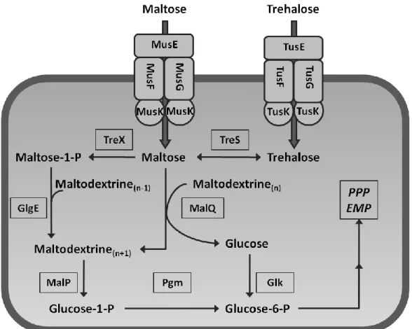
After trehalose uptake and the TreS catalysed conversion to maltose, trehalose is degraded via the same pathways as maltose (Figure 3), which can enter the cell via the ABC transporter MusFGK
2E (Henrich et al., 2013). It is then metabolised by the enzyme 4-α-glucanotransferase (MalQ), which forms maltodextrines and glucose, which is then phosphorylated by a glucokinase (Glk) to glucose-6-phosphate. A maltodextrine phosphory- lase (MalP) phosphorolytically cleaves maltodextrines to form glucose-1-phosphate, which is then isomerised to glucose-6-phosphate by phosphoglucomutase (Pgm). The latter is then degraded via reactions of central carbon metabolism.
Figure 3: Uptake and degradation of trehalose and maltose in C. glutamicum. PPP: pentose phosphate pathway; EMP: Embden-Meyerhof pathway. For further abbreviations see text.
The presence of a second pathway for maltose and trehalose degradation in C. glutamicum
has been shown (Henrich, 2011) in succession to its identification in M. tuberculosis and
M. smegmatis as the sole pathway for trehalose degradation in these bacteria (Kalscheuer et
al., 2010). Accordingly, maltose is phosphorylated to maltose-1-phosphate by a maltokinase
(TreX) and incorporated into α-glucans, intermediates of glycogen metabolism in C.
glutamicum (Seibold & Eikmanns, 2007), by a maltosyltransferase (GlgE) (Figure 3). However, it is not yet understood under which conditions this pathway is active and what impact it has on the metabolism of maltose and trehalose.
In contradiction to its function as carbon source, trehalose accumulation as byproduct is frequently observed during bioreactor cultivation of C. glutamicum (Vallino &
Stephanopoulos, 1993; Gourdon & Lindley, 1999; Wittmann & Heinzle, 2001). Considering the presence of an uptake system for trehalose and enzymes for its degradation, this observation was unexpected. This also indicates the presence of a trehalose export system in C. glutamicum but neither the mechanism of trehalose excretion to the medium nor a regulatory mechanism, which could be expected to hinder the uptake of trehalose, has been investigated in C. glutamicum yet.
1.5 Trehalose as precursor for TMM synthesis
Further support for the presence of a trehalose export system comes from the investigation of TMM and TDM synthesis in C. glutamicum. The main features of this pathway are supposed to be similar in different members of the Corynebacterineae, albeit additional steps for the synthesis of more complex mycolic acid derivatives are required in some members of this group. In contrast to mycobacteria, corynebacterial mutants defective in mycolic acid synthesis or transport are viable and thus, the latter are important model organisms for the examination of mycolic acid synthesis and transport.
The precursors for mycolic acid synthesis are two fatty acids. C. glutamicum possesses two type-I fatty acid synthases, FAS-IA and FAS-IB (Radmacher et al., 2005a). Products of these enzymes are mostly C
16– C
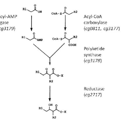
18fatty acids during growth on glucose as substrate (Radmacher et al., 2005a). In M. smegmatis and M. tuberculosis, a FAS-II enzyme complex is present that further elongates FAS-I products (Bloch, 1977). As shown in Figure 4, mycolic acid synthesis then proceeds with the activation of two fatty acids via adenylation and carboxylation, respectively (Trivedi et al., 2004; Portevin et al., 2005; Gande et al., 2007). A polyketide synthase encoded by cg3178 in C. glutamicum catalyses the decarboxylative condensation of the two activated precursors to an α-alkyl, β-keto-acyl intermediate (Gande et al., 2004;
Portevin et al., 2004). During condensation, the substrates and products are covalently
linked to the polyketide synthase domains via thioester bonds (Gande et al., 2004) and the
condensation product is probably transferred to a mannosylphosphopolyprenol carrier (‘X’ in Figure 4) that has been identified as a TMM precursor in M. smegmatis (Besra et al., 1994).
The final α-alkyl, β-hydroxy mycolic acid motif is then formed by the reduction of the precursor (Lea-Smith et al., 2007).
Figure 4: Final steps of mycolic acid synthesis. Gene numbers for C. glutamicum genes encoding the presented enzymes are given in brackets. X denotes the mannosylphosphopolyprenol carrier identified as a putative mycolic acid carrying precursor for TMM in M. smegmatis. Modified after Portevin et al. (2005).
The lasts steps of TMM synthesis in Corynebacterineae are still discussed controversially in
the literature. Especially the localisation of the TMM forming transfer of mycolic acids onto
trehalose remains uncertain. Nevertheless, TMM has been shown to be the mycolic acid
donor for the formation of TDM and the mycolylation of arabinogalactane. Thereby, one
molecule of trehalose is released to the periplasm per reaction cycle, which is catalysed by
six mycolyltransferases with partial redundancy in C. glutamicum (Brand et al., 2003; De
Sousa-D'Auria et al., 2003). ABC trehalose uptake systems recently identified in
mycobacteria (Kalscheuer et al., 2010) and in C. glutamicum (Henrich, 2011) are probably
important for the re-uptake of trehalose released to the periplasm during TDM synthesis.
Based on studies in M. tuberculosis and C. matruchotii, a pathway has been proposed, in which the mycolic acid moiety is transferred from a mannosylphosphopolyprenol carrier to trehalose-6-phosphate, yielding TMM-phosphate, by a hypothetical mycolyltransferase.
TMM-phosphate could then be dephosphorylated by a hypothetical phosphatase in the cytosol (Shimakata & Minatogawa, 2000; Takayama et al., 2005). This model is supported by the observation that [
14C]-labelled trehalose was only incorporated into trehalose glycolipids after its uptake in M. tuberculosis (Kalscheuer et al., 2010).
Furthermore, it was recently published by several groups that inhibition of MmpL3 led to the accumulation of TMM in M. tuberculosis and M. smegmatis (Grzegorzewicz et al., 2012; La Rosa et al., 2012; Tahlan et al., 2012; Varela et al., 2012). MmpL proteins (mycobacterial membrane protein large
)belong to the RND (restriction, nodulation, cell division) family, secondary active efflux transporters with broad substrate spectrum, which are found in all domains of life. These proteins have 12 transmembrane domains (TMD) and two large periplasmic domains between TMD 1 and 2 and TMD 7 and 8, which are responsible for substrate specificity (Yu et al., 2005). In M. tuberculosis, a total of 13 mmpL-genes are present. Identified substrates for MmpL transporters in M. tuberculosis comprise lipids found in the cell envelope like phthiocerol dimycocerosate for MmpL7 (Camacho et al., 2001) and diacyltrehalose sulfate, a precursor for sulfolipid-I synthesis, for MmpL8 (Converse et al., 2003; Domenech et al., 2004). Four mmpL-genes are also present in C. glutamicum and simultaneous inactivation of two of these genes led to the absence of mycolic acids in the cell envelope. In contrast to the situation in mycobacteria, no accumulation of TMM was observed in this mutant (Varela et al., 2012). Thus, also transport was not shown biochemically, MmpL3 and homologous proteins in C. glutamicum were suggested to export TMM from the cytosol to the periplasm (Grzegorzewicz et al., 2012; La Rosa et al., 2012;
Tahlan et al., 2012; Varela et al., 2012). Nevertheless, the role of MmpL transporters in the glycolipid metabolism of C. glutamicum remains uncertain.
In contrast, a second hypothesis assumes that trehalose and a mycolic acid precursor are
exported independently and react to TMM in the periplasm. Trehalose is assumed to be the
precursor for TMM synthesis rather than trehalose-6-phosphate since trehalose synthesis via
either of three pathways in C. glutamicum is sufficient to enable TMM synthesis (Tzvetkov et
al., 2003; Wolf et al., 2003). Most importantly, in C. glutamicum ΔotsA ΔtreS ΔtreY Δtus, a
mutant which is unable to synthesise and to take up trehalose, trehalose mycolate synthesis
could be restored by the addition of trehalose to the medium (Henrich, 2011). Further proof for this theory was given by the identification of an ABC transporter in C. matruchotii, which probably exports short-chain mycolic acids to the periplasm (Wang et al., 2006). Besides the export of an activated mycolic acid precursor, a prerequisite for TMM synthesis in the periplasm is the export of trehalose from the cytosol to the periplasm. Although trehalose is known to accumulate in the culture supernatant of C. glutamicum, the mechanism of trehalose excretion has not been investigated yet and the export system is not known.
1.6 Aims of this project
The model of periplasmic TMM synthesis in C. glutamicum from trehalose and a mycolic acid precursor requires the export of both a mycolic acid precursors and trehalose to the periplasm. This model is supported by the ability of C. glutamicum ΔotsA ΔtreS ΔtreY Δtus to use external trehalose for TMM synthesis but was challenged by the recent identification of MmpL proteins in C. glutamicum and related species and their assignment to the export of TMM to the periplasm.
To validate the assumption of periplasmic TMM formation, trehalose excretion in C. glutamicum should be investigated qualitatively and quantitatively in this work.
C. glutamicum test strains should be constructed to investigate trehalose export. Analytical techniques for the sensitive and specific quantification of intra- and extracellular trehalose concentrations should be developed to allow the mechanistic analysis of trehalose excretion.
Besides testing unspecific mechanism probably contributing to trehalose accumulation in the culture supernatant, a set of genes putatively encoding sugar export systems in C. glutamicum should be tested for their contribution to trehalose excretion. In parallel, the identification of the assumed trehalose export system should be achieved by the screening of a C. glutamicum mutant library. To enable the identification of mutants with altered intracellular trehalose concentrations, a trehalose sensor based on the trehalose binding protein of C. glutamicum should be constructed.
Further, trehalose transport should be investigated in C. glutamicum to resolve the apparent
contradiction of trehalose accumulation in the supernatant of C. glutamicum cultures in
spite of the presence of an uptake system in this bacterium. In this context, the
biotechnological significance of engineering trehalose uptake in C. glutamicum should be
addressed.
2 Material and Methods
2.1 Bacterial strains, plasmids, and oligonucleotides
All bacterial strains used in this work are shown in Table
1.
Table 1: Strains used in this work.
C. glutamicum Genotype or application Reference
ATCC 13032 Wild type (Abe et al., 1967)
Δcg0284 Deletion of cg0284 This study
Δcg3174 Deletion of cg3174 This study
Δcg3174 Δcg0284 Deletion of cg3174 and cg0284 This study
IMcg2893 Insertion of pDrive in cg2893 This study
DM1729 Lysine overproduction strain, pycP458S, homV59A, lysCT311I (Georgi et al., 2005) DM1729 IMcg0834 DM1729 with insertion of pDrive in cg0834 This study
ΔmalQ Deletion of malQ (Henrich, 2011)
ΔmalQ ΔtreX Deletion of malQ and treX This study
ΔmalQ ΔtreX Δtus Deletion of malQ, treX, and the cg0831 – cg0835 gene cluster This study ΔmalQ ΔtreX Δtus IMcg0206 Deletion of malQ, treX, the cg0831 – cg0835 gene cluster, and
insertion of pDrive in cg0206
This study
ΔmalQ ΔtreX Δtus IMcg0340 Deletion of malQ, treX, the cg0831 – cg0835 gene cluster, and insertion of pDrive in cg0340
This study
ΔmalQ ΔtreX Δtus IMcg0501 Deletion of malQ, treX, the cg0831 – cg0835 gene cluster, and insertion of pDrive in cg0501
This study
ΔmalQ ΔtreX Δtus IMcg0772 Deletion of malQ, treX, the cg0831 – cg0835 gene cluster, and insertion of pDrive in cg0772
This study
ΔmalQ ΔtreX Δtus IMcg1212 Deletion of malQ, treX, the cg0831 – cg0835 gene cluster, and insertion of pDrive in cg1212
This study
ΔmalQ ΔtreX Δtus IMcg1289 Deletion of malQ, treX, the cg0831 – cg0835 gene cluster, and insertion of pDrive in cg1289
This study
ΔmalQ ΔtreX Δtus IMcg1399 Deletion of malQ, treX, the cg0831 – cg0835 gene cluster, and insertion of pDrive in cg1399
This study
ΔmalQ ΔtreX Δtus IMcg1526 Deletion of malQ, treX, the cg0831 – cg0835 gene cluster, and insertion of pDrive in cg1526
This study
ΔmalQ ΔtreX Δtus IMcg2618 Deletion of malQ, treX, the cg0831 – cg0835 gene cluster, and insertion of pDrive in cg2618
This study
ΔmalQ ΔtreX Δtus IMcg2739 Deletion of malQ, treX, the cg0831 – cg0835 gene cluster, and insertion of pDrive in cg2739
This study
ΔmalQ ΔtreX Δtus IMcg2893 Deletion of malQ, treX, the cg0831 – cg0835 gene cluster, and insertion of pDrive in cg2893
This study
ΔmalQ ΔtreX Δtus IMcg2895 Deletion of malQ, treX, the cg0831 – cg0835 gene cluster, and insertion of pDrive in cg2895
This study
ΔmalQ ΔtreX Δtus IMcg2971 Deletion of malQ, treX, the cg0831 – cg0835 gene cluster, and insertion of pDrive in cg2971
This study
ΔmalQ ΔtreX Δtus IMcg3038 Deletion of malQ, treX, the cg0831 – cg0835 gene cluster, and insertion of pDrive in cg3038
This study
C. glutamicum Genotype or application Reference ΔmalQ ΔtreX Δtus Δcg3178 Deletion of malQ, treX, the cg0831 – cg0835 gene cluster, and
cg3178
This study
ΔmalQ ΔtreX Δtus IMcg3226 Deletion of malQ, treX, the cg0831 – cg0835 gene cluster, and insertion of pDrive in cg3226
This study
ΔmalQ ΔtreX Δtus IMcg3240 Deletion of malQ, treX, the cg0831 – cg0835 gene cluster, and insertion of pDrive in cg3240
This study
ΔmalQ ΔtreX Δtus IMcg3245 Deletion of malQ, treX, the cg0831 – cg0835 gene cluster, and insertion of pDrive in cg3245
This study
ΔmalQ ΔtreX Δtus IMcg3301 Deletion of malQ, treX, the cg0831 – cg0835 gene cluster, and insertion of pDrive in cg3301
This study
ΔmalQ ΔtreX Δtus IMcg3334 Deletion of malQ, treX, the cg0831 – cg0835 gene cluster, and insertion of pDrive in cg3334
This study
ΔmalQ ΔtreX Δtus IMcg3353 Deletion of malQ, treX, the cg0831 – cg0835 gene cluster, and insertion of pDrive in cg3353
This study
ΔmalQ ΔtreX Δtus IMcg3387 Deletion of malQ, treX, the cg0831 – cg0835 gene cluster, and insertion of pDrive in cg3387
This study
ΔmalQ ΔtreX Δtus IMcg3395 Deletion of malQ, treX, the cg0831 – cg0835 gene cluster, and insertion of pDrive in cg3395
This study
ΔmalQ ΔmscCG ΔmscL Deletion of malQ, mscCG, and mscL (Henrich, 2011) ΔmalQ ΔtreX ΔmscCG ΔmscL Deletion of malQ, treX, mscCG, and mscL This study
ΔmalQ ΔtreX ΔtreS Deletion of malQ, treX, and treS This study
Δmus Deletion of the cg2703 – cg2708 gene cluster (Henrich, 2011)
ΔtreS Deletion of treS (Wolf et al., 2003)
ΔtreS Δtus Deletion of treS and the cg0831 – cg0835 gene cluster This study ΔotsA ΔtreS ΔtreY Deletion of otsA, treS, and treY (Wolf et al., 2003) ΔotsA ΔtreS ΔtreY Δtus Deletion of otsA, treS, treY, and the cg0831 –cg0835 gene
cluster
(Henrich, 2011)
E. coli Genotype or application Reference
DH5α-mcr endA1 supE44 thi-1 λ-recA1 gyrA96 relA1 deoR Δ(lacZYA-argF) U196 Φ80lacZΔM15 mcrA Δ(mmr hsdRMS mcrBC)
(Grant et al., 1990) BL21 (DE3) F- dcm ompT hsdS(rB-
mB-
) gal λ(DE3) (Studier & Moffatt, 1986)
JM109 endA1 recA1 gyrA96 thi-1 hsdR17 (rK - mK
+) relA1 supE44 Δ(lac- proAB) [F’ traD36 proAB laqIq lacZΔM15]
(Yanisch-Perron et al., 1985)
All plasmids used in this work are shown in Table
2.
Table 2: Plasmids used in this work.
Plasmid Relevant characteristic or application Reference pBB1 Constitutive overexpression of genes in E. coli and
C. glutamicum. Ptac, CmR
(Krause et al., 2010)
pET29b(+) IPTG-inducible overexpression of genes in E. coli BL21 (DE3). PT7, KanR, C-terminal 6×Histag, lacI.
Novagen, Darmstadt
pTre1 Trehalase gene from R. marinus ligated to pET23a(+) (Jorge et al., 2007) pET29b_tre_rmr Overexpression of a trehalase gene from R. marinus in E.
coli BL21 (DE3)
This study pET29b_tusE Overexpression of tusE in E. coli BL21 (DE3) This study
Plasmid Relevant characteristic or application Reference pET29b_tusEΔN25 Overexpression of tusEΔN25 in E. coli BL21 (DE3) This study pET29b_tusEΔN32 Overexpression of tusEΔN32 in E. coli BL21 (DE3) This study pET29b_TreSen1-34 Overexpression of trehalose nanosensors TreSen1 - 34 in
E. coli BL21 (DE3)
This study
pDrive Cloning vector. KanR, AmpR, lacZα. Qiagen, Hilden
pJET Cloning vector. AmpR. Thermo Scientific ,USA
pK19mobsacB Plasmid for gene deletion in C. glutamicum. KanR, oriVE.
coli, oriT, mob, sacB
(Schäfer et al., 1994)
pK19mobsacB_Δcg0284 Plasmid for deletion of cg0284 in C. glutamcium This study pK19mobsacB_Δcg3174 Plasmid for deletion of cg3174 in C. glutamcium This study pK19mobsacB_Δcg3178 Plasmid for deletion of cg3178 in C. glutamcium This study pK19mobsacB_ΔtreX Plasmid for deletion of treX in C. glutamcium This study pK19mobsacB_ΔtreStreX Plasmid for deletion of treS and treX in C. glutamcium This study pK19mobsacB_Δtus Plasmid for deletion of the cg0831 – cg0835 gene cluster (Henrich, 2011) pRSETb_AT1.03,
pRSETb_AT1.03YEMK, pRSETb_AT3.10MGK, pRSETb_AT1.03R122K/R126K
Overexpression of AT1.03, AT1.03YEMK, AT3.10MGK, and AT103R122K/R126K
in E. coli
(Imamura et al., 2009)
pBB1_AT1.03, pBB1_AT1.03YEMK, pBB1_AT3.10MGK,
pBB1_AT1.03R122K/R126K
Overexpression of AT1.03, AT1.03YEMK, AT3.10MGK, and AT103R122K/R126K
in C. glutamicum
This study
pRSETb_FLIPmal-2µ, Overexpression of FLIPmal-2µ in E. coli (Fehr et al., 2002) pXMJ19 IPTG-inducible overexpression of genes in E. coli and C.
glutamicum. Ptac, lacIq, CmR.
(Jakoby et al., 1999)
pXMJ19_tus Overexpression of the cg0831 – cg0835 gene cluster in C. glutamicum.
(Henrich, 2011)
pXMJ19_otsBAE. coli Overexpression of the otsBA-operon from E. coli JM109 in C. glutamicum.
(Padilla et al., 2004a)
pXMJ19_cg2893 Overexpression of cg2893 in C. glutamicum. This study pXMJ19_cg2895 Overexpression of cg2895 in C. glutamicum. This study
All oligonucleotides used in this work are shown in Table 3.
Table 3: Oligonucleotides used in this work. Restriction sites are underlined and the corresponding restriction endonucleases are given in brackets.
Oligonucleotide Sequence Application
delcg0284 fw a CCCGGGTCGCAAGTGGATTGTG (XmaI) Deletion of cg0284 delcg0284 rv a CCAACTTCCATACTCCATTCCATATTCTGCGGCC
TCAAC
Deletion of cg0284
delcg0284 fw b GGAATGGAGTATGGAAGTTGGCCTTCATGCCG ATCTTCC
Deletion of cg0284
delcg0284 rv b GGATCCCTGCTTCTCCTCCCATTC (BamHI) Deletion of cg0284
delcg0284 check fw CCGCAGGTAAGTTAGTG Test-primer for deletion of cg0284 delcg0284 check rv GACATAGGTGCCAAGTC Test-primer for deletion of cg0284 delcg3174 fw a GAATTCCCAGGTGGCTCTGATGT (EcoRI) Deletion of cg3174
delcg3174 rv a CCAACTTCCATACTCCATTCCCGGCGATGACGA Deletion of cg3174
Oligonucleotide Sequence Application CTAAC
delcg3174 fw b GGAATGGAGTATGGAAGTTGGCGATACCGTGG CATCTC
Deletion of cg3174
delcg3174 rv b GGATCCACCAGACGGTCGATAGG (BamHI) Deletion of cg3174
delcg3174 check fw AAGGCCCTGAAAGTGTC Test-primer for deletion of cg3174 delcg3174 check rv CTTCCACTGCGGTCATA Test-primer for deletion of cg3174 delcg3178 fw a CCCGGGTGGTTGAGGCGGATAG (XmaI) Deletion of cg3178
delcg3178 rv a CCAACTTCCATACTCCATTCCAACGGCCAATCG GTAG
Deletion of cg3178
delcg3178 fw b GGAATGGAGTATGGAAGTTGGGACTTCGCCAA GAAGACC
Deletion of cg3178
delcg3178 rv b CCCGGGCCGTCAGCTCTCCGTTAC (XmaI) Deletion of cg3178
delcg3178 check fw CTCCTCAGCCTCGTT Test-primer for deletion of cg3178 delcg3178 check rv TTCCGTCAGCTCTCC Test-primer for deletion of cg3178 deltreSX fw a GGATCCTGGCCTGGAGAATTCGGATA (BamHI) Deletion of treS and treX
deltreSX rv a CCAACTTCCATACTCCATTCCTCTTCCGATTCGT GCTCAAC
Deletion of treS and treX
deltreSX fw b GGAATGGAGTATGGAAGTTGGCGTTGTACGAG GTTGCCTAT
Deletion of treS and treX
deltreSX rv b CTGCAGTAAGGACACGCCTCTGCATT (PstI) Deletion of treS and treX
deltreSX check fw TTGCTCGCCGCTACTTC Test-primer for deletion of treS and treX deltreSX check rv TCTGCGGTGGAGGAAGA Test-primer for deletion of treS and treX deltreX fw a GAATTCATCCTGAACGCCTGTACCTT (EcoRI) Deletion of treX
deltreX rv a CCAACTTCCATACTCCATTCCAAGTGTTCTCGCC ACATTCG
Deletion of treX
deltreX fw b GGAATGGAGTATGGAAGTTGGCGTTGTACGAG GTTGCCTAT
Deletion of treX
deltreX rv b CTGCAGTAAGGACACGCCTCTGCATT (PstI) Deletion of treX
deltreX check fw AGCTGGAACTCCTTCAC Test-primer for deletion of treX deltreX check rv TCTGCGGTGGAGGAAGA Test-primer for deletion of treX IMcg0206 fw TCAACGTGCCCTTAGGAATC Integration mutagenesis cg0206 IMcg0206 rv AATACCAAGGCGCCAGCAGC Integration mutagenesis cg0206 IMcg0206 check GCAATGATCGGTCCTTTAGC Test-primer for integration mutagenesis IMcg0340 fw TGGCATCCACATGCATCG Integration mutagenesis cg0340 IMcg0340 rv TGCGTAGCTGATGGAGAAGG Integration mutagenesis cg0340 IMcg0340 check ATCCTGCGAGACTTCAC Test-primer for integration mutagenesis IMcg0501 fw CTTTGGAGCACGCCGATGAG Integration mutagenesis cg0501 IMcg0501 rv ACACTGGTCGGCGACCAATG Integration mutagenesis cg0501 IMcg0501 check ACAGGACTGCAGACAGAAAC Test-primer for integration mutagenesis
IMcg0772 fw CTTCATCGCTGGTCTG Integration mutagenesis cg0772
IMcg0772 rv CCACCGACGTAGTTTC Integration mutagenesis cg0772
IMcg0772 check CGGCGGACATATCAAAC Test-primer for integration mutagenesis IMcg1212 fw CCTCCACGGTGTTGGATACG Integration mutagenesis cg1212 IMcg1212 rv CCCGTAATCAGATCGCGTTC Integration mutagenesis cg1212 IMcg1212 check AGCTTGGTCGAACTCTTCTG Test-primer for integration mutagenesis IMcg1289 fw CCAGTTATCGGTGGTGTTC Integration mutagenesis cg1289 IMcg1289 rv AGGCGATCCACCAATCGTC Integration mutagenesis cg1289 IMcg1289 check TGCCAACGGCGAAAGTC Test-primer for integration mutagenesis IMcg1399 fw CTTCCATGGCGTTCATTCCG Integration mutagenesis cg1399
Oligonucleotide Sequence Application
IMcg1399 rv GGTGCAGCTCTTGGAGTGTG Integration mutagenesis cg1399 IMcg1399 check GGCGACCTTTAAGGCTTTAC Test-primer for integration mutagenesis IMcg1526 fw TATCGCTCCAGCGACAC Integration mutagenesis cg1526 IMcg1526 rv ACGGAGCCAACCAGAAG Integration mutagenesis cg1526 IMcg1526 check CAGTAGTACCGGCATAAG Test-primer for integration mutagenesis IMcg2618 fw TGCATCACAGTGGTGATCAG Integration mutagenesis cg2618 IMcg2618 rv GATGATGCCCTGCTGTTCTG Integration mutagenesis cg2618 IMcg2618 check CCGCGATGTGTTCTTCTCTG Test-primer for integration mutagenesis IMcg2739 fw ATACCGCCGATCACGAGTCC Integration mutagenesis cg2739 IMcg2739 rv GTGTGGCTGGTTTCGTACTC Integration mutagenesis cg2739 IMcg2739 check GAAGGAACTTCCCGGCTGTG Test-primer for integration mutagenesis
IMcg2893 fw GCGTTTGCTCCAACTG Integration mutagenesis cg2893
IMcg2893 rv AACCACACCGCCTAAG Integration mutagenesis cg2893
IMcg2893 check CCGACCATTCGTTACAG Test-primer for integration mutagenesis IMcg2895 fw AGTACTATGCGCAGCTCTCC Integration mutagenesis cg2895 IMcg2895 rv ACATCACCGGCAGATAGTCC Integration mutagenesis cg2895 IMcg2895 check ACACTTGTCACCGTGTTC Test-primer for integration mutagenesis IMcg2971 fw TGCTGATGACGGTTACG Integration mutagenesis cg2971 IMcg2971 rv TGGTGGTCAAGGAGAAG Integration mutagenesis cg2971 IMcg2971 check GCTTCATGCTGTGAAAGG Test-primer for integration mutagenesis
IMcg3038 fw CGTGCGTGGTGATTG Integration mutagenesis cg3038
IMcg3038 rv GATGCGGTGTGGATG Integration mutagenesis cg3038
IMcg3038 check GAAAGGCCTGCTCGTTG Test-primer for integration mutagenesis IMcg3226 fw ATCGGAATTGGCGGCGAATG Integration mutagenesis cg3226 IMcg3226 rv TGTAGGTGCCGAAGATGTAG Integration mutagenesis cg3226 IMcg3226 check AGGTGACACGCCTTACATTC Test-primer for integration mutagenesis IMcg3240 fw TCCGGCGTCGATACTTTC Integration mutagenesis cg3240 IMcg3240 rv AGGATTCCGCCAATGAGC Integration mutagenesis cg3240 IMcg3240 check TGGCGCATGATGAACTC Test-primer for integration mutagenesis IMcg3245 fw CTGCACCTGCATTCGAAATC Integration mutagenesis cg3245 IMcg3245 rv TGCTGCTATTGCCCAGTTTG Integration mutagenesis cg3245 IMcg3245 check AAGTAGCTGCTGGTCGAGTC Test-primer for integration mutagenesis IMcg3301 fw GCCGCATGGTCGTTATCCTG Integration mutagenesis cg3301 IMcg3301 rv GCAACCGCATAGCCTGGAAG Integration mutagenesis cg3301 IMcg3301 check TGCTTATCGACGCCCACTTC Test-primer for integration mutagenesis IMcg3334 fw CCGCACTATCGCAGCATTG Integration mutagenesis cg3334 IMcg3334 rv TTCGGGTTGCCACTGTCAC Integration mutagenesis cg3334 IMcg3334 check AACAGCCCGATTCAAG Test-primer for integration mutagenesis IMcg3353 fw AAAGCAGCGGTGATTGG Integration mutagenesis cg3353 IMcg3353 rv GAGCAGGGTGACAAATGTG Integration mutagenesis cg3353 IMcg3353 check ACCAGAATCGGGAGGAC Test-primer for integration mutagenesis IMcg3387 fw TCTACGGACCAGGATTTG Integration mutagenesis cg3387
IMcg3387 rv ACGAACCAGCCATTTG Integration mutagenesis cg3387
IMcg3387 check GGGCTAAATGCTCCAAAC Test-primer for integration mutagenesis IMcg3395 fw CACGCGGTCACTCCACATAC Integration mutagenesis cg3395 IMcg3395 rv GAGGTTTCTTCGGAGCTTTC Integration mutagenesis cg3395 IMcg3395 check GCGGGCTTTACGGTTATGCG Test-primer for integration mutagenesis M13 fw TGTAAAACGACGGCCAGT Test-primer for integration mutagenesis
M13 rv CAGGAAACAGCTATGAC Test-primer for integration mutagenesis
![Figure 11: Trehalose export experiment with [ 14 C]-maltose as substrate. 300 µM [ 14 C]-labelled maltose was added at 0 min and the time courses of the intracellular amount of radiolabel (●) and of the total amount of radiolabel ( ■ ) w](https://thumb-eu.123doks.com/thumbv2/1library_info/3654380.1503470/46.892.157.735.281.763/figure-trehalose-experiment-substrate-labelled-intracellular-radiolabel-radiolabel.webp)
